Heme
Heme Oxygenase (Decyclizing)
Heme Oxygenase-1
Hemeproteins
Protoporphyrins
Carbon Monoxide
Myoglobin
Porphyrins
Iron
Oxidation-Reduction
Spectrum Analysis, Raman
Spectrophotometry
Ferrochelatase
Metalloporphyrins
Cytochrome c Group
Hemopexin
Hemoglobins
Electron Spin Resonance Spectroscopy
Aminolevulinic Acid
Cytochrome b Group
Cytochromes
Whales
Electron Transport Complex IV
Apoproteins
Deuteroporphyrins
Porphobilinogen Synthase
Oxygen
Peroxidases
Enzyme Induction
Protein Binding
Metmyoglobin
Protein Conformation
Mesoporphyrins
Binding Sites
Electron Transport
Mixed Function Oxygenases
Ligands
Cytochrome P-450 Enzyme System
Cyanides
Cytochrome-c Peroxidase
Oxidoreductases
Molecular Sequence Data
Tetrapyrroles
Porphyrias
Amino Acid Sequence
Internal electron transfer between hemes and Cu(II) bound at cysteine beta93 promotes methemoglobin reduction by carbon monoxide. (1/4934)
Previous studies showed that CO/H2O oxidation provides electrons to drive the reduction of oxidized hemoglobin (metHb). We report here that Cu(II) addition accelerates the rate of metHb beta chain reduction by CO by a factor of about 1000. A mechanism whereby electron transfer occurs via an internal pathway coupling CO/H2O oxidation to Fe(III) and Cu(II) reduction is suggested by the observation that the copper-induced rate enhancement is inhibited by blocking Cys-beta93 with N-ethylmaleimide. Furthermore, this internal electron-transfer pathway is more readily established at low Cu(II) concentrations in Hb Deer Lodge (beta2His --> Arg) and other species lacking His-beta2 than in Hb A0. This difference is consistent with preferential binding of Cu(II) in Hb A0 to a high affinity site involving His-beta2, which is ineffective in promoting electron exchange between Cu(II) and the beta heme iron. Effective electron transfer is thus affected by Hb type but is not governed by the R left arrow over right arrow T conformational equilibrium. The beta hemes in Cu(II)-metHb are reduced under CO at rates close to those observed for cytochrome c oxidase, where heme and copper are present together in the oxygen-binding site and where internal electron transfer also occurs. (+info)Negligible amount of copper in hepatic L-tryptophan 2,3-dioxygenase. (2/4934)
During the purification of L-tryptophan 2,3-dioxygenase, a protohemoprotein from rat liver, both copper and heme contents of the preparations were found to be progressively increased as purification proceeded. However, the greater part of copper was removed in the late stages of the purification giving a copper to heme ratio less than 0.4. The small amounts of copper could further be reduced by one-half, by a mild treatment of enzyme with chelators such as ethylenedi aminetetraacetate, without any accompanying decrease in enzymatic activity. Since the turnover number of these enzyme preparations expressed per mol of enzyme-bound heme, 200 to 277 min-1 at 25 degrees, were either comparable to or slightly higher than those reported with homogeneous enzyme preparations, the heme in the preparation was considered to be of fully active L-tryptophan 2,3-dioxygenase and, therefore, such a small ratio of copper to heme, 0.1 to 0.3, indicated that copper is not a constituent of L-tryptophan 2,3-dioxygenase of rat liver. The findings were thus inconsistent with the results of Brady et al. (Brady, F. O., Monaco, M. E. Forman, H. J. Schutz, G., and Feigelson, P. (1972) J. Biol. Chem. 247, 7915-7922), who found that L-tryptophan 2,3-dioxygenase contained 2 g atoms of copper and 2 mol of heme/mol of enzyme. Possible reasons for this discrepancy have been discussed. (+info)The role of Saccharomyces cerevisiae Met1p and Met8p in sirohaem and cobalamin biosynthesis. (3/4934)
MET1 and MET8 mutants of Saccharomyces cerevisiae can be complemented by Salmonella typhimurium cysG, indicating that the genes are involved in the transformation of uroporphyrinogen III into sirohaem. In the present study, we have demonstrated complementation of defined cysG mutants of Sal. typhimurium and Escherichia coli, with either MET1 or MET8 cloned in tandem with Pseudomonas denitrificans cobA. The conclusion drawn from these experiments is that MET1 encodes the S-adenosyl-l-methionine uroporphyrinogen III transmethylase activity, and MET8 encodes the dehydrogenase and chelatase activities (all three functions are encoded by Sal. typhimurium and E. coli cysG). MET8 was further cloned into pET14b to allow expression of the protein with an N-terminal His-tag. After purification, the functions of the His-tagged Met8p were studied in vitro by assay with precorrin-2 in the presence of NAD+ and Co2+. The results demonstrated that Met8p acts as a dehydrogenase and chelatase in the biosynthesis of sirohaem. Moreover, despite the fact that S. cerevisiae does not make cobalamins de novo, we have shown also that MET8 is able to complement cobalamin cobaltochelatase mutants and have revealed a subtle difference in the early stages of the anaerobic cobalamin biosynthetic pathways between Sal. typhimurium and Bacillus megaterium. (+info)Structure of a cytochrome P450-redox partner electron-transfer complex. (4/4934)
The crystal structure of the complex between the heme- and FMN-binding domains of bacterial cytochrome P450BM-3, a prototype for the complex between eukaryotic microsomal P450s and P450 reductase, has been determined at 2.03 A resolution. The flavodoxin-like flavin domain is positioned at the proximal face of the heme domain with the FMN 4.0 and 18.4 A from the peptide that precedes the heme-binding loop and the heme iron, respectively. The heme-binding peptide represents the most efficient and coupled through-bond electron pathway to the heme iron. Substantial differences between the FMN-binding domains of P450BM-3 and microsomal P450 reductase, observed around the flavin-binding sites, are responsible for different redox properties of the FMN, which, in turn, control electron flow to the P450. (+info)Cloning of Bacillus stearothermophilus ctaA and heme A synthesis with the CtaA protein produced in Escherichia coli. (5/4934)
The Bacillus stearothermophilus ctaA gene, which is required for heme A synthesis, was found upstream of the ctaBCDEF/caaEABCD gene cluster as in B. subtilis and B. firmus. The deduced protein sequence indicate that CtaA is a 35-kDa intrinsic membrane protein with seven hydrophobic segments. Alignment of CtaA sequences showed conserved residues including histidines that may be involved in heme B binding and substrate binding. Expression of ctaA in E. coli resulted in increased formation of a membrane-bound b-type cytochrome, heme A production, and severe growth inhibition. Furthermore, B. stearothermophilus CtaA produced in E. coli was found to catalyze the conversion of heme O to heme A in vitro. (+info)Coupling of the oxygen-linked interaction energy for inositol hexakisphosphate and bezafibrate binding to human HbA0. (6/4934)
The energetics of signal propagation between different functional domains (i.e. the binding sites for O2, inositol hexakisphospate (IHP), and bezafibrate (BZF)) of human HbA0 was analyzed at different heme ligation states and through the use of a stable, partially heme ligated intermediate. Present data allow three main conclusions to be drawn, and namely: (i) IHP and BZF enhance each others binding as the oxygenation proceeds, the coupling free energy going from close to zero in the deoxy state to -3.4 kJ/mol in the oxygenated form; (ii) the simultaneous presence of IHP and BZF stabilizes the hemoglobin T quaternary structure at very low O2 pressures, but as oxygenation proceeds it does not impair the transition toward the R structure, which indeed occurs also under these conditions; (iii) under room air pressure (i.e. pO2 = 150 torr), IHP and BZF together induce the formation of an asymmetric dioxygenated hemoglobin tetramer, whose features appear reminiscent of those suggested for transition state species (i.e. T- and R-like tertiary conformation(s) within a quaternary R-like structure). (+info)Chlamydomonas chloroplast ferrous hemoglobin. Heme pocket structure and reactions with ligands. (7/4934)
We report the optical and resonance Raman spectral characterization of ferrous recombinant Chlamydomonas LI637 hemoglobin. We show that it is present in three pH-dependent equilibrium forms including a 4-coordinate species at acid pH, a 5-coordinate high spin species at neutral pH, and a 6-coordinate low spin species at alkaline pH. The proximal ligand to the heme is the imidazole group of a histidine. Kinetics of the reactions with ligands were determined by stopped-flow spectroscopy. At alkaline pH, combination with oxygen, nitric oxide, and carbon monoxide displays a kinetic behavior that is interpreted as being rate-limited by conversion of the 6-coordinate form to a reactive 5-coordinate form. At neutral pH, combination rates of the 5-coordinate form with oxygen and carbon monoxide were much faster (>10(7) microM-1 s-1). The dissociation rate constant measured for oxygen is among the slowest known, 0.014 s-1, and is independent of pH. Replacement of the tyrosine 63 (B10) by leucine or of the putative distal glutamine by glycine increases the dissociation rate constant 70- and 30-fold and increases the rate of autoxidation 20- and 90-fold, respectively. These results are consistent with at least two hydrogen bonds stabilizing the bound oxygen molecule, one from tyrosine B10 and the other from the distal glutamine. In addition, the high frequency (232 cm-1) of the iron-histidine bond suggests a structure that lacks any proximal strain thus contributing to high ligand affinity. (+info)Modulation of the remote heme site geometry of recombinant mouse neuronal nitric-oxide synthase by the N-terminal hook region. (8/4934)
The role of two essential residues at the N-terminal hook region of neuronal nitric-oxide synthase (nNOS) in nitric-oxide synthase activity was investigated. Full-length mouse nNOS proteins containing single-point mutations of Thr-315 and Asp-314 to alanine were produced in the Escherichia coli and baculovirus-insect cell expression systems. The molecular properties of the mutant proteins were analyzed in detail by biochemical, optical, and electron paramagnetic resonance spectroscopic techniques and compared with those of the wild-type enzyme. Replacement of Asp-314 by Ala altered the geometry around the heme site and the substrate-binding pocket of the heme domain and abrogated the ability of nNOS to form catalytically active dimers. Replacement of Thr-315 by Ala reduced the protein stability and altered the geometry around the heme site, especially in the absence of bound (6R)-5,6,7, 8-tetrahydro-L-biopterin cofactor. These results suggest that Asp-314 and Thr-315 both play critical structural roles in stabilizing the heme domain and subunit interactions in mouse nNOS. (+info)Heme is not a medical term per se, but it is a term used in the field of medicine and biology. Heme is a prosthetic group found in hemoproteins, which are proteins that contain a heme iron complex. This complex plays a crucial role in various biological processes, including oxygen transport (in hemoglobin), electron transfer (in cytochromes), and chemical catalysis (in peroxidases and catalases).
The heme group consists of an organic component called a porphyrin ring, which binds to a central iron atom. The iron atom can bind or release electrons, making it essential for redox reactions in the body. Heme is also vital for the formation of hemoglobin and myoglobin, proteins responsible for oxygen transport and storage in the blood and muscles, respectively.
In summary, heme is a complex organic-inorganic structure that plays a critical role in several biological processes, particularly in electron transfer and oxygen transport.
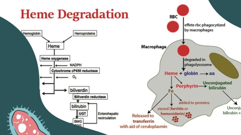
Heme Oxygenase-1 (HO-1) is an inducible enzyme that catalyzes the degradation of heme into biliverdin, iron, and carbon monoxide. It is a rate-limiting enzyme in the oxidative degradation of heme. HO-1 is known to play a crucial role in cellular defense against oxidative stress and inflammation. It is primarily located in the microsomes of many tissues, including the spleen, liver, and brain. Induction of HO-1 has been shown to have cytoprotective effects, while deficiency in HO-1 has been associated with several pathological conditions, such as vascular diseases, neurodegenerative disorders, and cancer.
Heme proteins are a type of protein that contain a heme group, which is a prosthetic group composed of an iron atom contained in the center of a large organic ring called a porphyrin. The heme group gives these proteins their characteristic red color. Hemeproteins have various important functions in biological systems, including oxygen transport (e.g., hemoglobin), electron transfer (e.g., cytochromes), and enzymatic catalysis (e.g., peroxidases and catalases). The heme group can bind and release gases, such as oxygen and carbon monoxide, and can participate in redox reactions due to the ease with which iron can change its oxidation state.
Protoporphyrins are organic compounds that are the immediate precursors to heme in the porphyrin synthesis pathway. They are composed of a porphyrin ring, which is a large, complex ring made up of four pyrrole rings joined together, with an acetate and a propionate side chain at each pyrrole. Protoporphyrins are commonly found in nature and are important components of many biological systems, including hemoglobin, the protein in red blood cells that carries oxygen throughout the body.
There are several different types of protoporphyrins, including protoporphyrin IX, which is the most common form found in humans and other animals. Protoporphyrins can be measured in the blood or other tissues as a way to diagnose or monitor certain medical conditions, such as lead poisoning or porphyrias, which are rare genetic disorders that affect the production of heme. Elevated levels of protoporphyrins in the blood or tissues can indicate the presence of these conditions and may require further evaluation and treatment.
Carbon monoxide (CO) is a colorless, odorless, and tasteless gas that is slightly less dense than air. It is toxic to hemoglobic animals when encountered in concentrations above about 35 ppm. This compound is a product of incomplete combustion of organic matter, and is a major component of automobile exhaust.
Carbon monoxide is poisonous because it binds to hemoglobin in red blood cells much more strongly than oxygen does, forming carboxyhemoglobin. This prevents the transport of oxygen throughout the body, which can lead to suffocation and death. Symptoms of carbon monoxide poisoning include headache, dizziness, weakness, nausea, vomiting, confusion, and disorientation. Prolonged exposure can lead to unconsciousness and death.
Carbon monoxide detectors are commonly used in homes and other buildings to alert occupants to the presence of this dangerous gas. It is important to ensure that these devices are functioning properly and that they are placed in appropriate locations throughout the building. Additionally, it is essential to maintain appliances and heating systems to prevent the release of carbon monoxide into living spaces.
Myoglobin is a protein found in the muscle tissue, particularly in red or skeletal muscles. It belongs to the globin family and has a similar structure to hemoglobin, another oxygen-binding protein found in red blood cells. Myoglobin's primary function is to store oxygen within the muscle cells, making it readily available for use during periods of increased oxygen demand, such as during physical exertion.
Myoglobin contains heme groups that bind to and release oxygen molecules. The protein has a higher affinity for oxygen than hemoglobin, allowing it to maintain its bound oxygen even in low-oxygen environments. When muscle cells are damaged or undergo necrosis (cell death), myoglobin is released into the bloodstream and can be detected in serum or urine samples. Elevated levels of myoglobin in the blood or urine may indicate muscle injury, trauma, or diseases affecting muscle integrity, such as rhabdomyolysis or muscular dystrophies.
Porphyrins are complex organic compounds that contain four pyrrole rings joined together by methine bridges (=CH-). They play a crucial role in the biochemistry of many organisms, as they form the core structure of various heme proteins and other metalloproteins. Some examples of these proteins include hemoglobin, myoglobin, cytochromes, and catalases, which are involved in essential processes such as oxygen transport, electron transfer, and oxidative metabolism.
In the human body, porphyrins are synthesized through a series of enzymatic reactions known as the heme biosynthesis pathway. Disruptions in this pathway can lead to an accumulation of porphyrins or their precursors, resulting in various medical conditions called porphyrias. These disorders can manifest as neurological symptoms, skin lesions, and gastrointestinal issues, depending on the specific type of porphyria and the site of enzyme deficiency.
It is important to note that while porphyrins are essential for life, their accumulation in excessive amounts or at inappropriate locations can result in pathological conditions. Therefore, understanding the regulation and function of porphyrin metabolism is crucial for diagnosing and managing porphyrias and other related disorders.
In the context of medicine, iron is an essential micromineral and key component of various proteins and enzymes. It plays a crucial role in oxygen transport, DNA synthesis, and energy production within the body. Iron exists in two main forms: heme and non-heme. Heme iron is derived from hemoglobin and myoglobin in animal products, while non-heme iron comes from plant sources and supplements.
The recommended daily allowance (RDA) for iron varies depending on age, sex, and life stage:
* For men aged 19-50 years, the RDA is 8 mg/day
* For women aged 19-50 years, the RDA is 18 mg/day
* During pregnancy, the RDA increases to 27 mg/day
* During lactation, the RDA for breastfeeding mothers is 9 mg/day
Iron deficiency can lead to anemia, characterized by fatigue, weakness, and shortness of breath. Excessive iron intake may result in iron overload, causing damage to organs such as the liver and heart. Balanced iron levels are essential for maintaining optimal health.
Oxidation-Reduction (redox) reactions are a type of chemical reaction involving a transfer of electrons between two species. The substance that loses electrons in the reaction is oxidized, and the substance that gains electrons is reduced. Oxidation and reduction always occur together in a redox reaction, hence the term "oxidation-reduction."
In biological systems, redox reactions play a crucial role in many cellular processes, including energy production, metabolism, and signaling. The transfer of electrons in these reactions is often facilitated by specialized molecules called electron carriers, such as nicotinamide adenine dinucleotide (NAD+/NADH) and flavin adenine dinucleotide (FAD/FADH2).
The oxidation state of an element in a compound is a measure of the number of electrons that have been gained or lost relative to its neutral state. In redox reactions, the oxidation state of one or more elements changes as they gain or lose electrons. The substance that is oxidized has a higher oxidation state, while the substance that is reduced has a lower oxidation state.
Overall, oxidation-reduction reactions are fundamental to the functioning of living organisms and are involved in many important biological processes.
Spectrum analysis in the context of Raman spectroscopy refers to the measurement and interpretation of the Raman scattering spectrum of a material or sample. Raman spectroscopy is a non-destructive analytical technique that uses the inelastic scattering of light to examine the vibrational modes of molecules.
When a monochromatic light source, typically a laser, illuminates a sample, a small fraction of the scattered light undergoes a shift in frequency due to interactions with the molecular vibrations of the sample. This shift in frequency is known as the Raman shift and is unique to each chemical bond or functional group within a molecule.
In a Raman spectrum, the intensity of the scattered light is plotted against the Raman shift, which is expressed in wavenumbers (cm-1). The resulting spectrum provides a "fingerprint" of the sample's molecular structure and composition, allowing for the identification and characterization of various chemical components within the sample.
Spectrum analysis in Raman spectroscopy can reveal valuable information about the sample's crystallinity, phase transitions, polymorphism, molecular orientation, and other properties. This technique is widely used across various fields, including materials science, chemistry, biology, pharmaceuticals, and forensics, to analyze a diverse range of samples, from simple liquids and solids to complex biological tissues and nanomaterials.
Spectrophotometry is a technical analytical method used in the field of medicine and science to measure the amount of light absorbed or transmitted by a substance at specific wavelengths. This technique involves the use of a spectrophotometer, an instrument that measures the intensity of light as it passes through a sample.
In medical applications, spectrophotometry is often used in laboratory settings to analyze various biological samples such as blood, urine, and tissues. For example, it can be used to measure the concentration of specific chemicals or compounds in a sample by measuring the amount of light that is absorbed or transmitted at specific wavelengths.
In addition, spectrophotometry can also be used to assess the properties of biological tissues, such as their optical density and thickness. This information can be useful in the diagnosis and treatment of various medical conditions, including skin disorders, eye diseases, and cancer.
Overall, spectrophotometry is a valuable tool for medical professionals and researchers seeking to understand the composition and properties of various biological samples and tissues.
Ferrochelatase is a medical/biochemical term that refers to an enzyme called Fe-chelatase or heme synthase. This enzyme plays a crucial role in the biosynthesis of heme, which is a vital component of hemoglobin, cytochromes, and other important biological molecules.
Ferrochelatase functions by catalyzing the insertion of ferrous iron (Fe2+) into protoporphyrin IX, the final step in heme biosynthesis. This enzyme is located within the inner mitochondrial membrane of cells and is widely expressed in various tissues, with particularly high levels found in erythroid precursor cells, liver, and brain.
Defects or mutations in the ferrochelatase gene can lead to a rare genetic disorder called erythropoietic protoporphyria (EPP), which is characterized by an accumulation of protoporphyrin IX in red blood cells, plasma, and other tissues. This accumulation results in photosensitivity, skin lesions, and potential complications such as liver dysfunction and gallstones.
Heptanoates are chemical compounds that contain the functional group of heptanoic acid. Heptanoic acid, also known as n-caproic acid, is a type of carboxylic acid with a 7-carbon chain and the molecular formula C7H15COOH.
Heptanoates are commonly used in the production of various chemicals, including flavors, fragrances, and pharmaceuticals. In medicine, heptanoates may be used as esters in the formulation of drugs to improve their solubility, absorption, and stability. For example, some injectable forms of medications may use heptanoate salts or esters to enhance their delivery into the body.
It's important to note that specific medical definitions for "heptanoates" may vary depending on the context and application.
Metalloporphyrins are a type of porphyrin molecule that contain a metal ion at their center. Porphyrins are complex organic compounds containing four modified pyrrole rings connected to form a planar, aromatic ring known as a porphine. When a metal ion is incorporated into the center of the porphyrin ring, it forms a metalloporphyrin.
These molecules have great biological significance, as they are involved in various essential processes within living organisms. For instance, heme, a type of iron-containing porphyrin, plays a crucial role in oxygen transport and storage in the body by forming part of hemoglobin and myoglobin molecules. Chlorophyll, another metalloporphyrin with magnesium at its center, is essential for photosynthesis in plants, algae, and some bacteria.
Metalloporphyrins have also found applications in several industrial and medical fields, including catalysis, sensors, and pharmaceuticals. Their unique structure and properties make them valuable tools for researchers and scientists to study and utilize in various ways.
Cytochrome c is a small protein that is involved in the electron transport chain, a key part of cellular respiration in which cells generate energy in the form of ATP. Cytochrome c contains a heme group, which binds to and transports electrons. The cytochrome c group refers to a class of related cytochromes that have similar structures and functions. These proteins are found in the mitochondria of eukaryotic cells (such as those of plants and animals) and in the inner membranes of bacteria. They play a crucial role in the production of energy within the cell, and are also involved in certain types of programmed cell death (apoptosis).
Hemopexin is a protein found in blood plasma. It's primary function is to bind and transport heme, a potentially toxic molecule that is released when hemoglobin from red blood cells is broken down. Hemopexin helps to prevent damage to tissues and organs by keeping free heme levels low in the bloodstream. It also plays a role in the immune response and has antioxidant properties. A deficiency in hemopexin can lead to increased risk of tissue damage and inflammation.
Hemoglobin (Hb or Hgb) is the main oxygen-carrying protein in the red blood cells, which are responsible for delivering oxygen throughout the body. It is a complex molecule made up of four globin proteins and four heme groups. Each heme group contains an iron atom that binds to one molecule of oxygen. Hemoglobin plays a crucial role in the transport of oxygen from the lungs to the body's tissues, and also helps to carry carbon dioxide back to the lungs for exhalation.
There are several types of hemoglobin present in the human body, including:
* Hemoglobin A (HbA): This is the most common type of hemoglobin, making up about 95-98% of total hemoglobin in adults. It consists of two alpha and two beta globin chains.
* Hemoglobin A2 (HbA2): This makes up about 1.5-3.5% of total hemoglobin in adults. It consists of two alpha and two delta globin chains.
* Hemoglobin F (HbF): This is the main type of hemoglobin present in fetal life, but it persists at low levels in adults. It consists of two alpha and two gamma globin chains.
* Hemoglobin S (HbS): This is an abnormal form of hemoglobin that can cause sickle cell disease when it occurs in the homozygous state (i.e., both copies of the gene are affected). It results from a single amino acid substitution in the beta globin chain.
* Hemoglobin C (HbC): This is another abnormal form of hemoglobin that can cause mild to moderate hemolytic anemia when it occurs in the homozygous state. It results from a different single amino acid substitution in the beta globin chain than HbS.
Abnormal forms of hemoglobin, such as HbS and HbC, can lead to various clinical disorders, including sickle cell disease, thalassemia, and other hemoglobinopathies.
Electron Spin Resonance (ESR) Spectroscopy, also known as Electron Paramagnetic Resonance (EPR) Spectroscopy, is a technique used to investigate materials with unpaired electrons. It is based on the principle of absorption of energy by the unpaired electrons when they are exposed to an external magnetic field and microwave radiation.
In this technique, a sample is placed in a magnetic field and microwave radiation is applied. The unpaired electrons in the sample absorb energy and change their spin state when the energy of the microwaves matches the energy difference between the spin states. This absorption of energy is recorded as a function of the magnetic field strength, producing an ESR spectrum.
ESR spectroscopy can provide information about the number, type, and behavior of unpaired electrons in a sample, as well as the local environment around the electron. It is widely used in physics, chemistry, and biology to study materials such as free radicals, transition metal ions, and defects in solids.
Aminolevulinic acid (ALA) is a naturally occurring compound in the human body and is a key precursor in the biosynthesis of heme, which is a component of hemoglobin in red blood cells. It is also used as a photosensitizer in dermatology for the treatment of certain types of skin conditions such as actinic keratosis and basal cell carcinoma.
In medical terms, ALA is classified as an α-keto acid and a porphyrin precursor. It is synthesized in the mitochondria from glycine and succinyl-CoA in a reaction catalyzed by the enzyme aminolevulinic acid synthase. After its synthesis, ALA is transported to the cytosol where it undergoes further metabolism to form porphyrins, which are then used for heme biosynthesis in the mitochondria.
In dermatology, topical application of ALA followed by exposure to a specific wavelength of light can lead to the production of reactive oxygen species that destroy abnormal cells in the skin while leaving healthy cells unharmed. This makes it an effective treatment for precancerous and cancerous lesions on the skin.
It is important to note that ALA can cause photosensitivity, which means that patients who have undergone ALA-based treatments should avoid exposure to sunlight or other sources of bright light for a period of time after the treatment to prevent adverse reactions.
Cytochrome b is a type of cytochrome, which is a class of proteins that contain heme as a cofactor and are involved in electron transfer. Cytochromes are classified based on the type of heme they contain and their absorption spectra.
The cytochrome b group includes several subfamilies of cytochromes, including cytochrome b5, cytochrome b2, and cytochrome bc1 (also known as complex III). These cytochromes are involved in various biological processes, such as fatty acid desaturation, steroid metabolism, and the electron transport chain.
The electron transport chain is a series of protein complexes in the inner mitochondrial membrane that generates most of the ATP (adenosine triphosphate) required for cellular energy production. Cytochrome bc1 is a key component of the electron transport chain, where it functions as a dimer and catalyzes the transfer of electrons from ubiquinol to cytochrome c while simultaneously pumping protons across the membrane. This creates an electrochemical gradient that drives ATP synthesis.
Deficiencies or mutations in cytochrome b genes can lead to various diseases, such as mitochondrial disorders and cancer.
Cytochromes are a type of hemeprotein found in the mitochondria and other cellular membranes of organisms. They contain a heme group, which is a prosthetic group composed of an iron atom surrounded by a porphyrin ring. This structure allows cytochromes to participate in redox reactions, acting as electron carriers in various biological processes.
There are several types of cytochromes, classified based on the type of heme they contain and their absorption spectra. Some of the most well-known cytochromes include:
* Cytochrome c: a small, mobile protein found in the inner mitochondrial membrane that plays a crucial role in the electron transport chain during cellular respiration.
* Cytochrome P450: a large family of enzymes involved in the metabolism of drugs, toxins, and other xenobiotics. They are found in various tissues, including the liver, lungs, and skin.
* Cytochrome b: a component of several electron transport chains, including those found in mitochondria, bacteria, and chloroplasts.
Cytochromes play essential roles in energy production, detoxification, and other metabolic processes, making them vital for the survival and function of living organisms.
I believe there may be some confusion in your question. Whales are not a medical term but rather large marine mammals. They belong to the Cetacean family, which includes dolphins and porpoises. If you're asking about a medical condition or something similar that might be associated with the word "whales," I would need more information to provide an accurate response.
Electron Transport Complex IV is also known as Cytochrome c oxidase. It is the last complex in the electron transport chain, located in the inner mitochondrial membrane of eukaryotic cells and the plasma membrane of prokaryotic cells. This complex contains 13 subunits, two heme groups (a and a3), and three copper centers (A, B, and C).
In the electron transport chain, Complex IV receives electrons from cytochrome c and transfers them to molecular oxygen, reducing it to water. This process is accompanied by the pumping of protons across the membrane, contributing to the generation of a proton gradient that drives ATP synthesis via ATP synthase (Complex V). The overall reaction catalyzed by Complex IV can be summarized as follows:
4e- + 4H+ + O2 → 2H2O
Defects in Cytochrome c oxidase can lead to various diseases, including mitochondrial encephalomyopathies and neurodegenerative disorders.
Histidine is an essential amino acid, meaning it cannot be synthesized by the human body and must be obtained through dietary sources. Its chemical formula is C6H9N3O2. Histidine plays a crucial role in several physiological processes, including:
1. Protein synthesis: As an essential amino acid, histidine is required for the production of proteins, which are vital components of various tissues and organs in the body.
2. Hemoglobin synthesis: Histidine is a key component of hemoglobin, the protein in red blood cells responsible for carrying oxygen throughout the body. The imidazole side chain of histidine acts as a proton acceptor/donor, facilitating the release and uptake of oxygen by hemoglobin.
3. Acid-base balance: Histidine is involved in maintaining acid-base homeostasis through its role in the biosynthesis of histamine, which is a critical mediator of inflammatory responses and allergies. The decarboxylation of histidine results in the formation of histamine, which can increase vascular permeability and modulate immune responses.
4. Metal ion binding: Histidine has a high affinity for metal ions such as zinc, copper, and iron. This property allows histidine to participate in various enzymatic reactions and maintain the structural integrity of proteins.
5. Antioxidant defense: Histidine-containing dipeptides, like carnosine and anserine, have been shown to exhibit antioxidant properties by scavenging reactive oxygen species (ROS) and chelating metal ions. These compounds may contribute to the protection of proteins and DNA from oxidative damage.
Dietary sources of histidine include meat, poultry, fish, dairy products, and wheat germ. Histidine deficiency is rare but can lead to growth retardation, anemia, and impaired immune function.
In the context of medicine and pharmacology, "kinetics" refers to the study of how a drug moves throughout the body, including its absorption, distribution, metabolism, and excretion (often abbreviated as ADME). This field is called "pharmacokinetics."
1. Absorption: This is the process of a drug moving from its site of administration into the bloodstream. Factors such as the route of administration (e.g., oral, intravenous, etc.), formulation, and individual physiological differences can affect absorption.
2. Distribution: Once a drug is in the bloodstream, it gets distributed throughout the body to various tissues and organs. This process is influenced by factors like blood flow, protein binding, and lipid solubility of the drug.
3. Metabolism: Drugs are often chemically modified in the body, typically in the liver, through processes known as metabolism. These changes can lead to the formation of active or inactive metabolites, which may then be further distributed, excreted, or undergo additional metabolic transformations.
4. Excretion: This is the process by which drugs and their metabolites are eliminated from the body, primarily through the kidneys (urine) and the liver (bile).
Understanding the kinetics of a drug is crucial for determining its optimal dosing regimen, potential interactions with other medications or foods, and any necessary adjustments for special populations like pediatric or geriatric patients, or those with impaired renal or hepatic function.
Apoproteins are the protein components of lipoprotein complexes, which are responsible for transporting fat molecules, such as cholesterol and triglycerides, throughout the body. Apoproteins play a crucial role in the metabolism of lipids by acting as recognition signals that allow lipoproteins to interact with specific receptors on cell surfaces.
There are several different types of apoproteins, each with distinct functions. For example, apolipoprotein A-1 (apoA-1) is the major protein component of high-density lipoproteins (HDL), which are responsible for transporting excess cholesterol from tissues to the liver for excretion. Apolipoprotein B (apoB) is a large apoprotein found in low-density lipoproteins (LDL), very low-density lipoproteins (VLDL), and lipoprotein(a). ApoB plays a critical role in the assembly and secretion of VLDL from the liver, and it also mediates the uptake of LDL by cells.
Abnormalities in apoprotein levels or function can contribute to the development of various diseases, including cardiovascular disease, diabetes, and Alzheimer's disease. Therefore, measuring apoprotein levels in the blood can provide valuable information for diagnosing and monitoring these conditions.
Deuteroporphyrins are porphyrin derivatives that contain two carboxylic acid side chains. They are intermediates in the biosynthesis of heme and chlorophyll, which are essential molecules for biological processes such as oxygen transport and photosynthesis, respectively.
Deuteroporphyrins can be further classified into isomers based on the position of the carboxylic acid side chains. The most common isomer is deuteroporphyrin IX, which has the carboxylic acid side chains located at positions 1 and 2 relative to the pyrrole nitrogen atoms.
Deuteroporphyrins have been studied in various medical contexts, including as potential markers of porphyria, a group of metabolic disorders characterized by the accumulation of porphyrin precursors. Additionally, deuteroporphyrins and their derivatives have been investigated for their potential use in photodynamic therapy, a treatment modality that uses light-activated drugs to destroy cancer cells.
Porphobilinogen Synthase (also known as PBGD or hydroxymethylbilane synthase) is an enzyme that catalyzes the second step in the heme biosynthesis pathway. This enzyme is responsible for converting two molecules of porphobilinogen into a linear tetrapyrrole called hydroxymethylbilane, which is then converted into uroporphyrinogen III by uroporphyrinogen III synthase.
Deficiency in Porphobilinogen Synthase can lead to a rare genetic disorder known as acute intermittent porphyria (AIP), which is characterized by the accumulation of porphobilinogen and other precursors in the heme biosynthesis pathway, resulting in neurovisceral symptoms such as abdominal pain, vomiting, neuropathy, and psychiatric disturbances.
Levulinic acid is not specifically a medical term, but it is a chemical compound with the formula C5H8O2. It is a white crystalline solid that is used in the production of various chemicals and materials. However, I can provide you with some general information about levulinic acid:
Levulinic acid is a saturated carboxylic acid, which means it contains a carboxyl group (-COOH) and is fully saturated with hydrogen atoms. It is an alpha-beta unsaturated carboxylic acid due to the presence of a carbon-carbon double bond (C=C) between the second and third carbon atoms in its structure.
Levulinic acid can be found naturally in small amounts in various fruits, such as apples and grapes, and is also present in some fermented foods like beer and wine. It can be produced industrially from biomass sources, such as cellulose or lignocellulosic materials, through a process called acid hydrolysis.
In the medical field, levulinic acid may have potential applications as an antimicrobial agent due to its ability to inhibit the growth of certain bacteria and fungi. It is also used in the synthesis of pharmaceuticals and other chemical products. However, it is not a substance that is typically directly associated with medical treatment or diagnosis.
Oxygen is a colorless, odorless, tasteless gas that constitutes about 21% of the earth's atmosphere. It is a crucial element for human and most living organisms as it is vital for respiration. Inhaled oxygen enters the lungs and binds to hemoglobin in red blood cells, which carries it to tissues throughout the body where it is used to convert nutrients into energy and carbon dioxide, a waste product that is exhaled.
Medically, supplemental oxygen therapy may be provided to patients with conditions such as chronic obstructive pulmonary disease (COPD), pneumonia, heart failure, or other medical conditions that impair the body's ability to extract sufficient oxygen from the air. Oxygen can be administered through various devices, including nasal cannulas, face masks, and ventilators.
Peroxidases are a group of enzymes that catalyze the oxidation of various substrates using hydrogen peroxide (H2O2) as the electron acceptor. These enzymes contain a heme prosthetic group, which plays a crucial role in their catalytic activity. Peroxidases are widely distributed in nature and can be found in plants, animals, and microorganisms. They play important roles in various biological processes, including defense against oxidative stress, lignin degradation, and host-pathogen interactions. Some common examples of peroxidases include glutathione peroxidase, which helps protect cells from oxidative damage, and horseradish peroxidase, which is often used in laboratory research.
Enzyme induction is a process by which the activity or expression of an enzyme is increased in response to some stimulus, such as a drug, hormone, or other environmental factor. This can occur through several mechanisms, including increasing the transcription of the enzyme's gene, stabilizing the mRNA that encodes the enzyme, or increasing the translation of the mRNA into protein.
In some cases, enzyme induction can be a beneficial process, such as when it helps the body to metabolize and clear drugs more quickly. However, in other cases, enzyme induction can have negative consequences, such as when it leads to the increased metabolism of important endogenous compounds or the activation of harmful procarcinogens.
Enzyme induction is an important concept in pharmacology and toxicology, as it can affect the efficacy and safety of drugs and other xenobiotics. It is also relevant to the study of drug interactions, as the induction of one enzyme by a drug can lead to altered metabolism and effects of another drug that is metabolized by the same enzyme.
Protein binding, in the context of medical and biological sciences, refers to the interaction between a protein and another molecule (known as the ligand) that results in a stable complex. This process is often reversible and can be influenced by various factors such as pH, temperature, and concentration of the involved molecules.
In clinical chemistry, protein binding is particularly important when it comes to drugs, as many of them bind to proteins (especially albumin) in the bloodstream. The degree of protein binding can affect a drug's distribution, metabolism, and excretion, which in turn influence its therapeutic effectiveness and potential side effects.
Protein-bound drugs may be less available for interaction with their target tissues, as only the unbound or "free" fraction of the drug is active. Therefore, understanding protein binding can help optimize dosing regimens and minimize adverse reactions.
Metmyoglobin is the oxidized form of myoglobin, a protein found in muscle tissue that binds and stores oxygen. When myoglobin is exposed to oxidizing agents or when muscle tissue is damaged (such as during exercise or after death), it can become oxidized and transform into metmyoglobin. This form of the protein cannot bind or store oxygen, and its presence in food (particularly in meats) can lead to off-flavors, discoloration, and reduced shelf life. In medical contexts, metmyoglobin may be used as a marker for muscle damage or hypoxia (lack of oxygen).
Bilirubin is a yellowish pigment that is produced by the liver when it breaks down old red blood cells. It is a normal byproduct of hemoglobin metabolism and is usually conjugated (made water-soluble) in the liver before being excreted through the bile into the digestive system. Elevated levels of bilirubin can cause jaundice, a yellowing of the skin and eyes. Increased bilirubin levels may indicate liver disease or other medical conditions such as gallstones or hemolysis. It is also measured to assess liver function and to help diagnose various liver disorders.
Protein conformation refers to the specific three-dimensional shape that a protein molecule assumes due to the spatial arrangement of its constituent amino acid residues and their associated chemical groups. This complex structure is determined by several factors, including covalent bonds (disulfide bridges), hydrogen bonds, van der Waals forces, and ionic bonds, which help stabilize the protein's unique conformation.
Protein conformations can be broadly classified into two categories: primary, secondary, tertiary, and quaternary structures. The primary structure represents the linear sequence of amino acids in a polypeptide chain. The secondary structure arises from local interactions between adjacent amino acid residues, leading to the formation of recurring motifs such as α-helices and β-sheets. Tertiary structure refers to the overall three-dimensional folding pattern of a single polypeptide chain, while quaternary structure describes the spatial arrangement of multiple folded polypeptide chains (subunits) that interact to form a functional protein complex.
Understanding protein conformation is crucial for elucidating protein function, as the specific three-dimensional shape of a protein directly influences its ability to interact with other molecules, such as ligands, nucleic acids, or other proteins. Any alterations in protein conformation due to genetic mutations, environmental factors, or chemical modifications can lead to loss of function, misfolding, aggregation, and disease states like neurodegenerative disorders and cancer.
Bacterial proteins are a type of protein that are produced by bacteria as part of their structural or functional components. These proteins can be involved in various cellular processes, such as metabolism, DNA replication, transcription, and translation. They can also play a role in bacterial pathogenesis, helping the bacteria to evade the host's immune system, acquire nutrients, and multiply within the host.
Bacterial proteins can be classified into different categories based on their function, such as:
1. Enzymes: Proteins that catalyze chemical reactions in the bacterial cell.
2. Structural proteins: Proteins that provide structural support and maintain the shape of the bacterial cell.
3. Signaling proteins: Proteins that help bacteria to communicate with each other and coordinate their behavior.
4. Transport proteins: Proteins that facilitate the movement of molecules across the bacterial cell membrane.
5. Toxins: Proteins that are produced by pathogenic bacteria to damage host cells and promote infection.
6. Surface proteins: Proteins that are located on the surface of the bacterial cell and interact with the environment or host cells.
Understanding the structure and function of bacterial proteins is important for developing new antibiotics, vaccines, and other therapeutic strategies to combat bacterial infections.
Mesoporphyrins are a type of porphyrin, which are organic compounds containing four pyrrole rings connected by methine bridges in a cyclic arrangement. Porphyrins are important components of various biological molecules such as hemoglobin, myoglobin, and cytochromes.
Mesoporphyrins have a specific structure with two propionic acid side chains and two acetic acid side chains attached to the pyrrole rings. They are intermediates in the biosynthesis of heme, which is a complex formed by the insertion of iron into protoporphyrin IX, a type of porphyrin.
Mesoporphyrins have been used in medical research and clinical settings as photosensitizers for photodynamic therapy (PDT), a treatment that uses light to activate a photosensitizing agent to destroy abnormal cells or tissues. In particular, mesoporphyrin IX has been used for the PDT treatment of various types of cancer, such as bladder, esophageal, and lung cancer, as well as for the treatment of age-related macular degeneration (AMD), a leading cause of vision loss in older adults.
It is important to note that mesoporphyrins are not typically used as a diagnostic tool or a therapeutic agent in routine clinical practice, but rather as part of experimental research and clinical trials.
In the context of medical and biological sciences, a "binding site" refers to a specific location on a protein, molecule, or cell where another molecule can attach or bind. This binding interaction can lead to various functional changes in the original protein or molecule. The other molecule that binds to the binding site is often referred to as a ligand, which can be a small molecule, ion, or even another protein.
The binding between a ligand and its target binding site can be specific and selective, meaning that only certain ligands can bind to particular binding sites with high affinity. This specificity plays a crucial role in various biological processes, such as signal transduction, enzyme catalysis, or drug action.
In the case of drug development, understanding the location and properties of binding sites on target proteins is essential for designing drugs that can selectively bind to these sites and modulate protein function. This knowledge can help create more effective and safer therapeutic options for various diseases.
The Electron Transport Chain (ETC) is a series of complexes in the inner mitochondrial membrane that are involved in the process of cellular respiration. It is the final pathway for electrons derived from the oxidation of nutrients such as glucose, fatty acids, and amino acids to be transferred to molecular oxygen. This transfer of electrons drives the generation of a proton gradient across the inner mitochondrial membrane, which is then used by ATP synthase to produce ATP, the main energy currency of the cell.
The electron transport chain consists of four complexes (I-IV) and two mobile electron carriers (ubiquinone and cytochrome c). Electrons from NADH and FADH2 are transferred to Complex I and Complex II respectively, which then pass them along to ubiquinone. Ubiquinone then transfers the electrons to Complex III, which passes them on to cytochrome c. Finally, cytochrome c transfers the electrons to Complex IV, where they combine with oxygen and protons to form water.
The transfer of electrons through the ETC is accompanied by the pumping of protons from the mitochondrial matrix to the intermembrane space, creating a proton gradient. The flow of protons back across the inner membrane through ATP synthase drives the synthesis of ATP from ADP and inorganic phosphate.
Overall, the electron transport chain is a crucial process for generating energy in the form of ATP in the cell, and it plays a key role in many metabolic pathways.
Mixed Function Oxygenases (MFOs) are a type of enzyme that catalyze the addition of one atom each from molecular oxygen (O2) to a substrate, while reducing the other oxygen atom to water. These enzymes play a crucial role in the metabolism of various endogenous and exogenous compounds, including drugs, carcinogens, and environmental pollutants.
MFOs are primarily located in the endoplasmic reticulum of cells and consist of two subunits: a flavoprotein component that contains FAD or FMN as a cofactor, and an iron-containing heme protein. The most well-known example of MFO is cytochrome P450, which is involved in the oxidation of xenobiotics and endogenous compounds such as steroids, fatty acids, and vitamins.
MFOs can catalyze a variety of reactions, including hydroxylation, epoxidation, dealkylation, and deamination, among others. These reactions often lead to the activation or detoxification of xenobiotics, making MFOs an important component of the body's defense system against foreign substances. However, in some cases, these reactions can also produce reactive intermediates that may cause toxicity or contribute to the development of diseases such as cancer.
A ligand, in the context of biochemistry and medicine, is a molecule that binds to a specific site on a protein or a larger biomolecule, such as an enzyme or a receptor. This binding interaction can modify the function or activity of the target protein, either activating it or inhibiting it. Ligands can be small molecules, like hormones or neurotransmitters, or larger structures, like antibodies. The study of ligand-protein interactions is crucial for understanding cellular processes and developing drugs, as many therapeutic compounds function by binding to specific targets within the body.
The Cytochrome P-450 (CYP450) enzyme system is a group of enzymes found primarily in the liver, but also in other organs such as the intestines, lungs, and skin. These enzymes play a crucial role in the metabolism and biotransformation of various substances, including drugs, environmental toxins, and endogenous compounds like hormones and fatty acids.
The name "Cytochrome P-450" refers to the unique property of these enzymes to bind to carbon monoxide (CO) and form a complex that absorbs light at a wavelength of 450 nm, which can be detected spectrophotometrically.
The CYP450 enzyme system is involved in Phase I metabolism of xenobiotics, where it catalyzes oxidation reactions such as hydroxylation, dealkylation, and epoxidation. These reactions introduce functional groups into the substrate molecule, which can then undergo further modifications by other enzymes during Phase II metabolism.
There are several families and subfamilies of CYP450 enzymes, each with distinct substrate specificities and functions. Some of the most important CYP450 enzymes include:
1. CYP3A4: This is the most abundant CYP450 enzyme in the human liver and is involved in the metabolism of approximately 50% of all drugs. It also metabolizes various endogenous compounds like steroids, bile acids, and vitamin D.
2. CYP2D6: This enzyme is responsible for the metabolism of many psychotropic drugs, including antidepressants, antipsychotics, and beta-blockers. It also metabolizes some endogenous compounds like dopamine and serotonin.
3. CYP2C9: This enzyme plays a significant role in the metabolism of warfarin, phenytoin, and nonsteroidal anti-inflammatory drugs (NSAIDs).
4. CYP2C19: This enzyme is involved in the metabolism of proton pump inhibitors, antidepressants, and clopidogrel.
5. CYP2E1: This enzyme metabolizes various xenobiotics like alcohol, acetaminophen, and carbon tetrachloride, as well as some endogenous compounds like fatty acids and prostaglandins.
Genetic polymorphisms in CYP450 enzymes can significantly affect drug metabolism and response, leading to interindividual variability in drug efficacy and toxicity. Understanding the role of CYP450 enzymes in drug metabolism is crucial for optimizing pharmacotherapy and minimizing adverse effects.
Cyanides are a group of chemical compounds that contain the cyano group, -CN, which consists of a carbon atom triple-bonded to a nitrogen atom. They are highly toxic and can cause rapid death due to the inhibition of cellular respiration. Cyanide ions (CN-) bind to the ferric iron in cytochrome c oxidase, a crucial enzyme in the electron transport chain, preventing the flow of electrons and the production of ATP, leading to cellular asphyxiation.
Common sources of cyanides include industrial chemicals such as hydrogen cyanide (HCN) and potassium cyanide (KCN), as well as natural sources like certain fruits, nuts, and plants. Exposure to high levels of cyanides can occur through inhalation, ingestion, or skin absorption, leading to symptoms such as headache, dizziness, nausea, vomiting, rapid heartbeat, seizures, coma, and ultimately death. Treatment for cyanide poisoning typically involves the use of antidotes that bind to cyanide ions and convert them into less toxic forms, such as thiosulfate and rhodanese.
Cytochrome-c peroxidase is an enzyme found in the inner membrane of mitochondria, which are the energy-producing structures in cells. It plays a crucial role in the electron transport chain, a series of complexes that generate energy in the form of ATP through a process called oxidative phosphorylation.
The enzyme's primary function is to catalyze the conversion of hydrogen peroxide (H2O2) into water (H2O) and oxygen (O2). This reaction helps protect the cell from the harmful effects of hydrogen peroxide, which can damage proteins, lipids, and DNA if left unchecked.
Cytochrome-c peroxidase contains a heme group, which is a prosthetic group consisting of an iron atom surrounded by a porphyrin ring. This heme group is responsible for the enzyme's ability to undergo redox reactions, where it cycles between its oxidized and reduced states during the catalytic cycle.
The medical relevance of cytochrome-c peroxidase lies in its role in cellular metabolism and energy production. Dysfunctions in the electron transport chain or oxidative phosphorylation processes, including those involving cytochrome-c peroxidase, can lead to various mitochondrial disorders and diseases, such as neurodegenerative conditions, muscle weakness, and metabolic abnormalities. However, it is essential to note that the study of this enzyme and its role in health and disease is still an active area of research.
Oxidoreductases are a class of enzymes that catalyze oxidation-reduction reactions, which involve the transfer of electrons from one molecule (the reductant) to another (the oxidant). These enzymes play a crucial role in various biological processes, including energy production, metabolism, and detoxification.
The oxidoreductase-catalyzed reaction typically involves the donation of electrons from a reducing agent (donor) to an oxidizing agent (acceptor), often through the transfer of hydrogen atoms or hydride ions. The enzyme itself does not undergo any permanent chemical change during this process, but rather acts as a catalyst to lower the activation energy required for the reaction to occur.
Oxidoreductases are classified and named based on the type of electron donor or acceptor involved in the reaction. For example, oxidoreductases that act on the CH-OH group of donors are called dehydrogenases, while those that act on the aldehyde or ketone groups are called oxidases. Other examples include reductases, peroxidases, and catalases.
Understanding the function and regulation of oxidoreductases is important for understanding various physiological processes and developing therapeutic strategies for diseases associated with impaired redox homeostasis, such as cancer, neurodegenerative disorders, and cardiovascular disease.
Molecular sequence data refers to the specific arrangement of molecules, most commonly nucleotides in DNA or RNA, or amino acids in proteins, that make up a biological macromolecule. This data is generated through laboratory techniques such as sequencing, and provides information about the exact order of the constituent molecules. This data is crucial in various fields of biology, including genetics, evolution, and molecular biology, allowing for comparisons between different organisms, identification of genetic variations, and studies of gene function and regulation.
Tetrapyrroles are a class of organic compounds that contain four pyrrole rings joined together in a macrocyclic structure. They are important in biology because they form the core structure of many essential cofactors and prosthetic groups in proteins, including heme, chlorophyll, and cobalamin (vitamin B12).
Heme is a tetrapyrrole that contains iron and is a crucial component of hemoglobin, the protein responsible for oxygen transport in red blood cells. Chlorophyll is another tetrapyrrole that contains magnesium and plays a vital role in photosynthesis, the process by which plants convert light energy into chemical energy. Cobalamin contains cobalt and is essential for DNA synthesis, fatty acid metabolism, and neurotransmitter synthesis.
Abnormalities in tetrapyrrole biosynthesis can lead to various diseases, such as porphyrias, which are characterized by the accumulation of toxic intermediates in the heme biosynthetic pathway.
Ferric compounds are inorganic compounds that contain the iron(III) cation, Fe3+. Iron(III) is a transition metal and can form stable compounds with various anions. Ferric compounds are often colored due to the d-d transitions of the iron ion. Examples of ferric compounds include ferric chloride (FeCl3), ferric sulfate (Fe2(SO4)3), and ferric oxide (Fe2O3). Ferric compounds have a variety of uses, including as catalysts, in dye production, and in medical applications.
Porphyrias are a group of rare genetic disorders that affect the production of heme, a component in hemoglobin that carries oxygen in the blood. The diseases are caused by mutations in the genes involved in the production of heme, leading to the buildup of porphyrins or their precursors in the body. These substances can be toxic and can cause various symptoms depending on the specific type of porphyria. Symptoms may include abdominal pain, neurological problems, and skin issues. Porphyrias are typically divided into two categories: acute porphyrias, which affect the nervous system, and cutaneous porphyrias, which primarily affect the skin.
An amino acid sequence is the specific order of amino acids in a protein or peptide molecule, formed by the linking of the amino group (-NH2) of one amino acid to the carboxyl group (-COOH) of another amino acid through a peptide bond. The sequence is determined by the genetic code and is unique to each type of protein or peptide. It plays a crucial role in determining the three-dimensional structure and function of proteins.
Ferrous compounds are inorganic substances that contain iron (Fe) in its +2 oxidation state. The term "ferrous" is derived from the Latin word "ferrum," which means iron. Ferrous compounds are often used in medicine, particularly in the treatment of iron-deficiency anemia due to their ability to provide bioavailable iron to the body.
Examples of ferrous compounds include ferrous sulfate, ferrous gluconate, and ferrous fumarate. These compounds are commonly found in dietary supplements and multivitamins. Ferrous sulfate is one of the most commonly used forms of iron supplementation, as it has a high iron content and is relatively inexpensive.
It's important to note that ferrous compounds can be toxic in large doses, so they should be taken under the guidance of a healthcare professional. Overdose can lead to symptoms such as nausea, vomiting, diarrhea, abdominal pain, and potentially fatal consequences if left untreated.
I'm sorry for any confusion, but "horses" is not a medical term. It is the common name given to the species *Equus ferus caballus*, which are large domesticated mammals used for transportation, work, and recreation. If you have any questions about horses or a related topic that you would like a medical perspective on, please let me know and I'd be happy to help!
Cytochrome4
- Cytochrome a refers to the heme A in specific combination with membrane protein forming a portion of cytochrome c oxidase. (wikipedia.org)
- Cytochrome c554 (cyt c554), a tetra-heme cytochrome from Nitrosomonas europaea, is an essential component in the biological nitrification pathway. (rcsb.org)
- Heme oxygenase-1 (HO-1) or HSP32 is the inducible isoform of heme oxygenase which catalyzes the NADPH, O2 and cytochrome P450 reductase dependent oxidation of heme to carbon monoxide, iron and billiverdin that is immediately reduced to bilirubin. (ihcworld.com)
- Surface enhanced Raman spectra (SERS) excited at 785 nm are reported for hemoglobin, myoglobin, two heme B proteins, and cytochrome c, a heme C protein, solubilized in a variety of solvent systems and then placed on Au and Ag substrates. (ojp.gov)
Proteins18
- Among the metalloporphyrins deployed by metalloproteins as prosthetic groups, heme is one of the most widely used and defines a family of proteins known as hemoproteins. (wikipedia.org)
- other important types include heme A and heme C. Isolated hemes are commonly designated by capital letters while hemes bound to proteins are designated by lower case letters. (wikipedia.org)
- When comparing proteomes of MVs between iron-limiting and iron-rich conditions, we found that under iron-limiting conditions, heme-binding proteins are enriched. (nature.com)
- Soluble guanylate cyclases are nitric oxide-responsive signaling proteins in which the nitric oxide sensor is a heme-binding domain of unknown structure that we have termed the heme-NO and oxygen binding (H-NOX) domain. (nih.gov)
- The approach allows efficient incorporation of heme analogs using a widely available bacterial strain and offers an attractive alternative to present reconstitution methods that subject proteins to harsh, denaturing conditions. (nih.gov)
- corresponding heme arrangements are observed in other multi-heme proteins. (rcsb.org)
- Heme is a critical component of iron-containing proteins called hemoproteins. (xshotpix.com)
- To obtain the most intense SERS spectra of heme B proteins (Mb and Hb) it is not sufficient to be in a low pH (pH ~ 2) denaturing environment. (ojp.gov)
- A hydrophobic solvent component is additionally required in order to efficiently solubilize the cleaved heme moiety and consequently observe intense SERS spectra indicative of these heme B proteins. (ojp.gov)
- Although both heme B proteins exhibit virtually identical, metal (Au, Ag) specific SERS spectra, Cyt c is much weaker in the low pH environments and only displays a Ag-like SERS spectrum on both metals indicating that this heme C protein is not cleaved in any of these solvation environments. (ojp.gov)
- Motif FoodWorks has suffered a setback in its dispute with Impossible Foods over meaty-tasting heme proteins in meat alternatives after the US Patent and Trademark Office (USPTO) rejected its request for an inter partes review of the patent at the center of a legal dispute between the pair. (foodnavigator-usa.com)
- The dispute centers on the use of meaty-tasting heme proteins in plant-based meat alternatives, an area in which both companies play. (foodnavigator-usa.com)
- In a lawsuit filed in March 2022, Impossible accused Motif of infringing its '761 patent, which covers the application of heme proteins in meat alternatives. (foodnavigator-usa.com)
- Despite the clear and unambiguous exclusion of animal proteins, Impossible alleges that Motif infringes because its products contain an animal heme-containing protein. (foodnavigator-usa.com)
- While the biochemical effects of ITCs have been well characterized, less information is available regarding their effects on the accumulation of stress-inducible heme oxygenase-1 (HO-1) and heat shock proteins (HSPs) as well as the possible formation of aggregated protein due to thiol modification. (uwaterloo.ca)
- Analyses of the temperature dependences and shapes of nuclear magnetic relaxation dispersion (NMRD) signals of site-directed mutants of human myoglobin indicate that a water molecule is bound to the sixth coordination site of the ferric heme in proteins in which the valine at position 68 is changed to either aspartate (Val68Asp) or asparagine (Val68Asn). (caltech.edu)
- In the study discussed here, Chergui's lab looked at a more biologically relevant class of heme proteins, the ferrous myoglobins, which carry oxygen to muscles. (nccr-must.ch)
- C. Bacteroides fragilis requires the ferrous‐iron transporter FeoAB and the CobN‐like proteins BtuS1 and BtuS2 for assimilation of iron released from heme. (utah.edu)
Enzyme heme2
- Upon loss of attachment, ATF4 activated a coordinated program of cytoprotective autophagy and antioxidant responses, including induced expression of the major antioxidant enzyme heme oxygenase 1 (HO-1). (jci.org)
- This lung damage induced the cytoprotective enzyme heme oxygenase-1, which is an enzyme that catalyzes heme degradation into CO, biliverdin, and Fe2+. (umsystem.edu)
Mononuclear non-heme iron1
- Scholars@Duke publication: Reaction mechanisms of mononuclear non-heme iron oxygenases. (duke.edu)
Involved in heme synthesis2
- The organs mainly involved in heme synthesis are the liver and the bone marrow, although every cell requires heme to function properly. (wikipathways.org)
- Porphyria is the common term for a group of syndromes, largely hereditary, that result from defects in porphyrins (the enzymes involved in heme synthesis). (medscape.com)
Hemin3
- The strong SERS signals of hemoglobin and myoglobin are due to hemin and ferric acetate heme formed in pH 2 HCl/ethanol and pH 2/50% acetic acid solutions respectively. (ojp.gov)
- Hemin (plasma-derived intravenous heme) is the definitive treatment and mainstay of management. (medscape.com)
- Reconstitute it with human serum albumin 25% (132 mL of 25% human serum albumin to 1 vial of hemin [301 mg heme]) and infuse it into large vein to reduce the risk of thrombophlebitis. (medscape.com)
Biosynthesis5
- The core will measure activity of the enzymes responsible for heme biosynthesis. (utah.edu)
- The core specializes in the UPLC quantification of heme and porphyrin levels, and the measurement of activity of enzymes responsible for heme biosynthesis. (utah.edu)
- Erythropoietin signaling regulates heme biosynthesis. (utah.edu)
- Reductions in the mitochondrial ABC transporter Abcb10 affect the transcriptional profile of heme biosynthesis genes. (utah.edu)
- Promoting Heme and Phycocyanin Biosynthesis in Synechocystis sp. (bvsalud.org)
Environment of the heme2
- Hemoproteins achieve their remarkable functional diversity by modifying the environment of the heme macrocycle within the protein matrix. (wikipedia.org)
- Environment of the Heme in Myoglobins. (caltech.edu)
Synthesis5
- The enzymatic process that produces heme is properly called porphyrin synthesis, as all the intermediates are tetrapyrroles that are chemically classified are porphyrins. (wikipathways.org)
- Recently, we reported the synthesis of a carboxylate-rich non-heme diiron enzyme model compound [Fe 2 (μ-O 2 CAr Tol ) 4 (4-CNPy) 2 ] ( 1 ), where - O 2 CAr Tol is 2,6-di- p -tolylbenzoate and 4-CNPy is 4-cyanopyridine (Yoon, S. (figshare.com)
- For d1 heme synthesis this intermediate has to undergo the replacement of two propionate side chains with oxygen functionalities and the introduction of a double bond into a further peripheral side chain. (kent.ac.uk)
- For heme synthesis didecarboxysiroheme is converted into Fe-coproporphyrin by oxidative loss of two acetic acid side chains. (kent.ac.uk)
- Infusion of hemelike agents rapidly restores the free-heme pool in hepatocytes, thereby exerting negative feedback repression on the rate-limiting enzyme of heme synthesis. (medscape.com)
Electron transfer6
- The heme iron serves as a source or sink of electrons during electron transfer or redox chemistry. (wikipedia.org)
- Heme b(D) is a distinctive feature of the succinate:menaquinone reductases, but the role of this heme in electron transfer to quinone has not previously been analyzed. (lu.se)
- The H113M mutant enzyme contains heme b(D) with raised midpoint potential and is impaired in electron transfer to menaquinone. (lu.se)
- The results support a model in which menaquinone is reduced on the positive side of the membrane and the transmembrane electrochemical potential provides driving force for electron transfer from succinate via heme b(P) and heme b(D) to menaquinone. (lu.se)
- The team reports exceptionally high rates for heme-to-heme electron transfer, on the order of 1 billion per second. (thomasyoungcentre.org)
- The study confirmed that electron transfer indeed takes place between tryptophan and the heme molecule, producing an ionic form of the latter that matched theoretical predictions. (nccr-must.ch)
Eukaryotic Initia1
- Phosphorylation of Eukaryotic Initiation Factor 2 by Heme-Regulated Inhibitor Kinase-Related Protein Kinases in Schizosaccharomyces pombe Is Important for Resistance to Environmental Stresses. (en-academic.com)
Carbon monoxide3
- Heme oxygenase, an essential enzyme in heme catabolism, cleaves heme to form biliverdin, which is subsequently converted to bilirubin by biliverdin reductase, and carbon monoxide, a putative neurotransmitter. (genetex.com)
- Heme oxygenase-1 (Hmox1) catalyzes the conversion of heme to biliverdin, carbon monoxide (CO), and ferrous iron (Fe 2+ ). (springer.com)
- Note: carbon monoxide can be produced endogenously as a byproduct of heme metabolism. (cdc.gov)
Pathway9
- Together, our findings strongly suggest that MVs act as a newly identified pathway for heme recycling, and represent a public good shared between phylogenetically closely related species. (nature.com)
- In humans, this pathway serves almost exclusively to form heme. (wikipathways.org)
- There is recent data which indicates the ability of peroxynitrite (ONOO-) to modulate the expression of HO-1 and suggests that the heme oxygenase pathway contributes to protection against the cytotoxic action of ONOO- which is a potent oxidizing agent generated by the interaction of nitric oxide (NO) and the superoxide anion. (ihcworld.com)
- How are porphyrias related to the heme pathway? (xshotpix.com)
- The genes for all the enzymes in the heme pathway have been identified. (xshotpix.com)
- What causes mutations in the heme biosynthetic pathway? (xshotpix.com)
- In the porphyrias, these mutations are in the genes involved in a certain chemical pathway, called the heme biosynthetic pathway. (xshotpix.com)
- Each type of porphyria is caused by a defect in a specific enzyme in the heme biosynthetic pathway. (xshotpix.com)
- Clinical manifestations depend on the step in the heme production pathway in which the enzymatic defect occurs. (medscape.com)
Porphyrins1
- Place an Order The Iron and Heme Core provides analysis of metals, precursor porphyrins and heme. (utah.edu)
Free Heme1
- Free Heme and Amyloid-β: A Fatal Liaison in Alzheimer's Disease. (j-alz.com)
Oxygenase activity2
- Heme oxygenase activity is induced by its substrate heme and by various nonheme substances. (genetex.com)
- Our results indicate that an 18 hr exposure to low concentrations of PAT (5-25 uM) increases cellular glutathione levels, heat shock protein 70 and heme oxygenase activity in cardiac myocytes in a concentration-dependent manner. (cdc.gov)
Peroxidases5
- In general, diatomic gases only bind to the reduced heme, as ferrous Fe(II) while most peroxidases cycle between Fe(III) and Fe(IV) and hemeproteins involved in mitochondrial redox, oxidation-reduction, cycle between Fe(II) and Fe(III). (wikipedia.org)
- plant peroxidases incorporate heme B. Lactoperoxidase and eosinophil peroxidase are protective enzymes responsible for the destruction of invading bacteria and virus. (wikipedia.org)
- Heme m contains the two ester bonds at the heme 1- and 5-methyl groups also present in heme l of other mammalian peroxidases, such as lactoperoxidase and eosinophil peroxidase. (wikipedia.org)
- Collectively, the data show that all three members of the class I heme peroxidases can support radical formation on the distal tryptophan and that the reactivity of this radical can be controlled either by the protein structure or by the nature of the compound I intermediate. (bris.ac.uk)
- Protonation of the proximal histidine ligand in heme peroxidases. (lu.se)
Porphyrin2
- In biochemical terms, heme is a coordination complex "consisting of an iron ion coordinated to a porphyrin acting as a tetradentate ligand, and to one or two axial ligands. (wikipedia.org)
- Heme/porphyrin/metal analysis was performed at the Iron and Heme Core facility at the University of Utah, supported in part by a grant from the NIH National Institute of Diabetes and Digestive and Kidney Diseases, Grant number U54DK110858. (utah.edu)
Membrane4
- Finally, we found that the heme carried in MVs is utilized by multiple species, and we further verified that membrane fusion efficiency and species evolutionary distance determine heme delivery. (nature.com)
- Wild-type (WT) and Heme Oxygenase 1 knockout (KO) HeLa cell extracts (30 μg) were separated by 12% SDS-PAGE, and the membrane was blotted with Heme Oxygenase 1 antibody (GTX101147) diluted at 1:500. (genetex.com)
- Various whole cell extracts (30 μg) were separated by 12% SDS-PAGE, and the membrane was blotted with Heme Oxygenase 1 antibody (GTX101147) diluted at 1:1500. (genetex.com)
- The succinate:menaquinone reductases all contain two heme groups in the membrane anchor of the enzyme: a proximal heme (heme b(P)) located close to the negative side of the membrane and a distal heme (heme b(D)) located close to the positive side of the membrane. (lu.se)
Catabolism2
- Heme is seen as an intermediate molecule in catabolism of haemoglobin in the process of bilirubin metabolism. (wikipathways.org)
- Heme oxygenase-1 (Hmox1) is a stress-inducible protein crucial in heme catabolism. (springer.com)
Hemoglobin5
- Heme (American English), or haem (Commonwealth English, both pronounced /hi:m/ HEEM), is a precursor to hemoglobin, which is necessary to bind oxygen in the bloodstream. (wikipedia.org)
- Hemes are most commonly recognized as components of hemoglobin, the red pigment in blood, but are also found in a number of other biologically important hemoproteins such as myoglobin, cytochromes, catalases, heme peroxidase, and endothelial nitric oxide synthase. (wikipedia.org)
- For example, the ability of hemoglobin to effectively deliver oxygen to tissues is due to specific amino acid residues located near the heme molecule. (wikipedia.org)
- Heme is a compound that the body needs to make hemoglobin and there are several steps to make this compound in the body. (xshotpix.com)
- results when heme cannot be produced normally, and therefore not enough hemoglobin is made. (medlineplus.gov)
Hemoproteins2
- Next, we proved that MVs participate in extracellular heme capture and transport, especially in heme recycling from environmental hemoproteins. (nature.com)
- Heme b is an iron-containing co-factor in hemoproteins. (sun.ac.za)
Covalently attached2
- Heme l is the derivative of heme B which is covalently attached to the protein of lactoperoxidase, eosinophil peroxidase, and thyroid peroxidase. (wikipedia.org)
- The crystal structure of cyt c554 at 2.6 A resolution shows a predominantly alpha-helical protein with four covalently attached hemes. (rcsb.org)
Tryptophan3
- Heme-based tryptophan dioxygenases are established immunosuppressive metalloproteins with significant biomedical interest. (ku.edu)
- This combined synthetic, biochemical, and biophysical study establishes the catalytic role of the α-amino group of the substrate during the O atom transfer reactions and thus represents a substantial advance to the mechanistic comprehension of the heme-based tryptophan dioxygenases. (ku.edu)
- The scientists studied them in their physiological state to see if tryptophan transferred electron to the myoglobin's oxygen-binding heme molecule. (nccr-must.ch)
Concentrations are low1
- When heme concentrations are low in the RBC , it shuts off production of globin chains. (en-academic.com)
Binds3
- In the transportation or detection of diatomic gases, the gas binds to the heme iron. (wikipedia.org)
- A1M binds heme and the crystal structure suggests that C34 and H123 participate in a heme binding site. (lu.se)
- The resultant protein (BfrB), which assembles into a 445.3 kDa complex0020from 24 identical subunits, binds 12 molecules of heme axially coordinated by two Met residues. (ku.edu)
Residues2
- The addition of peroxide with the glutamyl-375 and aspartyl-225 of lactoperoxidase forms ester bonds between these amino acid residues and the heme 1- and 5-methyl groups, respectively. (wikipedia.org)
- We have investigated the involvement of these five residues in the interactions with heme. (lu.se)
Precursors3
- Where do heme precursors accumulate in the body? (xshotpix.com)
- If one of the essential enzymes in heme production is deficient, certain precursors may accumulate in tissues (especially in the bone marrow or liver), appear in excess in the blood, and get excreted in the urine or stool. (xshotpix.com)
- Analysis and quantification of heme and its precursors can be obtained for cell pellets, tissue, whole blood, urine, feces and other complex biological materials. (utah.edu)
Ferrous1
- Results from additional experimentation indicate that Bfd must bind to BfrB to promote heme mediation of electrons from the surface to the core to support the efficient mobilization of ferrous ions from BfrB. (ku.edu)
Arginate2
- This mechanism is of therapeutic importance: infusion of heme arginate or hematin can abort attacks of porphyria in patients with an inborn error of metabolism of this process, by reducing transcription of ALA synthase. (wikipathways.org)
- Heme arginate is not currently available in the United States. (medscape.com)
Myoglobin1
- while Motif FoodWorks' HEMAMI heme protein is identical to bovine myoglobin, which is found in the muscle tissue of cows. (foodnavigator-usa.com)
HMOX11
- HMOX1 and HMOX2 belong to the heme oxygenase family. (genetex.com)
Ligand1
- During the detection of diatomic gases, the binding of the gas ligand to the heme iron induces conformational changes in the surrounding protein. (wikipedia.org)
Polypeptide2
- Iron Heme Polypep-Folic Acid Tablet is a medication that combines iron, heme polypeptide, and folic acid to support red blood cell production, improve iron levels, and prevent or treat iron-deficiency anemia. (imedix.com)
- Heme iron polypeptide is a form of iron supplement. (imedix.com)
HMOX21
- Heme Oxygenase 2 antibody LS-C776072 is an RPE-conjugated rabbit polyclonal antibody to Heme Oxygenase 2 (HMOX2) from human. (lsbio.com)
Degradation2
- Here we show that near-infrared autofluorescence is associated with the presence of intraplaque hemorrhage and heme degradation products, particularly bilirubin by using our recently created mouse model, which uniquely reflects plaque instability as seen in humans, and human carotid endarterectomy samples. (nature.com)
- In addition to human CEA samples, this model allowed us to identify intraplaque hemorrhage and heme degradation products as sources of the detected NIRAF signal. (nature.com)
Antibody4
- Heme Oxygenase 1 antibody detects Heme Oxygenase 1 protein at endoplasmic reticulum and nucleus by immunofluorescent analysis. (genetex.com)
- Green: Heme Oxygenase 1 stained by Heme Oxygenase 1 antibody (GTX101147) diluted at 1:500. (genetex.com)
- Heme Oxygenase 1 antibody detects Heme Oxygenase 1 protein at cytoplasm on human renal carcinoma by immunohistochemical analysis. (genetex.com)
- Heme Oxygenase 1 antibody detects Heme Oxygenase 1 protein at cytoplasm by immunohistochemical analysis. (genetex.com)
NADPH2
- In the presence of sodium dithionite or in the presence of P. aeruginosa ferredoxin NADP reductase (FPR) and NADPH the heme in BfrB remains oxidized and the core iron mineral is mobilized sluggishly. (ku.edu)
- In stark contrast, addition of NADPH to a solution containing BfrB, FPR and the apo-form of P. aeruginosa bacterioferritin associated ferredoxin (apo-Bfd) results in rapid reduction of the heme in BfrB and in the efficient mobilization of the core iron mineral. (ku.edu)
Kinase2
- Heme-regulated inhibitor (HRI) kinase is a member of the eIF-2{alpha} kinase family. (en-academic.com)
- Low concentrations of heme activate Heme-Regulated Inhibitor Kinase, which phosphorylates the Serine-51 of eIF-2{alpha}, and makes its interaction with eIF-2B nonproductive(eIF-2{alpha} sequesters eIF-2B). (en-academic.com)
Nonheme2
Iron10
- DQ12-45-1b, to analyze the function of its MVs in heme-iron recycling and sharing between species. (nature.com)
- The rate-limiting enzyme responsible for this reaction, ALA synthase, is strictly regulated by intracellular iron levels and heme concentration. (wikipathways.org)
- We examined the relationship between heme b abundance and the microbial community composition, and its utility for mapping iron limited phytoplankton. (sun.ac.za)
- Hence, heme b depletion and regulation is likely to occur during bloom progression when phytoplankton class-dependent absolute iron requirements exceed the available ambient concentration of DFe. (sun.ac.za)
- Thus, heme b provides a snapshot of the cellular activity in situ and could both be used as indicator of iron limitation and contribute to understanding phytoplankton adaptation mechanisms to changing iron supplies. (sun.ac.za)
- The Iron and Heme Core is at Wintrobe room 621. (utah.edu)
- You are invited to join a weekly Zoom meeting of of the Iron and Heme Core, where answers for questions regarding sample submission, appropriate testing, data analysis and how to run assays in your home lab will be answered by Hector Bergonia, Director of the core. (utah.edu)
- The Iron and Heme Core is part of the NIDDK sponsored, Center for Iron and Heme Disorders. (utah.edu)
- ABCB7 mutations slightly alter the structure of the ABCB7 protein, disrupting its usual role in heme production and iron homeostasis . (medlineplus.gov)
- 4) Wherever "Iron (As Fe)" is used as a source of nutrient, heme iron shall not be used in any form in any article of food. (who.int)
Bone marrow1
- Heme is biosynthesized in both the bone marrow and the liver. (wikipedia.org)
Bound2
- Heme m is the derivative of heme B covalently bound at the active site of myeloperoxidase. (wikipedia.org)
- Particularly striking is the structure of the protoporphyrin IX group, which is distorted from planarity to an extent not seen before in protein-bound heme groups. (nih.gov)
Histidine1
- a histidine residue, located adjacent to the heme group, becomes positively charged under acidic conditions (which are caused by dissolved CO2 in working muscles, etc.), releasing oxygen from the heme group. (wikipedia.org)
Oxidative2
- Hesperetin attenuated acetaminophen-induced hepatotoxicity by inhibiting hepatocyte necrosis and apoptosis, oxidative stress and inflammatory response via upregulation of heme oxygenase-1 expression. (stembook.org)
- Fe-coproporphyrin is then transformed into heme by the oxidative decarboxylation of two propionate side chains. (kent.ac.uk)
Prosthetic1
- This process entails the hijacking of siroheme, the prosthetic group of sulfite and nitrite reductase, and its processing into heme and d1 heme. (kent.ac.uk)
Metalloporphyrins1
- HO-1 or HSP32, a major heat shock/stress response protein is ubiquitous and its mRNA as well as its activity can be increased several-fold by heme, other metalloporphyrins, transition metals and stimuli that induce cellular stress. (ihcworld.com)
Soluble1
- Targeting heme-oxidised/heme-free soluble guanylyl cyclise to identify diabetic patients at risk of development of vascular disease. (monash.edu)
Biochemical1
- Structural and biochemical characterization of two heme binding sites on α1-microglobulin using site directed mutagenesis and molecular simulation. (lu.se)
Biological1
- Modified tetrapyrroles such as chlorophyll, heme, siroheme, vitamin B12, coenzyme F430, and heme d1 underpin a wide range of essential biological functions in all domains of life, and it is therefore surprising that the syntheses of many of these life pigments remain poorly understood. (kent.ac.uk)
Compounds2
Reductase2
Induction1
- The stress protein heme oxygenase is especially susceptible to induction by metals. (cdc.gov)
Protein expression1
- In the present study, sublethal concentrations of PAT were examined in neonatal rat cardiac myocytes for PAT-induced changes in cellular glutathione levels, heme oxygenase and stress protein expression. (cdc.gov)