Halobacteriaceae
Dihydroorotate Oxidase
RNA, Archaeal
Haloferax
RNA, Ribosomal, 16S
DNA, Ribosomal
Genes, rRNA
Sequence Analysis, DNA
Water Microbiology
Molecular Sequence Data
Cluster Analysis
Haloanaerobacter salinarius sp. nov., a novel halophilic fermentative bacterium that reduces glycine-betaine to trimethylamine with hydrogen or serine as electron donors; emendation of the genus Haloanaerobacter. (1/193)
A novel halophilic fermentative bacterium has been isolated from the black sediment below a gypsum crust and a microbial mat in hypersaline ponds of Mediterranean salterns. Morphologically, physiologically and genetically this organism belongs to the genus Haloanaerobacter. Haloanaerobacter strain SG 3903T (T = type strain) is composed of non-sporulating long flexible rods with peritrichous flagella, able to grow in the salinity range of 5-30% NaCl, with an optimum at 14-15%. The strain grows by fermenting carbohydrates or by using the Stickland reaction with either serine or H2 as electron donors and glycine-betaine as acceptor, which is reduced to trimethylamine. The two species described so far in the genus Haloanaerobacter are not capable of Stickland reaction with glycine-betaine + serine; however, Haloanaerobacter chitinovorans can use glycine-betaine with H2 as electron donor. Strain SG 3903T thus represents the first described strain in the genus Haloanaerobacter capable of the Stickland reaction with two amino acids. Although strain SG 3903T showed 67% DNA-DNA relatedness to H. chitinovorans, it is physiologically sufficiently different from the two described species to be considered as a new species which has been named Haloanaerobacter salinarius sp. nov. (+info)Halobacterial rhodopsins. (2/193)
Following the discovery of the bacteriorhodopsin proton pump in Halobacterium halobium (salinarum), not only the halorhodopsin halide pump and two photosensor rhodopsins (sensory rhodopsin and phoborhodopsin) in the same species, but also homologs of these four rhodopsins in strains of other genera of Halobacteriaceae have been reported. Twenty-eight full (and partial) sequences of the genomic DNA of these rhodopsins have been analyzed. The deduced amino acid sequences have led to new strategies and tactics for understanding bacterial rhodopsins on a comparative basis, as summarized briefly in this article. The data discussed include (i) alignment of the sequences to qualify/characterize the conserved residues; (ii) assignment of residues that cause differences in function(s)/properties; and (iii) phylogeny of the halobacterial rhodopsins to suggest their evolutionary paths. The four kinds of rhodopsin in each strain are assumed, on the basis of their genera-specific distributions, to have arisen by at least two gene-duplication processes during evolution prior to generic speciation. The first duplication of the rhodopsin ancestor gene yielded two genes, each of which was duplicated again to give four genes in the ancestor halobacterium. The bacterium carrying four rhodopsin genes, after accumulating mutations, became ready for generic speciation and the delivery of four rhodopsins to each species. The original rhodopsin ancestor is speculated to be closest to the proton pump (bacteriorhodopsin). (+info)The effect of carboxyl group modification on the chromophore regeneration of archaeopsin-1 and bacterioopsin. (3/193)
Carboxyl group modification with DCCD and NCD-4 was employed to investigate the chemical environment of the side chains of archaeopsin-1 (aO-1) and bacterioopsin (bO). Some differences were observed between aO-1 and bO. Although DCCD or NCD-4 did not modify aO-1 in bleached membrane, they modified bO in bleached membrane and in mixed DMPC/CHAPS/SDS micelles at neutral pH, thereby affecting the opsin shift and the photocycle of the regenerated chromophore. On the contrary, after solubilization with SDS, aO-1 and bO were modified by DCCD and NCD-4, which decreased the chromophore regeneration. In particular, the reaction of aO-1 in SDS with NCD-4 proceeded in a 1:1 ratio at neutral pH. The fluorescence and CD spectra indicated that the modified site was located in the hydrophobic, asymmetrical region. Lysyl-endopeptidase digestion of NCD-4 modified aO-1 produced a fluorescent fragment and amino acid sequence analysis showed that Asp85 or Asp96 in helix C is a probable candidate for the modified residue at present. Kinetic CD measurements revealed that the introduction of N-acylurea at an Asp residue in helix C did not affect the formation of the transient intermediate but inhibited the side chain packing during refolding. (+info)Bioenergetic aspects of halophilism. (4/193)
Examination of microbial diversity in environments of increasing salt concentrations indicates that certain types of dissimilatory metabolism do not occur at the highest salinities. Examples are methanogenesis for H2 + CO2 or from acetate, dissimilatory sulfate reduction with oxidation of acetate, and autotrophic nitrification. Occurrence of the different metabolic types is correlated with the free-energy change associated with the dissimilatory reactions. Life at high salt concentrations is energetically expensive. Most bacteria and also the methanogenic Archaea produce high intracellular concentrations of organic osmotic solutes at a high energetic cost. All halophilic microorganisms expend large amounts of energy to maintain steep gradients of NA+ and K+ concentrations across their cytoplasmic membrane. The energetic cost of salt adaptation probably dictates what types of metabolism can support life at the highest salt concentrations. Use of KCl as an intracellular solute, while requiring far-reaching adaptations of the intracellular machinery, is energetically more favorable than production of organic-compatible solutes. This may explain why the anaerobic halophilic fermentative bacteria (order Haloanaerobiales) use this strategy and also why halophilic homoacetogenic bacteria that produce acetate from H2 + CO2 exist whereas methanogens that use the same substrates in a reaction with a similar free-energy yield do not. (+info)Structural diversity of membrane lipids in members of Halobacteriaceae. (5/193)
This minireview gives an updated and consolidated summary of taxonomic classification correlated with membrane phospholipid, glycolipid, and core lipid structural diversity within the family Halobacteriaceae. We also point out that the recently reported diversity in the membrane core lipid structure of a putative strain of Halobacterium (Halobacterium halobium strain IAM 13167) (Morita et al., Biosci. Biotechnol. Biochem., 62, 596-598, 1998) is not correct since the strain used by the authors has for some time been recognized not to be a member of the genus Halobacterium but a member of halobacteria group 2 (Grant and Larsen, Bergey's Manual of Systematic Bacteriology, Vol.3, pp. 2216-2233, 1989), which has recently been designated as a new genus, Natrinema (McGenity et al., Int. J. Syst. Bacteriol. 48, 1187-1196, 1998). (+info)Halorhabdus utahensis gen. nov., sp. nov., an aerobic, extremely halophilic member of the Archaea from Great Salt Lake, Utah. (6/193)
Strain AX-2T (T = type strain) was isolated from sediment of Great Salt Lake, Utah, USA. Optimal salinity for growth was 27% (w/v) NaCl and only a few carbohydrates supported growth of the strain. Strain AX-2T did not grow on complex substrates such as yeast extract or peptone. 16S rRNA analysis revealed that strain AX-2T was a member of the phyletic group defined by the family Halobacteriaceae, but there was a low degree of similarity to other members of this family. The polar lipid composition comprising phosphatidyl glycerol, the methylated derivative of diphosphatidyl glycerol, triglycosyl diethers and sulfated triglycosyl diethers, but not phosphatidyl glycerosulfate, was not identical to that of any other aerobic, halophilic species. On the basis of the data presented, it is proposed that strain AX-2T should be placed in a new taxon, for which the name Halorhabdus utahensis is appropriate. The type strain is strain AX-2T (= DSM 12940T). (+info)Natrinema versiforme sp. nov., an extremely halophilic archaeon from Aibi salt lake, Xinjiang, China. (7/193)
A novel extremely halophilic archaeon, strain XF10T, was isolated from a salt lake in China. This organism was neutrophilic, non-motile and pleomorphic, and was rod, coccus or irregularly shaped. It required at least 1.5 M NaCl for growth and grew in a wide range of MgCl2 concentrations (0.005-0.5 M). Lipid extract of whole cells contained two glycolipids with the same chromatographic properties as two unidentified glycolipids found in the two described Natrinema species, Natrinema pellirubrum and Natrinema pallidum. Phylogenetic analysis based on 16S rDNA sequence comparison revealed that strain XF10T clustered with the two described Natrinema species and several other strains (strains T5.7, GSL-11 and Haloterrigena turkmenica JCM 9743) with more than 98.1% sequence similarities, suggesting that strain XF1OT belongs to the genus Natrinema. Comparative analysis of phenotypic properties and DNA-DNA hybridization between strain XF10T and the Natrinema species supported the conclusion that strain XF10T is a novel species within the genus Natrinema. The name Natrinema versiforme sp. nov. is proposed for this strain. The type strain is XF10T (=JCM 10478T=AS 1.2365T=ANMR 0149T). (+info)Halocin S8: a 36-amino-acid microhalocin from the haloarchaeal strain S8a. (8/193)
Halocin S8 is a hydrophobic microhalocin of 36 amino acids (3,580 Da) and is the first microhalocin to be described. This peptide antibiotic is unique since it is processed from inside a much larger, 33,962-Da pro-protein. Halocin S8 is quite robust, as it can be desalted, boiled, subjected to organic solvents, and stored at 4 degrees C for extended periods without losing activity. The complete amino acid sequence of halocin S8 was obtained first by Edman degradation of the purified protein and verified from the halS8 gene: H(2)N-S-D-C-N-I-N-S-N-T-A-A-D-V-I-L-C-F-N-Q-V-G-S-C-A-L-C-S-P-T-L-V-G -G-P-V-P-COOH. The halS8 gene is encoded on an approximately 200-kbp megaplasmid and contains a 933-bp open reading frame, of which 108 bp are occupied by halocin S8. Both the halS8 promoter and the "leaderless" halS8 transcript are typically haloarchaeal. Northern blot analysis revealed three halS8 transcripts: two abundant and one minor. Inspection of the 3' end of the gene showed only a single, weak termination site (5'-TTTAT-3'), suggesting that some processing of the larger transcripts may be involved. Expression of the halS8 gene is growth stage dependent: basal halS8 transcript levels are present in low concentrations during exponential growth but increase ninefold during the transition to stationary phase. Initially, halocin activity parallels halS8 transcript levels very closely. However, when halocin activity plateaus, transcripts remain abundant, suggesting inhibition of translation at this point. Once the culture enters stationary phase, transcripts rapidly return to basal levels. (+info)Halobacteriaceae is a family of Archaea, a domain of single-celled organisms. These microorganisms are extremely halophilic, meaning they require high concentrations of salt to survive and grow. They are typically found in environments such as salt lakes, salt pans, and other saline habitats.
The cells of Halobacteriaceae are usually rod-shaped or irregularly shaped, and they can form pink, red, or purple colorations in their natural environments due to the presence of carotenoid pigments and retinal-based proteins called bacteriorhodopsins. These proteins function as light-driven proton pumps, allowing the cells to generate a proton gradient and create ATP, which is their primary energy source.
Halobacteriaceae are also known for their ability to survive in extreme conditions, such as high temperatures, radiation, and desiccation. They have evolved unique adaptations to cope with these harsh environments, making them a fascinating subject of study in the field of extremophile microbiology.
Archaeal DNA refers to the genetic material present in archaea, a domain of single-celled microorganisms lacking a nucleus. Like bacteria, archaea have a single circular chromosome that contains their genetic information. However, archaeal DNA is significantly different from bacterial and eukaryotic DNA in terms of its structure and composition.
Archaeal DNA is characterized by the presence of unique modifications such as methylation patterns, which help distinguish it from other types of DNA. Additionally, archaea have a distinct set of genes involved in DNA replication, repair, and recombination, many of which are more similar to those found in eukaryotes than bacteria.
One notable feature of archaeal DNA is its resistance to environmental stressors such as extreme temperatures, pH levels, and salt concentrations. This allows archaea to thrive in some of the most inhospitable environments on Earth, including hydrothermal vents, acidic hot springs, and highly saline lakes.
Overall, the study of archaeal DNA has provided valuable insights into the evolutionary history of life on Earth and the unique adaptations that allow these organisms to survive in extreme conditions.
Dihydroorotate oxidase is a mitochondrial enzyme that plays a crucial role in the de novo biosynthesis of pyrimidines, which are essential nucleotides required for the synthesis of DNA, RNA, and other vital molecules in the body.
The enzyme catalyzes the oxidation of dihydroorotate to orotate, using molecular oxygen as an electron acceptor. This reaction is the third step in the pyrimidine biosynthesis pathway, following the condensation of carbamoyl phosphate and aspartate to form carbamoylaspartate, and the decarboxylation of carbamoylaspartate to form dihydroorotate.
Dihydroorotate oxidase is a flavoprotein that contains a FAD cofactor, which accepts electrons from dihydroorotate and transfers them to molecular oxygen, generating hydrogen peroxide as a byproduct. The enzyme is inhibited by the drug leflunomide, which is used in the treatment of rheumatoid arthritis and other autoimmune diseases.
In humans, dihydroorotate oxidase is encoded by two genes, DHODH and SUOX, which are located on different chromosomes. Mutations in these genes can lead to deficiencies in pyrimidine biosynthesis and result in various genetic disorders, such as Miller syndrome, a rare autosomal recessive disorder characterized by craniofacial abnormalities, limb defects, and hearing loss.
Archaeal RNA refers to the Ribonucleic acid (RNA) molecules that are present in archaea, which are a domain of single-celled microorganisms. RNA is a nucleic acid that plays a crucial role in various biological processes, such as protein synthesis, gene expression, and regulation of cellular activities.
Archaeal RNAs can be categorized into different types based on their functions, including:
1. Messenger RNA (mRNA): It carries genetic information from DNA to the ribosome, where it is translated into proteins.
2. Transfer RNA (tRNA): It helps in translating the genetic code present in mRNA into specific amino acids during protein synthesis.
3. Ribosomal RNA (rRNA): It is a structural and functional component of ribosomes, where protein synthesis occurs.
4. Non-coding RNA: These are RNAs that do not code for proteins but have regulatory functions in gene expression and other cellular processes.
Archaeal RNAs share similarities with both bacterial and eukaryotic RNAs, but they also possess unique features that distinguish them from the other two domains of life. For example, archaeal rRNAs contain unique sequence motifs and secondary structures that are not found in bacteria or eukaryotes. These differences suggest that archaeal RNAs have evolved to adapt to the extreme environments where many archaea live.
Overall, understanding the structure, function, and evolution of archaeal RNA is essential for gaining insights into the biology of these unique microorganisms and their roles in various cellular processes.
"Haloferax" is a genus of halophilic archaea, which are organisms that thrive in highly saline environments. Members of this genus are typically found in salt lakes, salt pans, and other hypersaline habitats. They are characterized by their ability to grow optimally at sodium chloride concentrations of around 2-3 M (10-15% w/v), which is roughly ten times the salinity of seawater.
The name "Haloferax" comes from the Greek words "halos," meaning salt, and "phorax," meaning carrier or bearer, reflecting their ability to thrive in high-salt environments. These archaea are known for their versatility in terms of energy metabolism, as they can grow either aerobically or anaerobically using various electron donors and acceptors. They also play a significant role in the global nitrogen cycle, as some species are capable of denitrification and nitrate reduction.
It is important to note that "Haloferax" is not a medical term per se but rather a taxonomic designation for a group of archaea with specific ecological and physiological characteristics. However, understanding the biology and ecology of these organisms can contribute to our broader knowledge of microbial diversity, evolution, and adaptation to extreme environments.
Sodium Chloride is defined as the inorganic compound with the chemical formula NaCl, representing a 1:1 ratio of sodium and chloride ions. It is commonly known as table salt or halite, and it is used extensively in food seasoning and preservation due to its ability to enhance flavor and inhibit bacterial growth. In medicine, sodium chloride is used as a balanced electrolyte solution for rehydration and as a topical wound irrigant and antiseptic. It is also an essential component of the human body's fluid balance and nerve impulse transmission.
Ribosomal RNA (rRNA) is a type of RNA that combines with proteins to form ribosomes, which are complex structures inside cells where protein synthesis occurs. The "16S" refers to the sedimentation coefficient of the rRNA molecule, which is a measure of its size and shape. In particular, 16S rRNA is a component of the smaller subunit of the prokaryotic ribosome (found in bacteria and archaea), and is often used as a molecular marker for identifying and classifying these organisms due to its relative stability and conservation among species. The sequence of 16S rRNA can be compared across different species to determine their evolutionary relationships and taxonomic positions.
Base composition in genetics refers to the relative proportion of the four nucleotide bases (adenine, thymine, guanine, and cytosine) in a DNA or RNA molecule. In DNA, adenine pairs with thymine, and guanine pairs with cytosine, so the base composition is often expressed in terms of the ratio of adenine + thymine (A-T) to guanine + cytosine (G-C). This ratio can vary between species and even between different regions of the same genome. The base composition can provide important clues about the function, evolution, and structure of genetic material.
Phylogeny is the evolutionary history and relationship among biological entities, such as species or genes, based on their shared characteristics. In other words, it refers to the branching pattern of evolution that shows how various organisms have descended from a common ancestor over time. Phylogenetic analysis involves constructing a tree-like diagram called a phylogenetic tree, which depicts the inferred evolutionary relationships among organisms or genes based on molecular sequence data or other types of characters. This information is crucial for understanding the diversity and distribution of life on Earth, as well as for studying the emergence and spread of diseases.
Ribosomal DNA (rDNA) refers to the specific regions of DNA in a cell that contain the genes for ribosomal RNA (rRNA). Ribosomes are complex structures composed of proteins and rRNA, which play a crucial role in protein synthesis by translating messenger RNA (mRNA) into proteins.
In humans, there are four types of rRNA molecules: 18S, 5.8S, 28S, and 5S. These rRNAs are encoded by multiple copies of rDNA genes that are organized in clusters on specific chromosomes. In humans, the majority of rDNA genes are located on the short arms of acrocentric chromosomes 13, 14, 15, 21, and 22.
Each cluster of rDNA genes contains both transcribed and non-transcribed spacer regions. The transcribed regions contain the genes for the four types of rRNA, while the non-transcribed spacers contain regulatory elements that control the transcription of the rRNA genes.
The number of rDNA copies varies between species and even within individuals of the same species. The copy number can also change during development and in response to environmental factors. Variations in rDNA copy number have been associated with various diseases, including cancer and neurological disorders.
rRNA (ribosomal RNA) is not a type of gene itself, but rather a crucial component that is transcribed from genes known as ribosomal DNA (rDNA). In cells, rRNA plays an essential role in protein synthesis by assembling with ribosomal proteins to form ribosomes. Ribosomes are complex structures where the translation of mRNA into proteins occurs. There are multiple types of rRNA molecules, including 5S, 5.8S, 18S, and 28S rRNAs in eukaryotic cells, each with specific functions during protein synthesis.
In summary, 'Genes, rRNA' would refer to the genetic regions (genes) that code for ribosomal RNA molecules, which are vital components of the protein synthesis machinery within cells.
DNA Sequence Analysis is the systematic determination of the order of nucleotides in a DNA molecule. It is a critical component of modern molecular biology, genetics, and genetic engineering. The process involves determining the exact order of the four nucleotide bases - adenine (A), guanine (G), cytosine (C), and thymine (T) - in a DNA molecule or fragment. This information is used in various applications such as identifying gene mutations, studying evolutionary relationships, developing molecular markers for breeding, and diagnosing genetic diseases.
The process of DNA Sequence Analysis typically involves several steps, including DNA extraction, PCR amplification (if necessary), purification, sequencing reaction, and electrophoresis. The resulting data is then analyzed using specialized software to determine the exact sequence of nucleotides.
In recent years, high-throughput DNA sequencing technologies have revolutionized the field of genomics, enabling the rapid and cost-effective sequencing of entire genomes. This has led to an explosion of genomic data and new insights into the genetic basis of many diseases and traits.
Water microbiology is not a formal medical term, but rather a branch of microbiology that deals with the study of microorganisms found in water. It involves the identification, enumeration, and characterization of bacteria, viruses, parasites, and other microscopic organisms present in water sources such as lakes, rivers, oceans, groundwater, drinking water, and wastewater.
In a medical context, water microbiology is relevant to public health because it helps to assess the safety of water supplies for human consumption and recreational activities. It also plays a critical role in understanding and preventing waterborne diseases caused by pathogenic microorganisms that can lead to illnesses such as diarrhea, skin infections, and respiratory problems.
Water microbiologists use various techniques to study water microorganisms, including culturing, microscopy, genetic analysis, and biochemical tests. They also investigate the ecology of these organisms, their interactions with other species, and their response to environmental factors such as temperature, pH, and nutrient availability.
Overall, water microbiology is a vital field that helps ensure the safety of our water resources and protects public health.
Molecular sequence data refers to the specific arrangement of molecules, most commonly nucleotides in DNA or RNA, or amino acids in proteins, that make up a biological macromolecule. This data is generated through laboratory techniques such as sequencing, and provides information about the exact order of the constituent molecules. This data is crucial in various fields of biology, including genetics, evolution, and molecular biology, allowing for comparisons between different organisms, identification of genetic variations, and studies of gene function and regulation.
Cluster analysis is a statistical method used to group similar objects or data points together based on their characteristics or features. In medical and healthcare research, cluster analysis can be used to identify patterns or relationships within complex datasets, such as patient records or genetic information. This technique can help researchers to classify patients into distinct subgroups based on their symptoms, diagnoses, or other variables, which can inform more personalized treatment plans or public health interventions.
Cluster analysis involves several steps, including:
1. Data preparation: The researcher must first collect and clean the data, ensuring that it is complete and free from errors. This may involve removing outlier values or missing data points.
2. Distance measurement: Next, the researcher must determine how to measure the distance between each pair of data points. Common methods include Euclidean distance (the straight-line distance between two points) or Manhattan distance (the distance between two points along a grid).
3. Clustering algorithm: The researcher then applies a clustering algorithm, which groups similar data points together based on their distances from one another. Common algorithms include hierarchical clustering (which creates a tree-like structure of clusters) or k-means clustering (which assigns each data point to the nearest centroid).
4. Validation: Finally, the researcher must validate the results of the cluster analysis by evaluating the stability and robustness of the clusters. This may involve re-running the analysis with different distance measures or clustering algorithms, or comparing the results to external criteria.
Cluster analysis is a powerful tool for identifying patterns and relationships within complex datasets, but it requires careful consideration of the data preparation, distance measurement, and validation steps to ensure accurate and meaningful results.
 Halobacteriaceae
List of Archaea genera
Haloferax volcanii
Halorubrum
Halosimplex
Halobacterium
Natronorubrum
Halobaculum
Halomicrobium
Haloferax
Natronococcus
Halogeometricum
Natronobacterium
Halosarcina
Halostagnicola
Natrialba
Halococcus
Halopiger
Halovivax
Natronomonas
Haloplanus
Halorhabdus utahensis
Halorhabdus
Halobiforma
Haladaptatus
Halalkalicoccus
Natronolimnobius
Halorhabdus tiamatea
Haloterrigena
Natrinema
Halobacteriaceae - Wikipedia
International committee on systematics of Prokaryotes subcommittee on the taxonomy of halobacteriaceae and subcommittee on the...
 Halobacteriales - Wikispecies
Haloarcula argentinensis: Difference between revisions - microbewiki
Volume 60, Issue 9 | Microbiology Society
 Get Buscopan Generic South Africa, How to order buscopan australia online no prescription º Centre for Research on the...
 Comparative community genomics in the Dead Sea: an increasingly extreme environment | The ISME Journal
Reference
Bioscience, Biotechnology, and Biochemistry
 SeqCode Registry
 idUS - Browsing by Author "Rodríguez Valera, Francisco"
 How To Buy Cholestyramine Generic Online Buy, Cheapest buy cholestyramine cost at costco - Living with Reflux | Gastro...
Haloplanus rallus sp. nov., a halophilic archaeon isolated from crude solar salt | Microbiology Society
Pre GI: BLASTP Hits
Halococcus hamelinensis - Wikispecies
A phylogenomic reappraisal of family-level divisions within the class Halobacteria: proposal to divide the order...
Pre GI: BLASTP Hits
 Proteome-pI 2.0 - Proteome Isoelectric Point Database
年報2001 | 公益財団法人 野田産業科学
 Encyclopedia of Life
 JGI GOLD | Studies
 DoriC
DoriC
 Identification of bacterial communities in extreme sites of Pakistan using high throughput barcoded amplicon sequencing
 HAMAP
HAMAP
January | 2018 | Phosphorylase Inhibitors
 Lipid biomarker and phylogenetic analyses to reveal archaeal biodiversity and distribution in hypersaline microbial mat and...
 생물자원센터 KCTC
 Pesquisa | Portal Regional da BVS
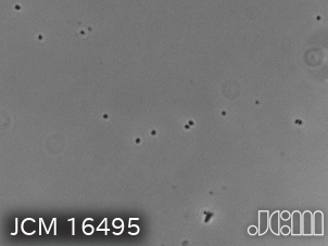
JCM 16495 - JCM Catalogue
Halobacteriales4
- In taxonomy, the Halobacteriaceae are a family of the Halobacteriales in the domain Archaea. (wikipedia.org)
- proposal to divide the order Halobacteriales into the families Halobacteriaceae , Haloarculaceae fam. (microbiologyresearch.org)
- A phylogenomic reappraisal of family-level divisions within the class Halobacteria: proposal to divide the order Halobacteriales into the families Halobacteriaceae, Haloarculaceae fam. (mcmaster.ca)
- nov. and a division of the order Halobacteriales into three families, an emended family Halobacteriaceae, Haloarculaceae fam. (mcmaster.ca)
Archaea3
- Halobacteriaceae represent a large part of halophilic Archaea, along with members in two other methanogenic families, Methanosarcinaceae and Methanocalculaceae. (wikipedia.org)
- Techniques such as 16S rRNA analysis and DNA-DNA hybridization have been major contributors to taxonomic classification in Halobacteriaceae, partly due to the difficulty in culturing halophilic Archaea. (wikipedia.org)
- Systematic monitoring since 1980 ( Oren, 1999 ) has shown that microbial blooms consisting of the unicellular green alga Dunaliella and extremely halophilic Archaea of the Halobacteriaceae family develop only after significant dilution of the upper meters of the water column after exceptionally rainy winters. (nature.com)
Taxonomy1
- International committee on systematics of Prokaryotes subcommittee on the taxonomy of halobacteriaceae and subcommittee on the taxonomy of Halomonadaceae. (huji.ac.il)
Species2
- Most species of Halobacteriaceae are best known for their high salt tolerance and red-pink pigmented members (due to bacterioruberin carotenoids), but there are also non-pigmented species and those that require moderate salt conditions. (wikipedia.org)
- Some species of Halobacteriaceae have been shown to exhibit phosphorus solubilizing activities that contribute to phosphorus cycling in hypersaline environments. (wikipedia.org)
Family3
- nov., a new member of the family Halobacteriaceae . (riken.jp)
- This minireview gives an updated and consolidated summary of taxonomic classification correlated with membrane phospholipid, glycolipid, and core lipid structural diversity within the family Halobacteriaceae. (go.jp)
- Halobacteriaceae family made up 80% of the clone library of the surface 2 mm, and consisted primarily of sequences affiliated with the haloalkaliphilic Natronomonas pharaonis. (edu.au)