Corneal Transplantation
Eye
Keratoplasty, Penetrating
Tissue Preservation
Descemet Stripping Endothelial Keratoplasty
Tissue Donors
Cornea
Fuchs' Endothelial Dystrophy
Organ Preservation
Corneal Stroma
Macula Lutea
Organ Culture Techniques
Cell Count
Eye Injuries
Dry Eye Syndromes
Eye Enucleation
Arvicolinae
Visual Acuity
Ocular Physiological Phenomena
Compound Eye, Arthropod
Eye Protective Devices
Retina
Biological Specimen Banks
Ophthalmic Solutions
Fixation, Ocular
Posterior Eye Segment
Glaucoma
Vitreous Body
National Practitioner Data Bank
Sclera
Eye Infections
Axial Length, Eye
Netherlands Antilles
Anterior Chamber
Myopia
Molecular Sequence Data
Aqueous Humor
Iris
Lens, Crystalline
Visual Fields
Eye Infections, Bacterial
Cataract
Ciliary Body
Pursuit, Smooth
Sequence Analysis, DNA
Eye Infections, Viral
Refractive Errors
Photic Stimulation
Vision Disorders
Databases, Protein
Choroid
Tomography, Optical Coherence
Ophthalmology
Diagnostic Techniques, Ophthalmological
Fluorescein Angiography
Conjunctiva
Oculomotor Muscles
Vision, Ocular
Retinal Detachment
Fundus Oculi
Photoreceptor Cells, Invertebrate
Eye Infections, Fungal
Eye Infections, Parasitic
Eye Pain
Eye Evisceration
Sensory Deprivation
Vitrectomy
Strabismus
Photography
Macaca mulatta
Retinal Diseases
Macular Degeneration
Blindness
Amblyopia
Optic Disk
Hyperopia
Ocular Hypertension
Corneal Topography
Tonometry, Ocular
Glaucoma, Open-Angle
Base Sequence
Optic Nerve
Optic Nerve Diseases
Head Movements
Visual Perception
Reflex, Vestibulo-Ocular
United Nations
Eyeglasses
Lens Implantation, Intraocular
Fovea Centralis
Amino Acid Sequence
Reproducibility of Results
Databases, Factual
Blood Donors
Visual Field Tests
Uveitis
Internet
Retinal Ganglion Cells
Astigmatism
Administration, Topical
Eye Diseases, Hereditary
Phacoemulsification
Visual Pathways
Proteins
Ocular Motility Disorders
Corneal Opacity
Fluorophotometry
Optometry
Endophthalmitis
Software
Epithelium, Corneal
Rabbits
Uvea
Macaca fascicularis
Diabetic Retinopathy
Conjunctivitis
Aphakia
Nystagmus, Optokinetic
Glaucoma, Angle-Closure
Dominance, Ocular
Sleep, REM
Vitreous Detachment
Algorithms
Retrospective Studies
Nystagmus, Pathologic
Contig Mapping
Nerve Fibers
Biometry
Vision Disparity
Retinal Perforations
Drosophila Proteins
Keratoconus
Visual Cortex
Blinking
Psychomotor Performance
Lasers, Excimer
Cloning, Molecular
Sequence Alignment
User-Computer Interface
Fluorescein
Blood Preservation
Oculomotor Nerve
Computational Biology
Rotation
Choroidal Neovascularization
Retinoscopy
Keratomileusis, Laser In Situ
Genome
Contrast Sensitivity
Macular Edema
Gonioscopy
Head
Fluorescent Antibody Technique, Indirect
Vision Screening
A new surgical technique for deep stromal, anterior lamellar keratoplasty. (1/81)
AIMS: To describe a new surgical technique for deep stromal anterior lamellar keratoplasty. METHODS: In eye bank eyes and sighted human eyes, aqueous was exchanged by air, to visualise the posterior corneal surface--that is, the "air to endothelium" interface. Through a 5.0 mm scleral incision, a deep stromal pocket was created across the cornea, using the air to endothelium interface as a reference plane for dissection depth. The pocket was filled with viscoelastic, and an anterior corneal lamella was excised. A full thickness donor button was sutured into the recipient bed after stripping its Descemet's membrane. RESULTS: In 25 consecutive human eye bank eyes, a 12% microperforation rate was found. Corneal dissection depth averaged 95.4% (SD 2.7%). Six patient eyes had uneventful surgeries; in a seventh eye, perforation of the lamellar bed occurred. All transplants cleared. Central pachymetry ranged from 0.62 to 0.73 mm. CONCLUSION: With this technique a deep stromal anterior lamellar keratoplasty can be performed with the donor to recipient interface just anterior to the posterior corneal surface. The technique has the advantage that the dissection can be completed in the event of inadvertent microperforation, or that the procedure can be aborted to perform a planned penetrating keratoplasty. (+info)Evaluation of potential organ culture media for eye banking using human donor corneas. (2/81)
AIM: To evaluate the ability of different commercially available cell culture solutions to preserve human donor corneas during 3 weeks of "closed system" organ culture at physiological temperature. This screening was performed in an attempt to establish a rational basis for the development of a serum-free organ culture medium for eye banking. METHODS: 72 normal human donor corneas were organ cultured for 21 days at 31 degrees C in eight different test media (nine corneas in each group). The basic culture solutions included: minimal essential medium (MEM), MEM with stabilised L-glutamine, M199, DIF-1000, SFM, F99, and F99 with ascorbic acid, insulin, bFGF, transferrin, selenium, and lipids (termed F99-Sr). All media were supplemented with 2% fetal calf serum (FCS), except for MEM, which was also studied at 8% FCS. The evaluation parameters included: (1) the endothelial cell loss as evaluated using trypan blue staining; (2) the ability of keratocytes and endothelial cells to incorporate tritiated uridine into RNA as evaluated using autoradiography and digital image analysis; (3) the leakage of immunogenic keratan sulphate as assessed using ELISA; and (4) changes in storage medium pH, glucose, and lactate content. RESULTS: SFM induced the lowest endothelial cell loss of 14% (SD 2%) and the highest RNA synthesis rates of all test solutions supplemented with 2% FCS. Corneas stored in SFM also showed the least leakage of keratan sulphate and the highest glucose consumption and lactate production. In five media (MEM with 2% FCS, MEM with stabilised L-glutamine, M199, F99, and F99-Sr), comparable and intermediate potentials for organ culture were observed with endothelial cell loss of 16-19%. By contrast, 29% (4%) of the endothelium was lost after storage in DIF-1000. Interestingly, the use of 8% FCS (in MEM) had a marked protective effect on the endothelium, which showed the highest RNA synthetic activity combined with a cell loss of only 11% (4%), compared with 19% (6%) at 2% FCS (p<0.05). CONCLUSION: Among the present test solutions, SFM appears to be the most prominent candidate for a new corneal organ culture medium and should be further tested and possibly refined to effectively substitute serum addition. (+info)Automated tri-image analysis of stored corneal endothelium. (3/81)
BACKGROUND: Endothelial examination of organ culture stored corneas is usually done manually and on several mosaic zones. Some banks use an image analyser that takes account of only one zone. This method is restricted by image quality, and may be inaccurate if endothelial cell density (ECD) within the mosaic is not homogeneous. The authors have developed an analyser that has tools for automatic error detection and correction, and can measure ECD and perform morphometry on multiple zones of three images of the endothelial mosaic. METHODS: 60 human corneas were divided into two equal groups: group 1 with homogeneous mosaics, group 2 with heterogeneous ones. Three standard microscopy video images of the endothelium, graded by quality, were analysed either in isolation (so called mono-image analysis) or simultaneously (so called tri-image analysis), with 50 or 300 endothelial cells (ECs) counted. The automated analysis was compared with the manual analysis, which concerned 10 non-adjacent zones and about 300 cells. For each analysis method, failures and durations were studied according to image quality. RESULTS: All corneas were able to undergo analysis, in about 2 or 7.5 minutes for 50 and 300 ECs respectively. The tri-image analysis did not increase analysis time and never failed, even with mediocre images. The tri-image analysis of 300 ECs was always most highly correlated with the manual count, particularly in the heterogeneous cornea group (r=0.94, p<0.001) and prevented serious count errors. CONCLUSIONS: This analyser allows reliable and rapid analysis of ECD, even for heterogeneous endothelia mosaics and mediocre images. (+info)Enhancing eye donation rates. Training students to be motivators. (4/81)
PURPOSE: Medical professionals could enhance eye donation rates by reminding relatives during grief counseling at the time of patient's death. This study was designed to assess the knowledge and attitudes of final year medical students (future doctors) towards eye donation, prior to instruction in eye banking. METHODS: The responses of 49 final-year medical students to a questionnaire on eye donation were compared with 24 non-medical students (controls). The results were analysed statistically using the chi-square test. RESULTS: More than one-third of students and controls were unaware that eyes are removed within six hours of death. Eight (16.3%) students and 6 (25.0%) controls felt that a close relative's eyes could be donated after death only if he had indicated willingness (P = 0.05). Three (6.1%) students and 3 (12.5%) controls were undecided about donating their own eyes. Nineteen (38.8%) students and 6 (25%) controls did not know where to go in order to pledge/donate eyes. The controls had poorer knowledge of ocular and systemic contraindications, and they did not know that storage could be prolonged (P < 0.001). Only 27 (55.1%) students had knowledge of corneal storage. CONCLUSIONS: Controls were poorly informed about various aspects of eye donation suggesting inadequate dissemination of information by the media. Students and controls alike had misconceptions regarding donation of relatives' eyes and hesitation regarding their own. These aspects should be emphasized during undergraduate teaching to dispel misgivings regarding wastage of donor eyes and to encourage future doctors to promote eye donation. (+info)Sensitivity and rapidity of blood culture bottles in the detection of cornea organ culture media contamination by bacteria and fungi. (5/81)
AIMS: To test the bactericidal activity of standard organ culture medium, and to compare the sensitivity and rapidity of blood culture bottles with conventional microbiological methods for detection of bacteria and fungi inoculated in a standard cornea organ culture medium. METHODS: The bactericidal activity of contaminated standard organ culture medium containing 100 IU/ml penicillin, 0.1 mg/ml streptomycin, and 0.25 micro g/ml amphotericin B was evaluated after 48 hours of incubation at 31 degrees C with five inocula of 14 bacteria. Two yeasts (Candida spp) and one Aspergillus were also tested. Contaminated media were then inoculated in three blood bottles (aerobic, anaerobic, fungal) placed in a Bactec 9240 automat; three conventional microbiological broths were the control. Changes in colour of organ culture medium and growth on conventional broth were screened daily by visual inspection. The sensitivity and rapidity of detection of contamination were compared between the three methods: blood bottle, conventional, and visual. RESULTS: Organ culture medium eradicated five bacteria irrespective of the starting inoculums: Streptococcus pneumoniae, Branhamella catarrhalis, Escherichia coli, Propionibacterium acnes, and Haemophilus influenzae. For micro-organisms where the medium was ineffective or bactericidal only (methicillin resistant Staphylococcus aureus, methicillin sensitive Staphylococcus aureus, Staphylococcus epidermidis, Staphylococcus haemolyticus, Pseudomonas aeruginosa, Acinetobacter baumannii, Bacillus subtilis, Klebsiella pneumoniae, Enterococcus faecalis, Candida albicans, Candida kruzei, Aspergillus fumigatus), the blood bottle, conventional, and visual methods detected microbial growth in 100%, 76.5%, and 70% of cases respectively. Mean detection time using blood bottles was 15.1 hours (SD 13.8, range 2-52). In cases of detection by the blood bottle method and the conventional method, the former was always faster: 95.5% against 65.2% detection within 24 hours (p=0.022) respectively. CONCLUSIONS: Blood bottles detect more efficiently and more rapidly a wider range of bacteria and fungi than the conventional microbiological method and the visual inspection of organ culture media. (+info)Awareness of eye donation in an adult population of southern India. A pilot study. (6/81)
PURPOSE: To determine "awareness of eye donation" and corneal transplantation in an adult population of southern India. METHODS: 507 participants chosen by systematic random sampling were interviewed using a structured questionnaire. Participants were selected among patients attending two community outreach programmes at different sites, and from patients presenting directly to the hospital. RESULTS: 257 participants (50.69%) were aware of eye donations. The major source of awareness was publicity campaigns (n=105). Only 22 (4.34%) participants were aware that eye donation had to be done within 6 hours of death. Four hundred and three (79.50%) participants were not aware of corneal transplantation. Illiteracy and rural residence were more likely predictors of ignorance. CONCLUSION: Although multiple strategies are currently followed to increase awareness of eye donations and corneal transplants, more innovative strategies have to be developed, especially to target illiterate and rural populations. (+info)Stability of RNA from the retina and retinal pigment epithelium in a porcine model simulating human eye bank conditions. (7/81)
PURPOSE: To assess RNA stability after death in a porcine model to simulate current human eye bank techniques. METHODS: Eye bank time interval data were collected from 191 donor specimens: death to refrigeration, enucleation, and tissue processing. A control porcine eye was enucleated, retina and RPE isolated, and specimens frozen (-80 degrees C). Fourteen porcine eyes remained at room temperature for 2 hours and then cooled to 4 degrees C. Retina and RPE were isolated and frozen (-80 degrees C) at 5, 12, 24, 29, 36, 48, and 72 hours. Four globes remained in a moist chamber, five whole and five sectioned globes were immersed in RNAlater (Ambion, Austin, TX) at 5, 12, 24, or 48 hours. RNA was isolated. The 28S and 18S rRNA peaks were analyzed by electrophoresis. RT-PCR was performed on each sample. Messenger RNA for GAPDH, beta-actin, mouse rhodopsin from retina (mRHO), and RPE-65 (from RPE) were analyzed with gel electrophoresis. RESULTS: The average time from death to refrigeration was 4.2 hours, to enucleation 6.4 hours, and to tissue processing 10.7 hours. RT-PCR gel electrophoresis patterns from retinal tissue had bands of similar intensity at each interval from beta-actin, GAPDH, and RHO. Band patterns from RPE demonstrated decay of the RT-PCR gene products after 5 hours. This decay was delayed by at least 24 hours with the use of RNAlater. The 28S rRNA decay was similar for retina and RPE. CONCLUSIONS: Retinal tissue RNA can be analyzed within the time constraints of current eye bank tissue processing, whereas analysis of RPE necessitates either rapid processing or use of RNAlater. These results should aid in future studies in which eye bank tissue is used for RNA analysis. (+info)Is manual counting of corneal endothelial cell density in eye banks still acceptable? The French experience. (8/81)
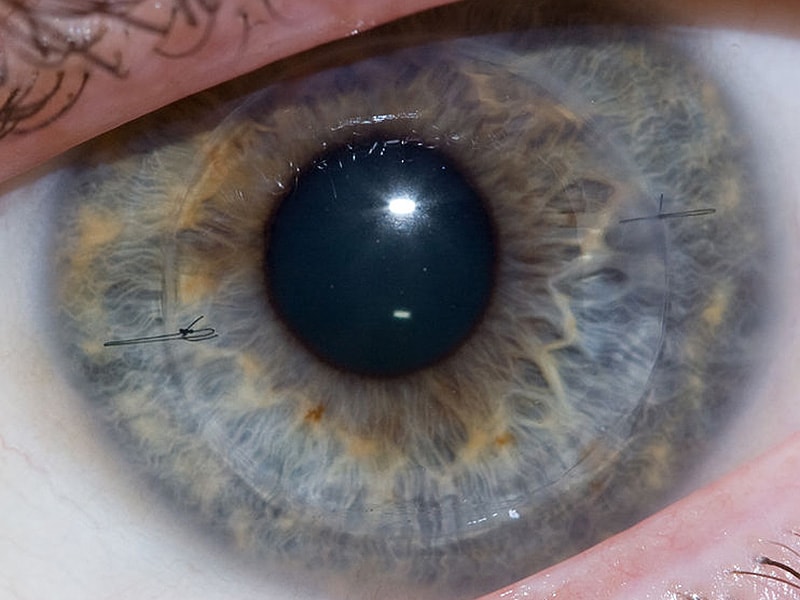
AIM: To examine the differences in manual endothelial cell counting methods in French eye banks and to analyse whether these differences could explain some substantial discrepancies observed in endothelial cell density (ECD) for corneas made available for transplant. METHODS: A questionnaire was sent to the 22 eye banks asking for details of the technical features of the light microscopes used, the microscope calibration, strategy for cell counting, the technical staff, and the method of presenting endothelial data. RESULTS: All eye banks responded and 91% (20/22) used only manual counting methods, in real time, directly through a microscope, and 62 different technicians, with varying experience, were involved in such counting. Counting of cells within the borders of a grid that were in contact with two adjacent borders was the most common method (17/22, 77%). Of the eight banks (8/22, 36%) that did not calibrate their microscopes, six reported the highest ECD values. Of the 14 others (64%), six applied a "magnification correcting factor" to the initial cell counts. In five of these cases, the corrected ECD was lower than estimated on initial count. Most of the banks (12/22, 55%) counted 100 cells or less in one to six non-adjacent zones of the mosaic. 14 of the banks (14/22, 64%) also graded cell polymegethism while seven (7/22, 32%) also graded pleomorphism ("hexagonality"). CONCLUSIONS: Lack of microscope calibration appears to be the leading cause of variance in ECD estimates in French eye banks. Other factors such as differences in counting strategy, the evaluation of smaller numbers of cells, and the different extent of experience of the technicians may also contribute to intraobserver and interobserver variability. Further comparative studies, including cross checking and the outcome of repeated counts from manual methods, are clearly needed with cross calibration to a computer based image archiving and analysis system. (+info)Corneal transplantation, also known as keratoplasty, is a surgical procedure in which all or part of a damaged or diseased cornea is replaced with healthy corneal tissue from a deceased donor. The cornea is the clear, dome-shaped surface at the front of the eye that plays an important role in focusing vision. When it becomes cloudy or misshapen due to injury, infection, or inherited conditions, vision can become significantly impaired.
During the procedure, the surgeon carefully removes a circular section of the damaged cornea and replaces it with a similarly sized piece of donor tissue. The new cornea is then stitched into place using very fine sutures that are typically removed several months after surgery.
Corneal transplantation has a high success rate, with more than 90% of procedures resulting in improved vision. However, as with any surgical procedure, there are risks involved, including infection, rejection of the donor tissue, and bleeding. Regular follow-up care is essential to monitor for any signs of complications and ensure proper healing.
The eye is the organ of sight, primarily responsible for detecting and focusing on visual stimuli. It is a complex structure composed of various parts that work together to enable vision. Here are some of the main components of the eye:
1. Cornea: The clear front part of the eye that refracts light entering the eye and protects the eye from harmful particles and microorganisms.
2. Iris: The colored part of the eye that controls the amount of light reaching the retina by adjusting the size of the pupil.
3. Pupil: The opening in the center of the iris that allows light to enter the eye.
4. Lens: A biconvex structure located behind the iris that further refracts light and focuses it onto the retina.
5. Retina: A layer of light-sensitive cells (rods and cones) at the back of the eye that convert light into electrical signals, which are then transmitted to the brain via the optic nerve.
6. Optic Nerve: The nerve that carries visual information from the retina to the brain.
7. Vitreous: A clear, gel-like substance that fills the space between the lens and the retina, providing structural support to the eye.
8. Conjunctiva: A thin, transparent membrane that covers the front of the eye and the inner surface of the eyelids.
9. Extraocular Muscles: Six muscles that control the movement of the eye, allowing for proper alignment and focus.
The eye is a remarkable organ that allows us to perceive and interact with our surroundings. Various medical specialties, such as ophthalmology and optometry, are dedicated to the diagnosis, treatment, and management of various eye conditions and diseases.
The endothelium of the cornea is the thin, innermost layer of cells that lines the inner surface of the cornea, which is the clear, dome-shaped structure at the front of the eye. This single layer of specialized cells is essential for maintaining the transparency and proper hydration of the cornea, allowing light to pass through it and focus on the retina.
The endothelial cells are hexagonal in shape and have tight junctions between them, creating a semi-permeable barrier that controls the movement of water and solutes between the corneal stroma (the middle layer of the cornea) and the anterior chamber (the space between the cornea and the iris). The endothelial cells actively pump excess fluid out of the cornea, maintaining a delicate balance of hydration that is critical for corneal clarity.
Damage to or dysfunction of the corneal endothelium can result in corneal edema (swelling), cloudiness, and loss of vision. Factors contributing to endothelial damage include aging, eye trauma, intraocular surgery, and certain diseases such as Fuchs' dystrophy and glaucoma.
An Eye Bank is an organization that collects, stores, and distributes donated human eyes for corneal transplantation and other ocular medical research purposes. The eye bank's primary function is to ensure the quality of the donated tissue and make it available for those in need of sight-restoring procedures.
The cornea, the clear front part of the eye, can be surgically transplanted from a deceased donor to a recipient with corneal damage or disease, thereby improving or restoring their vision. The eye bank's role includes obtaining consent for donation, retrieving the eyes from the donor, evaluating the tissue for suitability, preserving it properly, and then allocating it to surgeons for transplantation.
Eye banks follow strict medical guidelines and adhere to ethical standards to ensure the safety and quality of the donated tissues. The process involves screening potential donors for infectious diseases and other conditions that may affect the quality or safety of the cornea. Once deemed suitable, the corneas are carefully removed, preserved in specific solutions, and stored until they are needed for transplantation.
In addition to corneal transplants, eye banks also support research and education in ophthalmology by providing human eye tissues for various studies aimed at advancing our understanding of eye diseases and developing new treatments.
Penetrating keratoplasty (PK) is a type of corneal transplant surgery where the entire thickness of the host's damaged or diseased cornea is removed and replaced with a similar full-thickness portion of a healthy donor's cornea. The procedure aims to restore visual function, alleviate pain, and improve the structural integrity of the eye. It is typically performed for conditions such as severe keratoconus, corneal scarring, or corneal ulcers that cannot be treated with other, less invasive methods. Following the surgery, patients may require extended recovery time and rigorous postoperative care to minimize the risk of complications and ensure optimal visual outcomes.
Tissue preservation is the process of preventing decomposition or autolysis (self-digestion) of tissues after they have been removed from a living organism. This is typically achieved through the use of fixatives, such as formaldehyde or glutaraldehyde, which stabilize proteins and other cellular structures by creating cross-links between them. Other methods of tissue preservation include freezing, dehydration, and embedding in paraffin or plastic resins. Properly preserved tissues can be stored for long periods of time and used for various research and diagnostic purposes, such as histology, immunohistochemistry, and molecular biology studies.
Descemet Stripping Endothelial Keratoplasty (DSEK) is a type of corneal transplant surgery that involves replacing the damaged endothelium (inner layer) of the cornea with healthy endothelial cells from a donor. In this procedure, the surgeon removes the patient's Descemet's membrane (a thin, clear tissue beneath the endothelium) along with the damaged endothelium. Then, a thin disc of donor tissue, which includes both the endothelium and a small portion of the adjacent corneal stroma, is inserted into the eye and positioned using an air bubble. The new endothelial cells help to pump excess fluid out of the cornea, allowing it to become clear again. DSEK typically results in faster visual recovery and lower rejection rates compared to traditional full-thickness corneal transplantation.
A Tissue Bank is a specialized facility that collects, stores, and distributes human tissues for medical research, transplantation, or therapeutic purposes. These tissues can include organs, bones, skin, heart valves, tendons, and other bodily tissues that can be used for various medical applications.
Tissue banks follow strict regulations and guidelines to ensure the safety and quality of the tissues they handle. They implement rigorous screening and testing procedures to minimize the risk of disease transmission and maintain the integrity of the tissues. The tissues are stored under specific conditions, such as temperature and humidity, to preserve their function and viability until they are needed for use.
Tissue banks play a critical role in advancing medical research and improving patient outcomes by providing researchers and clinicians with access to high-quality human tissues for study and transplantation.
A tissue donor is an individual who has agreed to allow organs and tissues to be removed from their body after death for the purpose of transplantation to restore the health or save the life of another person. The tissues that can be donated include corneas, heart valves, skin, bone, tendons, ligaments, veins, and cartilage. These tissues can enhance the quality of life for many recipients and are often used in reconstructive surgeries. It is important to note that tissue donation does not interfere with an open casket funeral or other cultural or religious practices related to death and grieving.
The cornea is the clear, dome-shaped surface at the front of the eye. It plays a crucial role in focusing vision. The cornea protects the eye from harmful particles and microorganisms, and it also serves as a barrier against UV light. Its transparency allows light to pass through and get focused onto the retina. The cornea does not contain blood vessels, so it relies on tears and the fluid inside the eye (aqueous humor) for nutrition and oxygen. Any damage or disease that affects its clarity and shape can significantly impact vision and potentially lead to blindness if left untreated.
Fuchs' Endothelial Dystrophy is a medical condition that affects the eye's cornea. It is a slowly progressing disorder that causes the endothelium, a thin layer of cells lining the inner surface of the cornea, to deteriorate and eventually fail to function properly. This results in swelling of the cornea, leading to cloudy vision, distorted vision, and sensitivity to light.
The condition is typically inherited and tends to affect both eyes. It is more common in women than in men and usually becomes apparent after the age of 50. There is no cure for Fuchs' Endothelial Dystrophy, but treatments such as corneal transplantation can help improve vision and alleviate symptoms.
Organ preservation is a medical technique used to maintain the viability and functionality of an organ outside the body for a certain period, typically for transplantation purposes. This process involves cooling the organ to slow down its metabolic activity and prevent tissue damage, while using specialized solutions that help preserve the organ's structure and function. Commonly preserved organs include hearts, livers, kidneys, lungs, and pancreases. The goal of organ preservation is to ensure that the transplanted organ remains in optimal condition until it can be successfully implanted into a recipient.
The corneal stroma, also known as the substantia propria, is the thickest layer of the cornea, which is the clear, dome-shaped surface at the front of the eye. The cornea plays a crucial role in focusing vision.
The corneal stroma makes up about 90% of the cornea's thickness and is composed of parallel bundles of collagen fibers that are arranged in regular, repeating patterns. These fibers give the cornea its strength and transparency. The corneal stroma also contains a small number of cells called keratocytes, which produce and maintain the collagen fibers.
Disorders that affect the corneal stroma can cause vision loss or other eye problems. For example, conditions such as keratoconus, in which the cornea becomes thin and bulges outward, can distort vision and make it difficult to see clearly. Other conditions, such as corneal scarring or infection, can also affect the corneal stroma and lead to vision loss or other eye problems.
The macula lutea, often simply referred to as the macula or fovea centralis, is a part of the eye that is responsible for central vision and color perception. It's located in the center of the retina, the light-sensitive tissue at the back of the eye. The macula contains a high concentration of pigments called xanthophylls, which give it a yellowish color and protect the photoreceptor cells in this area from damage by blue light.
The central part of the macula is called the fovea, which is a small depression that contains only cones, the photoreceptor cells responsible for color vision and high visual acuity. The fovea is surrounded by the parafovea and the perifovea, which contain both cones and rods, the photoreceptor cells responsible for low-light vision and peripheral vision.
Damage to the macula can result in a loss of central vision and color perception, a condition known as age-related macular degeneration (AMD), which is a leading cause of blindness in older adults. Other conditions that can affect the macula include macular edema, macular holes, and macular pucker.
Corneal diseases are a group of disorders that affect the cornea, which is the clear, dome-shaped surface at the front of the eye. The cornea plays an important role in focusing vision, and any damage or disease can cause significant visual impairment or loss. Some common types of corneal diseases include:
1. Keratoconus: A progressive disorder in which the cornea thins and bulges outward into a cone shape, causing distorted vision.
2. Fuchs' dystrophy: A genetic disorder that affects the inner layer of the cornea called the endothelium, leading to swelling, cloudiness, and decreased vision.
3. Dry eye syndrome: A condition in which the eyes do not produce enough tears or the tears evaporate too quickly, causing discomfort, redness, and blurred vision.
4. Corneal ulcers: Open sores on the cornea that can be caused by infection, trauma, or other factors.
5. Herpes simplex keratitis: A viral infection of the cornea that can cause recurrent episodes of inflammation, scarring, and vision loss.
6. Corneal dystrophies: Inherited disorders that affect the structure and clarity of the cornea, leading to visual impairment or blindness.
7. Bullous keratopathy: A condition in which the endothelium fails to pump fluid out of the cornea, causing it to swell and form blisters.
8. Corneal trauma: Injury to the cornea caused by foreign objects, chemicals, or other factors that can lead to scarring, infection, and vision loss.
Treatment for corneal diseases varies depending on the specific condition and severity of the disease. Options may include eyedrops, medications, laser surgery, corneal transplantation, or other treatments.
Organ culture techniques refer to the methods used to maintain or grow intact organs or pieces of organs under controlled conditions in vitro, while preserving their structural and functional characteristics. These techniques are widely used in biomedical research to study organ physiology, pathophysiology, drug development, and toxicity testing.
Organ culture can be performed using a variety of methods, including:
1. Static organ culture: In this method, the organs or tissue pieces are placed on a porous support in a culture dish and maintained in a nutrient-rich medium. The medium is replaced periodically to ensure adequate nutrition and removal of waste products.
2. Perfusion organ culture: This method involves perfusing the organ with nutrient-rich media, allowing for better distribution of nutrients and oxygen throughout the tissue. This technique is particularly useful for studying larger organs such as the liver or kidney.
3. Microfluidic organ culture: In this approach, microfluidic devices are used to create a controlled microenvironment for organ cultures. These devices allow for precise control over the flow of nutrients and waste products, as well as the application of mechanical forces.
Organ culture techniques can be used to study various aspects of organ function, including metabolism, secretion, and response to drugs or toxins. Additionally, these methods can be used to generate three-dimensional tissue models that better recapitulate the structure and function of intact organs compared to traditional two-dimensional cell cultures.
"Cell count" is a medical term that refers to the process of determining the number of cells present in a given volume or sample of fluid or tissue. This can be done through various laboratory methods, such as counting individual cells under a microscope using a specialized grid called a hemocytometer, or using automated cell counters that use light scattering and electrical impedance techniques to count and classify different types of cells.
Cell counts are used in a variety of medical contexts, including hematology (the study of blood and blood-forming tissues), microbiology (the study of microscopic organisms), and pathology (the study of diseases and their causes). For example, a complete blood count (CBC) is a routine laboratory test that includes a white blood cell (WBC) count, red blood cell (RBC) count, hemoglobin level, hematocrit value, and platelet count. Abnormal cell counts can indicate the presence of various medical conditions, such as infections, anemia, or leukemia.
Eye diseases are a range of conditions that affect the eye or visual system, causing damage to vision and, in some cases, leading to blindness. These diseases can be categorized into various types, including:
1. Refractive errors: These include myopia (nearsightedness), hyperopia (farsightedness), astigmatism, and presbyopia, which affect the way light is focused on the retina and can usually be corrected with glasses or contact lenses.
2. Cataracts: A clouding of the lens inside the eye that leads to blurry vision, glare, and decreased contrast sensitivity. Cataract surgery is the most common treatment for this condition.
3. Glaucoma: A group of diseases characterized by increased pressure in the eye, leading to damage to the optic nerve and potential blindness if left untreated. Treatment includes medications, laser therapy, or surgery.
4. Age-related macular degeneration (AMD): A progressive condition that affects the central part of the retina called the macula, causing blurry vision and, in advanced stages, loss of central vision. Treatment may include anti-VEGF injections, laser therapy, or nutritional supplements.
5. Diabetic retinopathy: A complication of diabetes that affects the blood vessels in the retina, leading to bleeding, leakage, and potential blindness if left untreated. Treatment includes laser therapy, anti-VEGF injections, or surgery.
6. Retinal detachment: A separation of the retina from its underlying tissue, which can lead to vision loss if not treated promptly with surgery.
7. Amblyopia (lazy eye): A condition where one eye does not develop normal vision, often due to a misalignment or refractive error in childhood. Treatment includes correcting the underlying problem and encouraging the use of the weaker eye through patching or other methods.
8. Strabismus (crossed eyes): A misalignment of the eyes that can lead to amblyopia if not treated promptly with surgery, glasses, or other methods.
9. Corneal diseases: Conditions that affect the transparent outer layer of the eye, such as keratoconus, Fuchs' dystrophy, and infectious keratitis, which can lead to vision loss if not treated promptly.
10. Uveitis: Inflammation of the middle layer of the eye, which can cause vision loss if not treated promptly with anti-inflammatory medications or surgery.
Eye movements, also known as ocular motility, refer to the voluntary or involuntary motion of the eyes that allows for visual exploration of our environment. There are several types of eye movements, including:
1. Saccades: rapid, ballistic movements that quickly shift the gaze from one point to another.
2. Pursuits: smooth, slow movements that allow the eyes to follow a moving object.
3. Vergences: coordinated movements of both eyes in opposite directions, usually in response to a three-dimensional stimulus.
4. Vestibulo-ocular reflex (VOR): automatic eye movements that help stabilize the gaze during head movement.
5. Optokinetic nystagmus (OKN): rhythmic eye movements that occur in response to large moving visual patterns, such as when looking out of a moving vehicle.
Abnormalities in eye movements can indicate neurological or ophthalmological disorders and are often assessed during clinical examinations.
A blood bank is a facility that collects, tests, stores, and distributes blood and blood components for transfusion purposes. It is a crucial part of the healthcare system, as it ensures a safe and adequate supply of blood products to meet the needs of patients undergoing various medical procedures or treatments. The term "blood bank" comes from the idea that collected blood is "stored" or "banked" until it is needed for transfusion.
The primary function of a blood bank is to ensure the safety and quality of the blood supply. This involves rigorous screening and testing of donated blood to detect any infectious diseases, such as HIV, hepatitis B and C, syphilis, and West Nile virus. Blood banks also perform compatibility tests between donor and recipient blood types to minimize the risk of transfusion reactions.
Blood banks offer various blood products, including whole blood, red blood cells, platelets, plasma, and cryoprecipitate. These products can be used to treat a wide range of medical conditions, such as anemia, bleeding disorders, cancer, and trauma. In addition, some blood banks may also provide specialized services, such as apheresis (a procedure that separates specific blood components) and therapeutic phlebotomy (the removal of excess blood).
Blood banks operate under strict regulations and guidelines to ensure the safety and quality of their products and services. These regulations are established by national and international organizations, such as the American Association of Blood Banks (AABB), the World Health Organization (WHO), and the U.S. Food and Drug Administration (FDA).
Eye injuries refer to any damage or trauma caused to the eye or its surrounding structures. These injuries can vary in severity and may include:
1. Corneal abrasions: A scratch or scrape on the clear surface of the eye (cornea).
2. Chemical burns: Occurs when chemicals come into contact with the eye, causing damage to the cornea and other structures.
3. Eyelid lacerations: Cuts or tears to the eyelid.
4. Subconjunctival hemorrhage: Bleeding under the conjunctiva, the clear membrane that covers the white part of the eye.
5. Hyphema: Accumulation of blood in the anterior chamber of the eye, which is the space between the cornea and iris.
6. Orbital fractures: Breaks in the bones surrounding the eye.
7. Retinal detachment: Separation of the retina from its underlying tissue, which can lead to vision loss if not treated promptly.
8. Traumatic uveitis: Inflammation of the uvea, the middle layer of the eye, caused by trauma.
9. Optic nerve damage: Damage to the optic nerve, which transmits visual information from the eye to the brain.
Eye injuries can result from a variety of causes, including accidents, sports-related injuries, violence, and chemical exposure. It is important to seek medical attention promptly for any suspected eye injury to prevent further damage and potential vision loss.
Dry eye syndrome, also known as keratoconjunctivitis sicca, is a condition characterized by insufficient lubrication and moisture of the eyes. This occurs when the tears produced by the eyes are not sufficient in quantity or quality to keep the eyes moist and comfortable. The medical definition of dry eye syndromes includes the following symptoms:
1. A gritty or sandy sensation in the eyes
2. Burning or stinging sensations
3. Redness and irritation
4. Blurred vision that improves with blinking
5. Light sensitivity
6. A feeling of something foreign in the eye
7. Stringy mucus in or around the eyes
8. Difficulty wearing contact lenses
9. Watery eyes, which may seem contradictory but can be a response to dryness
10. Eye fatigue and discomfort after prolonged screen time or reading
The causes of dry eye syndromes can include aging, hormonal changes, certain medical conditions (such as diabetes, rheumatoid arthritis, lupus, Sjogren's syndrome), medications (antihistamines, decongestants, antidepressants, birth control pills), environmental factors (dry air, wind, smoke, dust), and prolonged screen time or reading.
Treatment for dry eye syndromes depends on the severity of the condition and its underlying causes. It may include artificial tears, lifestyle changes, prescription medications, and in some cases, surgical procedures to improve tear production or drainage.
Eye abnormalities refer to any structural or functional anomalies that affect the eye or its surrounding tissues. These abnormalities can be present at birth (congenital) or acquired later in life due to various factors such as injury, disease, or aging. Some examples of eye abnormalities include:
1. Strabismus: Also known as crossed eyes, strabismus is a condition where the eyes are misaligned and point in different directions.
2. Nystagmus: This is an involuntary movement of the eyes that can be horizontal, vertical, or rotatory.
3. Cataracts: A cataract is a clouding of the lens inside the eye that can cause vision loss.
4. Glaucoma: This is a group of eye conditions that damage the optic nerve and can lead to vision loss.
5. Retinal disorders: These include conditions such as retinal detachment, macular degeneration, and diabetic retinopathy.
6. Corneal abnormalities: These include conditions such as keratoconus, corneal ulcers, and Fuchs' dystrophy.
7. Orbital abnormalities: These include conditions such as orbital tumors, thyroid eye disease, and Graves' ophthalmopathy.
8. Ptosis: This is a condition where the upper eyelid droops over the eye.
9. Color blindness: A condition where a person has difficulty distinguishing between certain colors.
10. Microphthalmia: A condition where one or both eyes are abnormally small.
These are just a few examples of eye abnormalities, and there are many others that can affect the eye and its functioning. If you suspect that you have an eye abnormality, it is important to consult with an ophthalmologist for proper diagnosis and treatment.
Eye burns typically refer to injuries or damage to the eyes caused by exposure to harmful substances, extreme temperatures, or radiation. This can result in a variety of symptoms, including redness, pain, tearing, swelling, and blurred vision.
Chemical eye burns can occur when the eyes come into contact with strong acids, alkalis, or other irritants. These substances can cause damage to the cornea, conjunctiva, and other structures of the eye. The severity of the burn will depend on the type and concentration of the chemical, as well as the length of time it was in contact with the eye.
Thermal eye burns can result from exposure to hot or cold temperatures, such as steam, flames, or extreme cold. These types of burns can cause damage to the surface of the eye and may require medical attention to prevent further complications.
Radiation eye burns can occur after exposure to high levels of ultraviolet (UV) light, such as from welding torches, sun lamps, or tanning beds. Prolonged exposure to these sources can cause damage to the cornea and other structures of the eye, leading to symptoms like pain, redness, and sensitivity to light.
If you experience symptoms of an eye burn, it is important to seek medical attention as soon as possible. Treatment may include flushing the eyes with water or saline solution, administering medication to relieve pain and inflammation, or in severe cases, surgery to repair damaged tissue.
Eye enucleation is a surgical procedure that involves the removal of the entire eyeball, leaving the eye muscles, eyelids, and orbital structures intact. This procedure is typically performed to treat severe eye conditions or injuries, such as uncontrollable pain, blindness, cancer, or trauma. After the eyeball is removed, an implant may be placed in the socket to help maintain its shape and appearance. The optic nerve and other surrounding tissues are cut during the enucleation procedure, which means that vision cannot be restored in the affected eye. However, the remaining eye structures can still function normally, allowing for regular blinking, tear production, and eyelid movement.
Eye color is a characteristic determined by variations in a person's genes. The color of the eyes depends on the amount and type of pigment called melanin found in the eye's iris.
There are three main types of eye colors: brown, blue, and green. Brown eyes have the most melanin, while blue eyes have the least. Green eyes have a moderate amount of melanin combined with a golden tint that reflects light to give them their unique color.
Eye color is a polygenic trait, which means it is influenced by multiple genes. The two main genes responsible for eye color are OCA2 and HERC2, both located on chromosome 15. These genes control the production, transport, and storage of melanin in the iris.
It's important to note that eye color can change during infancy and early childhood due to the development of melanin in the iris. Additionally, some medications or medical conditions may also cause changes in eye color over time.
A sperm bank is a facility that collects, stores, and distributes semen from donors for the purpose of artificial insemination. The sperm samples are typically collected through masturbation and then frozen in liquid nitrogen to preserve them for long-term storage. Potential donors undergo rigorous screening processes, including medical examinations, genetic testing, and background checks, to ensure that their sperm is healthy and free from infectious diseases.
Sperm banks may be used by individuals or couples who are unable to conceive naturally due to male infertility, same-sex female couples, single women, or those with genetic disorders who wish to avoid passing on certain genetic conditions to their offspring. Recipients can choose a donor based on various factors such as physical characteristics, ethnicity, education level, and personality traits.
It is important to note that the regulations governing sperm banks vary by country and even by state or province within countries. Therefore, it is essential to research and understand the specific laws and guidelines that apply in your location before using a sperm bank.
Arvicolinae is a subfamily of rodents that includes voles, lemmings, and muskrats. These small mammals are characterized by their short legs, rounded bodies, and short tails. They are primarily found in the northern hemisphere, with the majority of species living in North America and Eurasia.
Arvicolines are known for their high reproductive rate and ability to survive in a variety of habitats, including grasslands, forests, tundra, and wetlands. They have a unique set of teeth called hypsodont teeth, which continue to grow throughout their lives. This adaptation allows them to wear down their teeth as they gnaw on tough plant material.
Many arvicoline species are important prey animals for larger predators, such as hawks, owls, and foxes. Some species, like the muskrat, are also hunted by humans for their fur or meat. In recent years, some arvicoline populations have experienced dramatic fluctuations in size due to changes in their habitats and food supplies, leading to concerns about their conservation status.
A milk bank, also known as a human milk bank or breastmilk bank, is a service that collects, screens, pasteurizes, and stores donated human breast milk. The milk is then distributed to hospitals, outpatient facilities, or directly to individuals in need, such as premature infants or those with medical conditions that prevent them from receiving their own mother's milk. Milk banks follow strict protocols to ensure the safety and quality of the donated milk, including blood tests for disease screening and pasteurization to kill any potential viruses or bacteria. The goal of a milk bank is to provide a safe and reliable source of human breast milk to promote the health and well-being of vulnerable infants.
Visual acuity is a measure of the sharpness or clarity of vision. It is usually tested by reading an eye chart from a specific distance, such as 20 feet (6 meters). The standard eye chart used for this purpose is called the Snellen chart, which contains rows of letters that decrease in size as you read down the chart.
Visual acuity is typically expressed as a fraction, with the numerator representing the testing distance and the denominator indicating the smallest line of type that can be read clearly. For example, if a person can read the line on the eye chart that corresponds to a visual acuity of 20/20, it means they have normal vision at 20 feet. If their visual acuity is 20/40, it means they must be as close as 20 feet to see what someone with normal vision can see at 40 feet.
It's important to note that visual acuity is just one aspect of overall vision and does not necessarily reflect other important factors such as peripheral vision, depth perception, color vision, or contrast sensitivity.
Eye neoplasms, also known as ocular tumors or eye cancer, refer to abnormal growths of tissue in the eye. These growths can be benign (non-cancerous) or malignant (cancerous). Eye neoplasms can develop in various parts of the eye, including the eyelid, conjunctiva, cornea, iris, ciliary body, choroid, retina, and optic nerve.
Benign eye neoplasms are typically slow-growing and do not spread to other parts of the body. They may cause symptoms such as vision changes, eye pain, or a noticeable mass in the eye. Treatment options for benign eye neoplasms include monitoring, surgical removal, or radiation therapy.
Malignant eye neoplasms, on the other hand, can grow and spread rapidly to other parts of the body. They may cause symptoms such as vision changes, eye pain, floaters, or flashes of light. Treatment options for malignant eye neoplasms depend on the type and stage of cancer but may include surgery, radiation therapy, chemotherapy, or a combination of these treatments.
It is important to note that early detection and treatment of eye neoplasms can improve outcomes and prevent complications. Regular eye exams with an ophthalmologist are recommended for early detection and prevention of eye diseases, including eye neoplasms.
"Ocular physiological phenomena" is not a standardized medical term with a specific definition. However, I can provide some examples of ocular physiological phenomena, which refer to various normal functions and processes that occur in the eye. Here are a few examples:
1. Accommodation: The ability of the eye to change optical power to maintain a clear image or focus on an object as its distance varies. This is primarily achieved by changing the curvature of the lens through the action of the ciliary muscles.
2. Pupillary reflex: The automatic adjustment of the pupil's size in response to changes in light intensity. In bright light, the pupil constricts (miosis), while in dim light, it dilates (mydriasis). This reflex helps regulate the amount of light that enters the eye.
3. Tear production: The continuous secretion of tears by the lacrimal glands to keep the eyes moist and protected from dust, microorganisms, and other foreign particles.
4. Extraocular muscle function: The coordinated movement of the six extraocular muscles that control eyeball rotation and enable various gaze directions.
5. Color vision: The ability to perceive and distinguish different colors based on the sensitivity of photoreceptor cells (cones) in the retina to specific wavelengths of light.
6. Dark adaptation: The process by which the eyes adjust to low-light conditions, improving visual sensitivity primarily through changes in the rod photoreceptors' sensitivity and pupil dilation.
7. Light adaptation: The ability of the eye to adjust to different levels of illumination, mainly through alterations in pupil size and photoreceptor cell response.
These are just a few examples of ocular physiological phenomena. There are many more processes and functions that occur within the eye, contributing to our visual perception and overall eye health.
A compound eye is a characteristic type of eye found in arthropods, including insects, crustaceans, and some extinct fossil groups. Each eye is composed of numerous individual photoreceptor units called ommatidia, which function together to provide a wide field of vision and excellent motion detection capabilities.
In an arthropod compound eye, each ommatidium contains a group of visual cells (called retinula cells) surrounding a central rhabdomere, which is the light-sensitive structure that converts light into electrical signals. The number of ommatidia in a compound eye can vary greatly between species and even within different regions of an individual's eye, ranging from just a few to tens of thousands.
Compound eyes offer several advantages for arthropods:
1. Wide Field of Vision: Compound eyes provide a panoramic view of the environment, allowing arthropods to detect predators, prey, or mates from various directions simultaneously.
2. Motion Detection: The apposition-type compound eye (one type of compound eye structure) is particularly adept at detecting motion due to the neural processing of signals between adjacent ommatidia. This allows arthropods to respond quickly to potential threats or opportunities.
3. Light Adaptation: Compound eyes can adapt to different light conditions, allowing arthropods to function effectively in both bright daylight and dimly lit environments. Some species have specialized regions within their compound eyes that are optimized for specific light conditions, such as the dorsal rim area in insects, which is sensitive to polarized skylight.
4. UV Sensitivity: Many arthropods can detect ultraviolet (UV) light due to the presence of photopigments within their ommatidia that absorb UV wavelengths. This ability allows them to perceive patterns and cues in their environment that are invisible to humans, such as floral guides in bees or mate-recognition signals in certain insects.
Despite their limitations in terms of resolution and image quality compared to vertebrate eyes, compound eyes have evolved to serve the unique needs and ecological roles of arthropods effectively.
Eye protective devices are specialized equipment designed to protect the eyes from various hazards and injuries. They include items such as safety glasses, goggles, face shields, welding helmets, and full-face respirators. These devices are engineered to provide a barrier between the eyes and potential dangers like chemical splashes, impact particles, radiation, and other environmental hazards.
Safety glasses are designed to protect against flying debris, dust, and other airborne particles. They typically have side shields to prevent objects from entering the eye from the sides. Goggles offer a higher level of protection than safety glasses as they form a protective seal around the eyes, preventing liquids and fine particles from reaching the eyes.
Face shields and welding helmets are used in industrial settings to protect against radiation, sparks, and molten metal during welding or cutting operations. Full-face respirators are used in environments with harmful airborne particles or gases, providing protection for both the eyes and the respiratory system.
It is essential to choose the appropriate eye protective device based on the specific hazard present to ensure adequate protection.
A bacterial genome is the complete set of genetic material, including both DNA and RNA, found within a single bacterium. It contains all the hereditary information necessary for the bacterium to grow, reproduce, and survive in its environment. The bacterial genome typically includes circular chromosomes, as well as plasmids, which are smaller, circular DNA molecules that can carry additional genes. These genes encode various functional elements such as enzymes, structural proteins, and regulatory sequences that determine the bacterium's characteristics and behavior.
Bacterial genomes vary widely in size, ranging from around 130 kilobases (kb) in Mycoplasma genitalium to over 14 megabases (Mb) in Sorangium cellulosum. The complete sequencing and analysis of bacterial genomes have provided valuable insights into the biology, evolution, and pathogenicity of bacteria, enabling researchers to better understand their roles in various diseases and potential applications in biotechnology.
The retina is the innermost, light-sensitive layer of tissue in the eye of many vertebrates and some cephalopods. It receives light that has been focused by the cornea and lens, converts it into neural signals, and sends these to the brain via the optic nerve. The retina contains several types of photoreceptor cells including rods (which handle vision in low light) and cones (which are active in bright light and are capable of color vision).
In medical terms, any pathological changes or diseases affecting the retinal structure and function can lead to visual impairment or blindness. Examples include age-related macular degeneration, diabetic retinopathy, retinal detachment, and retinitis pigmentosa among others.
A Biological Specimen Bank, also known as a biobank or tissue bank, is a type of medical facility that collects, stores, and distributes biological samples for research purposes. These samples can include tissues, cells, DNA, blood, and other bodily fluids, and are often collected during medical procedures or from donors who have given their informed consent. The samples are then cataloged and stored in specialized conditions to preserve their quality and integrity.
Biobanks play a critical role in advancing medical research by providing researchers with access to large numbers of well-characterized biological samples. This allows them to study the underlying causes of diseases, develop new diagnostic tests and treatments, and evaluate the safety and effectiveness of drugs and other therapies. Biobanks may be established for specific research projects or as part of larger, more comprehensive efforts to build biomedical research infrastructure.
It is important to note that the use of biological specimens in research is subject to strict ethical guidelines and regulations, which are designed to protect the privacy and interests of donors and ensure that the samples are used responsibly and for legitimate scientific purposes.
Intraocular pressure (IOP) is the fluid pressure within the eye, specifically within the anterior chamber, which is the space between the cornea and the iris. It is measured in millimeters of mercury (mmHg). The aqueous humor, a clear fluid that fills the anterior chamber, is constantly produced and drained, maintaining a balance that determines the IOP. Normal IOP ranges from 10-21 mmHg, with average values around 15-16 mmHg. Elevated IOP is a key risk factor for glaucoma, a group of eye conditions that can lead to optic nerve damage and vision loss if not treated promptly and effectively. Regular monitoring of IOP is essential in diagnosing and managing glaucoma and other ocular health issues.
Penetrating eye injuries are a type of ocular trauma where a foreign object or substance pierces the outer layers of the eye and damages the internal structures. This can result in serious harm to various parts of the eye, such as the cornea, iris, lens, or retina, and may potentially cause vision loss or blindness if not promptly treated.
The severity of a penetrating eye injury depends on several factors, including the type and size of the object that caused the injury, the location of the wound, and the extent of damage to the internal structures. Common causes of penetrating eye injuries include sharp objects, such as metal shards or glass fragments, projectiles, such as pellets or bullets, and explosive materials.
Symptoms of a penetrating eye injury may include pain, redness, sensitivity to light, blurred vision, floaters, or the presence of a foreign body in the eye. If you suspect that you have sustained a penetrating eye injury, it is essential to seek immediate medical attention from an ophthalmologist or other healthcare professional with experience in treating eye trauma.
Treatment for penetrating eye injuries may include removing any foreign objects or substances from the eye, repairing damaged tissues, and administering medications to prevent infection and reduce inflammation. In some cases, surgery may be necessary to repair the injury and restore vision. Preventing eye injuries is crucial, and appropriate protective eyewear should be worn when engaging in activities that pose a risk of eye trauma.
Ophthalmic solutions are sterile, single-use or multi-dose preparations in a liquid form that are intended for topical administration to the eye. These solutions can contain various types of medications, such as antibiotics, anti-inflammatory agents, antihistamines, or lubricants, which are used to treat or prevent ocular diseases and conditions.
The pH and osmolarity of ophthalmic solutions are carefully controlled to match the physiological environment of the eye and minimize any potential discomfort or irritation. The solutions may be packaged in various forms, including drops, sprays, or irrigations, depending on the intended use and administration route.
It is important to follow the instructions for use provided by a healthcare professional when administering ophthalmic solutions, as improper use can lead to eye injury or reduced effectiveness of the medication.
Foreign bodies in the eye refer to any object or particle that is not normally present in the eye and becomes lodged in it. These foreign bodies can range from small particles like sand or dust to larger objects such as metal shavings or glass. They can cause irritation, pain, redness, watering, and even vision loss if they are not removed promptly and properly.
The symptoms of an eye foreign body may include:
* A feeling that something is in the eye
* Pain or discomfort in the eye
* Redness or inflammation of the eye
* Watering or tearing of the eye
* Sensitivity to light
* Blurred vision or difficulty seeing
If you suspect that you have a foreign body in your eye, it is important to seek medical attention immediately. An eye care professional can examine your eye and determine the best course of treatment to remove the foreign body and prevent any further damage to your eye.
Eye movement measurements, also known as oculometry, refer to the measurement and analysis of eye movements. This can include assessing the direction, speed, range, and patterns of eye movement. These measurements are often used in research and clinical settings to understand various aspects of vision, perception, and cognition. They can be used to diagnose and monitor conditions that affect eye movement, such as strabismus (crossed eyes), amblyopia (lazy eye), or neurological disorders. Additionally, eye movement measurements are also used in areas such as human-computer interaction, marketing research, and virtual reality to understand how individuals interact with their environment.
Ocular fixation is a term used in ophthalmology and optometry to refer to the ability of the eyes to maintain steady gaze or visual focus on an object. It involves the coordinated movement of the extraocular muscles that control eye movements, allowing for clear and stable vision.
In medical terminology, fixation specifically refers to the state in which the eyes are aligned and focused on a single point in space. This is important for maintaining visual perception and preventing blurring or double vision. Ocular fixation can be affected by various factors such as muscle weakness, nerve damage, or visual processing disorders.
Assessment of ocular fixation is often used in eye examinations to evaluate visual acuity, eye alignment, and muscle function. Abnormalities in fixation may indicate the presence of underlying eye conditions or developmental delays that require further investigation and treatment.
The posterior segment of the eye refers to the back portion of the interior of the eye, including the vitreous, retina, choroid, and optic nerve. This region is responsible for processing visual information and transmitting it to the brain. The retina contains photoreceptor cells that convert light into electrical signals, which are then sent through the optic nerve to the brain for interpretation as images. Disorders of the posterior eye segment can lead to vision loss or blindness.
Glaucoma is a group of eye conditions that damage the optic nerve, often caused by an abnormally high pressure in the eye (intraocular pressure). This damage can lead to permanent vision loss or even blindness if left untreated. The most common type is open-angle glaucoma, which has no warning signs and progresses slowly. Angle-closure glaucoma, on the other hand, can cause sudden eye pain, redness, nausea, and vomiting, as well as rapid vision loss. Other less common types of glaucoma also exist. While there is no cure for glaucoma, early detection and treatment can help slow or prevent further vision loss.
The vitreous body, also known simply as the vitreous, is the clear, gel-like substance that fills the space between the lens and the retina in the eye. It is composed mainly of water, but also contains collagen fibers, hyaluronic acid, and other proteins. The vitreous helps to maintain the shape of the eye and provides a transparent medium for light to pass through to reach the retina. With age, the vitreous can become more liquefied and may eventually separate from the retina, leading to symptoms such as floaters or flashes of light.
The National Practitioner Data Bank (NPDB) is not a medical term per se, but rather a legislative creation established by the Health Care Quality Improvement Act of 1986 (HCQIA). It is a vast electronic repository that contains reports on medical malpractice payments, adverse actions related to licensure and clinical privileges, Medicare and Medicaid exclusions, and other negative clinical outcomes associated with healthcare practitioners.
The NPDB's primary objective is to improve the quality of healthcare by collecting and disseminating information that can assist healthcare entities in making informed decisions about healthcare practitioners' competence and conduct. The database is confidential and accessible only to specific authorized entities, including hospitals, medical boards, professional societies, and other healthcare organizations, for the purpose of conducting background checks, credentialing, and privileging determinations.
Healthcare practitioners themselves cannot access their own records in the NPDB directly but can request self-query reports to learn about any information reported about them. The NPDB is a crucial tool in maintaining patient safety and promoting transparency within the healthcare system.
The sclera is the tough, white, fibrous outer coating of the eye in humans and other vertebrates, covering about five sixths of the eyeball's surface. It provides protection for the delicate inner structures of the eye and maintains its shape. The sclera is composed mainly of collagen and elastic fiber, making it strong and resilient. Its name comes from the Greek word "skleros," which means hard.
Eye infections, also known as ocular infections, are conditions characterized by the invasion and multiplication of pathogenic microorganisms in any part of the eye or its surrounding structures. These infections can affect various parts of the eye, including the conjunctiva (conjunctivitis), cornea (keratitis), eyelid (blepharitis), or the internal structures of the eye (endophthalmitis, uveitis). The symptoms may include redness, pain, discharge, itching, blurred vision, and sensitivity to light. The cause can be bacterial, viral, fungal, or parasitic, and the treatment typically involves antibiotics, antivirals, or antifungals, depending on the underlying cause.
Axial length, in the context of the eye, refers to the measurement of the distance between the front and back portions of the eye, specifically from the cornea (the clear front "window" of the eye) to the retina (the light-sensitive tissue at the back of the eye). This measurement is typically expressed in millimeters (mm).
The axial length of the eye is an important factor in determining the overall refractive power of the eye and can play a role in the development of various eye conditions, such as myopia (nearsightedness) or hyperopia (farsightedness). Changes in axial length, particularly elongation, are often associated with an increased risk of developing myopia. Regular monitoring of axial length can help eye care professionals track changes in the eye and manage these conditions more effectively.
I'm sorry for any confusion, but "Netherlands Antilles" is not a medical term. It refers to a former political entity in the Caribbean that was composed of several islands including Curacao, Sint Maarten, Bonaire, Saba, and Sint Eustatius. These islands are now special municipalities of the Netherlands or part of the Kingdom of the Netherlands. I'm here to help with medical information, so if you have any health-related questions, feel free to ask!
The anterior chamber is the front portion of the eye, located between the cornea (the clear front "window" of the eye) and the iris (the colored part of the eye). It is filled with a clear fluid called aqueous humor that provides nutrients to the structures inside the eye and helps maintain its shape. The anterior chamber plays an important role in maintaining the overall health and function of the eye.
Myopia, also known as nearsightedness, is a common refractive error of the eye. It occurs when the eye is either too long or the cornea (the clear front part of the eye) is too curved. As a result, light rays focus in front of the retina instead of directly on it, causing distant objects to appear blurry while close objects remain clear.
Myopia typically develops during childhood and can progress gradually or rapidly until early adulthood. It can be corrected with glasses, contact lenses, or refractive surgery such as LASIK. Regular eye examinations are essential for people with myopia to monitor any changes in their prescription and ensure proper correction.
While myopia is generally not a serious condition, high levels of nearsightedness can increase the risk of certain eye diseases, including cataracts, glaucoma, retinal detachment, and myopic degeneration. Therefore, it's crucial to manage myopia effectively and maintain regular follow-ups with an eye care professional.
Molecular sequence data refers to the specific arrangement of molecules, most commonly nucleotides in DNA or RNA, or amino acids in proteins, that make up a biological macromolecule. This data is generated through laboratory techniques such as sequencing, and provides information about the exact order of the constituent molecules. This data is crucial in various fields of biology, including genetics, evolution, and molecular biology, allowing for comparisons between different organisms, identification of genetic variations, and studies of gene function and regulation.
Aqueous humor is a clear, watery fluid that fills the anterior and posterior chambers of the eye. It is produced by the ciliary processes in the posterior chamber and circulates through the pupil into the anterior chamber, where it provides nutrients to the cornea and lens, maintains intraocular pressure, and helps to shape the eye. The aqueous humor then drains out of the eye through the trabecular meshwork and into the canal of Schlemm, eventually reaching the venous system.
In medical terms, the iris refers to the colored portion of the eye that surrounds the pupil. It is a circular structure composed of thin, contractile muscle fibers (radial and circumferential) arranged in a regular pattern. These muscles are controlled by the autonomic nervous system and can adjust the size of the pupil in response to changes in light intensity or emotional arousal. By constricting or dilating the iris, the amount of light entering the eye can be regulated, which helps maintain optimal visual acuity under various lighting conditions.
The color of the iris is determined by the concentration and distribution of melanin pigments within the iris stroma. The iris also contains blood vessels, nerves, and connective tissue that support its structure and function. Anatomically, the iris is continuous with the ciliary body and the choroid, forming part of the uveal tract in the eye.
The crystalline lens is a biconvex transparent structure in the eye that helps to refract (bend) light rays and focus them onto the retina. It is located behind the iris and pupil and is suspended by small fibers called zonules that connect it to the ciliary body. The lens can change its shape to accommodate and focus on objects at different distances, a process known as accommodation. With age, the lens may become cloudy or opaque, leading to cataracts.
Visual fields refer to the total area in which objects can be seen while keeping the eyes focused on a central point. It is the entire area that can be observed using peripheral (side) vision while the eye gazes at a fixed point. A visual field test is used to detect blind spots or gaps (scotomas) in a person's vision, which could indicate various medical conditions such as glaucoma, retinal damage, optic nerve disease, brain tumors, or strokes. The test measures both the central and peripheral vision and maps the entire area that can be seen when focusing on a single point.
Bacterial eye infections, also known as bacterial conjunctivitis or bacterial keratitis, are caused by the invasion of bacteria into the eye. The most common types of bacteria that cause these infections include Staphylococcus aureus, Streptococcus pneumoniae, and Haemophilus influenzae.
Bacterial conjunctivitis is an inflammation of the conjunctiva, the thin membrane that covers the white part of the eye and the inner surface of the eyelids. Symptoms include redness, swelling, pain, discharge, and a gritty feeling in the eye. Bacterial keratitis is an infection of the cornea, the clear front part of the eye. Symptoms include severe pain, sensitivity to light, tearing, and decreased vision.
Bacterial eye infections are typically treated with antibiotic eye drops or ointments. It is important to seek medical attention promptly if you suspect a bacterial eye infection, as untreated infections can lead to serious complications such as corneal ulcers and vision loss. Preventive measures include good hygiene practices, such as washing your hands frequently and avoiding touching or rubbing your eyes.
A cataract is a clouding of the natural lens in the eye that affects vision. This clouding can cause vision to become blurry, faded, or dim, making it difficult to see clearly. Cataracts are a common age-related condition, but they can also be caused by injury, disease, or medication use. In most cases, cataracts develop gradually over time and can be treated with surgery to remove the cloudy lens and replace it with an artificial one.
The ciliary body is a part of the eye's internal structure that is located between the choroid and the iris. It is composed of muscle tissue and is responsible for adjusting the shape of the lens through a process called accommodation, which allows the eye to focus on objects at varying distances. Additionally, the ciliary body produces aqueous humor, the clear fluid that fills the anterior chamber of the eye and helps to nourish the eye's internal structures. The ciliary body is also responsible for maintaining the shape and position of the lens within the eye.
In medical terms, "tears" are a clear, salty liquid that is produced by the tear glands (lacrimal glands) in our eyes. They serve to keep the eyes moist, protect against dust and other foreign particles, and help to provide clear vision by maintaining a smooth surface on the front of the eye. Tears consist of water, oil, and mucus, which help to prevent evaporation and ensure that the tears spread evenly across the surface of the eye. Emotional or reflexive responses, such as crying or yawning, can also stimulate the production of tears.
In the context of medical terminology, I believe you may be referring to "pursuit" as it relates to neurological tests. A smooth pursuit is a type of eye movement in which the eyes smoothly and slowly follow a moving object. It requires coordination between the extraocular muscles, vestibular system, and visual system. If there are issues with any of these systems, smooth pursuit can be affected, leading to abnormalities such as jerky or saccadic movements.
Therefore, "smooth pursuit" is a medical term used to describe the normal, coordinated movement of the eyes that allows for the tracking of moving objects in a smooth and continuous manner.
DNA Sequence Analysis is the systematic determination of the order of nucleotides in a DNA molecule. It is a critical component of modern molecular biology, genetics, and genetic engineering. The process involves determining the exact order of the four nucleotide bases - adenine (A), guanine (G), cytosine (C), and thymine (T) - in a DNA molecule or fragment. This information is used in various applications such as identifying gene mutations, studying evolutionary relationships, developing molecular markers for breeding, and diagnosing genetic diseases.
The process of DNA Sequence Analysis typically involves several steps, including DNA extraction, PCR amplification (if necessary), purification, sequencing reaction, and electrophoresis. The resulting data is then analyzed using specialized software to determine the exact sequence of nucleotides.
In recent years, high-throughput DNA sequencing technologies have revolutionized the field of genomics, enabling the rapid and cost-effective sequencing of entire genomes. This has led to an explosion of genomic data and new insights into the genetic basis of many diseases and traits.
Binocular vision refers to the ability to use both eyes together to create a single, three-dimensional image of our surroundings. This is achieved through a process called binocular fusion, where the images from each eye are aligned and combined in the brain to form a unified perception.
The term "binocular vision" specifically refers to the way that our visual system integrates information from both eyes to create depth perception and enhance visual clarity. When we view an object with both eyes, they focus on the same point in space and send slightly different images to the brain due to their slightly different positions. The brain then combines these images to create a single, three-dimensional image that allows us to perceive depth and distance.
Binocular vision is important for many everyday activities, such as driving, reading, and playing sports. Disorders of binocular vision can lead to symptoms such as double vision, eye strain, and difficulty with depth perception.
Viral eye infections are caused by viruses that invade different parts of the eye, leading to inflammation and irritation. Some common types of viral eye infections include conjunctivitis (pink eye), keratitis, and dendritic ulcers. These infections can cause symptoms such as redness, watering, soreness, sensitivity to light, and discharge. In some cases, viral eye infections can also lead to complications like corneal scarring and vision loss if left untreated. They are often highly contagious and can spread through contact with contaminated surfaces or respiratory droplets. Antiviral medications may be used to treat certain types of viral eye infections, but in many cases, the infection will resolve on its own over time. Preventive measures such as good hygiene and avoiding touching the eyes can help reduce the risk of viral eye infections.
Refractive errors are a group of vision conditions that include nearsightedness (myopia), farsightedness (hyperopia), astigmatism, and presbyopia. These conditions occur when the shape of the eye prevents light from focusing directly on the retina, causing blurred or distorted vision.
Myopia is a condition where distant objects appear blurry while close-up objects are clear. This occurs when the eye is too long or the cornea is too curved, causing light to focus in front of the retina instead of directly on it.
Hyperopia, on the other hand, is a condition where close-up objects appear blurry while distant objects are clear. This happens when the eye is too short or the cornea is not curved enough, causing light to focus behind the retina.
Astigmatism is a condition that causes blurred vision at all distances due to an irregularly shaped cornea or lens.
Presbyopia is a natural aging process that affects everyone as they get older, usually around the age of 40. It causes difficulty focusing on close-up objects and can be corrected with reading glasses, bifocals, or progressive lenses.
Refractive errors can be diagnosed through a comprehensive eye exam and are typically corrected with eyeglasses, contact lenses, or refractive surgery such as LASIK.
Photic stimulation is a medical term that refers to the exposure of the eyes to light, specifically repetitive pulses of light, which is used as a method in various research and clinical settings. In neuroscience, it's often used in studies related to vision, circadian rhythms, and brain function.
In a clinical context, photic stimulation is sometimes used in the diagnosis of certain medical conditions such as seizure disorders (like epilepsy). By observing the response of the brain to this light stimulus, doctors can gain valuable insights into the functioning of the brain and the presence of any neurological disorders.
However, it's important to note that photic stimulation should be conducted under the supervision of a trained healthcare professional, as improper use can potentially trigger seizures in individuals who are susceptible to them.
Vision disorders refer to a wide range of conditions that affect the visual system and result in various symptoms, such as blurry vision, double vision, distorted vision, impaired depth perception, and difficulty with visual tracking or focusing. These disorders can be categorized into several types, including:
1. Refractive errors: These occur when the shape of the eye prevents light from focusing directly on the retina, resulting in blurry vision. Examples include myopia (nearsightedness), hyperopia (farsightedness), astigmatism, and presbyopia (age-related loss of near vision).
2. Strabismus: Also known as crossed eyes or walleye, strabismus is a misalignment of the eyes where they point in different directions, which can lead to double vision or loss of depth perception.
3. Amblyopia: Often called lazy eye, amblyopia is a condition where one eye has reduced vision due to lack of proper visual development during childhood. It may be caused by strabismus, refractive errors, or other factors that interfere with normal visual development.
4. Accommodative disorders: These involve problems with the focusing ability of the eyes, such as convergence insufficiency (difficulty focusing on close objects) and accommodative dysfunction (inability to maintain clear vision at different distances).
5. Binocular vision disorders: These affect how the eyes work together as a team, leading to issues like poor depth perception, eye strain, and headaches. Examples include convergence insufficiency, divergence excess, and suppression.
6. Ocular motility disorders: These involve problems with eye movement, such as nystagmus (involuntary eye movements), strabismus, or restricted extraocular muscle function.
7. Visual processing disorders: These affect the brain's ability to interpret and make sense of visual information, even when the eyes themselves are healthy. Symptoms may include difficulty with reading, recognizing shapes and objects, and understanding spatial relationships.
8. Low vision: This term refers to significant visual impairment that cannot be fully corrected with glasses, contact lenses, medication, or surgery. It includes conditions like macular degeneration, diabetic retinopathy, glaucoma, and cataracts.
9. Blindness: Complete loss of sight in both eyes, which can be caused by various factors such as injury, disease, or genetic conditions.
A protein database is a type of biological database that contains information about proteins and their structures, functions, sequences, and interactions with other molecules. These databases can include experimentally determined data, such as protein sequences derived from DNA sequencing or mass spectrometry, as well as predicted data based on computational methods.
Some examples of protein databases include:
1. UniProtKB: a comprehensive protein database that provides information about protein sequences, functions, and structures, as well as literature references and links to other resources.
2. PDB (Protein Data Bank): a database of three-dimensional protein structures determined by experimental methods such as X-ray crystallography and nuclear magnetic resonance (NMR) spectroscopy.
3. BLAST (Basic Local Alignment Search Tool): a web-based tool that allows users to compare a query protein sequence against a protein database to identify similar sequences and potential functional relationships.
4. InterPro: a database of protein families, domains, and functional sites that provides information about protein function based on sequence analysis and other data.
5. STRING (Search Tool for the Retrieval of Interacting Genes/Proteins): a database of known and predicted protein-protein interactions, including physical and functional associations.
Protein databases are essential tools in proteomics research, enabling researchers to study protein function, evolution, and interaction networks on a large scale.
The choroid is a layer of the eye that contains blood vessels that supply oxygen and nutrients to the outer layers of the retina. It lies between the sclera (the white, protective coat of the eye) and the retina (the light-sensitive tissue at the back of the eye). The choroid is essential for maintaining the health and function of the retina, particularly the photoreceptor cells that detect light and transmit visual signals to the brain. Damage to the choroid can lead to vision loss or impairment.
Optical coherence tomography (OCT) is a non-invasive imaging technique that uses low-coherence light to capture high-resolution cross-sectional images of biological tissues, particularly the retina and other ocular structures. OCT works by measuring the echo time delay of light scattered back from different depths within the tissue, creating a detailed map of the tissue's structure. This technique is widely used in ophthalmology to diagnose and monitor various eye conditions such as macular degeneration, diabetic retinopathy, and glaucoma.
Ophthalmology is a branch of medicine that deals with the diagnosis, treatment, and prevention of diseases and disorders of the eye and visual system. It is a surgical specialty, and ophthalmologists are medical doctors who complete additional years of training to become experts in eye care. They are qualified to perform eye exams, diagnose and treat eye diseases, prescribe glasses and contact lenses, and perform eye surgery. Some subspecialties within ophthalmology include cornea and external disease, glaucoma, neuro-ophthalmology, pediatric ophthalmology, retina and vitreous, and oculoplastics.
Diagnostic techniques in ophthalmology refer to the various methods and tests used by eye specialists (ophthalmologists) to examine, evaluate, and diagnose conditions related to the eyes and visual system. Here are some commonly used diagnostic techniques:
1. Visual Acuity Testing: This is a basic test to measure the sharpness of a person's vision. It typically involves reading letters or numbers from an eye chart at a specific distance.
2. Refraction Test: This test helps determine the correct lens prescription for glasses or contact lenses by measuring how light is bent as it passes through the cornea and lens.
3. Slit Lamp Examination: A slit lamp is a microscope that allows an ophthalmologist to examine the structures of the eye, including the cornea, iris, lens, and retina, in great detail.
4. Tonometry: This test measures the pressure inside the eye (intraocular pressure) to detect conditions like glaucoma. Common methods include applanation tonometry and non-contact tonometry.
5. Retinal Imaging: Several techniques are used to capture images of the retina, including fundus photography, fluorescein angiography, and optical coherence tomography (OCT). These tests help diagnose conditions like macular degeneration, diabetic retinopathy, and retinal detachments.
6. Color Vision Testing: This test evaluates a person's ability to distinguish between different colors, which can help detect color vision deficiencies or neurological disorders affecting the visual pathway.
7. Visual Field Testing: This test measures a person's peripheral (or side) vision and can help diagnose conditions like glaucoma, optic nerve damage, or brain injuries.
8. Pupillary Reactions Tests: These tests evaluate how the pupils respond to light and near objects, which can provide information about the condition of the eye's internal structures and the nervous system.
9. Ocular Motility Testing: This test assesses eye movements and alignment, helping diagnose conditions like strabismus (crossed eyes) or nystagmus (involuntary eye movement).
10. Corneal Topography: This non-invasive imaging technique maps the curvature of the cornea, which can help detect irregularities, assess the fit of contact lenses, and plan refractive surgery procedures.
Fluorescein angiography is a medical diagnostic procedure used in ophthalmology to examine the blood flow in the retina and choroid, which are the inner layers of the eye. This test involves injecting a fluorescent dye, Fluorescein, into a patient's arm vein. As the dye reaches the blood vessels in the eye, a specialized camera takes rapid sequences of photographs to capture the dye's circulation through the retina and choroid.
The images produced by fluorescein angiography can help doctors identify any damage to the blood vessels, leakage, or abnormal growth of new blood vessels. This information is crucial in diagnosing and managing various eye conditions such as age-related macular degeneration, diabetic retinopathy, retinal vein occlusions, and inflammatory eye diseases.
It's important to note that while fluorescein angiography is a valuable diagnostic tool, it does carry some risks, including temporary side effects like nausea, vomiting, or allergic reactions to the dye. In rare cases, severe adverse reactions can occur, so patients should discuss these potential risks with their healthcare provider before undergoing the procedure.
The conjunctiva is the mucous membrane that lines the inner surface of the eyelids and covers the front part of the eye, also known as the sclera. It helps to keep the eye moist and protected from irritants. The conjunctiva can become inflamed or infected, leading to conditions such as conjunctivitis (pink eye).
Cataract extraction is a surgical procedure that involves removing the cloudy lens (cataract) from the eye. This procedure is typically performed to restore vision impairment caused by cataracts and improve overall quality of life. There are two primary methods for cataract extraction:
1. Phacoemulsification: This is the most common method used today. It involves making a small incision in the front part of the eye (cornea), inserting an ultrasonic probe to break up the cloudy lens into tiny pieces, and then removing those pieces with suction. After removing the cataract, an artificial intraocular lens (IOL) is inserted to replace the natural lens and help focus light onto the retina.
2. Extracapsular Cataract Extraction: In this method, a larger incision is made on the side of the cornea, allowing the surgeon to remove the cloudy lens in one piece without breaking it up. The back part of the lens capsule is left intact to support the IOL. This technique is less common and typically reserved for more advanced cataracts or when phacoemulsification cannot be performed.
Recovery from cataract extraction usually involves using eye drops to prevent infection and inflammation, as well as protecting the eye with a shield or glasses during sleep for a few weeks after surgery. Most people experience improved vision within a few days to a week following the procedure.
The oculomotor muscles are a group of extraocular muscles that control the movements of the eye. They include:
1. Superior rectus: This muscle is responsible for elevating the eye and helping with inward rotation (intorsion) when looking downwards.
2. Inferior rectus: It depresses the eye and helps with outward rotation (extorsion) when looking upwards.
3. Medial rectus: This muscle adducts, or moves, the eye towards the midline of the face.
4. Inferior oblique: The inferior oblique muscle intorts and elevates the eye.
5. Superior oblique: It extorts and depresses the eye.
These muscles work together to allow for smooth and precise movements of the eyes, enabling tasks such as tracking moving objects, reading, and maintaining visual fixation on a single point in space.
Monocular vision refers to the ability to see and process visual information using only one eye. It is the type of vision that an individual has when they are using only one eye to look at something, while the other eye may be covered or not functioning. This can be contrasted with binocular vision, which involves the use of both eyes working together to provide depth perception and a single, combined visual field.
Monocular vision is important for tasks that only require the use of one eye, such as when looking through a microscope or using a telescope. However, it does not provide the same level of depth perception and spatial awareness as binocular vision. In some cases, individuals may have reduced visual acuity or other visual impairments in one eye, leading to limited monocular vision in that eye. It is important for individuals with monocular vision to have regular eye exams to monitor their eye health and ensure that any visual impairments are detected and treated promptly.
Ocular vision refers to the ability to process and interpret visual information that is received by the eyes. This includes the ability to see clearly and make sense of the shapes, colors, and movements of objects in the environment. The ocular system, which includes the eye and related structures such as the optic nerve and visual cortex of the brain, works together to enable vision.
There are several components of ocular vision, including:
* Visual acuity: the clarity or sharpness of vision
* Field of vision: the extent of the visual world that is visible at any given moment
* Color vision: the ability to distinguish different colors
* Depth perception: the ability to judge the distance of objects in three-dimensional space
* Contrast sensitivity: the ability to distinguish an object from its background based on differences in contrast
Disorders of ocular vision can include refractive errors such as nearsightedness or farsightedness, as well as more serious conditions such as cataracts, glaucoma, and macular degeneration. These conditions can affect one or more aspects of ocular vision and may require medical treatment to prevent further vision loss.
Retinal detachment is a serious eye condition that occurs when the retina, a thin layer of tissue at the back of the eye responsible for processing light and sending visual signals to the brain, pulls away from its normal position. This can lead to significant vision loss or even blindness if not promptly treated. Retinal detachment can be caused by various factors such as aging, trauma, eye disease, or an inflammatory condition. Symptoms of retinal detachment may include sudden flashes of light, floaters, a shadow in the peripheral vision, or a curtain-like covering over part of the visual field. Immediate medical attention is necessary to prevent further damage and preserve vision.
"Fundus Oculi" is a medical term that refers to the back part of the interior of the eye, including the optic disc, macula, fovea, retinal vasculature, and peripheral retina. It is the area where light is focused and then transmitted to the brain via the optic nerve, forming visual images. Examinations of the fundus oculi are crucial for detecting various eye conditions such as diabetic retinopathy, macular degeneration, glaucoma, and other retinal diseases. The examination is typically performed using an ophthalmoscope or a specialized camera called a retinal camera.
Photoreceptor cells in invertebrates are specialized sensory neurons that convert light stimuli into electrical signals. These cells are primarily responsible for the ability of many invertebrates to detect and respond to light, enabling behaviors such as phototaxis (movement towards or away from light) and vision.
Invertebrate photoreceptor cells typically contain light-sensitive pigments that absorb light at specific wavelengths. The most common type of photopigment is rhodopsin, which consists of a protein called opsin and a chromophore called retinal. When light hits the photopigment, it changes the conformation of the chromophore, triggering a cascade of molecular events that ultimately leads to the generation of an electrical signal.
Invertebrate photoreceptor cells can be found in various locations throughout the body, depending on their function. For example, simple eyespots containing a few photoreceptor cells may be scattered over the surface of the body in some species, while more complex eyes with hundreds or thousands of photoreceptors may be present in other groups. In addition to their role in vision, photoreceptor cells can also serve as sensory organs for regulating circadian rhythms, detecting changes in light intensity, and mediating social behaviors.
Fungal eye infections, also known as fungal keratitis or ocular fungal infections, are caused by the invasion of fungi into the eye. The most common types of fungi that cause these infections include Fusarium, Aspergillus, and Candida. These infections can affect any part of the eye, including the cornea, conjunctiva, sclera, and vitreous humor.
Fungal eye infections often present with symptoms such as redness, pain, sensitivity to light, tearing, blurred vision, and discharge. In severe cases, they can lead to corneal ulcers, perforation of the eye, and even blindness if left untreated. Risk factors for fungal eye infections include trauma to the eye, contact lens wear, immunosuppression, and pre-existing eye conditions such as dry eye or previous eye surgery.
Diagnosis of fungal eye infections typically involves a thorough eye examination, including visual acuity testing, slit lamp examination, and sometimes corneal scrapings for microbiological culture and sensitivity testing. Treatment usually involves topical antifungal medications, such as natamycin or amphotericin B, and in some cases may require oral or intravenous antifungal therapy. In severe cases, surgical intervention may be necessary to remove infected tissue or repair any damage caused by the infection.
Parasitic eye infections are conditions characterized by the invasion and infestation of the eye or its surrounding structures by parasites. These can be protozoans, helminths, or ectoparasites. Examples of such infections include Acanthamoeba keratitis, which is caused by a free-living amoeba found in water and soil; Toxoplasmosis, which is caused by the protozoan Toxoplasma gondii; Loiasis, which is caused by the parasitic filarial worm Loa loa; and Demodicosis, which is caused by the mite Demodex folliculorum. Symptoms can vary depending on the type of parasite but often include redness, pain, discharge, and vision changes. Treatment typically involves antiparasitic medications and sometimes surgery to remove the parasites or damaged tissue. Prevention measures include good hygiene practices and avoiding contact with contaminated water or soil.
Eye pain is defined as discomfort or unpleasant sensations in the eye. It can be sharp, throbbing, stabbing, burning, or aching. The pain may occur in one or both eyes and can range from mild to severe. Eye pain can result from various causes, including infection, inflammation, injury, or irritation of the structures of the eye, such as the cornea, conjunctiva, sclera, or uvea. Other possible causes include migraines, optic neuritis, and glaucoma. It is essential to seek medical attention if experiencing sudden, severe, or persistent eye pain, as it can be a sign of a serious underlying condition that requires prompt treatment.
Eyelids are the thin folds of skin that cover and protect the front surface (cornea) of the eye when closed. They are composed of several layers, including the skin, muscle, connective tissue, and a mucous membrane called the conjunctiva. The upper and lower eyelids meet at the outer corner of the eye (lateral canthus) and the inner corner of the eye (medial canthus).
The main function of the eyelids is to protect the eye from foreign particles, light, and trauma. They also help to distribute tears evenly over the surface of the eye through blinking, which helps to keep the eye moist and healthy. Additionally, the eyelids play a role in facial expressions and non-verbal communication.
Eye evisceration is a surgical procedure in which the contents of the eye are removed, leaving the sclera (the white part of the eye) and the eyelids intact. This procedure is typically performed to treat severe eye injuries or infections, as well as to alleviate pain in blind eyes. After the eye contents are removed, an orbital implant is placed in the eye socket to restore its shape and volume. The eyelids are then closed over the implant, creating a smooth appearance. It's important to note that although the eye appears to have some cosmetic normality after the procedure, vision cannot be restored.
A pupil, in medical terms, refers to the circular opening in the center of the iris (the colored part of the eye) that allows light to enter and reach the retina. The size of the pupil can change involuntarily in response to light intensity and emotional state, as well as voluntarily through certain eye exercises or with the use of eye drops. Pupillary reactions are important in clinical examinations as they can provide valuable information about the nervous system's functioning, particularly the brainstem and cranial nerves II and III.
Sensory deprivation, also known as perceptual isolation or sensory restriction, refers to the deliberate reduction or removal of stimuli from one or more of the senses. This can include limiting input from sight, sound, touch, taste, and smell. The goal is to limit a person's sensory experiences in order to study the effects on cognition, perception, and behavior.
In a clinical context, sensory deprivation can occur as a result of certain medical conditions or treatments, such as blindness, deafness, or pharmacological interventions that affect sensory processing. Prolonged sensory deprivation can lead to significant psychological and physiological effects, including hallucinations, delusions, and decreased cognitive function.
It's important to note that sensory deprivation should not be confused with meditation or relaxation techniques that involve reducing external stimuli in a controlled manner to promote relaxation and focus.
A vitrectomy is a surgical procedure that involves the removal of some or all of the vitreous humor, which is the clear gel-like substance filling the center of the eye. This surgery is often performed to treat various retinal disorders such as diabetic retinopathy, retinal detachment, macular hole, and vitreous hemorrhage.
During a vitrectomy, the ophthalmologist makes small incisions in the sclera (the white part of the eye) to access the vitreous cavity. The surgeon then uses specialized instruments to remove the cloudy or damaged vitreous and may also repair any damage to the retina or surrounding tissues. Afterward, a clear saline solution is injected into the eye to maintain its shape and help facilitate healing.
In some cases, a gas bubble or silicone oil may be placed in the eye after the vitrectomy to help hold the retina in place while it heals. These substances will gradually be absorbed or removed during follow-up appointments. The body naturally produces a new, clear vitreous to replace the removed material over time.
Vitrectomy is typically performed under local anesthesia and may require hospitalization or outpatient care depending on the individual case. Potential risks and complications include infection, bleeding, cataract formation, retinal detachment, and increased eye pressure. However, with proper care and follow-up, most patients experience improved vision after a successful vitrectomy procedure.
Strabismus is a condition of the ocular muscles where the eyes are not aligned properly and point in different directions. One eye may turn inward, outward, upward, or downward while the other one remains fixed and aligns normally. This misalignment can occur occasionally or constantly. Strabismus is also commonly referred to as crossed eyes or walleye. The condition can lead to visual impairments such as amblyopia (lazy eye) and depth perception problems if not treated promptly and effectively, usually through surgery, glasses, or vision therapy.
An eye hemorrhage, also known as subconjunctival hemorrhage, is a condition where there is bleeding in the eye, specifically under the conjunctiva which is the clear membrane that covers the white part of the eye (sclera). This membrane has tiny blood vessels that can rupture and cause blood to accumulate, leading to a visible red patch on the surface of the eye.
Eye hemorrhages are usually painless and harmless, and they often resolve on their own within 1-2 weeks without any treatment. However, if they occur frequently or are accompanied by other symptoms such as vision changes, pain, or sensitivity to light, it is important to seek medical attention as they could indicate a more serious underlying condition. Common causes of eye hemorrhages include trauma, high blood pressure, blood thinners, and aging.
I'm sorry for any confusion, but "photography" is not a term typically used in medical definitions. Photography refers to the art, application, or process of creating images by recording light or other electromagnetic radiation, either electronically by means of an image sensor, or chemically by means of a light-sensitive material such as photographic film.
If you're looking for a medical term related to imaging, there are several terms that might be relevant, such as:
1. Radiography: This is a technique using X-rays to visualize the internal structures of the body.
2. Ultrasonography: Also known as ultrasound, this is a diagnostic imaging technique using high-frequency sound waves to create images of the inside of the body.
3. Computed Tomography (CT): A type of imaging that uses X-rays to create detailed cross-sectional images of the body.
4. Magnetic Resonance Imaging (MRI): A type of imaging that uses magnetic fields and radio waves to create detailed images of the organs and tissues within the body.
5. Nuclear Medicine: This is a branch of medical imaging that uses small amounts of radioactive material to diagnose and treat diseases.
If you have any questions related to medical definitions or topics, feel free to ask!
"Macaca mulatta" is the scientific name for the Rhesus macaque, a species of monkey that is native to South, Central, and Southeast Asia. They are often used in biomedical research due to their genetic similarity to humans.
Retinal diseases refer to a group of conditions that affect the retina, which is the light-sensitive tissue located at the back of the eye. The retina is responsible for converting light into electrical signals that are sent to the brain and interpreted as visual images. Retinal diseases can cause vision loss or even blindness, depending on their severity and location in the retina.
Some common retinal diseases include:
1. Age-related macular degeneration (AMD): A progressive disease that affects the central part of the retina called the macula, causing blurred or distorted vision.
2. Diabetic retinopathy: A complication of diabetes that can damage the blood vessels in the retina, leading to vision loss.
3. Retinal detachment: A serious condition where the retina becomes separated from its underlying tissue, requiring immediate medical attention.
4. Macular edema: Swelling or thickening of the macula due to fluid accumulation, which can cause blurred vision.
5. Retinitis pigmentosa: A group of inherited eye disorders that affect the retina's ability to respond to light, causing progressive vision loss.
6. Macular hole: A small break in the macula that can cause distorted or blurry vision.
7. Retinal vein occlusion: Blockage of the retinal veins that can lead to bleeding, swelling, and potential vision loss.
Treatment for retinal diseases varies depending on the specific condition and its severity. Some treatments include medication, laser therapy, surgery, or a combination of these options. Regular eye exams are essential for early detection and treatment of retinal diseases.
Macular degeneration, also known as age-related macular degeneration (AMD), is a medical condition that affects the central part of the retina, called the macula. The macula is responsible for sharp, detailed vision, which is necessary for activities such as reading, driving, and recognizing faces.
In AMD, there is a breakdown or deterioration of the macula, leading to gradual loss of central vision. There are two main types of AMD: dry (atrophic) and wet (exudative). Dry AMD is more common and progresses more slowly, while wet AMD is less common but can cause rapid and severe vision loss if left untreated.
The exact causes of AMD are not fully understood, but risk factors include age, smoking, family history, high blood pressure, obesity, and exposure to sunlight. While there is no cure for AMD, treatments such as vitamin supplements, laser therapy, and medication injections can help slow its progression and reduce the risk of vision loss.
Blindness is a condition of complete or near-complete vision loss. It can be caused by various factors such as eye diseases, injuries, or birth defects. Total blindness means that a person cannot see anything at all, while near-complete blindness refers to having only light perception or the ability to perceive the direction of light, but not able to discern shapes or forms. Legal blindness is a term used to define a certain level of visual impairment that qualifies an individual for government assistance and benefits; it usually means best corrected visual acuity of 20/200 or worse in the better eye, or a visual field no greater than 20 degrees in diameter.
Amblyopia is a medical condition that affects the visual system, specifically the way the brain and eyes work together. It is often referred to as "lazy eye" and is characterized by reduced vision in one or both eyes that is not correctable with glasses or contact lenses alone. This occurs because the brain favors one eye over the other, causing the weaker eye to become neglected and underdeveloped.
Amblyopia can result from various conditions such as strabismus (eye misalignment), anisometropia (significant difference in prescription between the two eyes), or deprivation (such as a cataract that blocks light from entering the eye). Treatment for amblyopia typically involves correcting any underlying refractive errors, patching or blurring the stronger eye to force the weaker eye to work, and/or vision therapy. Early intervention is crucial to achieve optimal visual outcomes.
Ophthalmoscopy is a medical examination technique used by healthcare professionals to observe the interior structures of the eye, including the retina, optic disc, and vitreous humor. This procedure typically involves using an ophthalmoscope, a handheld device that consists of a light and magnifying lenses. The healthcare provider looks through the ophthalmoscope and directly observes the internal structures of the eye by illuminating them.
There are several types of ophthalmoscopy, including direct ophthalmoscopy, indirect ophthalmoscopy, and slit-lamp biomicroscopy. Each type has its own advantages and disadvantages, and they may be used in different situations depending on the specific clinical situation and the information needed.
Ophthalmoscopy is an important diagnostic tool for detecting and monitoring a wide range of eye conditions, including diabetic retinopathy, glaucoma, age-related macular degeneration, and other retinal disorders. It can also provide valuable information about the overall health of the individual, as changes in the appearance of the retina or optic nerve may indicate the presence of systemic diseases such as hypertension or diabetes.
The optic disk, also known as the optic nerve head, is the point where the optic nerve fibers exit the eye and transmit visual information to the brain. It appears as a pale, circular area in the back of the eye, near the center of the retina. The optic disk has no photoreceptor cells (rods and cones), so it is insensitive to light. It is an important structure to observe during eye examinations because changes in its appearance can indicate various ocular diseases or conditions, such as glaucoma, optic neuritis, or papilledema.
Hyperopia, also known as farsightedness, is a refractive error in which the eye does not focus light directly on the retina when looking at a distant object. Instead, light is focused behind the retina, causing close-up objects to appear blurry. This condition usually results from the eyeball being too short or the cornea having too little curvature. It can be corrected with eyeglasses, contact lenses, or refractive surgery.
Ocular convergence is the normal, inward movement of both eyes towards each other to focus on a nearby object. This coordinated action allows for single, clear vision (binocular vision) of the object. It is an important component of visual function and is controlled by the brain receiving input from the muscles that move the eyes.
Convergence insufficiency is a common condition where the eyes have difficulty maintaining alignment during close work, such as reading or using a computer. This can result in eye strain, double vision, and difficulty concentrating. Treatment for convergence insufficiency may include vision therapy, exercises to improve convergence ability, and/or the use of prism lenses.
Ocular hypertension is a medical condition characterized by elevated pressure within the eye (intraocular pressure or IOP), which is higher than normal but not necessarily high enough to cause any visible damage to the optic nerve or visual field loss. It serves as a significant risk factor for developing glaucoma, a sight-threatening disease.
The normal range of intraocular pressure is typically between 10-21 mmHg (millimeters of mercury). Ocular hypertension is often defined as an IOP consistently above 21 mmHg, although some studies suggest that even pressures between 22-30 mmHg may not cause damage in all individuals. Regular monitoring and follow-up with an ophthalmologist are essential for people diagnosed with ocular hypertension to ensure early detection and management of any potential glaucomatous changes. Treatment options include medications, laser therapy, or surgery to lower the IOP and reduce the risk of glaucoma onset.
Electroretinography (ERG) is a medical test used to evaluate the functioning of the retina, which is the light-sensitive tissue located at the back of the eye. The test measures the electrical responses of the retina to light stimulation.
During the procedure, a special contact lens or electrode is placed on the surface of the eye to record the electrical activity generated by the retina's light-sensitive cells (rods and cones) and other cells in the retina. The test typically involves presenting different levels of flashes of light to the eye while the electrical responses are recorded.
The resulting ERG waveform provides information about the overall health and function of the retina, including the condition of the photoreceptors, the integrity of the inner retinal layers, and the health of the retinal ganglion cells. This test is often used to diagnose and monitor various retinal disorders, such as retinitis pigmentosa, macular degeneration, and diabetic retinopathy.
Corneal topography is a non-invasive medical imaging technique used to create a detailed map of the surface curvature of the cornea, which is the clear, dome-shaped surface at the front of the eye. This procedure provides valuable information about the shape and condition of the cornea, helping eye care professionals assess various eye conditions such as astigmatism, keratoconus, and other corneal abnormalities. It can also be used in contact lens fitting, refractive surgery planning, and post-surgical evaluation.
Ocular tonometry is a medical test used to measure the pressure inside the eye, also known as intraocular pressure (IOP). This test is an essential part of diagnosing and monitoring glaucoma, a group of eye conditions that can cause vision loss and blindness due to damage to the optic nerve from high IOP.
The most common method of ocular tonometry involves using a tonometer device that gently touches the front surface of the eye (cornea) with a small probe or prism. The device measures the amount of force required to flatten the cornea slightly, which correlates with the pressure inside the eye. Other methods of ocular tonometry include applanation tonometry, which uses a small amount of fluorescein dye and a blue light to measure the IOP, and rebound tonometry, which uses a lightweight probe that briefly touches the cornea and then bounces back to determine the IOP.
Regular ocular tonometry is important for detecting glaucoma early and preventing vision loss. It is typically performed during routine eye exams and may be recommended more frequently for individuals at higher risk of developing glaucoma, such as those with a family history of the condition or certain medical conditions like diabetes.
Open-angle glaucoma is a chronic, progressive type of glaucoma characterized by the gradual loss of optic nerve fibers and resulting in visual field defects. It is called "open-angle" because the angle where the iris meets the cornea (trabecular meshwork) appears to be normal and open on examination. The exact cause of this condition is not fully understood, but it is associated with increased resistance to the outflow of aqueous humor within the trabecular meshwork, leading to an increase in intraocular pressure (IOP). This elevated IOP can cause damage to the optic nerve and result in vision loss.
The onset of open-angle glaucoma is often asymptomatic, making regular comprehensive eye examinations crucial for early detection and management. Treatment typically involves lowering IOP using medications, laser therapy, or surgery to prevent further optic nerve damage and preserve vision.
A base sequence in the context of molecular biology refers to the specific order of nucleotides in a DNA or RNA molecule. In DNA, these nucleotides are adenine (A), guanine (G), cytosine (C), and thymine (T). In RNA, uracil (U) takes the place of thymine. The base sequence contains genetic information that is transcribed into RNA and ultimately translated into proteins. It is the exact order of these bases that determines the genetic code and thus the function of the DNA or RNA molecule.
The optic nerve, also known as the second cranial nerve, is the nerve that transmits visual information from the retina to the brain. It is composed of approximately one million nerve fibers that carry signals related to vision, such as light intensity and color, from the eye's photoreceptor cells (rods and cones) to the visual cortex in the brain. The optic nerve is responsible for carrying this visual information so that it can be processed and interpreted by the brain, allowing us to see and perceive our surroundings. Damage to the optic nerve can result in vision loss or impairment.
Ophthalmologic surgical procedures refer to various types of surgeries performed on the eye and its surrounding structures by trained medical professionals called ophthalmologists. These procedures aim to correct or improve vision, diagnose and treat eye diseases or injuries, and enhance the overall health and functionality of the eye. Some common examples of ophthalmologic surgical procedures include:
1. Cataract Surgery: This procedure involves removing a cloudy lens (cataract) from the eye and replacing it with an artificial intraocular lens (IOL).
2. LASIK (Laser-Assisted In Situ Keratomileusis): A type of refractive surgery that uses a laser to reshape the cornea, correcting nearsightedness, farsightedness, and astigmatism.
3. Glaucoma Surgery: Several surgical options are available for treating glaucoma, including laser trabeculoplasty, traditional trabeculectomy, and various drainage device implantations. These procedures aim to reduce intraocular pressure (IOP) and prevent further optic nerve damage.
4. Corneal Transplant: This procedure involves replacing a damaged or diseased cornea with a healthy donor cornea to restore vision and improve the eye's appearance.
5. Vitreoretinal Surgery: These procedures focus on treating issues within the vitreous humor (gel-like substance filling the eye) and the retina, such as retinal detachment, macular holes, or diabetic retinopathy.
6. Strabismus Surgery: This procedure aims to correct misalignment of the eyes (strabismus) by adjusting the muscles responsible for eye movement.
7. Oculoplastic Surgery: These procedures involve reconstructive, cosmetic, and functional surgeries around the eye, such as eyelid repair, removal of tumors, or orbital fracture repairs.
8. Pediatric Ophthalmologic Procedures: Various surgical interventions are performed on children to treat conditions like congenital cataracts, amblyopia (lazy eye), or blocked tear ducts.
These are just a few examples of ophthalmic surgical procedures. The specific treatment plan will depend on the individual's condition and overall health.
An injection is a medical procedure in which a medication, vaccine, or other substance is introduced into the body using a needle and syringe. The substance can be delivered into various parts of the body, including into a vein (intravenous), muscle (intramuscular), under the skin (subcutaneous), or into the spinal canal (intrathecal or spinal).
Injections are commonly used to administer medications that cannot be taken orally, have poor oral bioavailability, need to reach the site of action quickly, or require direct delivery to a specific organ or tissue. They can also be used for diagnostic purposes, such as drawing blood samples (venipuncture) or injecting contrast agents for imaging studies.
Proper technique and sterile conditions are essential when administering injections to prevent infection, pain, and other complications. The choice of injection site depends on the type and volume of the substance being administered, as well as the patient's age, health status, and personal preferences.
In the field of medicine, "time factors" refer to the duration of symptoms or time elapsed since the onset of a medical condition, which can have significant implications for diagnosis and treatment. Understanding time factors is crucial in determining the progression of a disease, evaluating the effectiveness of treatments, and making critical decisions regarding patient care.
For example, in stroke management, "time is brain," meaning that rapid intervention within a specific time frame (usually within 4.5 hours) is essential to administering tissue plasminogen activator (tPA), a clot-busting drug that can minimize brain damage and improve patient outcomes. Similarly, in trauma care, the "golden hour" concept emphasizes the importance of providing definitive care within the first 60 minutes after injury to increase survival rates and reduce morbidity.
Time factors also play a role in monitoring the progression of chronic conditions like diabetes or heart disease, where regular follow-ups and assessments help determine appropriate treatment adjustments and prevent complications. In infectious diseases, time factors are crucial for initiating antibiotic therapy and identifying potential outbreaks to control their spread.
Overall, "time factors" encompass the significance of recognizing and acting promptly in various medical scenarios to optimize patient outcomes and provide effective care.
Optic nerve diseases refer to a group of conditions that affect the optic nerve, which transmits visual information from the eye to the brain. These diseases can cause various symptoms such as vision loss, decreased visual acuity, changes in color vision, and visual field defects. Examples of optic nerve diseases include optic neuritis (inflammation of the optic nerve), glaucoma (damage to the optic nerve due to high eye pressure), optic nerve damage from trauma or injury, ischemic optic neuropathy (lack of blood flow to the optic nerve), and optic nerve tumors. Treatment for optic nerve diseases varies depending on the specific condition and may include medications, surgery, or lifestyle changes.
Head movements refer to the voluntary or involuntary motion of the head in various directions. These movements can occur in different planes, including flexion (moving the head forward), extension (moving the head backward), rotation (turning the head to the side), and lateral bending (leaning the head to one side).
Head movements can be a result of normal physiological processes, such as when nodding in agreement or shaking the head to indicate disagreement. They can also be caused by neurological conditions, such as abnormal head movements in patients with Parkinson's disease or cerebellar disorders. Additionally, head movements may occur in response to sensory stimuli, such as turning the head toward a sound.
In a medical context, an examination of head movements can provide important clues about a person's neurological function and help diagnose various conditions affecting the brain and nervous system.
Visual perception refers to the ability to interpret and organize information that comes from our eyes to recognize and understand what we are seeing. It involves several cognitive processes such as pattern recognition, size estimation, movement detection, and depth perception. Visual perception allows us to identify objects, navigate through space, and interact with our environment. Deficits in visual perception can lead to learning difficulties and disabilities.
A vestibulo-ocular reflex (VOR) is a automatic motion of the eyes that helps to stabilize images on the retina during head movement. It is mediated by the vestibular system, which includes the semicircular canals and otolith organs in the inner ear.
When the head moves, the movement is detected by the vestibular system, which sends signals to the oculomotor nuclei in the brainstem. These nuclei then generate an eye movement that is equal and opposite to the head movement, allowing the eyes to remain fixed on a target while the head is moving. This reflex helps to maintain visual stability during head movements and is essential for activities such as reading, walking, and driving.
The VOR can be tested clinically by having the patient follow a target with their eyes while their head is moved passively. If the VOR is functioning properly, the eyes should remain fixed on the target despite the head movement. Abnormalities in the VOR can indicate problems with the vestibular system or the brainstem.
I'm sorry for any confusion, but the term "United Nations" is a political and international organization, not a medical concept or term. The United Nations (UN) is an intergovernmental organization that was established on October 24, 1945, to promote international cooperation and prevent conflicts. It is headquartered in New York City, and its member states work together to address various global issues, such as peace and security, climate change, human rights, and sustainable development.
If you have any questions related to medical terminology or health-related topics, I would be happy to help!
Eyeglasses are a medical device used to correct vision problems. Also known as spectacles, they consist of frames that hold one or more lenses through which a person looks to see clearly. The lenses may be made of glass or plastic and are designed to compensate for various visual impairments such as nearsightedness, farsightedness, astigmatism, or presbyopia. Eyeglasses can be custom-made to fit an individual's face and prescription, and they come in a variety of styles, colors, and materials. Some people wear eyeglasses all the time, while others may only need to wear them for certain activities such as reading or driving.
Intraocular lens (IOL) implantation is a surgical procedure that involves placing a small artificial lens inside the eye to replace the natural lens that has been removed. This procedure is typically performed during cataract surgery, where the cloudy natural lens is removed and replaced with an IOL to restore clear vision.
During the procedure, a small incision is made in the eye, and the cloudy lens is broken up and removed using ultrasound waves or laser energy. Then, the folded IOL is inserted through the same incision and positioned in the correct place inside the eye. Once in place, the IOL unfolds and is secured into position.
There are several types of IOLs available, including monofocal, multifocal, toric, and accommodating lenses. Monofocal lenses provide clear vision at one distance, while multifocal lenses offer clear vision at multiple distances. Toric lenses correct astigmatism, and accommodating lenses can change shape to focus on objects at different distances.
Overall, intraocular lens implantation is a safe and effective procedure that can help restore clear vision in patients with cataracts or other eye conditions that require the removal of the natural lens.
The fovea centralis, also known as the macula lutea, is a small pit or depression located in the center of the retina, an light-sensitive tissue at the back of the eye. It is responsible for sharp, detailed vision (central vision) and color perception. The fovea contains only cones, the photoreceptor cells that are responsible for color vision and high visual acuity. It has a higher concentration of cones than any other area in the retina, allowing it to provide the greatest detail and color discrimination. The center of the fovea is called the foveola, which contains the highest density of cones and is avascular, meaning it lacks blood vessels to avoid interfering with the light passing through to the photoreceptor cells.
Bacterial DNA refers to the genetic material found in bacteria. It is composed of a double-stranded helix containing four nucleotide bases - adenine (A), thymine (T), guanine (G), and cytosine (C) - that are linked together by phosphodiester bonds. The sequence of these bases in the DNA molecule carries the genetic information necessary for the growth, development, and reproduction of bacteria.
Bacterial DNA is circular in most bacterial species, although some have linear chromosomes. In addition to the main chromosome, many bacteria also contain small circular pieces of DNA called plasmids that can carry additional genes and provide resistance to antibiotics or other environmental stressors.
Unlike eukaryotic cells, which have their DNA enclosed within a nucleus, bacterial DNA is present in the cytoplasm of the cell, where it is in direct contact with the cell's metabolic machinery. This allows for rapid gene expression and regulation in response to changing environmental conditions.
An amino acid sequence is the specific order of amino acids in a protein or peptide molecule, formed by the linking of the amino group (-NH2) of one amino acid to the carboxyl group (-COOH) of another amino acid through a peptide bond. The sequence is determined by the genetic code and is unique to each type of protein or peptide. It plays a crucial role in determining the three-dimensional structure and function of proteins.
Reproducibility of results in a medical context refers to the ability to obtain consistent and comparable findings when a particular experiment or study is repeated, either by the same researcher or by different researchers, following the same experimental protocol. It is an essential principle in scientific research that helps to ensure the validity and reliability of research findings.
In medical research, reproducibility of results is crucial for establishing the effectiveness and safety of new treatments, interventions, or diagnostic tools. It involves conducting well-designed studies with adequate sample sizes, appropriate statistical analyses, and transparent reporting of methods and findings to allow other researchers to replicate the study and confirm or refute the results.
The lack of reproducibility in medical research has become a significant concern in recent years, as several high-profile studies have failed to produce consistent findings when replicated by other researchers. This has led to increased scrutiny of research practices and a call for greater transparency, rigor, and standardization in the conduct and reporting of medical research.
A factual database in the medical context is a collection of organized and structured data that contains verified and accurate information related to medicine, healthcare, or health sciences. These databases serve as reliable resources for various stakeholders, including healthcare professionals, researchers, students, and patients, to access evidence-based information for making informed decisions and enhancing knowledge.
Examples of factual medical databases include:
1. PubMed: A comprehensive database of biomedical literature maintained by the US National Library of Medicine (NLM). It contains citations and abstracts from life sciences journals, books, and conference proceedings.
2. MEDLINE: A subset of PubMed, MEDLINE focuses on high-quality, peer-reviewed articles related to biomedicine and health. It is the primary component of the NLM's database and serves as a critical resource for healthcare professionals and researchers worldwide.
3. Cochrane Library: A collection of systematic reviews and meta-analyses focused on evidence-based medicine. The library aims to provide unbiased, high-quality information to support clinical decision-making and improve patient outcomes.
4. OVID: A platform that offers access to various medical and healthcare databases, including MEDLINE, Embase, and PsycINFO. It facilitates the search and retrieval of relevant literature for researchers, clinicians, and students.
5. ClinicalTrials.gov: A registry and results database of publicly and privately supported clinical studies conducted around the world. The platform aims to increase transparency and accessibility of clinical trial data for healthcare professionals, researchers, and patients.
6. UpToDate: An evidence-based, physician-authored clinical decision support resource that provides information on diagnosis, treatment, and prevention of medical conditions. It serves as a point-of-care tool for healthcare professionals to make informed decisions and improve patient care.
7. TRIP Database: A search engine designed to facilitate evidence-based medicine by providing quick access to high-quality resources, including systematic reviews, clinical guidelines, and practice recommendations.
8. National Guideline Clearinghouse (NGC): A database of evidence-based clinical practice guidelines and related documents developed through a rigorous review process. The NGC aims to provide clinicians, healthcare providers, and policymakers with reliable guidance for patient care.
9. DrugBank: A comprehensive, freely accessible online database containing detailed information about drugs, their mechanisms, interactions, and targets. It serves as a valuable resource for researchers, healthcare professionals, and students in the field of pharmacology and drug discovery.
10. Genetic Testing Registry (GTR): A database that provides centralized information about genetic tests, test developers, laboratories offering tests, and clinical validity and utility of genetic tests. It serves as a resource for healthcare professionals, researchers, and patients to make informed decisions regarding genetic testing.
A blood donor is a person who voluntarily gives their own blood or blood components to be used for the benefit of another person in need. The blood donation process involves collecting the donor's blood, testing it for infectious diseases, and then storing it until it is needed by a patient. There are several types of blood donations, including:
1. Whole blood donation: This is the most common type of blood donation, where a donor gives one unit (about 450-500 milliliters) of whole blood. The blood is then separated into its components (red cells, plasma, and platelets) for transfusion to patients with different needs.
2. Double red cell donation: In this type of donation, the donor's blood is collected using a special machine that separates two units of red cells from the whole blood. The remaining plasma and platelets are returned to the donor during the donation process. This type of donation can be done every 112 days.
3. Platelet donation: A donor's blood is collected using a special machine that separates platelets from the whole blood. The red cells and plasma are then returned to the donor during the donation process. This type of donation can be done every seven days, up to 24 times a year.
4. Plasma donation: A donor's blood is collected using a special machine that separates plasma from the whole blood. The red cells and platelets are then returned to the donor during the donation process. This type of donation can be done every 28 days, up to 13 times a year.
Blood donors must meet certain eligibility criteria, such as being in good health, aged between 18 and 65 (in some countries, the upper age limit may vary), and weighing over 50 kg (110 lbs). Donors are also required to answer medical questionnaires and undergo a mini-physical examination before each donation. The frequency of blood donations varies depending on the type of donation and the donor's health status.
A visual field test is a method used to measure an individual's entire scope of vision, which includes what can be seen straight ahead and in peripheral (or side) vision. During the test, the person being tested is asked to focus on a central point while gradually identifying the appearance of objects moving into their peripheral vision. The visual field test helps detect blind spots (scotomas) or gaps in the visual field, which can be caused by various conditions such as glaucoma, brain injury, optic nerve damage, or retinal disorders. It's an essential tool for diagnosing and monitoring eye-related diseases and conditions.
Uveitis is the inflammation of the uvea, the middle layer of the eye between the retina and the white of the eye (sclera). The uvea consists of the iris, ciliary body, and choroid. Uveitis can cause redness, pain, and vision loss. It can be caused by various systemic diseases, infections, or trauma. Depending on the part of the uvea that's affected, uveitis can be classified as anterior (iritis), intermediate (cyclitis), posterior (choroiditis), or pan-uveitis (affecting all layers). Treatment typically includes corticosteroids and other immunosuppressive drugs to control inflammation.
I'm sorry for any confusion, but "Internet" is a term that pertains to the global network of interconnected computers and servers that enable the transmission and reception of data via the internet protocol (IP). It is not a medical term and does not have a specific medical definition. If you have any questions related to medicine or health, I'd be happy to try to help answer them for you!
Retinal Ganglion Cells (RGCs) are a type of neuron located in the innermost layer of the retina, the light-sensitive tissue at the back of the eye. These cells receive visual information from photoreceptors (rods and cones) via intermediate cells called bipolar cells. RGCs then send this visual information through their long axons to form the optic nerve, which transmits the signals to the brain for processing and interpretation as vision.
There are several types of RGCs, each with distinct morphological and functional characteristics. Some RGCs are specialized in detecting specific features of the visual scene, such as motion, contrast, color, or brightness. The diversity of RGCs allows for a rich and complex representation of the visual world in the brain.
Damage to RGCs can lead to various visual impairments, including loss of vision, reduced visual acuity, and altered visual fields. Conditions associated with RGC damage or degeneration include glaucoma, optic neuritis, ischemic optic neuropathy, and some inherited retinal diseases.
Astigmatism is a common eye condition that occurs when the cornea or lens has an irregular shape, causing blurred or distorted vision. The cornea and lens are typically smooth and curved uniformly in all directions, allowing light to focus clearly on the retina. However, if the cornea or lens is not smoothly curved and has a steeper curve in one direction than the other, it causes light to focus unevenly on the retina, leading to astigmatism.
Astigmatism can cause blurred vision at all distances, as well as eye strain, headaches, and fatigue. It is often present from birth and can be hereditary, but it can also develop later in life due to eye injuries or surgery. Astigmatism can be corrected with glasses, contact lenses, or refractive surgery such as LASIK.
Topical administration refers to a route of administering a medication or treatment directly to a specific area of the body, such as the skin, mucous membranes, or eyes. This method allows the drug to be applied directly to the site where it is needed, which can increase its effectiveness and reduce potential side effects compared to systemic administration (taking the medication by mouth or injecting it into a vein or muscle).
Topical medications come in various forms, including creams, ointments, gels, lotions, solutions, sprays, and patches. They may be used to treat localized conditions such as skin infections, rashes, inflammation, or pain, or to deliver medication to the eyes or mucous membranes for local or systemic effects.
When applying topical medications, it is important to follow the instructions carefully to ensure proper absorption and avoid irritation or other adverse reactions. This may include cleaning the area before application, covering the treated area with a dressing, or avoiding exposure to sunlight or water after application, depending on the specific medication and its intended use.
Vision tests are a series of procedures used to assess various aspects of the visual system, including visual acuity, accommodation, convergence, divergence, stereopsis, color vision, and peripheral vision. These tests help healthcare professionals diagnose and manage vision disorders, such as nearsightedness, farsightedness, astigmatism, amblyopia, strabismus, and eye diseases like glaucoma, cataracts, and macular degeneration. Common vision tests include:
1. Visual acuity test (Snellen chart or letter chart): Measures the sharpness of a person's vision at different distances.
2. Refraction test: Determines the correct lens prescription for glasses or contact lenses by assessing how light is bent as it passes through the eye.
3. Color vision test: Evaluates the ability to distinguish between different colors and color combinations, often using pseudoisochromatic plates or Ishihara tests.
4. Stereopsis test: Assesses depth perception and binocular vision by presenting separate images to each eye that, when combined, create a three-dimensional effect.
5. Cover test: Examines eye alignment and the presence of strabismus (crossed eyes or turned eyes) by covering and uncovering each eye while observing eye movements.
6. Ocular motility test: Assesses the ability to move the eyes in various directions and coordinate both eyes during tracking and convergence/divergence movements.
7. Accommodation test: Evaluates the ability to focus on objects at different distances by using lenses, prisms, or dynamic retinoscopy.
8. Pupillary response test: Examines the size and reaction of the pupils to light and near objects.
9. Visual field test: Measures the peripheral (side) vision using automated perimetry or manual confrontation techniques.
10. Slit-lamp examination: Inspects the structures of the front part of the eye, such as the cornea, iris, lens, and anterior chamber, using a specialized microscope.
These tests are typically performed by optometrists, ophthalmologists, or other vision care professionals during routine eye examinations or when visual symptoms are present.
Hereditary eye diseases refer to conditions that affect the eyes and are passed down from parents to their offspring through genetics. These diseases are caused by mutations or changes in an individual's DNA that are inherited from their parents. The mutations can occur in any of the genes associated with eye development, function, or health.
There are many different types of hereditary eye diseases, some of which include:
1. Retinitis Pigmentosa - a group of rare, genetic disorders that involve a breakdown and loss of cells in the retina.
2. Macular Degeneration - a progressive disease that damages the central portion of the retina, impairing vision.
3. Glaucoma - a group of eye conditions that damage the optic nerve, often caused by an increase in pressure inside the eye.
4. Cataracts - clouding of the lens inside the eye, which can lead to blurry vision and blindness.
5. Keratoconus - a progressive eye disease that causes the cornea to thin and bulge outward into a cone shape.
6. Color Blindness - a condition where an individual has difficulty distinguishing between certain colors.
7. Optic Neuropathy - damage to the optic nerve, which can result in vision loss.
The symptoms and severity of hereditary eye diseases can vary widely depending on the specific condition and the individual's genetic makeup. Some conditions may be present at birth or develop in early childhood, while others may not appear until later in life. Treatment options for these conditions may include medication, surgery, or lifestyle changes, and are often most effective when started early.
Phacoemulsification is a surgical procedure used in cataract removal. It involves using an ultrasonic device to emulsify (break up) the cloudy lens (cataract) into small pieces, which are then aspirated or sucked out through a small incision. This procedure allows for smaller incisions and faster recovery times compared to traditional cataract surgery methods. After the cataract is removed, an artificial intraocular lens (IOL) is typically implanted to replace the natural lens and restore vision.
Visual pathways, also known as the visual system or the optic pathway, refer to the series of specialized neurons in the nervous system that transmit visual information from the eyes to the brain. This complex network includes the retina, optic nerve, optic chiasma, optic tract, lateral geniculate nucleus, pulvinar, and the primary and secondary visual cortices located in the occipital lobe of the brain.
The process begins when light enters the eye and strikes the photoreceptor cells (rods and cones) in the retina, converting the light energy into electrical signals. These signals are then transmitted to bipolar cells and subsequently to ganglion cells, whose axons form the optic nerve. The fibers from each eye's nasal hemiretina cross at the optic chiasma, while those from the temporal hemiretina continue without crossing. This results in the formation of the optic tract, which carries visual information from both eyes to the opposite side of the brain.
The majority of fibers in the optic tract synapse with neurons in the lateral geniculate nucleus (LGN), a part of the thalamus. The LGN sends this information to the primary visual cortex, also known as V1 or Brodmann area 17, located in the occipital lobe. Here, simple features like lines and edges are initially processed. Further processing occurs in secondary (V2) and tertiary (V3-V5) visual cortices, where more complex features such as shape, motion, and depth are analyzed. Ultimately, this information is integrated to form our perception of the visual world.
Proteins are complex, large molecules that play critical roles in the body's functions. They are made up of amino acids, which are organic compounds that are the building blocks of proteins. Proteins are required for the structure, function, and regulation of the body's tissues and organs. They are essential for the growth, repair, and maintenance of body tissues, and they play a crucial role in many biological processes, including metabolism, immune response, and cellular signaling. Proteins can be classified into different types based on their structure and function, such as enzymes, hormones, antibodies, and structural proteins. They are found in various foods, especially animal-derived products like meat, dairy, and eggs, as well as plant-based sources like beans, nuts, and grains.
Ocular motility disorders refer to a group of conditions that affect the movement of the eyes. These disorders can result from nerve damage, muscle dysfunction, or brain injuries. They can cause abnormal eye alignment, limited range of motion, and difficulty coordinating eye movements. Common symptoms include double vision, blurry vision, strabismus (crossed eyes), nystagmus (involuntary eye movement), and difficulty tracking moving objects. Ocular motility disorders can be congenital or acquired and may require medical intervention to correct or manage the condition.
Intraocular lenses (IOLs) are artificial lens implants that are placed inside the eye during ophthalmic surgery, such as cataract removal. These lenses are designed to replace the natural lens of the eye that has become clouded or damaged, thereby restoring vision impairment caused by cataracts or other conditions.
There are several types of intraocular lenses available, including monofocal, multifocal, toric, and accommodative lenses. Monofocal IOLs provide clear vision at a single fixed distance, while multifocal IOLs offer clear vision at multiple distances. Toric IOLs are designed to correct astigmatism, and accommodative IOLs can change shape and position within the eye to allow for a range of vision.
The selection of the appropriate type of intraocular lens depends on various factors, including the patient's individual visual needs, lifestyle, and ocular health. The implantation procedure is typically performed on an outpatient basis and involves minimal discomfort or recovery time. Overall, intraocular lenses have become a safe and effective treatment option for patients with vision impairment due to cataracts or other eye conditions.
Phylogeny is the evolutionary history and relationship among biological entities, such as species or genes, based on their shared characteristics. In other words, it refers to the branching pattern of evolution that shows how various organisms have descended from a common ancestor over time. Phylogenetic analysis involves constructing a tree-like diagram called a phylogenetic tree, which depicts the inferred evolutionary relationships among organisms or genes based on molecular sequence data or other types of characters. This information is crucial for understanding the diversity and distribution of life on Earth, as well as for studying the emergence and spread of diseases.
Corneal opacity refers to a condition in which the cornea, the clear front part of the eye, becomes cloudy or opaque. This can occur due to various reasons such as injury, infection, degenerative changes, or inherited disorders. As a result, light is not properly refracted and vision becomes blurred or distorted. In some cases, corneal opacity can lead to complete loss of vision in the affected eye. Treatment options depend on the underlying cause and may include medication, corneal transplantation, or other surgical procedures.
Fluorophotometry is a medical diagnostic technique that measures the concentration of fluorescein dye in various tissues, particularly the eye. This technique utilizes a specialized instrument called a fluorophotometer which emits light at a specific wavelength that causes the fluorescein to emit light at a longer wavelength. The intensity of this emitted light is then measured and used to calculate the concentration of fluorescein in the tissue.
Fluorophotometry is often used in ophthalmology to assess the permeability of the blood-retinal barrier, which can be helpful in diagnosing and monitoring conditions such as diabetic retinopathy, age-related macular degeneration, and uveitis. It may also have applications in other medical fields for measuring the concentration of fluorescent markers in various tissues.
Motion perception is the ability to interpret and understand the movement of objects in our environment. It is a complex process that involves multiple areas of the brain and the visual system. In medical terms, motion perception refers to the specific function of the visual system to detect and analyze the movement of visual stimuli. This allows us to perceive and respond to moving objects in our environment, which is crucial for activities such as driving, sports, and even maintaining balance. Disorders in motion perception can lead to conditions like motion sickness or difficulty with depth perception.
Optometry is a healthcare profession that involves examining, diagnosing, and treating disorders related to vision. Optometrists are the primary healthcare practitioners who specialize in prescribing and fitting eyeglasses and contact lenses to correct refractive errors such as myopia (nearsightedness), hyperopia (farsightedness), astigmatism, and presbyopia. They also diagnose and manage various eye diseases, including glaucoma, cataracts, and age-related macular degeneration. Optometrists may provide low vision care services to individuals with visual impairments and can offer pre- and post-operative care for patients undergoing eye surgery.
Optometry is a regulated profession that requires extensive education and training, including the completion of a Doctor of Optometry (O.D.) degree program and passing national and state licensing exams. In some jurisdictions, optometrists may also prescribe certain medications to treat eye conditions and diseases.
Mydriatics are medications that cause mydriasis, which is the dilation of the pupil. These drugs work by blocking the action of the muscarinic receptors in the iris, leading to relaxation of the circular muscle and constriction of the radial muscle, resulting in pupil dilation. Mydriatics are often used in eye examinations to facilitate examination of the interior structures of the eye. Commonly used mydriatic agents include tropicamide, phenylephrine, and cyclopentolate. It is important to note that mydriatics can have side effects such as blurred vision, photophobia, and accommodation difficulties, so patients should be advised accordingly.
Endophthalmitis is a serious inflammatory eye condition that occurs when an infection develops inside the eyeball, specifically within the vitreous humor (the clear, gel-like substance that fills the space between the lens and the retina). This condition can be caused by bacteria, fungi, or other microorganisms that enter the eye through various means, such as trauma, surgery, or spread from another infected part of the body.
Endophthalmitis is often characterized by symptoms like sudden onset of pain, redness, decreased vision, and increased sensitivity to light (photophobia). If left untreated, it can lead to severe complications, including blindness. Treatment typically involves administering antibiotics or antifungal medications, either systemically or directly into the eye, and sometimes even requiring surgical intervention to remove infected tissues and relieve intraocular pressure.
Contact lenses are thin, curved plastic or silicone hydrogel devices that are placed on the eye to correct vision, replace a missing or damaged cornea, or for cosmetic purposes. They rest on the surface of the eye, called the cornea, and conform to its shape. Contact lenses are designed to float on a thin layer of tears and move with each blink.
There are two main types of contact lenses: soft and rigid gas permeable (RGP). Soft contact lenses are made of flexible hydrophilic (water-absorbing) materials that allow oxygen to pass through the lens to the cornea. RGP lenses are made of harder, more oxygen-permeable materials.
Contact lenses can be used to correct various vision problems, including nearsightedness, farsightedness, astigmatism, and presbyopia. They come in different shapes, sizes, and powers to suit individual needs and preferences. Proper care, handling, and regular check-ups with an eye care professional are essential for maintaining good eye health and preventing complications associated with contact lens wear.
I am not aware of a widely accepted medical definition for the term "software," as it is more commonly used in the context of computer science and technology. Software refers to programs, data, and instructions that are used by computers to perform various tasks. It does not have direct relevance to medical fields such as anatomy, physiology, or clinical practice. If you have any questions related to medicine or healthcare, I would be happy to try to help with those instead!
The corneal epithelium is the outermost layer of the cornea, which is the clear, dome-shaped surface at the front of the eye. It is a stratified squamous epithelium, consisting of several layers of flat, scale-like cells that are tightly packed together. The corneal epithelium serves as a barrier to protect the eye from microorganisms, dust, and other foreign particles. It also provides a smooth surface for the refraction of light, contributes to the maintenance of corneal transparency, and plays a role in the eye's sensitivity to touch and pain. The corneal epithelium is constantly being renewed through the process of cell division and shedding, with new cells produced by stem cells located at the limbus, the border between the cornea and the conjunctiva.
I believe there may be some confusion in your question. "Rabbits" is a common name used to refer to the Lagomorpha species, particularly members of the family Leporidae. They are small mammals known for their long ears, strong legs, and quick reproduction.
However, if you're referring to "rabbits" in a medical context, there is a term called "rabbit syndrome," which is a rare movement disorder characterized by repetitive, involuntary movements of the fingers, resembling those of a rabbit chewing. It is also known as "finger-chewing chorea." This condition is usually associated with certain medications, particularly antipsychotics, and typically resolves when the medication is stopped or adjusted.
The Uvea, also known as the uveal tract or vascular tunic, is the middle layer of the eye between the sclera (the white, protective outer coat) and the retina (the light-sensitive inner layer). It consists of three main parts: the iris (the colored part of the eye), the ciliary body (structures that control the lens shape and produce aqueous humor), and the choroid (a layer of blood vessels that provides oxygen and nutrients to the retina). Inflammation of the uvea is called uveitis.
"Macaca fascicularis" is the scientific name for the crab-eating macaque, also known as the long-tailed macaque. It's a species of monkey that is native to Southeast Asia. They are called "crab-eating" macaques because they are known to eat crabs and other crustaceans. These monkeys are omnivorous and their diet also includes fruits, seeds, insects, and occasionally smaller vertebrates.
Crab-eating macaques are highly adaptable and can be found in a wide range of habitats, including forests, grasslands, and wetlands. They are also known to live in close proximity to human settlements and are often considered pests due to their tendency to raid crops and steal food from humans.
These monkeys are social animals and live in large groups called troops. They have a complex social structure with a clear hierarchy and dominant males. Crab-eating macaques are also known for their intelligence and problem-solving abilities.
In medical research, crab-eating macaques are often used as animal models due to their close genetic relationship to humans. They are used in studies related to infectious diseases, neuroscience, and reproductive biology, among others.
Diabetic retinopathy is a diabetes complication that affects the eyes. It's caused by damage to the blood vessels of the light-sensitive tissue at the back of the eye (retina).
At first, diabetic retinopathy may cause no symptoms or only mild vision problems. Eventually, it can cause blindness. The condition usually affects both eyes.
There are two main stages of diabetic retinopathy:
1. Early diabetic retinopathy. This is when the blood vessels in the eye start to leak fluid or bleed. You might not notice any changes in your vision at this stage, but it's still important to get treatment because it can prevent the condition from getting worse.
2. Advanced diabetic retinopathy. This is when new, abnormal blood vessels grow on the surface of the retina. These vessels can leak fluid and cause severe vision problems, including blindness.
Diabetic retinopathy can be treated with laser surgery, injections of medication into the eye, or a vitrectomy (a surgical procedure to remove the gel-like substance that fills the center of the eye). It's important to get regular eye exams to detect diabetic retinopathy early and get treatment before it causes serious vision problems.
Conjunctivitis is an inflammation or infection of the conjunctiva, a thin, clear membrane that covers the inner surface of the eyelids and the outer surface of the eye. The condition can cause redness, itching, burning, tearing, discomfort, and a gritty feeling in the eyes. It can also result in a discharge that can be clear, yellow, or greenish.
Conjunctivitis can have various causes, including bacterial or viral infections, allergies, irritants (such as smoke, chlorine, or contact lens solutions), and underlying medical conditions (like dry eye or autoimmune disorders). Treatment depends on the cause of the condition but may include antibiotics, antihistamines, anti-inflammatory medications, or warm compresses.
It is essential to maintain good hygiene practices, like washing hands frequently and avoiding touching or rubbing the eyes, to prevent spreading conjunctivitis to others. If you suspect you have conjunctivitis, it's recommended that you consult an eye care professional for a proper diagnosis and treatment plan.
In medical terms, the orbit refers to the bony cavity or socket in the skull that contains and protects the eye (eyeball) and its associated structures, including muscles, nerves, blood vessels, fat, and the lacrimal gland. The orbit is made up of several bones: the frontal bone, sphenoid bone, zygomatic bone, maxilla bone, and palatine bone. These bones form a pyramid-like shape that provides protection for the eye while also allowing for a range of movements.
Aphakia is a medical condition that refers to the absence of the lens in the eye. This can occur naturally, but it's most commonly the result of surgery to remove a cataract, a cloudy lens that can cause vision loss. In some cases, the lens may not be successfully removed or may be accidentally lost during surgery, leading to aphakia. People with aphakia typically have significant vision problems and may require corrective measures such as glasses, contact lenses, or an intraocular lens implant to improve their vision.
Optokinetic nystagmus (OKN) is a type of involuntary eye movement that occurs in response to large moving visual patterns. It serves as a mechanism for stabilizing the image on the retina during head movement and helps in maintaining visual fixation.
In OKN, there are two phases of eye movement: a slow phase where the eyes follow or track the moving pattern, and a fast phase where the eyes quickly reset to the starting position. This results in a back-and-forth or "to-and-fro" motion of the eyes.
Optokinetic nystagmus can be elicited by observing a large moving object or a series of alternating visual stimuli, such as stripes on a rotating drum. It is often used in clinical settings to assess various aspects of the visual system, including oculomotor function and visual acuity.
Abnormalities in OKN can indicate problems with the vestibular system, brainstem, or cerebellum, and may be associated with conditions such as brain injury, multiple sclerosis, or cerebral palsy.
Angle-closure glaucoma is a type of glaucoma that is characterized by the sudden or gradually increasing pressure in the eye (intraocular pressure) due to the closure or narrowing of the angle between the iris and cornea. This angle is where the drainage system of the eye, called the trabecular meshwork, is located. When the angle becomes too narrow or closes completely, fluid cannot properly drain from the eye, leading to a buildup of pressure that can damage the optic nerve and cause permanent vision loss.
Angle-closure glaucoma can be either acute or chronic. Acute angle-closure glaucoma is a medical emergency that requires immediate treatment to prevent permanent vision loss. It is characterized by sudden symptoms such as severe eye pain, nausea and vomiting, blurred vision, halos around lights, and redness of the eye.
Chronic angle-closure glaucoma, on the other hand, develops more slowly over time and may not have any noticeable symptoms until significant damage has already occurred. It is important to diagnose and treat angle-closure glaucoma as early as possible to prevent vision loss. Treatment options include medications to lower eye pressure, laser treatment to create a new opening for fluid drainage, or surgery to improve the flow of fluid out of the eye.
Ocular dominance refers to the preference of one eye over the other in terms of visual perception and processing. In other words, it is the tendency for an individual to rely more heavily on the input from one particular eye when interpreting visual information. This can have implications in various visual tasks such as depth perception, aiming, and targeting.
Ocular dominance can be determined through a variety of tests, including the Miles test, the Porta test, or simply by observing which eye a person uses to align a visual target. It is important to note that ocular dominance does not necessarily indicate any sort of visual impairment or deficit; rather, it is a normal variation in the way that visual information is processed by the brain.
REM sleep, or Rapid Eye Movement sleep, is a stage of sleep characterized by rapid eye movements, low muscle tone, and active brain activity. It is one of the two main types of sleep along with non-REM sleep and is marked by vivid dreaming, increased brain metabolism, and altered brain wave patterns. REM sleep is often referred to as "paradoxical sleep" because of the seemingly contradictory nature of its characteristics - an active brain in a state of relaxation. It is thought to play a role in memory consolidation, learning, and mood regulation. A typical night's sleep cycle includes several episodes of REM sleep, with each episode becoming longer as the night progresses.
Vitreous detachment, also known as posterior vitreous detachment (PVD), is a common age-related eye condition characterized by the separation of the vitreous gel from the retina. The vitreous is a clear, gel-like substance that fills the space between the lens and the retina in the eye. As we age, the vitreous may change in consistency, becoming more liquefied, leading to the formation of pockets of liquid within the gel.
In vitreous detachment, the posterior part of the vitreous closest to the retina begins to pull away from the retinal surface due to the shrinkage and liquefaction of the vitreous gel. This separation can cause symptoms such as floaters (spots or strands in the field of vision), flashes of light, or a decrease in vision sharpness. While vitreous detachment is typically not a serious condition on its own, it can sometimes lead to complications like retinal tears or retinal detachment, which require immediate medical attention.
Pseudophakia is a medical term that refers to the condition where a person's natural lens in the eye has been replaced with an artificial one. This procedure is typically performed during cataract surgery, where the cloudy, natural lens is removed and replaced with a clear, artificial lens to improve vision. The prefix "pseudo" means false or fake, and "phakia" refers to the natural lens of the eye, hence the term "Pseudophakia" implies a false or artificial lens.
An algorithm is not a medical term, but rather a concept from computer science and mathematics. In the context of medicine, algorithms are often used to describe step-by-step procedures for diagnosing or managing medical conditions. These procedures typically involve a series of rules or decision points that help healthcare professionals make informed decisions about patient care.
For example, an algorithm for diagnosing a particular type of heart disease might involve taking a patient's medical history, performing a physical exam, ordering certain diagnostic tests, and interpreting the results in a specific way. By following this algorithm, healthcare professionals can ensure that they are using a consistent and evidence-based approach to making a diagnosis.
Algorithms can also be used to guide treatment decisions. For instance, an algorithm for managing diabetes might involve setting target blood sugar levels, recommending certain medications or lifestyle changes based on the patient's individual needs, and monitoring the patient's response to treatment over time.
Overall, algorithms are valuable tools in medicine because they help standardize clinical decision-making and ensure that patients receive high-quality care based on the latest scientific evidence.
Reaction time, in the context of medicine and physiology, refers to the time period between the presentation of a stimulus and the subsequent initiation of a response. This complex process involves the central nervous system, particularly the brain, which perceives the stimulus, processes it, and then sends signals to the appropriate muscles or glands to react.
There are different types of reaction times, including simple reaction time (responding to a single, expected stimulus) and choice reaction time (choosing an appropriate response from multiple possibilities). These measures can be used in clinical settings to assess various aspects of neurological function, such as cognitive processing speed, motor control, and alertness.
However, it is important to note that reaction times can be influenced by several factors, including age, fatigue, attention, and the use of certain medications or substances.
Retrospective studies, also known as retrospective research or looking back studies, are a type of observational study that examines data from the past to draw conclusions about possible causal relationships between risk factors and outcomes. In these studies, researchers analyze existing records, medical charts, or previously collected data to test a hypothesis or answer a specific research question.
Retrospective studies can be useful for generating hypotheses and identifying trends, but they have limitations compared to prospective studies, which follow participants forward in time from exposure to outcome. Retrospective studies are subject to biases such as recall bias, selection bias, and information bias, which can affect the validity of the results. Therefore, retrospective studies should be interpreted with caution and used primarily to generate hypotheses for further testing in prospective studies.
Retinal vessels refer to the blood vessels that are located in the retina, which is the light-sensitive tissue that lines the inner surface of the eye. The retina contains two types of blood vessels: arteries and veins.
The central retinal artery supplies oxygenated blood to the inner layers of the retina, while the central retinal vein drains deoxygenated blood from the retina. These vessels can be visualized during a routine eye examination using an ophthalmoscope, which allows healthcare professionals to assess their health and any potential abnormalities.
Retinal vessels are essential for maintaining the health and function of the retina, and any damage or changes to these vessels can affect vision and lead to various eye conditions such as diabetic retinopathy, retinal vein occlusion, and hypertensive retinopathy.
Pathological nystagmus is an abnormal, involuntary movement of the eyes that can occur in various directions (horizontal, vertical, or rotatory) and can be rhythmical or arrhythmic. It is typically a result of a disturbance in the vestibular system, central nervous system, or ocular motor pathways. Pathological nystagmus can cause visual symptoms such as blurred vision, difficulty with fixation, and oscillopsia (the sensation that one's surroundings are moving). The type, direction, and intensity of the nystagmus may vary depending on the underlying cause, which can include conditions such as brainstem or cerebellar lesions, multiple sclerosis, drug toxicity, inner ear disorders, and congenital abnormalities.
Contig mapping, short for contiguous mapping, is a process used in genetics and genomics to construct a detailed map of a particular region or regions of a genome. It involves the use of molecular biology techniques to physically join together, or "clone," overlapping DNA fragments from a specific region of interest in a genome. These joined fragments are called "contigs" because they are continuous and contiguous stretches of DNA that represent a contiguous map of the region.
Contig mapping is often used to study large-scale genetic variations, such as deletions, duplications, or rearrangements, in specific genomic regions associated with diseases or other traits. It can also be used to identify and characterize genes within those regions, which can help researchers understand their function and potential role in disease processes.
The process of contig mapping typically involves several steps, including:
1. DNA fragmentation: The genomic region of interest is broken down into smaller fragments using physical or enzymatic methods.
2. Cloning: The fragments are inserted into a vector, such as a plasmid or bacteriophage, which can be replicated in bacteria to produce multiple copies of each fragment.
3. Library construction: The cloned fragments are pooled together to create a genomic library, which contains all the DNA fragments from the region of interest.
4. Screening and selection: The library is screened using various methods, such as hybridization or PCR, to identify clones that contain overlapping fragments from the region of interest.
5. Contig assembly: The selected clones are ordered based on their overlapping regions to create a contiguous map of the genomic region.
6. Sequencing and analysis: The DNA sequence of the contigs is determined and analyzed to identify genes, regulatory elements, and other features of the genomic region.
Overall, contig mapping is an important tool for studying the structure and function of genomes, and has contributed significantly to our understanding of genetic variation and disease mechanisms.
Nerve fibers are specialized structures that constitute the long, slender processes (axons) of neurons (nerve cells). They are responsible for conducting electrical impulses, known as action potentials, away from the cell body and transmitting them to other neurons or effector organs such as muscles and glands. Nerve fibers are often surrounded by supportive cells called glial cells and are grouped together to form nerve bundles or nerves. These fibers can be myelinated (covered with a fatty insulating sheath called myelin) or unmyelinated, which influences the speed of impulse transmission.
Biometry, also known as biometrics, is the scientific study of measurements and statistical analysis of living organisms. In a medical context, biometry is often used to refer to the measurement and analysis of physical characteristics or features of the human body, such as height, weight, blood pressure, heart rate, and other physiological variables. These measurements can be used for a variety of purposes, including diagnosis, treatment planning, monitoring disease progression, and research.
In addition to physical measurements, biometry may also refer to the use of statistical methods to analyze biological data, such as genetic information or medical images. This type of analysis can help researchers and clinicians identify patterns and trends in large datasets, and make predictions about health outcomes or treatment responses.
Overall, biometry is an important tool in modern medicine, as it allows healthcare professionals to make more informed decisions based on data and evidence.
Vision disparity, also known as binocular vision disparity, refers to the difference in the image that is perceived by each eye. This can occur due to a variety of reasons such as misalignment of the eyes (strabismus), unequal refractive power in each eye (anisometropia), or abnormalities in the shape of the eye (astigmatism).
When there is a significant difference in the image that is perceived by each eye, the brain may have difficulty combining the two images into a single, three-dimensional perception. This can result in visual symptoms such as double vision (diplopia), eyestrain, headaches, and difficulty with depth perception.
Vision disparity can be detected through a comprehensive eye examination and may be treated with corrective lenses, prism lenses, vision therapy, or surgery, depending on the underlying cause and severity of the condition.
A retinal perforation is a full-thickness break or hole in the retina, which is the light-sensitive tissue that lines the inner surface of the eye. This condition can lead to a serious complication called retinal detachment, where the retina separates from the underlying tissue, potentially resulting in vision loss if not promptly treated. Retinal perforations may be caused by trauma, certain eye conditions, or invasive eye procedures. Immediate medical attention is required for retinal perforations to prevent further damage and preserve vision.
In the context of medical terminology, "light" doesn't have a specific or standardized definition on its own. However, it can be used in various medical terms and phrases. For example, it could refer to:
1. Visible light: The range of electromagnetic radiation that can be detected by the human eye, typically between wavelengths of 400-700 nanometers. This is relevant in fields such as ophthalmology and optometry.
2. Therapeutic use of light: In some therapies, light is used to treat certain conditions. An example is phototherapy, which uses various wavelengths of ultraviolet (UV) or visible light for conditions like newborn jaundice, skin disorders, or seasonal affective disorder.
3. Light anesthesia: A state of reduced consciousness in which the patient remains responsive to verbal commands and physical stimulation. This is different from general anesthesia where the patient is completely unconscious.
4. Pain relief using light: Certain devices like transcutaneous electrical nerve stimulation (TENS) units have a 'light' setting, indicating lower intensity or frequency of electrical impulses used for pain management.
Without more context, it's hard to provide a precise medical definition of 'light'.
'Drosophila proteins' refer to the proteins that are expressed in the fruit fly, Drosophila melanogaster. This organism is a widely used model system in genetics, developmental biology, and molecular biology research. The study of Drosophila proteins has contributed significantly to our understanding of various biological processes, including gene regulation, cell signaling, development, and aging.
Some examples of well-studied Drosophila proteins include:
1. HSP70 (Heat Shock Protein 70): A chaperone protein involved in protein folding and protection from stress conditions.
2. TUBULIN: A structural protein that forms microtubules, important for cell division and intracellular transport.
3. ACTIN: A cytoskeletal protein involved in muscle contraction, cell motility, and maintenance of cell shape.
4. BETA-GALACTOSIDASE (LACZ): A reporter protein often used to monitor gene expression patterns in transgenic flies.
5. ENDOGLIN: A protein involved in the development of blood vessels during embryogenesis.
6. P53: A tumor suppressor protein that plays a crucial role in preventing cancer by regulating cell growth and division.
7. JUN-KINASE (JNK): A signaling protein involved in stress response, apoptosis, and developmental processes.
8. DECAPENTAPLEGIC (DPP): A member of the TGF-β (Transforming Growth Factor Beta) superfamily, playing essential roles in embryonic development and tissue homeostasis.
These proteins are often studied using various techniques such as biochemistry, genetics, molecular biology, and structural biology to understand their functions, interactions, and regulation within the cell.
Keratoconus is a degenerative non-inflammatory disorder of the eye, primarily affecting the cornea. It is characterized by a progressive thinning and steepening of the central or paracentral cornea, causing it to assume a conical shape. This results in irregular astigmatism, myopia, and scattering of light leading to blurred vision, visual distortions, and sensitivity to glare. The exact cause of keratoconus is unknown, but it may be associated with genetics, eye rubbing, and certain medical conditions. It typically starts in the teenage years and progresses into the third or fourth decade of life. Treatment options include glasses, contact lenses, cross-linking, and corneal transplantation in advanced cases.
The visual cortex is the part of the brain that processes visual information. It is located in the occipital lobe, which is at the back of the brain. The visual cortex is responsible for receiving and interpreting signals from the retina, which are then transmitted through the optic nerve and optic tract.
The visual cortex contains several areas that are involved in different aspects of visual processing, such as identifying shapes, colors, and movements. These areas work together to help us recognize and understand what we see. Damage to the visual cortex can result in various visual impairments, such as blindness or difficulty with visual perception.
Blinking is the rapid and repetitive closing and reopening of the eyelids. It is a normal physiological process that helps to keep the eyes moist, protected and comfortable by spreading tears over the surface of the eye and removing any foreign particles or irritants that may have accumulated on the eyelid or the conjunctiva (the mucous membrane that covers the front of the eye and lines the inside of the eyelids).
Blinking is controlled by the facial nerve (cranial nerve VII), which sends signals to the muscles that control the movement of the eyelids. On average, people blink about 15-20 times per minute, but this rate can vary depending on factors such as mood, level of attention, and visual tasks. For example, people tend to blink less frequently when they are concentrating on a visual task or looking at a screen, which can lead to dry eye symptoms.
Psychomotor performance refers to the integration and coordination of mental processes (cognitive functions) with physical movements. It involves the ability to perform complex tasks that require both cognitive skills, such as thinking, remembering, and perceiving, and motor skills, such as gross and fine motor movements. Examples of psychomotor performances include driving a car, playing a musical instrument, or performing surgical procedures.
In a medical context, psychomotor performance is often used to assess an individual's ability to perform activities of daily living (ADLs) and instrumental activities of daily living (IADLs), such as bathing, dressing, cooking, cleaning, and managing medications. Deficits in psychomotor performance can be a sign of neurological or psychiatric disorders, such as dementia, Parkinson's disease, or depression.
Assessment of psychomotor performance may involve tests that measure reaction time, coordination, speed, precision, and accuracy of movements, as well as cognitive functions such as attention, memory, and problem-solving skills. These assessments can help healthcare professionals develop appropriate treatment plans and monitor the progression of diseases or the effectiveness of interventions.
Genomics is the scientific study of genes and their functions. It involves the sequencing and analysis of an organism's genome, which is its complete set of DNA, including all of its genes. Genomics also includes the study of how genes interact with each other and with the environment. This field of study can provide important insights into the genetic basis of diseases and can lead to the development of new diagnostic tools and treatments.
An excimer laser is a type of laser that is used in various medical procedures, particularly in ophthalmology and dermatology. The term "excimer" is derived from "excited dimer," which refers to a short-lived molecule formed when two atoms combine in an excited state.
Excimer lasers emit light at a specific wavelength that is determined by the type of gas used in the laser. In medical applications, excimer lasers typically use noble gases such as argon, krypton, or xenon, combined with halogens such as fluorine or chlorine. The most commonly used excimer laser in medical procedures is the excimer laser that uses a mixture of argon and fluoride gas to produce light at a wavelength of 193 nanometers (nm).
In ophthalmology, excimer lasers are primarily used for refractive surgery, such as LASIK and PRK, to correct vision problems like myopia, hyperopia, and astigmatism. The laser works by vaporizing tiny amounts of tissue from the cornea, reshaping its curvature to improve the way light is focused onto the retina.
In dermatology, excimer lasers are used for various skin conditions, including psoriasis, vitiligo, and atopic dermatitis. The laser works by emitting high-energy ultraviolet (UV) light that selectively targets and destroys the abnormal cells responsible for these conditions while leaving surrounding healthy tissue intact.
Excimer lasers are known for their precision, accuracy, and minimal side effects, making them a popular choice in medical procedures where fine detail and tissue preservation are critical.
Molecular cloning is a laboratory technique used to create multiple copies of a specific DNA sequence. This process involves several steps:
1. Isolation: The first step in molecular cloning is to isolate the DNA sequence of interest from the rest of the genomic DNA. This can be done using various methods such as PCR (polymerase chain reaction), restriction enzymes, or hybridization.
2. Vector construction: Once the DNA sequence of interest has been isolated, it must be inserted into a vector, which is a small circular DNA molecule that can replicate independently in a host cell. Common vectors used in molecular cloning include plasmids and phages.
3. Transformation: The constructed vector is then introduced into a host cell, usually a bacterial or yeast cell, through a process called transformation. This can be done using various methods such as electroporation or chemical transformation.
4. Selection: After transformation, the host cells are grown in selective media that allow only those cells containing the vector to grow. This ensures that the DNA sequence of interest has been successfully cloned into the vector.
5. Amplification: Once the host cells have been selected, they can be grown in large quantities to amplify the number of copies of the cloned DNA sequence.
Molecular cloning is a powerful tool in molecular biology and has numerous applications, including the production of recombinant proteins, gene therapy, functional analysis of genes, and genetic engineering.
In genetics, sequence alignment is the process of arranging two or more DNA, RNA, or protein sequences to identify regions of similarity or homology between them. This is often done using computational methods to compare the nucleotide or amino acid sequences and identify matching patterns, which can provide insight into evolutionary relationships, functional domains, or potential genetic disorders. The alignment process typically involves adjusting gaps and mismatches in the sequences to maximize the similarity between them, resulting in an aligned sequence that can be visually represented and analyzed.
A User-Computer Interface (also known as Human-Computer Interaction) refers to the point at which a person (user) interacts with a computer system. This can include both hardware and software components, such as keyboards, mice, touchscreens, and graphical user interfaces (GUIs). The design of the user-computer interface is crucial in determining the usability and accessibility of a computer system for the user. A well-designed interface should be intuitive, efficient, and easy to use, minimizing the cognitive load on the user and allowing them to effectively accomplish their tasks.
Fluorescein is not a medical condition or term, but rather a diagnostic dye used in various medical tests and procedures. Medically, it is referred to as Fluorescein Sodium, a fluorescent compound that absorbs light at one wavelength and emits light at another longer wavelength when excited.
In the field of ophthalmology (eye care), Fluorescein is commonly used in:
1. Fluorescein angiography: A diagnostic test to examine blood flow in the retina and choroid, often used to diagnose and manage conditions like diabetic retinopathy, age-related macular degeneration, and retinal vessel occlusions.
2. Tear film assessment: Fluorescein dye is used to evaluate the quality of tear film and diagnose dry eye syndrome by observing the staining pattern on the cornea.
3. Corneal abrasions/foreign body detection: Fluorescein dye can help identify corneal injuries, such as abrasions or foreign bodies, under a cobalt blue light.
In other medical fields, fluorescein is also used in procedures like:
1. Urinary tract imaging: To detect urinary tract abnormalities and evaluate kidney function.
2. Lymphangiography: A procedure to visualize the lymphatic system.
3. Surgical navigation: In some surgical procedures, fluorescein is used as a marker for better visualization of specific structures or areas.
Blood preservation refers to the process of keeping blood viable and functional outside of the body for transfusion purposes. This is typically achieved through the addition of various chemical additives, such as anticoagulants and nutrients, to a storage solution in which the blood is contained. The preserved blood is then refrigerated or frozen until it is needed for transfusion.
The goal of blood preservation is to maintain the structural integrity and functional capacity of the red blood cells, white blood cells, and platelets, as well as the coagulation factors, in order to ensure that the transfused blood is safe and effective. Different storage conditions and additives are used for the preservation of different components of blood, depending on their specific requirements.
It's important to note that while blood preservation extends the shelf life of donated blood, it does not last indefinitely. The length of time that blood can be stored depends on several factors, including the type of blood component and the storage conditions. Regular testing is performed to ensure that the preserved blood remains safe and effective for transfusion.
The oculomotor nerve, also known as the third cranial nerve (CN III), is a motor nerve that originates from the midbrain. It controls the majority of the eye muscles, including the levator palpebrae superioris muscle that raises the upper eyelid, and the extraocular muscles that enable various movements of the eye such as looking upward, downward, inward, and outward. Additionally, it carries parasympathetic fibers responsible for pupillary constriction and accommodation (focusing on near objects). Damage to this nerve can result in various ocular motor disorders, including strabismus, ptosis, and pupillary abnormalities.
Computational biology is a branch of biology that uses mathematical and computational methods to study biological data, models, and processes. It involves the development and application of algorithms, statistical models, and computational approaches to analyze and interpret large-scale molecular and phenotypic data from genomics, transcriptomics, proteomics, metabolomics, and other high-throughput technologies. The goal is to gain insights into biological systems and processes, develop predictive models, and inform experimental design and hypothesis testing in the life sciences. Computational biology encompasses a wide range of disciplines, including bioinformatics, systems biology, computational genomics, network biology, and mathematical modeling of biological systems.
Eye manifestations refer to any changes or abnormalities in the eye that can be observed or detected. These manifestations can be related to various medical conditions, diseases, or disorders affecting the eye or other parts of the body. They can include structural changes, such as swelling or bulging of the eye, as well as functional changes, such as impaired vision or sensitivity to light. Examples of eye manifestations include cataracts, glaucoma, diabetic retinopathy, macular degeneration, and uveitis.
In the context of medicine, particularly in anatomy and physiology, "rotation" refers to the movement of a body part around its own axis or the long axis of another structure. This type of motion is three-dimensional and can occur in various planes. A common example of rotation is the movement of the forearm bones (radius and ulna) around each other during pronation and supination, which allows the hand to be turned palm up or down. Another example is the rotation of the head during mastication (chewing), where the mandible moves in a circular motion around the temporomandibular joint.
Choroidal neovascularization (CNV) is a medical term that refers to the growth of new, abnormal blood vessels in the choroid layer of the eye, which is located between the retina and the sclera. This condition typically occurs as a complication of age-related macular degeneration (AMD), although it can also be caused by other eye diseases or injuries.
In CNV, the new blood vessels that grow into the choroid layer are fragile and can leak fluid or blood, which can cause distortion or damage to the retina, leading to vision loss. Symptoms of CNV may include blurred or distorted vision, a blind spot in the center of the visual field, or changes in color perception.
Treatment for CNV typically involves medications that are designed to stop the growth of new blood vessels, such as anti-VEGF drugs, which target a protein called vascular endothelial growth factor (VEGF) that is involved in the development of new blood vessels. Laser surgery or photodynamic therapy may also be used in some cases to destroy the abnormal blood vessels and prevent further vision loss.
In a medical context, "orientation" typically refers to an individual's awareness and understanding of their personal identity, place, time, and situation. It is a critical component of cognitive functioning and mental status. Healthcare professionals often assess a person's orientation during clinical evaluations, using tests that inquire about their name, location, the current date, and the circumstances of their hospitalization or visit.
There are different levels of orientation:
1. Person (or self): The individual knows their own identity, including their name, age, and other personal details.
2. Place: The individual is aware of where they are, such as the name of the city, hospital, or healthcare facility.
3. Time: The individual can accurately state the current date, day of the week, month, and year.
4. Situation or event: The individual understands why they are in the healthcare setting, what happened leading to their hospitalization or visit, and the nature of any treatments or procedures they are undergoing.
Impairments in orientation can be indicative of various neurological or psychiatric conditions, such as delirium, dementia, or substance intoxication or withdrawal. It is essential for healthcare providers to monitor and address orientation issues to ensure appropriate diagnosis, treatment, and patient safety.
Retinoscopy is a diagnostic technique used in optometry and ophthalmology to estimate the refractive error of the eye, or in other words, to determine the prescription for eyeglasses or contact lenses. This procedure involves shining a light into the patient's pupil and observing the reflection off the retina while introducing different lenses in front of the patient's eye. The examiner then uses specific movements and observations to determine the amount and type of refractive error, such as myopia (nearsightedness), hyperopia (farsightedness), astigmatism, or presbyopia. Retinoscopy is a fundamental skill for eye care professionals and helps ensure that patients receive accurate prescriptions for corrective lenses.
Intraocular injections are a type of medical procedure where medication is administered directly into the eye. This technique is often used to deliver drugs that treat various eye conditions, such as age-related macular degeneration, diabetic retinopathy, and endophthalmitis. The most common type of intraocular injection is an intravitreal injection, which involves injecting medication into the vitreous cavity, the space inside the eye filled with a clear gel-like substance called the vitreous humor. This procedure is typically performed by an ophthalmologist in a clinical setting and may be repeated at regular intervals depending on the condition being treated.
Laser In Situ Keratomileusis (LASIK) is a type of refractive surgery used to correct vision issues such as myopia (nearsightedness), hyperopia (farsightedness), and astigmatism. The procedure involves reshaping the cornea, which is the clear, dome-shaped surface at the front of the eye, using an excimer laser.
In LASIK, a thin flap is created on the surface of the cornea using a femtosecond or microkeratome laser. The flap is then lifted, and the excimer laser is used to reshape the underlying tissue. After the reshaping is complete, the flap is replaced, allowing for quicker healing and visual recovery compared to other refractive surgery procedures.
LASIK is an outpatient procedure that typically takes about 30 minutes or less per eye. Most people can expect to see improved vision within a few days of the procedure, although it may take several weeks for vision to fully stabilize. LASIK has a high success rate and is generally considered safe when performed by a qualified surgeon. However, as with any surgical procedure, there are risks involved, including dry eye, infection, and visual complications such as glare or halos around lights.
A genome is the complete set of genetic material (DNA, or in some viruses, RNA) present in a single cell of an organism. It includes all of the genes, both coding and noncoding, as well as other regulatory elements that together determine the unique characteristics of that organism. The human genome, for example, contains approximately 3 billion base pairs and about 20,000-25,000 protein-coding genes.
The term "genome" was first coined by Hans Winkler in 1920, derived from the word "gene" and the suffix "-ome," which refers to a complete set of something. The study of genomes is known as genomics.
Understanding the genome can provide valuable insights into the genetic basis of diseases, evolution, and other biological processes. With advancements in sequencing technologies, it has become possible to determine the entire genomic sequence of many organisms, including humans, and use this information for various applications such as personalized medicine, gene therapy, and biotechnology.
Contrast sensitivity is a measure of the ability to distinguish between an object and its background based on differences in contrast, rather than differences in luminance. Contrast refers to the difference in light intensity between an object and its immediate surroundings. Contrast sensitivity is typically measured using specially designed charts that have patterns of parallel lines with varying widths and contrast levels.
In clinical settings, contrast sensitivity is often assessed as part of a comprehensive visual examination. Poor contrast sensitivity can affect a person's ability to perform tasks such as reading, driving, or distinguishing objects from their background, especially in low-light conditions. Reduced contrast sensitivity is a common symptom of various eye conditions, including cataracts, glaucoma, and age-related macular degeneration.
Macular edema is a medical condition characterized by the accumulation of fluid in the macula, a small area in the center of the retina responsible for sharp, detailed vision. This buildup of fluid causes the macula to thicken and swell, which can distort central vision and lead to vision loss if not treated promptly. Macular edema is often a complication of other eye conditions such as diabetic retinopathy, age-related macular degeneration, retinal vein occlusion, or uveitis. It's important to note that while macular edema can affect anyone, it is more common in people with certain medical conditions like diabetes.
Gonioscopy is a diagnostic procedure in ophthalmology used to examine the anterior chamber angle, which is the area where the iris and cornea meet. This examination helps to evaluate the drainage pathways of the eye for conditions such as glaucoma. A special contact lens called a goniolens is placed on the cornea during the procedure to allow the healthcare provider to visualize the angle using a biomicroscope. The lens may be coupled with a mirrored or prismatic surface to enhance the view of the angle. Gonioscopy can help detect conditions like narrow angles, closed angles, neovascularization, and other abnormalities that might contribute to glaucoma development or progression.
In medical terms, the "head" is the uppermost part of the human body that contains the brain, skull, face, eyes, nose, mouth, and ears. It is connected to the rest of the body by the neck and is responsible for many vital functions such as sight, hearing, smell, taste, touch, and thought processing. The head also plays a crucial role in maintaining balance, speech, and eating.
Aphakia, postcataract is a medical condition that refers to the absence of the lens in the eye after cataract surgery. A cataract is a clouding of the natural lens inside the eye that can cause vision loss. During cataract surgery, the cloudy lens is removed and replaced with an artificial lens implant. However, if there is a complication during the procedure and the artificial lens is not placed in the eye or if it becomes dislocated after surgery, then the patient will develop aphakia, postcataract.
Patients with aphakia, postcataract have poor vision and may experience symptoms such as blurry vision, glare, and halos around lights. They are also at an increased risk of developing glaucoma and retinal detachment. To correct the vision in patients with aphakia, they can wear special contact lenses or glasses with high-powered lenses, or undergo a secondary surgical procedure to implant an artificial lens in the eye.
The Fluorescent Antibody Technique (FAT), Indirect is a type of immunofluorescence assay used to detect the presence of specific antigens in a sample. In this method, the sample is first incubated with a primary antibody that binds to the target antigen. After washing to remove unbound primary antibodies, a secondary fluorescently labeled antibody is added, which recognizes and binds to the primary antibody. This indirect labeling approach allows for amplification of the signal, making it more sensitive than direct methods. The sample is then examined under a fluorescence microscope to visualize the location and amount of antigen based on the emitted light from the fluorescent secondary antibody. It's commonly used in diagnostic laboratories for detection of various bacteria, viruses, and other antigens in clinical specimens.
Vision screening is a quick and cost-effective method used to identify individuals who are at risk of vision problems or eye diseases. It is not a comprehensive eye examination, but rather an initial evaluation that helps to determine if a further, more in-depth examination by an eye care professional is needed. Vision screenings typically involve tests for visual acuity, distance and near vision, color perception, depth perception, and alignment of the eyes. The goal of vision screening is to detect potential vision issues early on, so that they can be treated promptly and effectively, thereby preventing or minimizing any negative impact on a person's overall vision and quality of life.
Follow-up studies are a type of longitudinal research that involve repeated observations or measurements of the same variables over a period of time, in order to understand their long-term effects or outcomes. In medical context, follow-up studies are often used to evaluate the safety and efficacy of medical treatments, interventions, or procedures.
In a typical follow-up study, a group of individuals (called a cohort) who have received a particular treatment or intervention are identified and then followed over time through periodic assessments or data collection. The data collected may include information on clinical outcomes, adverse events, changes in symptoms or functional status, and other relevant measures.
The results of follow-up studies can provide important insights into the long-term benefits and risks of medical interventions, as well as help to identify factors that may influence treatment effectiveness or patient outcomes. However, it is important to note that follow-up studies can be subject to various biases and limitations, such as loss to follow-up, recall bias, and changes in clinical practice over time, which must be carefully considered when interpreting the results.
Prospective studies, also known as longitudinal studies, are a type of cohort study in which data is collected forward in time, following a group of individuals who share a common characteristic or exposure over a period of time. The researchers clearly define the study population and exposure of interest at the beginning of the study and follow up with the participants to determine the outcomes that develop over time. This type of study design allows for the investigation of causal relationships between exposures and outcomes, as well as the identification of risk factors and the estimation of disease incidence rates. Prospective studies are particularly useful in epidemiology and medical research when studying diseases with long latency periods or rare outcomes.
"Drosophila" is a genus of small flies, also known as fruit flies. The most common species used in scientific research is "Drosophila melanogaster," which has been a valuable model organism for many areas of biological and medical research, including genetics, developmental biology, neurobiology, and aging.
The use of Drosophila as a model organism has led to numerous important discoveries in genetics and molecular biology, such as the identification of genes that are associated with human diseases like cancer, Parkinson's disease, and obesity. The short reproductive cycle, large number of offspring, and ease of genetic manipulation make Drosophila a powerful tool for studying complex biological processes.
A trabeculectomy is a surgical procedure performed on the eye to treat glaucoma, an eye condition characterized by increased pressure within the eye that can lead to optic nerve damage and vision loss. The main goal of this operation is to create a new channel for the aqueous humor (the clear fluid inside the eye) to drain out, thus reducing the intraocular pressure (IOP).
During the trabeculectomy procedure, a small flap is made in the sclera (the white part of the eye), and a piece of the trabecular meshwork (a structure inside the eye that helps regulate the flow of aqueous humor) is removed. This opening allows the aqueous humor to bypass the obstructed drainage system and form a bleb, a small blister-like sac on the surface of the eye, which absorbs the fluid and reduces IOP.
The success of trabeculectomy depends on various factors, including the patient's age, type and severity of glaucoma, previous treatments, and overall health. Potential complications may include infection, bleeding, cataract formation, hypotony (abnormally low IOP), or failure to control IOP. Regular follow-up appointments with an ophthalmologist are necessary to monitor the eye's response to the surgery and manage any potential issues that may arise.
Species specificity is a term used in the field of biology, including medicine, to refer to the characteristic of a biological entity (such as a virus, bacterium, or other microorganism) that allows it to interact exclusively or preferentially with a particular species. This means that the biological entity has a strong affinity for, or is only able to infect, a specific host species.
For example, HIV is specifically adapted to infect human cells and does not typically infect other animal species. Similarly, some bacterial toxins are species-specific and can only affect certain types of animals or humans. This concept is important in understanding the transmission dynamics and host range of various pathogens, as well as in developing targeted therapies and vaccines.
A mutation is a permanent change in the DNA sequence of an organism's genome. Mutations can occur spontaneously or be caused by environmental factors such as exposure to radiation, chemicals, or viruses. They may have various effects on the organism, ranging from benign to harmful, depending on where they occur and whether they alter the function of essential proteins. In some cases, mutations can increase an individual's susceptibility to certain diseases or disorders, while in others, they may confer a survival advantage. Mutations are the driving force behind evolution, as they introduce new genetic variability into populations, which can then be acted upon by natural selection.
Developmental gene expression regulation refers to the processes that control the activation or repression of specific genes during embryonic and fetal development. These regulatory mechanisms ensure that genes are expressed at the right time, in the right cells, and at appropriate levels to guide proper growth, differentiation, and morphogenesis of an organism.
Developmental gene expression regulation is a complex and dynamic process involving various molecular players, such as transcription factors, chromatin modifiers, non-coding RNAs, and signaling molecules. These regulators can interact with cis-regulatory elements, like enhancers and promoters, to fine-tune the spatiotemporal patterns of gene expression during development.
Dysregulation of developmental gene expression can lead to various congenital disorders and developmental abnormalities. Therefore, understanding the principles and mechanisms governing developmental gene expression regulation is crucial for uncovering the etiology of developmental diseases and devising potential therapeutic strategies.
Microphthalmos is a medical condition where one or both eyes are abnormally small due to developmental anomalies in the eye. The size of the eye may vary from slightly smaller than normal to barely visible. This condition can occur in isolation or as part of a syndrome with other congenital abnormalities. It can also be associated with other ocular conditions such as cataracts, retinal disorders, and orbital defects. Depending on the severity, microphthalmos may lead to visual impairment or blindness.
Molecular models are three-dimensional representations of molecular structures that are used in the field of molecular biology and chemistry to visualize and understand the spatial arrangement of atoms and bonds within a molecule. These models can be physical or computer-generated and allow researchers to study the shape, size, and behavior of molecules, which is crucial for understanding their function and interactions with other molecules.
Physical molecular models are often made up of balls (representing atoms) connected by rods or sticks (representing bonds). These models can be constructed manually using materials such as plastic or wooden balls and rods, or they can be created using 3D printing technology.
Computer-generated molecular models, on the other hand, are created using specialized software that allows researchers to visualize and manipulate molecular structures in three dimensions. These models can be used to simulate molecular interactions, predict molecular behavior, and design new drugs or chemicals with specific properties. Overall, molecular models play a critical role in advancing our understanding of molecular structures and their functions.
The retinal pigment epithelium (RPE) is a single layer of cells located between the photoreceptor cells of the retina and the choroid, which is a part of the eye containing blood vessels. The RPE plays a crucial role in maintaining the health and function of the photoreceptors by providing them with nutrients, removing waste products, and helping to regulate the light-sensitive visual pigments within the photoreceptors.
The RPE cells contain pigment granules that absorb excess light to prevent scattering within the eye and improve visual acuity. They also help to form the blood-retina barrier, which restricts the movement of certain molecules between the retina and the choroid, providing an important protective function for the retina.
Damage to the RPE can lead to a variety of eye conditions, including age-related macular degeneration (AMD), which is a leading cause of vision loss in older adults.
I'm sorry for any confusion, but "India" is not a medical term or concept. It is a country in South Asia, the second-most populous country in the world, known for its rich history, diverse culture, and numerous contributions to various fields including medicine. If you have any questions related to medical topics, I would be happy to help answer them!
Depth perception is the ability to accurately judge the distance or separation of an object in three-dimensional space. It is a complex visual process that allows us to perceive the world in three dimensions and to understand the spatial relationships between objects.
Depth perception is achieved through a combination of monocular cues, which are visual cues that can be perceived with one eye, and binocular cues, which require input from both eyes. Monocular cues include perspective (the relative size of objects), texture gradients (finer details become smaller as distance increases), and atmospheric perspective (colors become less saturated and lighter in value as distance increases). Binocular cues include convergence (the degree to which the eyes must turn inward to focus on an object) and retinal disparity (the slight difference in the images projected onto the two retinas due to the slightly different positions of the eyes).
Deficits in depth perception can occur due to a variety of factors, including eye disorders, brain injuries, or developmental delays. These deficits can result in difficulties with tasks such as driving, sports, or navigating complex environments. Treatment for depth perception deficits may include vision therapy, corrective lenses, or surgery.
Uveal diseases refer to a group of medical conditions that affect the uvea, which is the middle layer of the eye located between the sclera (the white of the eye) and the retina (the light-sensitive tissue at the back of the eye). The uvea consists of the iris (the colored part of the eye), the ciliary body (which controls the lens), and the choroid (a layer of blood vessels that provides nutrients to the retina).
Uveal diseases can cause inflammation, damage, or tumors in the uvea, leading to symptoms such as eye pain, redness, light sensitivity, blurred vision, and floaters. Some common uveal diseases include uveitis (inflammation of the uvea), choroidal melanoma (a type of eye cancer that affects the choroid), and iris nevus (a benign growth on the iris). Treatment for uveal diseases depends on the specific condition and may include medications, surgery, or radiation therapy.
In the context of healthcare, an Information System (IS) is a set of components that work together to collect, process, store, and distribute health information. This can include hardware, software, data, people, and procedures that are used to create, process, and communicate information.
Healthcare IS support various functions within a healthcare organization, such as:
1. Clinical information systems: These systems support clinical workflows and decision-making by providing access to patient records, order entry, results reporting, and medication administration records.
2. Financial information systems: These systems manage financial transactions, including billing, claims processing, and revenue cycle management.
3. Administrative information systems: These systems support administrative functions, such as scheduling appointments, managing patient registration, and tracking patient flow.
4. Public health information systems: These systems collect, analyze, and disseminate public health data to support disease surveillance, outbreak investigation, and population health management.
Healthcare IS must comply with various regulations, including the Health Insurance Portability and Accountability Act (HIPAA), which governs the privacy and security of protected health information (PHI). Effective implementation and use of healthcare IS can improve patient care, reduce errors, and increase efficiency within healthcare organizations.
A Vitreous Hemorrhage is a medical condition where there is bleeding into the vitreous cavity of the eye. The vitreous cavity is the space in the eye that is filled with a clear, gel-like substance called the vitreous humor. This substance helps to maintain the shape of the eye and transmit light to the retina.
When a vitreous hemorrhage occurs, blood cells from the bleeding mix with the vitreous humor, causing it to become cloudy or hazy. As a result, vision can become significantly impaired, ranging from mildly blurry to complete loss of vision depending on the severity of the bleed.
Vitreous hemorrhages can occur due to various reasons such as trauma, retinal tears or detachments, diabetic retinopathy, age-related macular degeneration, and other eye conditions that affect the blood vessels in the eye. Treatment for vitreous hemorrhage depends on the underlying cause and may include observation, laser surgery, or vitrectomy (a surgical procedure to remove the vitreous humor and stop the bleeding).
Photorefractive Keratectomy (PRK) is a type of refractive surgery used to correct vision issues such as nearsightedness, farsightedness, and astigmatism. It works by reshaping the cornea using a laser, which alters how light enters the eye and focuses on the retina.
In PRK, the surgeon removes the thin outer layer of the cornea (epithelium) with an alcohol solution or a blunt surgical instrument before using the laser to reshape the underlying stromal layer. The epithelium then grows back during the healing process, which can take several days.
Compared to LASIK (another type of refractive surgery), PRK has a longer recovery time and may cause more discomfort in the first few days after surgery. However, it is an option for people who are not good candidates for LASIK due to thin corneas or other eye conditions.
It's important to note that while refractive surgeries like PRK can significantly improve vision and reduce dependence on glasses or contact lenses, they may not completely eliminate the need for corrective eyewear in all cases. Additionally, as with any surgical procedure, there are potential risks and complications associated with PRK, including infection, dry eye, and visual disturbances such as glare or halos around lights.
Paired box (PAX) transcription factors are a group of proteins that regulate gene expression during embryonic development and in some adult tissues. They are characterized by the presence of a paired box domain, a conserved DNA-binding motif that recognizes specific DNA sequences. PAX proteins play crucial roles in various developmental processes, such as the formation of the nervous system, eyes, and pancreas. Dysregulation of PAX genes has been implicated in several human diseases, including cancer.
Laser therapy, also known as phototherapy or laser photobiomodulation, is a medical treatment that uses low-intensity lasers or light-emitting diodes (LEDs) to stimulate healing, reduce pain, and decrease inflammation. It works by promoting the increase of cellular metabolism, blood flow, and tissue regeneration through the process of photobiomodulation.
The therapy can be used on patients suffering from a variety of acute and chronic conditions, including musculoskeletal injuries, arthritis, neuropathic pain, and wound healing complications. The wavelength and intensity of the laser light are precisely controlled to ensure a safe and effective treatment.
During the procedure, the laser or LED device is placed directly on the skin over the area of injury or discomfort. The non-ionizing light penetrates the tissue without causing heat or damage, interacting with chromophores in the cells to initiate a series of photochemical reactions. This results in increased ATP production, modulation of reactive oxygen species, and activation of transcription factors that lead to improved cellular function and reduced pain.
In summary, laser therapy is a non-invasive, drug-free treatment option for various medical conditions, providing patients with an alternative or complementary approach to traditional therapies.
Anisometropia is a medical term that refers to a condition where there is a significant difference in the refractive power between the two eyes. In other words, one eye has a significantly different optical prescription compared to the other eye. This condition can cause issues with binocular vision and depth perception, and can sometimes lead to amblyopia (lazy eye) if not corrected early in life. It is typically diagnosed through a comprehensive eye examination and can be corrected with glasses or contact lenses.
I'm sorry for any confusion, but "Macaca" is not a medical term. It is the name of a genus that includes several species of monkeys, commonly known as macaques. These primates are often used in biomedical research due to their similarities with humans in terms of genetics and physiology. If you have any questions related to medicine or health, I would be happy to try to help answer them.
A phenotype is the physical or biochemical expression of an organism's genes, or the observable traits and characteristics resulting from the interaction of its genetic constitution (genotype) with environmental factors. These characteristics can include appearance, development, behavior, and resistance to disease, among others. Phenotypes can vary widely, even among individuals with identical genotypes, due to differences in environmental influences, gene expression, and genetic interactions.
A corneal ulcer is a medical condition that affects the eye, specifically the cornea. It is characterized by an open sore or lesion on the surface of the cornea, which can be caused by various factors such as bacterial or fungal infections, viruses, or injury to the eye.
The cornea is a transparent tissue that covers the front part of the eye and protects it from harmful particles, bacteria, and other foreign substances. When the cornea becomes damaged or infected, it can lead to the development of an ulcer. Symptoms of a corneal ulcer may include pain, redness, tearing, sensitivity to light, blurred vision, and a white spot on the surface of the eye.
Corneal ulcers require prompt medical attention to prevent further damage to the eye and potential loss of vision. Treatment typically involves antibiotics or antifungal medications to eliminate the infection, as well as pain management and measures to protect the eye while it heals. In severe cases, surgery may be necessary to repair the damage to the cornea.
Protein sequence analysis is the systematic examination and interpretation of the amino acid sequence of a protein to understand its structure, function, evolutionary relationships, and other biological properties. It involves various computational methods and tools to analyze the primary structure of proteins, which is the linear arrangement of amino acids along the polypeptide chain.
Protein sequence analysis can provide insights into several aspects, such as:
1. Identification of functional domains, motifs, or sites within a protein that may be responsible for its specific biochemical activities.
2. Comparison of homologous sequences from different organisms to infer evolutionary relationships and determine the degree of similarity or divergence among them.
3. Prediction of secondary and tertiary structures based on patterns of amino acid composition, hydrophobicity, and charge distribution.
4. Detection of post-translational modifications that may influence protein function, localization, or stability.
5. Identification of protease cleavage sites, signal peptides, or other sequence features that play a role in protein processing and targeting.
Some common techniques used in protein sequence analysis include:
1. Multiple Sequence Alignment (MSA): A method to align multiple protein sequences to identify conserved regions, gaps, and variations.
2. BLAST (Basic Local Alignment Search Tool): A widely-used tool for comparing a query protein sequence against a database of known sequences to find similarities and infer function or evolutionary relationships.
3. Hidden Markov Models (HMMs): Statistical models used to describe the probability distribution of amino acid sequences in protein families, allowing for more sensitive detection of remote homologs.
4. Protein structure prediction: Methods that use various computational approaches to predict the three-dimensional structure of a protein based on its amino acid sequence.
5. Phylogenetic analysis: The construction and interpretation of evolutionary trees (phylogenies) based on aligned protein sequences, which can provide insights into the historical relationships among organisms or proteins.
Animal disease models are specialized animals, typically rodents such as mice or rats, that have been genetically engineered or exposed to certain conditions to develop symptoms and physiological changes similar to those seen in human diseases. These models are used in medical research to study the pathophysiology of diseases, identify potential therapeutic targets, test drug efficacy and safety, and understand disease mechanisms.
The genetic modifications can include knockout or knock-in mutations, transgenic expression of specific genes, or RNA interference techniques. The animals may also be exposed to environmental factors such as chemicals, radiation, or infectious agents to induce the disease state.
Examples of animal disease models include:
1. Mouse models of cancer: Genetically engineered mice that develop various types of tumors, allowing researchers to study cancer initiation, progression, and metastasis.
2. Alzheimer's disease models: Transgenic mice expressing mutant human genes associated with Alzheimer's disease, which exhibit amyloid plaque formation and cognitive decline.
3. Diabetes models: Obese and diabetic mouse strains like the NOD (non-obese diabetic) or db/db mice, used to study the development of type 1 and type 2 diabetes, respectively.
4. Cardiovascular disease models: Atherosclerosis-prone mice, such as ApoE-deficient or LDLR-deficient mice, that develop plaque buildup in their arteries when fed a high-fat diet.
5. Inflammatory bowel disease models: Mice with genetic mutations affecting intestinal barrier function and immune response, such as IL-10 knockout or SAMP1/YitFc mice, which develop colitis.
Animal disease models are essential tools in preclinical research, but it is important to recognize their limitations. Differences between species can affect the translatability of results from animal studies to human patients. Therefore, researchers must carefully consider the choice of model and interpret findings cautiously when applying them to human diseases.
In the context of medical terminology, "lenses" generally refers to optical lenses used in various medical devices and instruments. These lenses are typically made of glass or plastic and are designed to refract (bend) light in specific ways to help magnify, focus, or redirect images. Here are some examples:
1. In ophthalmology and optometry, lenses are used in eyeglasses, contact lenses, and ophthalmic instruments to correct vision problems like myopia (nearsightedness), hypermetropia (farsightedness), astigmatism, or presbyopia.
2. In surgical microscopes, lenses are used to provide a magnified and clear view of the operating field during microsurgical procedures like ophthalmic, neurosurgical, or ENT (Ear, Nose, Throat) surgeries.
3. In endoscopes and laparoscopes, lenses are used to transmit light and images from inside the body during minimally invasive surgical procedures.
4. In ophthalmic diagnostic instruments like slit lamps, lenses are used to examine various structures of the eye in detail.
In summary, "lenses" in medical terminology refer to optical components that help manipulate light to aid in diagnosis, treatment, or visual correction.
A coloboma is a congenital condition that results from incomplete closure of the optic fissure during fetal development. This results in a gap or hole in one or more structures of the eye, such as the iris, retina, choroid, or optic nerve. The size and location of the coloboma can vary widely, and it may affect one or both eyes.
Colobomas can cause a range of visual symptoms, depending on their size and location. Some people with colobomas may have no visual impairment, while others may experience reduced vision, double vision, or sensitivity to light. In severe cases, colobomas can lead to blindness.
Colobomas are usually diagnosed during routine eye exams and are typically not treatable, although some visual symptoms may be managed with glasses, contact lenses, or surgery in certain cases. Colobomas can occur as an isolated condition or as part of a genetic syndrome, so individuals with colobomas may benefit from genetic counseling to understand their risk of passing the condition on to their offspring.
Silicone oils are synthetic, polymerized forms of siloxane, which is a type of silicon-based compound. These oils are known for their stability, durability, and resistance to heat, chemicals, and aging. In the medical field, silicone oils are often used in various medical devices and procedures, such as:
1. Intraocular lenses: Silicone oils can be used as a temporary replacement for the vitreous humor (the gel-like substance that fills the eye) during vitreoretinal surgery, particularly when there is a retinal detachment or other serious eye conditions. The oil helps to reattach the retina and maintain its position until a permanent solution can be found.
2. Breast implants: Silicone oils are used as a filling material for breast implants due to their ability to mimic the feel of natural breast tissue. However, the use of silicone breast implants has been controversial due to concerns about potential health risks, including immune system disorders and cancer.
3. Drug delivery systems: Silicone oils can be used as a component in drug-eluting devices, which are designed to deliver medication slowly and consistently over an extended period. These devices can be used in various medical applications, such as wound healing or the treatment of chronic pain.
4. Medical adhesives: Silicone oils can be incorporated into medical adhesives to improve their flexibility, biocompatibility, and resistance to moisture and heat. These adhesives are often used in the manufacturing of medical devices and for securing bandages or dressings to the skin.
It is important to note that while silicone oils have many medical applications, they can also pose potential risks, such as migration, inflammation, or other complications. Therefore, their use should be carefully considered and monitored by healthcare professionals.
Retinal drusen are yellow-white, deposits of extracellular material that accumulate beneath the retina, most commonly in the macula. They are a common age-related finding and can also be seen in various other conditions such as inherited retinal diseases. Drusen can vary in size and number, and their presence is often associated with an increased risk of developing age-related macular degeneration (AMD), a leading cause of vision loss in older adults. However, not all individuals with drusen will develop AMD, and the significance of drusen depends on factors such as size, number, and location. It's important to monitor drusen and have regular eye examinations to assess any changes or progression that may indicate a higher risk for developing AMD.
I'm sorry for any confusion, but "Seed Dispersal" is not a term typically used in medical definitions. It is a concept from botany and ecology, referring to the various methods by which plants distribute their seeds away from the parent plant. This can occur through mechanisms such as wind, water, or animal dispersal. If you have any questions related to medical topics, I'd be happy to help with those instead!
Planococcaceae is a family of bacteria that belongs to the order Bacillales. These are Gram-positive, catalase-positive, and oxidase-negative bacteria, many of which are halophilic (salt-loving) or alkaliphilic (alkali-loving). The cells are typically coccoid (spherical) in shape and may occur singly, in pairs, or in clusters. Some members of this family can form endospores, which allow them to survive in harsh environments. Planococcaceae includes several genera, such as Planomicrobium, Pseudoplanococcus, and Salinicoccus, among others. These bacteria are commonly found in various environments, including soil, water, and food, and some of them can cause infections in humans and animals.
The abducens nerve, also known as the sixth cranial nerve (CN VI), is a motor nerve that controls the lateral rectus muscle of the eye. This muscle is responsible for moving the eye away from the midline (towards the temple) and enables the eyes to look towards the side while keeping them aligned. Any damage or dysfunction of the abducens nerve can result in strabismus, where the eyes are misaligned and point in different directions, specifically an adduction deficit, also known as abducens palsy or sixth nerve palsy.
 Eye bank - Wikipedia
Eye Magazine | Designer | Johnson Banks
 Banks Eye Options On Insurance Sales | PropertyCasualty360
 Fitch Keeping 'Negative' Eye on Deutsche Bank | CFO
 Shalby Hospitals eyes IPO, shortlists banks
 Bank of America eyes mobile banking expansion - Marketplace
 Investors keep eye on National Bank | eKathimerini.com
 A Partnership for Vision: Funeral Homes and Eye Banks Working Together - Funeral Professionals - UT Southwestern
 Wall Street eyes subdued open as bank worries persist | Kitco News
Why U.S. banks are keeping an eye on Cuba talks - Marketplace
 Westpac's new Banking as a Service boss eyes growth | Westpac
Eye-Deas 4: Photo Ideas Bank - Successful Blog
 Jordan Eye Bank
 Is JPMorgan Chase Eyeing a British Challenger Bank? | The Motley Fool
 Kredito Banks $6 Million as the Latin American Business Lender Eyes International Expansion - NextBillion
 Eye Bank Association of America - We restore sight worldwide
 The Eye, South Bank map
 Why central banks around the world have their eye on digital currencies - Insights | FIS
 Kazakhstan Development Bank eyes supporting several modernization projects in 2021 - Trend.Az
 Pound eyes upside after bank upgrade - TraderPlanet
 Jobs at risk as Deutsche Bank eyes stake in Craigs
 Through the eyes of Palestinian Americans: Settlers' rampage in West Bank village
 Development banks eye pension funds in new climate finance drive
AUD/USD Hits Post-Wedge Target. Bulls Eye Higher Ground Ahead of Central Bank Decision
 Forex Today: Kiwi weakest in Asia, Swiss National Bank, Bank of England rate decisions eyed
 All eyes on results of banking stress-tests - POLITICO
 World Bank eyes $170b crisis financing as Ukraine war hits global growth - News | Khaleej Times
Corneal transplantation7
- citation needed] The 1940s not only brought improvements to corneal transplantation, but also an incentive to mainstream those procedures into eye banking. (wikipedia.org)
- The establishment of the world's first eye bank was only the beginning of the great steps taken to improve corneal transplantation and to increase eye banking's influence in the transplantation community. (wikipedia.org)
- Establishes medical standards for evaluating and distributing eyes for corneal transplantation and research. (nih.gov)
- This is of course valid for a penetrating keratoplasty, which might be different for other types of corneal surgeries, such as endothelial corneal transplantation (partial thickness graft)," added Dr. Akpek, who is the national medical director of Tissue Banks International in the U.S. (medscape.com)
- The Eye-Bank benefits from the advice and counsel of a 24-member Medical Advisory Board of physicians and surgeons who provide our staff with invaluable experience and knowledge of medical issues regarding corneal transplantation and eye disease. (eyedonation.org)
- The Eye Bank Association of America (EBAA) recognized the absence of a scientific publication dedicated to eye banking and corneal transplantation. (wkadcenter.com)
- All met the Eye Bank Association of America's standards for human corneal transplantation, with ratings of good to excellent quality. (nih.gov)
20234
- The World Bank logo is seen at the 2023 Spring Meetings of the World Bank Group and the International Monetary Fund in Washington, U.S., April 13, 2023. (saltwire.com)
- Before the Hamas assault, 2023 already was the deadliest year for Palestinians in the West Bank in over two decades, with 250 Palestinians killed by Israeli fire, most during military operations. (dunyanews.tv)
- These guidelines set out a comprehensive control regime for banks to implement by December 31, 2023," it said. (dawn.com)
- Speaking at the 2023 Milken Institute Global Conference in Beverly Hills, California, Georgieva immediately addressed the biggest topic of the day - the banking crisis that has been unnerving investors for weeks. (zawya.com)
Corneas11
- Unlike other organs and tissues, corneas are in adequate supply for transplants in the United States, and excess tissue is exported internationally, where there are shortages in many countries, due to greater demand and a less-developed eye banking infrastructure. (wikipedia.org)
- In 2016, Vision Share eye banks provided more than half the supply of U.S. donor corneas with over 20,000 placements. (ophthalmologytimes.com)
- In fact, the organization's eye banks provide more than one-half the supply of donor corneas in the United States with more than 20,000 placements in 2016 alone. (ophthalmologytimes.com)
- Because the United States has more donor tissue than it requires for transplant procedures, the organization's network reaches beyond borders to coordinate with domestic and international eye banks and surgeons to distribute donor corneas around the world. (ophthalmologytimes.com)
- NHSBT has two Eye Banks .NHSBT investigated all our corneas discard due to contamination with the aim to review for any patterns. (bvsalud.org)
- NHSBT Eye Banks performs initial Microbiology sampling on all Corneas in Corneas in Organ Culture Media at 7 Days. (bvsalud.org)
- FILTON EYE BANK Contaminated Corneas in Organ Media 2018- 28 (0.91%) 2019 -45 (1.10%), 2020- 27 (1.03%), 2021- 39 (1.41%), 2022- 43 (2.1%) (until 15/08/22)Most common Identified Organisms C.Ablicans C. glabrata C.paraphilotisContaminated In Dextran Pre- Transplant 2018- 4 (0.17%) 2019 -6 (0.18%), 2020- 9 (0.46%), 2021- 0 (0%), 2022- 3 (0.3%) (until 15/08/22). (bvsalud.org)
- Most common Identified Organisms Bacillus speciesDavid Lucas Eye Bank Contaminated Corneas in Organ Media 2020- 20(1.8%), 2021- 37(1.96%), 2022- 21(1.4%) (until 15/08/22). (bvsalud.org)
- Donor corneas were provided by 43 participating eye banks. (nih.gov)
- Surgeons and patients now have scientific evidence that older donor corneas are suitable for transplantation," said Dr. Edward J. Holland, professor of ophthalmology at the University of Cincinnati, director of the Cornea Service at the Cincinnati Eye Institute and co-chair of the study. (nih.gov)
- The pressure on eye banks to provide corneas is increasing. (nih.gov)
Bank's8
- Large global commercial banks, such as JP Morgan, Citibank, Standard Chartered Bank and BNP Paribas have invested in such assets at an average of 7 billion baht, accounting for 0.43% of a bank's total capital, she said. (bangkokpost.com)
- Avenue Bank, the country's newest start-up bank, is raising fresh capital to fund it through to getting a full licence, which it expects by June 2022, as it eyes Judo Bank's success in targeting small businesses unsatisfied with major banks. (afr.com)
- The United States, the bank's largest shareholder, has been urging the bank for months to take bolder and faster steps to free up resources. (saltwire.com)
- The study, carried out by international finance analytics firm Risk Control, found the bank's two main lending arms, the International Bank for Reconstruction and Development (IBRD) and the International Development Association (IDA), have "significant headroom" to boost lending. (saltwire.com)
- Muhammad Nurul Islam, managing director/chief executive officer of Jaiz Bank, explained that wholesale transparency in the bank's regular operations at all levels has enabled it to move away from huge losses to break-even point in record time. (islamicfinance.de)
- Jordan Ahli Bank CEO and General Manager Dr. Ahmad Al-Hussein shared his excitement about this integration and the bank's commitment to innovative technologies, emphasizing the great impact AI has on making customer service more efficient. (eyeofriyadh.com)
- Jordan Ahli Bank's adoption of AI technologies in customer service is an important step towards strengthening its position as a leading bank in the region and confirms its commitment to providing modern banking innovations and solutions that exceed customer expectations. (eyeofriyadh.com)
- Given the now even starker contrast between the (central bank's) policy stance and central banks everywhere else in the world. (ibtimes.com)
Donor6
- Dr. Esen K. Akpek of the Wilmer Eye Institute of the Johns Hopkins University School of Medicine in Baltimore, Maryland, said in an email, "Surgeons place too much importance on the donor graft's endothelial cell count. (medscape.com)
- Cell count reported by the eye bank is not the most important criterion of eligibility of the donor graft, as the number might not be precise. (medscape.com)
- It also aims to establish a worldwide register of eye banks and develop global coding that will improve the traceability of donor materials. (cehjournal.org)
- To describe the causes and trends of corneal donor mortality from eye bank data in India during the coronavirus disease 2019 (COVID-19) pandemic. (nih.gov)
- 270 single eyes were examined, each from a different donor, obtained. (cdc.gov)
- At 5 years' follow-up in the Cornea Donor Study, 23% of subjects had at least one rejection event, and 37% of the eyes with a rejection event had graft failure. (medscape.com)
20182
- Best Domestic Bank in Sri Lanka for 2019' by Asiamoney Magazine, 'Bank of the Year in Sri Lanka 2018' presented by The Banker UK, 'Best Bank in Sri Lanka 2018' by Global Finance Magazine, USA, and Winner - Banking Category at the National Business Excellence Awards of the National Chamber of commerce Sri Lanka. (ndbbank.com)
- This retrospective eye bank-based study included 13,529 donors who donated their cornea between January 2018 and December 2021. (nih.gov)
20191
- A self-administered questionnaire was distributed to a convenience sample of dentists in the West Bank and Jerusalem between 12 March 2019 and 9 May 2019. (who.int)
EBAA5
- In October 1961, the Committee of Eye-Banks formed the Association during an organizational meeting in Chicago and named it the Eye Bank Association of America (EBAA). (wikipedia.org)
- The Eye Bank Association of America (EBAA) was established in 1961, and its members include eye banks that operate not only in the United States, but also in Canada, Europe, the Middle East, and Asia. (wikipedia.org)
- The EBAA has established comprehensive medical standards for eye banks, and the standardized the training and certification of eye bank technicians. (wikipedia.org)
- EBAA is the nationally-recognized accrediting and standards setting body for eye banks. (restoresight.org)
- Since 1961, EBAA member eye banks have provided tissue for more than 2 million sight restoring, life-changing corneal transplants. (restoresight.org)
20162
- The deal, which created a larger Westfield Bank, became official in October 2016. (westernmassedc.com)
- Islam also said that by 2016 end, the bank will have improved capital to enable it go outside the shores of Nigeria. (islamicfinance.de)
World's3
- In 1944, Paton established the world's first eye bank, the Eye-Bank for Sight Restoration, in New York. (wikipedia.org)
- All eyes were nearly one third of the world's human population. (cdc.gov)
- With the world's attention on Gaza and the humanitarian crisis there, the violence of war has also erupted in the West Bank. (dunyanews.tv)
America3
- Bank of America, Morgan Stanley, Deutsche Bank and others handled huge bond issues that kept Parmalat afloat -- and yielded handsome commissions for the banks and their managers. (wunc.org)
- The Global Alliance of Eye Bank Associations is formed of the Eye Bank Association of America, the European Eye Bank Association, the Association of Eye Banks of Asia, the Eye Banks Association of Australia and New Zealand, the Pan American Association of Eye Banks and the Eye Banks Association of India. (cehjournal.org)
- RMLEB recovers eye tissue from deceased donors in Colorado and Wyoming, is an accredited member of the Eye Bank Association of America, and is registered with the U.S. Food & Drug Administration. (corneas.org)
Expects5
- Dubai: With $374 billion (Dh1.37 trillion) of assets under management globally, Citi Private Bank (CBP) expects a 15 per cent growth in revevues and assets in the UAE this year, even as it eyes investment opportunities in private equity to fund small companies. (gulfnews.com)
- Kuwait's Gulf Bank expects to make an operating profit in 2010, which will help absorb additional provisions, after it narrowed its losses in 2009. (emirates247.com)
- It is testing a working capital product with a limited number of customers and expects to commence business banking services early next year. (afr.com)
- BEVERLY HILLS, California: I nternational Monetary Fund Managing Director Kristalina Georgieva said on Monday she expects more weaknesses to be exposed in the banking sector, only hours after First Republic Bank was rescued by JPMorgan Chase. (zawya.com)
- By the end of 2017, Javois expects the data resource bank to hold more than 15,000 specimens. (nih.gov)
Fintech3
- By Jon Hill (September 7, 2022, 8:46 PM EDT) -- The Office of the Comptroller of the Currency's acting chief said Wednesday that banks interested in getting into cryptocurrency should only do so with their 'eyes wide open' to the risks involved and painted bank-fintech partnerships as potential sources of instability that the agency wants to mitigate. (law360.com)
- Fintech Tyro was awarded a licence to provide business banking services in 2015, while ASX-listed Novatti and payments provider Zeller also have aspirations to become APRA-approved business banks. (afr.com)
- James Eyers writes on banking, fintech and technology. (afr.com)
India1
- It should be recognised/associated with the Eye Bank Association of India. (org.in)
Lender1
- We take a little bit more of a risk position in order for the lender to feel a little bit more comfortable,' said Tom Deyo, CEO of Montgomery County Green Bank in Rockville, Md., one of about 22 green banks in the U.S. (cbc.ca)
20171
- Its loans at $59 billion make up half of its assets, according to industry data on the commercial banking sector at March 2017. (jamaica-gleaner.com)
Surgeons6
- Into this process go years of work--not only by the surgeons carrying out sight-saving procedures, but also by eye banking leaders who have built a reliable, efficient system to speed corneal tissue from procurement to placement all over the world, every day. (ophthalmologytimes.com)
- To lead the world of eye banking, an organization must understand the clinical and practical needs of surgeons, even as procedures and techniques continually advance. (ophthalmologytimes.com)
- The Eye-Bank for Sight Restoration helps surgeons give their patients the beautiful gift of sight. (eyedonation.org)
- What is important to surgeons is what The Eye-Bank is all about. (eyedonation.org)
- NHS Blood and Transplant Tissue and Eye Services (TES) is a human multi- tissue , tissue bank supplying tissue for transplant to surgeons throughout the UK. (bvsalud.org)
- Funded by NIH's National Eye Institute (NEI) and several other sources, their study involved over 1,000 participants and 105 surgeons at 80 sites across the country. (nih.gov)
Joinville3
- T consecutively from deceased persons who donated to eye oxoplasma gondii is a protozoan parasite that infects banks, 15 in Joinville and 42 in São Paulo. (cdc.gov)
- To determine updated infection prevalence rates, we obtained 114 eyes collected during 2009 from eye banks in 2 disease-endemic regions of Brazil with different OT incidence rates, the south (Joinville, n = 15) and southeast (São Paulo, n = 42) ( Figure 2 ). (cdc.gov)
- The eyes were collected consecutively from deceased persons who donated to eye banks, 15 in Joinville and 42 in São Paulo. (cdc.gov)
Investors1
- Investors have said on the sidelines of the conference that they are worried more drama could lie ahead as investors could target other smaller vulnerable banks. (zawya.com)
Donors2
Operate3
- If banks or their units invest in digital asset companies and can operate them and control risk in compliance with central bank conditions, the regulator would consider increasing the 3% ceiling on a case-by-case basis, she said. (bangkokpost.com)
- The bank, which singled out the cities of Zhejiang, Jiangsu and Shanghai, said such a market would help banks use their capital more efficiently, but not provide any details on how such a market would operate. (institutionalinvestor.com)
- We anticipate that this branch model will be replicated throughout Jamaica into areas where we currently do not operate," the bank said via email. (jamaica-gleaner.com)
Amid1
- And on the bank, amid the mud, lies an enormous monster. (womenspost.ca)
Canada's1
- The French bank agreed to sell San Francisco-based Bank of the West to Canada's Bank of Montreal for $16.3 billion. (spglobal.com)
Ocular1
- Ocular tissue provided by The Eye-Bank for Sight Restoration is supported by more than 75 years of experience. (eyedonation.org)
19853
- Paulo Eye Bank consecutively were examined for macro- among the eyes examined macroscopically in 1985 and scopic chorioretinal lesions or histologic evidence of reti- consistent with previously published work ( 7 ). (cdc.gov)
- In 1985, as part of a study to determine prevalence of toxoplasmic retinochoroidal lesions among a random population, 270 archived eyes submitted to the São Paulo Eye Bank consecutively were examined for macroscopic chorioretinal lesions or histologic evidence of retinochoroiditis (R. Belfort, Jr., unpub. (cdc.gov)
- Cities where retinochoroidal scars consistent with Toxoplasma gondii infection were found on eyes donated to eye banks in Brazil during 1985 and 2009: São Paulo, São Paulo State, and its relative. (cdc.gov)
Ophthalmic1
- Designed to address the pressing needs for eye care and ophthalmic health in Bangladesh, Prime Bank Eye Hospital provides a wide range of treatment for those that cannot afford this care from elsewhere. (primebankeyehospital.org)
Technicians1
- Certifies eye banks and technicians. (nih.gov)
20202
- In 2020, Bank of the West contributed 7.36% of BNP Paribas' overall net income, according to S&P Global Market Intelligence data. (spglobal.com)
- It increased in size and cross-border in 2020 more than any other bank in the Financial Stability Board's list of global systemically important banks and must increase its capital buffers and meet a higher leverage ratio. (spglobal.com)
Japan's3
- Asia's economic calendar is jammed with top-tier releases on Tuesday, from Chinese purchasing managers index data to third quarter GDP figures from Hong Kong and Taiwan, but one stands above all others - the Bank of Japan's policy meeting. (yahoo.com)
- As we said on our Monday Macro podcast , Bank of Japan's Friday meeting has the most potential to surprise among a ton of central banks announcing policy decisions this week. (home.saxo)
- However, analysts warned the move was unlikely to have much long-term impact and the yen remained vulnerable owing to the Bank of Japan's refusal to tighten policy -- citing a need to boost the economy -- as the Fed ramps up rates. (ibtimes.com)
Country's1
- The Canadian Bankers Association says the country's six largest banks have allowed customers to defer payments on more than 10% of the mortgages in their portfolios as borrowers affected by Covid-19 seek financial help. (investmentexecutive.com)
Tissue bank1
- The NIA Aged Rodent Tissue Bank (ARTB) is a repository of tissue collected from mice and rats maintained in the NIA Aged Rodent Colonies . (nih.gov)
Endothelial3
- Very early endothelial cell loss is actually almost negligible in recipients, but the specular microscopy used by eye banks includes nonviable cells in its cell counts, the authors wrote online December 13 in British Journal of Ophthalmology. (medscape.com)
- In two complementary studies, Dr. Anne Sophie Gauthier of University Hospital in Besancon, France, and her colleagues compared the degree of post-op endothelial cell loss after keratoplasty as calculated by endothelial cell density at the eye bank (ebECD) and by post-op specular microscopy (SM), using the same organ-culture medium. (medscape.com)
- Descemet stripping endothelial keratoplasty (DSEK) or Descemet stripping automated endothelial keratoplasty (DSAEK) usually is indicated for eyes with endothelial disease alongside other comorbidities (eg, glaucoma, iris defect, history of vitrectomy). (medscape.com)
Reuters5
- The People's Bank of China has called on prosperous cities in the eastern region of the country to develop a market in which banks can sell their loans, reports Reuters. (institutionalinvestor.com)
- NEW YORK (Reuters Health) - Most very early cell loss after corneal transplant isn't due to surgical technique but to overestimated cell counts by eye banks, new research from France suggests. (medscape.com)
- Eye bank specular microscopy determines that cells are anatomically present but it cannot detect if cells are functionally deficient or dead," Dr. Sangita P. Patel of the Ross Eye Institute of the University at Buffalo, New York told Reuters Health. (medscape.com)
- LONDON (Reuters) - The World Bank aims to speed up the disbursement of loans by one third in as little as two years, a key pillar of an overhaul that aims to make the development organization more effective. (saltwire.com)
- We did not pin an exact date, but we will be looking to reduce by a third over perhaps a couple of years," said Ed Mountfield, Vice President for Operations Policy and Country Services at the World Bank, in an interview with Reuters in London. (saltwire.com)
Global13
- It is a huge week for global markets and policy - the BOJ's decision on Tuesday is the first of three major central bank pronouncements, with the U.S. Federal Reserve coming on Wednesday and the Bank of England on Thursday. (yahoo.com)
- Instead, the market may see a shift from monetary to fiscal policy, with a continued rise in risk due to populism, Eduardo C. Martinez, global head of investments at Citi Private Bank, told journalists at a round table. (gulfnews.com)
- The new Global Alliance of Eye Bank Associations aims to share knowledge and expertise, best practice guidelines, and information on scientific meetings and educational events. (cehjournal.org)
- If you work for an eye bank that is not currently a member of one of these associations, then the Global Alliance would like to hear from you. (cehjournal.org)
- The World Bank has launched a broader reform process that includes stretching its balance sheet as international threats from geopolitical tensions to global warming pile pressure on vulnerable nations. (saltwire.com)
- The bank is currently pressing for more grants and new capital from member countries, even as it leverages its balance sheet to scale up lending for responses to climate change and other global crises, such as pandemics and fragile states. (saltwire.com)
- http://businessdayonline.com/2015/01/jaiz-bank-eyeing-national-global-ma. (islamicfinance.de)
- They have picked up global market share in equities trading, but I don't think they are necessarily targeting the corporate and institutional bank as the most probable way forward for acquisitions," Peace said. (spglobal.com)
- BNP Paribas has also moved into a higher category under the global framework that determines how much capital large globally active banks should hold. (spglobal.com)
- NDB Bank which is the first and to date the only corporate in Sri Lanka to officially be certified with EDGE Certification, the leading and only global certification for gender equality at work place, is the parent company of the NDB Group, one of the fastest growing financial services conglomerates in Sri Lanka. (ndbbank.com)
- TOKYO: The safe-haven U.S. dollar remained on the back foot on Wednesday following two days of losses as global financial markets regained a measure of stability on hopes a full-blown banking crisis can be averted. (zawya.com)
- Asian markets fell again Friday as part of a global sell-off fuelled by recession fears after central banks around the world ramped up interest rates to fight decades-high inflation. (ibtimes.com)
- The National Eye Institute currently funds more than two dozen grants in 15 non-U.S. countries, supporting global health research in genetic, infectious and environmental eye diseases. (nih.gov)
Search1
- A Google search for 'Azealia Banks singing' also turns up results for Azealia singing Mariah! (papermag.com)
Programme3
- In addition to our ongoing $17bn programme, new operations worth $18bn will be prepared and presented to the World Bank Group board of directors in the next three years," Lopez told Turkey's Anadolu Agency. (middleeasteye.net)
- The SBP said it had been in constant consultation with the banking industry and other stakeholders to devise controls against sophisticated fraud techniques such as spoofing of banks' official helpline numbers, SIM swap attacks, identity theft, false registrations, etc. as well as focusing on consumer awareness programme by SBP and banks. (dawn.com)
- The National Programme for Control of Blindness of the Ministry of Health and Family Welfare provides assistance to organisations that want to set up eye banks. (org.in)
Establish2
- Sagicor Bank Jamaica plans to establish mini-branches in areas where the bank currently has no presence, and is extending its operating hours into Saturdays to carve out additional business in the commercial banking market as new players enter. (jamaica-gleaner.com)
- In the International Eye Bank of Prague, we decided to establish new protocol for lenticule withdrawal, storage and release for transplantation. (bmj.com)
Infrastructure3
- The committee in Tehran Chamber of Commerce has made suggestions for possible cooperation as part of the Iran-China 25-year agreement in banking, infrastructure, energy, development of ITC projects, agriculture, health, and environment, she said. (trend.az)
- The Canada Infrastructure Bank is a similar public-private initiative but has a much broader mandate and has been criticized for failing to attract sufficient investment and incentivizing the privatization of public services. (cbc.ca)
- It should have its own infrastructure to carry out the activities of an Eye Bank. (org.in)
Transplant3
- The first successful cornea transplant was performed in 1905 and the first eye bank was founded in 1944. (wikipedia.org)
- The "face" of Eye Donation Month: A Community of Compassion, is Zoe (front left), recipient of a corneal transplant at only 6 weeks old. (restoresight.org)
- Cord blood in public banks may be used by anyone who needs a cord blood transplant. (medlineplus.gov)
Settlers1
- Settlers claim the West Bank as their biblical birthright. (dunyanews.tv)
Paribas5
- BNP Paribas SA will have significant excess capital following the sale of its Bank of the West unit in the U.S., even after a €4 billion share buyback program. (spglobal.com)
- BNP Paribas will exit its retail and commercial banking activities in the U.S. but will maintain its investment banking operations there. (spglobal.com)
- BNP Paribas is the fifth large foreign bank to exit the highly competitive U.S. retail banking market in little over a year. (spglobal.com)
- The scale of domestic U.S. retail banks meant BNP Paribas saw exiting that market as a sensible option, Peace said. (spglobal.com)
- BNP Paribas felt that in the U.S., there was increasing consolidation creating larger banks that had economies of scale and better resources to digitalize their businesses, and BNP did not have the scale, the means or desire to scale up sufficiently to compete," Peace said. (spglobal.com)
Institutional bank1
- Liberty Financial's founder Sherman Ma sits on the Avenue board, while the CFO is Steve Kinsella, a former CFO for business and private banking at CBA-owned Bankwest, and a CFO for the institutional bank at CBA. (afr.com)
Assets4
- Bank of Thailand assistant governor for financial institutions policy group Roong Mallikamas made the remark at a seminar on digital assets held by Prachachart Turakij newspaper on Thursday. (bangkokpost.com)
- The central bank in collaboration with the Securities and Exchange Commission (SEC) held a media briefing on Thursday on the SEC prohibition on using digital assets as a means of payment for goods and services. (bangkokpost.com)
- With $115 billion in assets, it now ranks fourth in a market of eight banks and holds 10.5 per cent of the loan market. (jamaica-gleaner.com)
- It wouldn't be so bad, except we had been assured to no end that all was well at Barnes-up to, and including, the day the feds came in and seized the bank assets. (cityweekly.net)
Compliance1
- Providers in the north of West Bank and public sector workers were significantly associated with good compliance, P = 0.018 and P ≤ 0.0001, respectively. (who.int)
Stance2
- The Fed's hawkish stance, however, is coming in clear contrast with ECB and what is likely to come from the Bank of England as well, which suggests there is still room for the dollar to remain supported until US economic data starts to show clear signs of weakness. (home.saxo)
- Higher yield also continues to make the environment tougher for currencies where central bank stance remains divergent, such as the yen and the yuan. (home.saxo)
Toxoplasma1
- We extracted DNA from eyes to ascertain T. gondii prevalence and Toxoplasma genotype. (cdc.gov)
Coronavirus1
- Canadian banks announced a mortgage deferral program over two weeks ago in a move to help those hurt by the steps taken to slow the spread of the novel coronavirus. (investmentexecutive.com)
Ophthalmology1
- In 1955, 27 ophthalmologists (representing 12 eye banks), met with four major medical groups under the auspices of the American Academy of Ophthalmology and Otolaryngology (AAO&O). During that meeting, a Committee on Eye-Banks was formed and Paton was named Chairman. (wikipedia.org)
Methods2
- citation needed] Between 1956 and 1960, the Committee met numerous times, discussing various challenges shared by eye-banks, such as methods for increasing eye donations, the need for central clearing houses and the urgent need for uniform legislation in the eye-bank field. (wikipedia.org)
- Methods ReLEx (refractive lenticule extraction) SMILE (small incision lenticule extraction) is a common approach in laser eye surgery. (bmj.com)
SERVICES8
- Sagicor Bank said it was working on a suite of new products and services, but declined to comment on the size of the investment in its new initiatives and branch network. (jamaica-gleaner.com)
- During the Easter bank holiday and planned industrial action by junior doctors (7am 11 April to 7am 15 April), people are urged to use services wisely. (qvh.nhs.uk)
- ahliGPT" provides customers the opportunity to get quick and accurate answers to their queries as well as problems related to banking and financial services, saving time and effort for both bank clients and employees. (eyeofriyadh.com)
- It also marks an important turning point for the bank in its quest to lead in providing innovative and cutting-edge financial services, making it an inspiring model for banks and other financial institutions in the region to adopt modern technologies to improve customer experience. (eyeofriyadh.com)
- The financial services sector in the Middle East is expected to witness further innovations and developments driven by artificial intelligence and digital technology in the near future, with Jordan Ahli Bank playing a key role in encouraging this development and promoting competition and economic growth. (eyeofriyadh.com)
- These new measures are part of wider SBP objective to enhance digital financial inclusion and promote digital financial services by creating and enhancing customer trust in the safety, security and soundness of the digital banking ecosystem," it said. (dawn.com)
- With the increasing adoption and usage of digital banking in Pakistan by a large number of financial services users, fraudsters have been taking advantage of the lack of awareness among customers," it said. (dawn.com)
- The SBP rolled out a new and detailed set of guidelines for enhancing the security of digital banking products and services. (dawn.com)
NEWS1
- I missed the stunning news that City Weekly 's bank, Barnes Bank, will close. (cityweekly.net)
Provide3
- Currently, in the United States, eye banks provide tissue for over 80,000 cornea transplants each year to treat conditions such as keratoconus and corneal scarring. (wikipedia.org)
- The bank will provide an update to its strategic review Feb. 8 when it unveils full-year results. (spglobal.com)
- The World Bank has announced it will provide Turkey with an $18bn loan package, as the country moves to revamp its economic model after President Recep Tayyip Erdogan's re-election in May . (middleeasteye.net)
Equities3
- Meanwhile, on a medium-risk portfolio, Citi private bank is underweight equities and fixed income, and overweight gold and cash compared to the position four years ago, when it was overweight in equities, and underweight in fixed income. (gulfnews.com)
- In the past two years, it has taken on equities and prime brokerage businesses from Deutsche Bank AG and Credit Suisse Group AG, although it may cast the net wider when it comes to future investments. (spglobal.com)
- That came along with similar moves by banks in several other countries including Britain, Sweden, Norway, Switzerland, the Philippines and Indonesia -- all pointing to a dark outlook for equities. (ibtimes.com)
Timeframe1
- Adding resources to the implementation stage of projects backed by the bank is another way of reducing the timeframe of disbursements, Mountfield said. (saltwire.com)
Interventions2
- These interventions are considered major contributions to the current safety of eye transplantation. (wikipedia.org)
- In one of the major interventions to restrict fraudulently transferred funds from leaving the banking system, the SBP has directed banks offering branchless banking wallets to restrict cash-out, mobile top-up and or other online purchases from incoming fund transfers for two hours, the central bank said. (dawn.com)
Committee1
- However, in light of last week's bank failures, the committee may choose to remain conservative to ensure stability in the economy. (localnews8.com)
Infection1
- Repeated mass treatments with antibiotics can greatly reduce the occurrence of trachoma, a bacterial eye infection that causes blindness in millions of people, NIH-supported research has shown. (nih.gov)
Main2
World Bank1
- The World Bank official stated that the new package was a response to the Turkish government's determination to restore macroeconomic stability. (middleeasteye.net)
National4
- Together, the network and its members are able to meet local, national, and international eye tissue needs. (ophthalmologytimes.com)
- The proposed U.S. legislation is expected to allocate $7 billion US to state green banks and $20 billion to a national green bank, with $8 billion of that earmarked for low- to medium-income communities. (cbc.ca)
- The National Eye Institute at the NIH supports research to determine the most effective ways to deploy antibiotics to eliminate trachoma, which currently affects about 40 million people, mostly in the developing world. (nih.gov)
- Research funded by the NIH's National Eye Institute (NEI) is playing a crucial role by identifying effective antibiotic regimens to combat the scourge. (nih.gov)
Serum2
Hospital3
- R. Townley Paton, a renowned American ophthalmologist had become affiliated with Manhattan Eye, Ear & Throat Hospital, where he began performing corneal transplants with privately-acquired tissue. (wikipedia.org)
- A recovery technician is then dispatched to the hospital, funeral home, or medical examiner's office to recover the donor's eyes. (wikipedia.org)
- The data bank will be accessible to researchers who work with children in the hospital, as well as those who work in laboratories doing basic research with animal models. (nih.gov)
Central bank9
- The business groups of commercial banks planning to invest in digital asset businesses will have to keep sufficient capital reserves to handle possible risk, according to the central bank. (bangkokpost.com)
- The central bank wants to emphasise consumer protection, she said. (bangkokpost.com)
- Banks' holding companies and subsidiaries can invest more than 3% of total capital in digital asset businesses, assuming they prepare adequate capital to handle the risk of such investment, according to the central bank. (bangkokpost.com)
- On Wednesday, the central bank announced guidelines for digital asset investment regulations governing commercial banks and their business groups. (bangkokpost.com)
- The central bank plans to hold public hearings about the regulations, which are expected to be implemented in the middle of this year. (bangkokpost.com)
- The central bank has been developing Central Bank Digital Currency (CBDC) for retail users and plans to test it in the Bank of Thailand's regulatory sandbox in the second half of this year. (bangkokpost.com)
- Just a month ago the central bank left the bank rate at the record low of 0.1% and increased the size of the bond-buying program more than expected: from 150 billion to 875 billion pounds. (fbs.com)
- Since then he has reversed course on his unorthodox monetary policies, rallying support behind Simsek and recently appointed Central Bank Governor Hafize Gaye Erkan , who delivered an almost 17 percent benchmark interest rate rise to combat soaring inflation. (middleeasteye.net)
- Banks are required to compensate the customers due to delay on their part in taking timely remedial and control measures such as delay in blocking digital channels, delay in raising dispute requests, etc.," the central bank said in a statement on Thursday. (dawn.com)
Jordan2
- Jordan Ahli Bank launched its new artificial intelligence (AI) system "ahliGPT", becoming the first bank in the Middle East to embrace AI in its customer service. (eyeofriyadh.com)
- As the first bank in the region to adopt ahliGPT, Jordan Ahli Bank has paved the way for a new era of customer service in the Middle East banking industry. (eyeofriyadh.com)
Failures2
- Mortgage rates dropped this week in the wake of several bank failures, reversing course after rising half a percentage point over the past month. (localnews8.com)
- It said contagion risks had been contained by strong policy actions after the failures of two U.S. regional banks and the forced merger of Credit Suisse. (zawya.com)
Commitment1
- They continue to build on the organization's 20-year commitment to eye banking through a set of carefully considered goals. (ophthalmologytimes.com)
Researchers1
- Once the data bank is in place, researchers can collaborate and compare findings. (nih.gov)
Investment7
- As digital asset businesses have high levels of uncertainty, investment in them can cause a significant impact for banks, which have a duty to protect people's deposits. (bangkokpost.com)
- The Bank of Thailand is drafting new rules to supervise bank investment in digital asset businesses, including digital exchanges, digital asset brokerages and dealers. (bangkokpost.com)
- Clients still tend to favour lower risk capital preservation and income mandates over growth - but this may change with [US President Donald] Trump due to inject fiscal stimulus," said Mark Mills, head of Citi Private Bank, UAE, and head of Investment Counselling, Middle East. (gulfnews.com)
- The bank has added two investment analyst to support clients locally. (gulfnews.com)
- The investment commission in Tehran Chamber of Commerce has suggested the establishment of a joint bank between Iran and China, said the chairwoman of investment commission in Tehran Chamber of Commerce Ferial Mostofi, Trend reports citing ILNA. (trend.az)
- Avenue Bank will direct its investment to the smaller SME segment that has not had a similar level of attention. (afr.com)
- From its inception, the NDB Group which includes NDB Bank, NDB Capital, NDB Investment Banking, NDB Wealth, and NDB Securities has been a catalyst in the development of the nation, strengthening and empowering entrepreneurs, corporates and individuals from all strata of the economy. (ndbbank.com)
Year8
- Sagicor Bank gave up the No. 3 slot to new market entrant JN Bank in February of this year. (jamaica-gleaner.com)
- Eighteen (6.7%) eyes had chorioretinitis and 9 (3.3%) Paulo over a 25-year period. (cdc.gov)
- Donate £15 and send the journal to an eye care worker in a low-income country for a year. (cehjournal.org)
- Westfield Bank completed the transition and changed signs on all the branch locations in December of that year. (westernmassedc.com)
- In Q2, ICICI Bank reported a 30 per cent year-on-year jump in net profit aided by robust net interest income and other income as well as lower provisions. (business-standard.com)
- A hawkish surprise by the BOJ remains a low probability, high risk event, with expectations remaining aligned to bring no changes to the policy and keeping the short-term interest rate target unchanged at -0.1% and that for the 10-year bond yield around 0% after the bank tweaked the settings of its yield-curve-control policy at the last meeting in July. (home.saxo)
- However, at the end of the week, the failure and resulting bailout of Silicon Valley Bank led to heightened investor concern of additional bank closures, which pushed activity towards Treasury bonds, resulting in dropping yields on the 10-year Treasury and a decrease in mortgage rates," she said. (localnews8.com)
- Traders are keeping a close eye on developments following the Japanese finance ministry's intervention to support the yen, after it hit a new 24-year low of 146 against the dollar. (ibtimes.com)
Brazil1
- 10E-8).PCRDNAsequenceanalysiswasmoresensitive obtained 114 eyes collected during 2009 from eye banks atlocus NTS2 thanatlocus B1 ;ahighfrequencyofmixed in 2 disease-endemic regions of Brazil with different OT co-infectionswasdetected. (cdc.gov)