Encephalitozoon cuniculi
Encephalitozoonosis
Encephalitozoon
Microsporidia
Protozoan Infections
Apicomplexa
Enterocytozoon
Microsporida
Microsporidia, Unclassified
Spores, Protozoan
Spores
Protozoan Infections, Animal
Albendazole
Zoonoses
Rabbits
Role of gamma interferon in cellular immune response against murine Encephalitozoon cuniculi infection. (1/117)
Microsporidia are obligate intracellular protozoan parasites that cause a wide variety of opportunistic infection in patients with AIDS. Because it is able to grow in vitro, Encephalitozoon cuniculi is currently the best-studied microsporidian. T cells mediate protective immunity against this parasite. Splenocytes obtained from infected mice proliferate in vitro in response to irradiated parasites. A transient state of hyporesponsiveness to parasite antigen and mitogen was observed at day 17 postinfection. This downregulatory response could be partially reversed by addition of nitric oxide (NO) antagonist to the culture. Mice infected with E. cuniculi secrete significant levels of gamma interferon (IFN-gamma). Treatment with antibody to IFN-gamma or interleukin-2 (IL-12) was able to neutralize the resistance to the parasite. Mutant animals lacking the IFN-gamma or IL-12 gene were highly susceptible to infection. However, mice unable to secrete NO withstood high doses of parasite challenge, similar to normal wild-type animals. These studies describe an IFN-gamma-mediated protection against E. cuniculi infection that is independent of NO production. (+info)CD8+ CTLs are essential for protective immunity against Encephalitozoon cuniculi infection. (2/117)
Encephalitozoon cuniculi is a protozoan parasite that has been implicated recently as a cause of opportunistic infection in immunocompromised individuals. Protective immunity in the normal host is T cell-dependent. In the present study, the role of individual T cell subtypes in immunity against this parasite has been studied using gene knockout mice. Whereas CD4-/- animals resolved the infection, mice lacking CD8+ T cells or perforin gene succumbed to parasite challenge. The data obtained in these studies suggest that E. cuniculi infection induces a strong and early CD8+ T response that is important for host protection. The CD8+ T cell-mediated protection depends upon the CTL activity of this cell subset, as the host is rendered susceptible to infection in the absence of this function. This is the first report in which a strong dependence upon the cytolytic activity of host CD8+ T cells has been shown to be important in a parasite infection. (+info)Encephalitozoon cuniculi strain III is a cause of encephalitozoonosis in both humans and dogs. (3/117)
Microsporidia are obligate intracellular eukaryotic organisms found in a wide range of vertebrate and invertebrate hosts. Encephalitozoon cuniculi is commonly found in domestic rabbits and rodents and also occurs in dogs, other canids, and primates, including humans. DNA sequencing of the ribosomal RNA genes has been used to identify these parasites to a species level and to define E. cuniculi strains I, II, and III. Eight new dog isolates were characterized as E. cuniculi strain III by use of molecular methods. This strain has also been identified in isolates from immunocompromised humans, suggesting the zoonotic potential of this parasite species. Prolonged microsporidial spore shedding from asymptomatic dogs is also reported. (+info)Mammalian microsporidiosis. (4/117)
The phylum Microspora contains a diverse group of single-celled, obligate intracellular protozoa sharing a unique organelle, the polar filament, and parasitizing a wide variety of invertebrate and vertebrate animals, including insects, fish, birds, and mammals. Encephalitozoon cuniculi is the classic microsporidial parasite of mammals, and encephalitozoonosis in rabbits and rodents has been and continues to be recognized as a confounding variable in animal-based biomedical research. Although contemporary research colonies are screened for infection with this parasite, E. cuniculi remains a cause of morbidity and mortality in pet and conventionally raised rabbits. In addition, E. cuniculi is a potential pathogen of immature domestic dogs and farm-raised foxes. The recent discovery and identification of Encephalitozoon intestinalis, Encephalitozoon hellem, and Enterocytozoon bieneusi, in addition to E. cuniculi, as opportunistic pathogens of humans have renewed interest in the Microspora. Veterinary pathologists, trained in the comparative anatomy of multiple animal species and infectious disease processes, are in a unique position to contribute to the diagnosis and knowledge of the pathogenesis of these parasitic diseases. This review article covers the life cycle, ultrastructure, and biology of mammalian microsporaidia and the clinical disease and lesions seen in laboratory and domestic animals, particularly as they relate to Encephalitozoon species. Human microsporidial disease and animal models of human infection are also addressed. Often thought of as rabbit pathogens of historical importance, E. cuniculi and the related mammalian microsporidia are emerging as significant opportunistic pathogens of immunocompromised individuals. (+info)Developmental expression of a tandemly repeated, glycine- and serine-rich spore wall protein in the microsporidian pathogen Encephalitozoon cuniculi. (5/117)
Microsporidia are intracellular organisms of increasing importance as opportunistic pathogens in immunocompromised patients. Host cells are infected by the extrusion and injection of polar tubes located within spores. The spore is surrounded by a rigid spore wall which, in addition to providing mechanical resistance, might be involved in host cell recognition and initiation of the infection process. A 51-kDa outer spore wall protein was identified in Encephalitozoon cuniculi with the aid of a monoclonal antibody, and the corresponding gene, SWP1, was cloned by immunoscreening of a cDNA expression library. The cDNA encodes a protein of 450 amino acids which displays no significant similarities to known proteins in databases. The carboxy-terminal region consists of five tandemly arranged glycine- and serine-rich repetitive elements. SWP1 is a single-copy gene that is also present in the genomes of Encephalitozoon intestinalis and Encephalitozoon hellem as demonstrated by Southern analysis. Indirect immunofluorescence and immunoelectron microscopy revealed that SWP1 is differentially expressed during the infection cycle. The protein is absent in replicative meronts until 24 h postinfection, and its expression is first induced in early sporonts at a time when organisms translocate from the periphery to the center of the parasitophorous vacuole. Expression of SWP1 appears to be regulated at the mRNA level, as was shown by reverse transcriptase PCR analysis. Further identification and characterization of stage-specific genes might help to unravel the complex intracellular differentiation process of microsporidia. (+info)Encephalitozoon cuniculi (Microspora) genome: physical map and evidence for telomere-associated rDNA units on all chromosomes. (6/117)
A restriction map of the 2.8-Mb genome of the unicellular eukaryote Encephalitozoon cuniculi (phylum Microspora), a mammal-infecting intracellular parasite, has been constructed using two restriction enzymes with 6 bp recognition sites (Bss HII and Mlu I). The fragments resulting from either single digestions of the whole molecular karyotype or double digestions of 11 individual chromosomes have been separated by two-dimensional pulsed field gel electrophoresis (2D-PFGE) procedures. The average distance between successive restriction sites is approximately 19 kb. The terminal regions of the chromosomes show a common pattern covering approximately 15 kb and including one 16S-23S rDNA unit. Results of hybridisation and molecular combing experiments indicate a palindromic-like orientation of the two subtelomeric rDNA copies on each chromosome. We have also located 67 DNA markers (clones from a partial E. cuniculi genomic library) by hybridisation to restriction fragments. Partial or complete sequencing has revealed homologies with known protein-coding genes for 32 of these clones. Evidence for two homologous chromosomes III, with a size difference (3 kb) related to a subtelomeric deletion/insertion event, argues for diploidy of E.cuniculi. The physical map should be useful for both the whole genome sequencing project and studies on genome plasticity of this widespread parasite. (+info)Discrimination between viable and dead Encephalitozoon cuniculi (Microsporidian) spores by dual staining with sytox green and calcofluor white M2R. (7/117)
Microsporidia are obligate intracellular parasites, recognized as causing chronic diarrhea and systemic disease in AIDS patients, organ transplant recipients, travelers, and malnourished children. Species of microsporidia that infect humans have been detected in drinking-water sources, and methods are needed to ascertain if these microsporidia are viable and capable of causing infections. In this study, Calcofluor White M2R and Sytox Green stains were used in combination to differentiate between live (freshly harvested) and dead (boiled) Encephalitozoon cuniculi spores. Calcofluor White M2R binds to chitin in the microsporidian spore wall. Dual-stained live spores appeared as turquoise-blue ovals, while dead spores appeared as white-yellow ovals at an excitation wavelength of 395 to 415 nm used for viewing the Calcofluor stain. Sytox Green, a nuclear stain, is excluded by live spores but penetrates compromised spore membranes. Dual-stained dead spores fluoresced bright yellow-green when viewed at an excitation wavelength of 470 to 490 nm, whereas live spores failed to stain with Sytox Green. After live and dead spores were mixed at various ratios, the number of viably stained spores detected in the dual-staining procedure correlated (P = 0.0025) with the expected numbers of viable spores. Spore mixtures were also assayed for infectivity in a focus-forming assay, and a correlation (P = 0.0002) was measured between the percentage of focus-forming microsporidia and the percentage of expected infectious spores in each mixture. By analysis of variance, no statistically significant differences were measured between the percentage of viably stained microsporidia and the percentage of infectious microsporidia (P = 0.964) in each mixture. These results suggest that Calcofluor White M2R and Sytox Green stains, when used together, may facilitate studies to identify viable microsporidia. (+info)Lack of CD4(+) T cells does not affect induction of CD8(+) T-cell immunity against Encephalitozoon cuniculi infection. (8/117)
Cell-mediated immunity has been reported to play an important role in defense against Encephalitozoon cuniculi infection. Previous studies from our laboratory have underlined the importance of cytotoxic CD8(+) T lymphocytes (CTL) in survival of mice infected with E. cuniculi. In the present study, immune response against E. cuniculi infection in CD4(+) T-cell-deficient mice was evaluated. Similar to resistant wild-type animals, CD4(-/-) mice were able to resolve E. cuniculi infection even at a very high challenge dose (5 x 10(7) spores/mouse). Tissues from infected CD4(-/-) mice did not exhibit higher parasite loads in comparison to the parental wild-type mice. Conversely, at day 21 postinfection, susceptible CD8(-/-) mice had 10(14) times more parasites in the liver compared to control wild-type mice. Induction of the CD8(+) T-cell response in CD4(-/-) mice against E. cuniculi infection was studied. Interestingly, a normal antigen-specific CD8(+) T-cell response to E. cuniculi infection was observed in CD4(-/-) mice (precursor proliferation frequency, 1/2.5 x 10(4) versus 1/10(4) in wild-type controls). Lack of CD4(+) T cells did not alter the magnitude of the antigen-specific CTL response (precursor CTL frequency; 1/1.4 x 10(4) in CD4(-/-) mice versus 1/3 x 10(4) in control mice). Adoptive transfer of immune CD8(+) T cells from both CD4(-/-) and wild-type animals prevented the mortality in CD8(-/-) mice. E. cuniculi infection thus offers an example of an intracellular parasitic infection where CD8(+) T-cell immunity can be induced in the absence of CD4(+) T cells. (+info)'Encephalitozoon cuniculi' is a small, intracellular parasitic protozoan that belongs to the phylum Microspora. It is the causative agent of encephalitozoonosis, a disease that primarily affects rabbits but can also infect other animals including humans, particularly those with weakened immune systems.
In rabbits, E. cuniculi can cause a range of clinical signs, including neurological symptoms such as tremors, torticollis (wry neck), and hind limb paresis or paralysis. It can also lead to kidney disease and eye lesions. The parasite is typically transmitted through the ingestion of spores shed in the urine of infected animals.
In humans, E. cuniculi infection is usually asymptomatic but can cause serious complications in immunocompromised individuals, including encephalitis (inflammation of the brain), pneumonitis (inflammation of the lungs), and disseminated disease. It is typically transmitted through contact with infected animals or their feces, contaminated soil, or water.
Prevention measures include good hygiene practices, avoiding contact with infected animals, and proper handling and disposal of animal waste. In rabbits, vaccination and treatment with antiparasitic drugs may help reduce the risk of infection and transmission.
Encephalitozoonosis is a medical condition caused by infection with microsporidian parasites of the genus Encephalitozoon. The two most common species that cause disease in humans are Encephalitozoon cuniculi and Encephalitozoon intestinalis.
The infection typically occurs through the ingestion of spores present in contaminated food, water, or soil. Once inside the body, the spores can infect various organs, including the brain, lungs, eyes, and kidneys. The resulting disease can manifest as a wide range of symptoms, depending on the organ systems involved.
In the central nervous system, encephalitozoonosis can cause inflammation and damage to the brain and surrounding tissues, leading to symptoms such as headache, confusion, memory loss, and difficulty with coordination or balance. In the eyes, the infection can cause inflammation and scarring of the cornea, leading to vision loss. In the kidneys, encephalitozoonosis can cause interstitial nephritis, which can lead to kidney failure in severe cases.
Encephalitozoonosis is most commonly seen in immunocompromised individuals, such as those with HIV/AIDS or organ transplant recipients. However, it has also been reported in otherwise healthy individuals. Treatment typically involves the use of antimicrobial agents, such as albendazole or fumagillin, to eliminate the parasites from the body.
Encephalitozoon is a genus of intracellular parasites belonging to the phylum Microspora. The two species that are most relevant to human health are Encephalitozoon cuniculi and Encephalitozoon intestinalis (previously known as Septata intestinalis). These microscopic organisms are capable of infecting a wide range of hosts, including humans, and are often associated with opportunistic infections in immunocompromised individuals.
E. cuniculi is well-known for causing encephalitozoonosis, a disease that can lead to various symptoms depending on the infected organ. In humans, it primarily affects the central nervous system (CNS), leading to neurological issues such as seizures, cognitive impairment, and motor function loss. E. intestinalis, on the other hand, tends to infect the gastrointestinal tract, causing diarrhea and wasting syndrome.
Transmission of these parasites typically occurs through the ingestion of spores present in contaminated food, water, or soil. Once inside a host, the spores germinate and invade various cells, including intestinal epithelial cells, hepatocytes, and endothelial cells. The subsequent infection can lead to a range of clinical manifestations, from asymptomatic to severe, life-threatening disease.
Effective treatment for encephalitozoonosis involves the administration of antiparasitic drugs such as albendazole or nitazoxanide. In immunocompromised patients, improving immune function through appropriate therapy is also crucial to prevent recurrence and manage the infection effectively.
Microsporidia are a group of small, obligate intracellular parasites that belong to the kingdom Fungi. They are characterized by their spore stage, which contains a unique infection apparatus called the polar tube or coiled filament. These spores can infect a wide range of hosts, including humans, animals, and insects.
In humans, Microsporidia can cause chronic diarrhea and other gastrointestinal symptoms, particularly in individuals with weakened immune systems, such as those with HIV/AIDS. They can also infect various other tissues, including the eye, muscle, and kidney, leading to a variety of clinical manifestations.
Microsporidia were once considered to be protozoa but are now classified as fungi based on genetic and biochemical evidence. There are over 1,300 species of Microsporidia, with at least 14 species known to infect humans.
Microsporidiosis is an infection caused by microscopic, single-celled parasites belonging to the phylum Microspora. These parasites are primarily intracellular and can infect various organisms, including humans. Infection typically occurs through ingestion of spores present in contaminated food, water, or soil, or through inhalation of spores. Once inside a host, the spores germinate, releasing the infective sporoplasm that invades host cells and multiplies within them.
In humans, microsporidiosis can cause various symptoms depending on the species involved and the immune status of the host. In immunocompetent individuals, it may present as self-limiting diarrhea or mild gastrointestinal disturbances. However, in immunocompromised patients (e.g., those with HIV/AIDS, organ transplants, or using immunosuppressive medications), microsporidiosis can lead to severe and chronic diarrhea, wasting, and potentially life-threatening complications affecting various organs such as the eyes, kidneys, and respiratory system.
Diagnosis of microsporidiosis typically involves detecting the parasites in stool or tissue samples using specialized staining techniques (e.g., chromotrope stains) or molecular methods (e.g., PCR). Treatment usually includes antiparasitic drugs such as albendazole, which has activity against many microsporidian species. In severe cases or when the infection involves multiple organs, additional supportive care and management of underlying immunodeficiencies may be necessary.
Protozoan infections are diseases caused by microscopic, single-celled organisms known as protozoa. These parasites can enter the human body through contaminated food, water, or contact with an infected person or animal. Once inside the body, they can multiply and cause a range of symptoms depending on the type of protozoan and where it infects in the body. Some common protozoan infections include malaria, giardiasis, amoebiasis, and toxoplasmosis. Symptoms can vary widely but may include diarrhea, abdominal pain, fever, fatigue, and skin rashes. Treatment typically involves the use of antiprotozoal medications to kill the parasites and alleviate symptoms.
Apicomplexa is a phylum of single-celled, parasitic organisms that includes several medically important genera, such as Plasmodium (which causes malaria), Toxoplasma (which causes toxoplasmosis), and Cryptosporidium (which causes cryptosporidiosis). These organisms are characterized by the presence of a unique apical complex, which is a group of specialized structures at one end of the cell that are used during invasion and infection of host cells. They have a complex life cycle involving multiple stages, including sexual and asexual reproduction, often in different hosts. Many Apicomplexa are intracellular parasites, meaning they live and multiply inside the cells of their hosts.
Enterocytozoon is a genus of microsporidian parasites that are known to infect a variety of animals, including humans. The most well-known species in this genus is Enterocytozoon bieneusi, which is a common cause of diarrhea and other gastrointestinal symptoms in immunocompromised individuals, such as those with HIV/AIDS.
Enterocytozoon species infect the host by invading intestinal epithelial cells, specifically enterocytes, hence the name "enterocytozoon." Once inside the host cell, they replicate and can cause damage to the cell, leading to symptoms such as diarrhea, abdominal cramps, nausea, and vomiting.
Transmission of Enterocytozoon species typically occurs through ingestion of contaminated food or water, although sexual contact and mother-to-child transmission have also been reported. Diagnosis is usually made by detecting the parasite's DNA in stool samples using molecular techniques such as PCR. Treatment options for Enterocytozoon infections are limited, but antimicrobial drugs such as albendazole and fumagillin have shown some efficacy in reducing symptoms and clearing the infection.
Microsporidia are a group of small, spore-forming, obligate intracellular parasites that were once considered to be primitive protozoans but are now classified within the fungi. They are characterized by a unique infection mechanism called "polysporous invasion," where a single spore can infect multiple host cells and produce numerous progeny spores.
Microsporidia infect a wide range of hosts, including insects, fish, birds, and mammals, including humans. In humans, microsporidiosis is an opportunistic infection that primarily affects immunocompromised individuals, such as those with HIV/AIDS, organ transplant recipients, and those undergoing chemotherapy.
The most common Microsporidia species that infect humans are Enterocytozoon bieneusi and Encephalitozoon intestinalis, which can cause gastrointestinal symptoms such as diarrhea, abdominal pain, and weight loss. Other species can infect various organs, including the eyes, muscles, and respiratory system, causing a range of clinical manifestations.
Microsporidia have a complex life cycle that involves several developmental stages, including spores, meronts, and sporonts. The spores are highly resistant to environmental stresses and can survive for long periods outside the host, facilitating their transmission. Once inside the host cell, the spore releases its infectious contents, including a coiled tubular structure called the polar filament, which penetrates the host cell membrane and injects the parasite's genetic material into the host cytoplasm. The parasite then undergoes rapid multiplication, eventually producing numerous progeny spores that are released into the environment upon host cell lysis.
Microsporidia have been identified as potential bioterrorism agents due to their high infectivity, environmental resistance, and ability to cause severe disease in immunocompromised hosts. However, there are currently no effective vaccines or specific antimicrobial therapies available for microsporidiosis, and treatment is mainly supportive, focusing on managing symptoms and improving immune function.
'Microsporidia, unclassified' is not a medical definition itself, but rather a classification used in medical and scientific research to describe microsporidian species that have not yet been formally identified or classified into a specific genus or species within the phylum Microsporidia.
Microsporidia is a phylum of single-celled, spore-forming parasites that can infect various animals, including humans. They are obligate intracellular pathogens, meaning they need to live inside host cells to survive and replicate. Microsporidiosis, the infection caused by these organisms, primarily affects people with weakened immune systems, such as those with HIV/AIDS, organ transplant recipients, or individuals undergoing chemotherapy.
The term 'unclassified' is used when researchers identify a microsporidian species in a host but cannot assign it to a known genus or species due to insufficient information or unique characteristics that distinguish it from previously described Microsporidia. Further research and analysis are required to classify these unidentified Microsporidia properly.
Medical definitions for "spores" and "protozoan" are as follows:
1. Spores: These are typically single-celled reproductive units that are resistant to heat, drying, and chemicals. They are produced by certain bacteria, fungi, algae, and plants. In the context of infectious diseases, spores are particularly relevant in relation to certain types of bacteria such as Clostridium tetani (causes tetanus) and Bacillus anthracis (causes anthrax). These bacterial spores can survive for long periods in harsh environments and can cause illness if they germinate and multiply in a host.
2. Protozoan: This term refers to a diverse group of single-celled eukaryotic organisms, which are typically classified as animals rather than plants or fungi. Some protozoa can exist as free-living organisms, while others are parasites that require a host to complete their life cycle. Protozoa can cause various diseases in humans, such as malaria (caused by Plasmodium spp.), giardiasis (caused by Giardia lamblia), and amoebic dysentery (caused by Entamoeba histolytica).
Therefore, there isn't a specific medical definition for "spores, protozoan" as spores are produced by various organisms, including bacteria and fungi, while protozoa are single-celled organisms that can be free-living or parasitic. However, some protozoa do produce spores as part of their life cycle in certain species.
In the context of medicine, spores are typically discussed in relation to certain types of infections and diseases caused by microorganisms such as bacteria or fungi. Spores are a dormant, resistant form of these microorganisms that can survive under harsh environmental conditions, such as extreme temperatures, lack of nutrients, and exposure to chemicals.
Spores can be highly resistant to heat, radiation, and disinfectants, making them difficult to eliminate from contaminated surfaces or medical equipment. When the conditions are favorable, spores can germinate and grow into mature microorganisms that can cause infection.
Some examples of medically relevant spores include those produced by Clostridioides difficile (C. diff), a bacterium that can cause severe diarrhea and colitis in hospitalized patients, and Aspergillus fumigatus, a fungus that can cause invasive pulmonary aspergillosis in immunocompromised individuals.
It's worth noting that spores are not unique to medical contexts and have broader relevance in fields such as botany, mycology, and biology.
Ronidazole is an antiprotozoal and antibacterial medication. It is primarily used to treat infections caused by susceptible anaerobic bacteria and protozoa, including certain types of diarrhea, bacterial vaginosis, and amebiasis. Ronidazole works by interfering with the DNA of the microorganisms, which leads to their death.
The medical definition of Ronidazole is: "A nitroimidazole antimicrobial agent used in the treatment of infections caused by susceptible anaerobic bacteria and protozoa, including Trichomonas vaginalis, Giardia lamblia, Entamoeba histolytica, and certain anaerobic bacteria."
It is important to note that Ronidazole has potential side effects, such as nausea, vomiting, headache, and a metallic taste in the mouth. In rare cases, it can cause more serious side effects, including peripheral neuropathy and seizures. It should be used with caution and under the supervision of a healthcare professional.
Protozoan infections in animals refer to diseases caused by the invasion and colonization of one or more protozoan species in an animal host's body. Protozoa are single-celled eukaryotic organisms that can exist as parasites and can be transmitted through various modes, such as direct contact with infected animals, contaminated food or water, vectors like insects, and fecal-oral route.
Examples of protozoan infections in animals include:
1. Coccidiosis: It is a common intestinal disease caused by several species of the genus Eimeria that affects various animals, including poultry, cattle, sheep, goats, and pets like cats and dogs. The parasites infect the epithelial cells lining the intestines, causing diarrhea, weight loss, dehydration, and sometimes death in severe cases.
2. Toxoplasmosis: It is a zoonotic disease caused by the protozoan Toxoplasma gondii that can infect various warm-blooded animals, including humans, livestock, and pets like cats. The parasite forms cysts in various tissues, such as muscles, brain, and eyes, causing mild to severe symptoms depending on the host's immune status.
3. Babesiosis: It is a tick-borne disease caused by several species of Babesia protozoa that affect various animals, including cattle, horses, dogs, and humans. The parasites infect red blood cells, causing anemia, fever, weakness, and sometimes death in severe cases.
4. Leishmaniasis: It is a vector-borne disease caused by several species of Leishmania protozoa that affect various animals, including dogs, cats, and humans. The parasites are transmitted through the bite of infected sandflies and can cause skin lesions, anemia, fever, weight loss, and sometimes death in severe cases.
5. Cryptosporidiosis: It is a waterborne disease caused by the protozoan Cryptosporidium parvum that affects various animals, including humans, livestock, and pets like dogs and cats. The parasites infect the epithelial cells lining the intestines, causing diarrhea, abdominal pain, and dehydration.
Prevention and control of these diseases rely on various measures, such as vaccination, chemoprophylaxis, vector control, and environmental management. Public awareness and education are also essential to prevent the transmission and spread of these diseases.
Albendazole is an antiparasitic medication used to treat a variety of parasitic infections, including neurocysticercosis (a tapeworm infection that affects the brain), hydatid disease (a parasitic infection that can affect various organs), and other types of worm infestations such as pinworm, roundworm, hookworm, and whipworm infections.
Albendazole works by inhibiting the polymerization of beta-tubulin, a protein found in the microtubules of parasitic cells, which disrupts the parasite's ability to maintain its shape and move. This leads to the death of the parasite and elimination of the infection.
Albendazole is available in oral form and is typically taken two to three times a day with meals for several days or weeks, depending on the type and severity of the infection being treated. Common side effects of albendazole include nausea, vomiting, diarrhea, abdominal pain, and headache. Rare but serious side effects may include liver damage, bone marrow suppression, and neurological problems.
It is important to note that albendazole should only be used under the supervision of a healthcare provider, as it can have serious side effects and interactions with other medications. Additionally, it is not effective against all types of parasitic infections, so proper diagnosis is essential before starting treatment.
Fungal spores are defined as the reproductive units of fungi that are produced by specialized structures called hyphae. These spores are typically single-celled and can exist in various shapes such as round, oval, or ellipsoidal. They are highly resistant to extreme environmental conditions like heat, cold, and dryness, which allows them to survive for long periods until they find a suitable environment to germinate and grow into a new fungal organism. Fungal spores can be found in the air, water, soil, and on various surfaces, making them easily dispersible and capable of causing infections in humans, animals, and plants.
Zoonoses are infectious diseases that can be transmitted from animals to humans. They are caused by pathogens such as viruses, bacteria, parasites, or fungi that naturally infect non-human animals and can sometimes infect and cause disease in humans through various transmission routes like direct contact with infected animals, consumption of contaminated food or water, or vectors like insects. Some well-known zoonotic diseases include rabies, Lyme disease, salmonellosis, and COVID-19 (which is believed to have originated from bats). Public health officials work to prevent and control zoonoses through various measures such as surveillance, education, vaccination, and management of animal populations.
There doesn't seem to be a specific medical definition for "DNA, protozoan" as it is simply a reference to the DNA found in protozoa. Protozoa are single-celled eukaryotic organisms that can be found in various environments such as soil, water, and the digestive tracts of animals.
Protozoan DNA refers to the genetic material present in these organisms. It is composed of nucleic acids, including deoxyribonucleic acid (DNA) and ribonucleic acid (RNA), which contain the instructions for the development, growth, and reproduction of the protozoan.
The DNA in protozoa, like in other organisms, is made up of two strands of nucleotides that coil together to form a double helix. The four nucleotide bases that make up protozoan DNA are adenine (A), thymine (T), guanine (G), and cytosine (C). These bases pair with each other to form the rungs of the DNA ladder, with A always pairing with T and G always pairing with C.
The genetic information stored in protozoan DNA is encoded in the sequence of these nucleotide bases. This information is used to synthesize proteins, which are essential for the structure and function of the organism's cells. Protozoan DNA also contains other types of genetic material, such as regulatory sequences that control gene expression and repetitive elements with no known function.
Understanding the DNA of protozoa is important for studying their biology, evolution, and pathogenicity. It can help researchers develop new treatments for protozoan diseases and gain insights into the fundamental principles of genetics and cellular function.
Cyclohexanes are organic compounds that consist of a six-carbon ring arranged in a cyclic structure, with each carbon atom joined to two other carbon atoms by single bonds. This gives the molecule a shape that resembles a hexagonal ring. The carbons in the ring can be saturated, meaning that they are bonded to hydrogen atoms, or they can contain double bonds between some of the carbon atoms.
Cyclohexanes are important intermediates in the production of many industrial and consumer products, including plastics, fibers, dyes, and pharmaceuticals. They are also used as solvents and starting materials for the synthesis of other organic compounds.
One of the most well-known properties of cyclohexane is its ability to exist in two different conformations: a "chair" conformation and a "boat" conformation. In the chair conformation, the carbon atoms are arranged in such a way that they form a puckered ring, with each carbon atom bonded to two other carbons and two hydrogens. This conformation is more stable than the boat conformation, in which the carbon atoms form a flattened, saddle-shaped ring.
Cyclohexanes are relatively nonpolar and have low water solubility, making them useful as solvents for nonpolar substances. They also have a relatively high boiling point compared to other hydrocarbons of similar molecular weight, due to the fact that they can form weak intermolecular forces called London dispersion forces.
Cyclohexane is a flammable liquid with a mild, sweet odor. It is classified as a hazardous substance and should be handled with care. Exposure to cyclohexane can cause irritation of the eyes, skin, and respiratory tract, and prolonged exposure can lead to more serious health effects, including neurological damage.
I believe there may be some confusion in your question. "Rabbits" is a common name used to refer to the Lagomorpha species, particularly members of the family Leporidae. They are small mammals known for their long ears, strong legs, and quick reproduction.
However, if you're referring to "rabbits" in a medical context, there is a term called "rabbit syndrome," which is a rare movement disorder characterized by repetitive, involuntary movements of the fingers, resembling those of a rabbit chewing. It is also known as "finger-chewing chorea." This condition is usually associated with certain medications, particularly antipsychotics, and typically resolves when the medication is stopped or adjusted.
A fungal genome refers to the complete set of genetic material or DNA present in the cells of a fungus. It includes all the genes and non-coding regions that are essential for the growth, development, and survival of the organism. The fungal genome is typically haploid, meaning it contains only one set of chromosomes, unlike diploid genomes found in many animals and plants.
Fungal genomes vary widely in size and complexity, ranging from a few megabases to hundreds of megabases. They contain several types of genetic elements such as protein-coding genes, regulatory regions, repetitive elements, and mobile genetic elements like transposons. The study of fungal genomes can provide valuable insights into the evolution, biology, and pathogenicity of fungi, and has important implications for medical research, agriculture, and industrial applications.
 Encephalitozoon cuniculi
Gene density
Phagocyte
Hamiltosporidium
List of sequenced fungi genomes
List of sequenced eukaryotic genomes
Domestic rabbit
Rabbit
Enterocytozoon bieneusi
Ștefan S. Nicolau
Horizontal gene transfer in evolution
Microsporidiosis
List of MeSH codes (B05)
Zoonosis
Emerging infectious disease
Torticollis
Albendazole
Intron
Encephalitozoon intestinalis
Encephalitozoon cuniculi - Wikipedia
![SCOPe 2.07: Species: Fungus (Encephalitozoon cuniculi) [TaxId: 6035]](data:image/png;base64,iVBORw0KGgoAAAANSUhEUgAAABAAAAAQCAMAAAAoLQ9TAAABEVBMVEX////7/Pvh6Oq7yc3m6+zc5eKuv7nEz83z9vXz9feMoq4+X2w+XGPGzs+twbdYemwzVUo/XliEmJbt8PDFz9ccO1IYMEGZoKrS3dZGZlU+XE9nfXZed3Rxi4rM09KotME9T2QaKkk1P2CTrJuKmZDS1Nxnb4gaI0dgZXp/l4VFZE6msajBwtJ2epkkKVOHipmluak3XT1ZdFyNpIyAmn9gY4ZDRmdhZIBpgm1BY0JKbEqQopJ5iXC6v7fj6+CCmnTi39KYjmpqWzyJfWqZs5ZLcUQjPBY3SyuxtJ5FPSU+MBwoSSVYXTdHTy95Zk5oWlOXjoJkb1KYf3FUOCpXRDTJw75bgVhzgFetpqB6ZF5aSEOykn63AAAA2klEQVQYlTWO6VaCUBSFz02oa2ma1xInHIhMcIJsYLAk1DAih8CB93+Q4LI8/8639v7WBgBAZykGgD2/wEAvfXmVycJ1Ln9TIBQUb+9Kaa5cqdbqPAWNZqvdEKR7zCQBYMSHzmNXkgGdHL3+YDhSVPlpzCbg+eVVZN4UTdNlCpDIR2VidM1JUkHvH3F2an3a0/jHs/niy3SItVx+r1EEiO4akx+P+12tfTsGYKoYnA27/fP9gCp27ojbe/zWPx7oECSoiutt+GOYDI+nmpJM4BAGJwCYjWQkoMZ/kFkcIYaPAkoAAAAASUVORK5CYII=) SCOPe 2.07: Species: Fungus (Encephalitozoon cuniculi) [TaxId: 6035]
SCOPe 2.07: Species: Fungus (Encephalitozoon cuniculi) [TaxId: 6035]
SCOPe 2.06: Species: Fungus (Encephalitozoon cuniculi) [TaxId: 6035]
Encephalitozoon cuniculi and Extraintestinal Microsporidiosis in Bird Owners - Volume 28, Number 3-March 2022 - Emerging...
 ECU04 1310 (Encephalitozoon cuniculi GB-M1) | Gene Target - PubChem
 Serological screening of occurrence of antibodies to Encephalitozoon cuniculi in humans and animals in Eastern Slovakia
...
Serological screening of occurrence of antibodies to Encephalitozoon cuniculi in humans and animals in Eastern Slovakia
...
 Seroprevalens av antikroppar mot Encephalitozoon cuniculi hos friska tamkaniner - Epsilon Archive for Student Projects
 E.Cuniculi (Encephalitozoon Cuniculi)
 Prevalence of Antibodies to Trypanosoma cruzi, Toxoplasma gondii, Encephalitozoon cuniculi, Sarcocystis neurona, Besnoitia...
View of Slaughterhouse Seroprevalence of Encephalitozoon cuniculi in Meat Rabbits at Central Part of Thailand
 Microsporidiosis: Background, Pathophysiology, Epidemiology
 Altmetric - Subtelomere organization in the genome of the microsporidian Encephalitozoon cuniculi: patterns of repeated...
 Recombinant Candida sp. METAP2 Protein (ABIN1506178)
Albendazole - Wikipedia
 JBPC Publications: 2000 - 2010 | Marine Biological Laboratory
 Identification and comparative analysis of sixteen fungal peptidyl-prolyl cis/trans isomerase repertoires | BMC Genomics | Full...
 Kamil Sedlák - Search Results - PubMed
 Frontiers | Seroprevalence of Toxoplasma gondii and Neospora caninum in camels recently imported to Egypt from Sudan and a...
 PPT - MICROSPORIDIOSIS PowerPoint presentation | free to view - id: 1f10f7-ZDc1Z
 Medirabbit
 SMART: NDK domain annotation
 Flashcards - Mouse Strains
 Taconic Biosciences Implements Changes to its Animal Health Program | Taconic Biosciences
 Patches - Buckeye House Rabbit Society
3 Ecology and Evolution of Waterborne Pathogens and Indicator Organisms | Indicators for Waterborne Pathogens | The National...
 Coevolution of the CDCA7-HELLS ICF-related nucleosome remodeling complex and DNA methyltransferases
 Current Assays
 Bunny basics: Proper rabbit husbandry - Farm and Dairy
Spores4
- E. cuniculi spores are usually shed in urine, but can also be found in the feces and respiratory secretions of infected animals. (wikipedia.org)
- None of the samples from capybara reacted positively with L. infantum promastigotes or with spores of E. cuniculi. (vt.edu)
- Activity of bleach, ethanol and two commercial disinfectants against spores of Encephalitozoon cuniculi. (tci-thaijo.org)
- Infected rabbits shed E. cuniculi spores in urine, feces and respiratory secretions. (wormsandgermsblog.com)
Rabbits20
- An important cause of neurologic and renal disease in rabbits, E. cuniculi can also cause disease in immunocompromised people. (wikipedia.org)
- First identified in rabbits, E. cuniculi infections have been reported worldwide in over 20 mammalian species, including humans. (wikipedia.org)
- Up to 80% of rabbits in the United States and Europe are serologically positive for E. cuniculi, which indicates that they have been exposed to the organism. (wikipedia.org)
- In numerous countries, an epidemic of Encephalitozoon cuniculi ( E. cuniculi ) has been recorded in pet and meat rabbits. (tci-thaijo.org)
- Therefore, the goal of this survey is to look into the seroprevalence and influencing factors of E. cuniculi antibodies in pet rabbits from Kasetsart University Veterinary Teaching Hospital during 2017-2021 (n=288) and meat rabbits from a slaughterhouse in Kanchanaburi province (n=142) between June and August 2020. (tci-thaijo.org)
- According to the findings of this study, E. cuniculi seroprevalence in pet and meat rabbits was 67.1 and 70.4%, respectively. (tci-thaijo.org)
- Prophylactic and therapeutic effect of fenbendazole against Encephalitozoon cuniculi infection in immunosuppressed rabbits Original. (tci-thaijo.org)
- Seroprevalence of antibodies to Encephalitozoon cuniculi and Toxoplasma gondii in farmed domestic rabbits in Egypt. (tci-thaijo.org)
- Seroprevalence of Encephalitozoon cuniculi infection in pet rabbits in Brazil. (tci-thaijo.org)
- Serological survey for antibodies to Encephalitozoon cuniculi in pet rabbits in Italy. (tci-thaijo.org)
- Encephalitozoon cuniculi in rabbits in Germany: prevalence and sensitivity of antibody testing. (tci-thaijo.org)
- High Seroprevalence of Encephalitozoon cuniculi in Pet Rabbits in Japan. (tci-thaijo.org)
- Slaughterhouse seroprevalence of Encephalitozoon cuniculi in meat rabbits at central part of Thailand. (tci-thaijo.org)
- Molecular detection and genotype identification of E. cuniculi from pet rabbits. (tci-thaijo.org)
- Seroprevalence of antibodies to Encephalitozoon cuniculi in domestic rabbits in the United Kingdom. (tci-thaijo.org)
- Clinical signs, diagnosis, and treatment of Encephalitozoon cuniculi infection in rabbits. (tci-thaijo.org)
- Regardless, E. cuniculi is common in the pet rabbit population, causing neurological disease in some rabbits but living in many others without any signs of illness. (wormsandgermsblog.com)
- What is the outlook for rabbits with E. Cuniculi? (colerainevets.co.uk)
- A four-month study of the distribution of morphologic lesions relating to encephalitozoonosis and the staining proper-ties of Encephalitozoon cuniculi was carried out on 40 rabbits from a breeding colony of 100 White New Zealand rabbits. (tubitak.gov.tr)
- Many exotic pets may require testing for specific infectious diseases, such as Psittacosis in birds, Atadenovirus in bearded dragons, or Encephalitozoon cuniculi in rabbits. (thelittlehouse.ca)
Intestinalis5
- Microsporidia of the genus Encephalitozoon ( E. cuniculi , E. hellem , and E. intestinalis ) are intracellular pathogens infecting a wide range of animal species. (cdc.gov)
- Encephalitozoon cuniculi] [Encephalitozoon hellem] [Encephalitozoon intestinalis (syn. (cdc.gov)
- The most commonly found of these, Enterocytozoon bieneusi and the Encephalitozoon species (E. cuniculi, E. intestinalis, and E. hellem), infect humans and other animals. (usda.gov)
- Most infect invertebrates and fish, but 14 species in 8 genera infect humans and four of these, Enterocytozoon bieneusi and the Encephalitozoon species (E. cuniculi, E. intestinalis, and E. hellem), infect humans and other animals. (usda.gov)
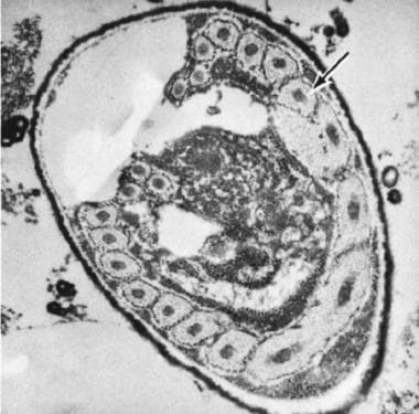
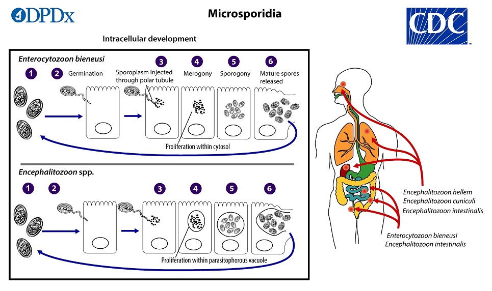
- The intracellular development of Enterocytozoon bieneusi and Encephalitozoon intestinalis are shown in sequence. (mhmedical.com)
Microsporidia3
- E. cuniculi is a spore-forming unicellular parasite belonging to the phylum Microsporidia. (wikipedia.org)
- Because the E. cuniculi genome contains genes related to some mitochondrial functions (for example, Fe-S cluster assembly), it is possible that microsporidia have retained a mitochondrion-derived organelle. (wikipedia.org)
- The infective form of microsporidia (E. cuniculi) is a resistant spore which can survive for a long time in the environment. (wikipedia.org)
Genotype1
- We identified Encephalitozoon cuniculi genotype II parasites as a cause of extraintestinal microsporidiosis in 2 owners of birds also infected with E. cuniculi . (cdc.gov)
Antibodies3
- Sera from 63 capybaras, from 6 counties in the state of Sao Paulo, Brazil, were examined for antibodies to Trypanosoma cruel, Leishmania infantum, Encephalitozoon cuniculi. (vt.edu)
- Wild arctic foxes ( Alopex lagopus ) from Greenland were tested for antibodies to Encephalitozoon cuniculi with an enzyme-linked immunosorbent assay and a carbon immunoassay. (bioone.org)
- An ELISA commercial test kit was used to detect antibodies against E. cuniculi . (tci-thaijo.org)
Nosema1
- Its current accepted name is Nosema cuniculi. (wikipedia.org)
Microsporidiosis2
- We describe the case of 2 bird owners in Poland who acquired E. cuniculi -caused microsporidiosis from their infected pet birds. (cdc.gov)
- Cases of donor-derived microsporidiosis ( Encephalitozoon cuniculi ) following bone marrow, kidney, liver, and heart transplantation have been confirmed. (cdc.gov)
Rabbit5
- Brain of a rabbit with intracellular cysts of Encephalitozoon cuniculi (H&E stain, 40X). (merckvetmanual.com)
- Encephalitozoon cuniculi is endemic in several captive and wild rabbit populations. (cdc.gov)
- When it can't be pinpointed to an ear infection, and e. cuniculi is also a possibility, rabbit vets will often treat for both until either can be determined as the definitive cause. (rabbitsonline.net)
- How can I stop my rabbit getting E. Cuniculi? (colerainevets.co.uk)
- Can I have my rabbit vaccinated against E. Cuniculi? (colerainevets.co.uk)
Species2
- Studies have isolated Encephalitozoon species in the urinary tract in those with disseminated infections, suggesting that sexual transmission is possible. (medscape.com)
- The ophthalmology department of the veterinary university Vienna has its research focus on the physiology and pathophysiology of the ocular surface and the infection with Encephalitozoon cunicucli and its ocular manifestations in all species. (ecvo.eu)
Parasite2
- Most common causes are an inner ear infection and the parasite Encephalitozoon_cuniculi. (rabbitsonline.net)
- Encephalitozoon Cuniculi (E. Cuniculi) is a protozoal parasite. (colerainevets.co.uk)
Urine1
- Western blot and immunofluorescence analysis of a human isolate of Encephalitozoon cuniculi established in culture from the urine of a patient with AIDS. (medscape.com)
Pathogen1
- Encephalitozoon cuniculi is a microsporidial pathogen of mammals with world-wide distribution. (wikipedia.org)
Genus1
- Infection with FUNGI of the genus ENCEPHALITOZOON. (bvsalud.org)
Immune response1
- 17. Encephalitozoon cuniculi takes advantage of efferocytosis to evade the immune response. (nih.gov)
Infections1
- Because encephalitozoons are opportunistic pathogens, the extraintestinal and disseminated infections and severe symptoms they cause are of concern in immunocompromised hosts, such as transplant recipients or persons living with HIV ( 2 , 3 ). (cdc.gov)
Diagnosis1
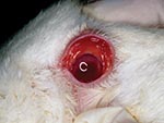
- The patient history and appearance of the lesion were compatible with Encephalitozoon cuniculi-induced phacoclastic uveitis, and a tentative diagnosis was made. (balkanvets.com)
Birds1
- E. cuniculi also infects rodents, and the organism has been detected in the feces of 13% of pet birds. (wikipedia.org)
Treatment1
- What is the treatment for E. Cuniculi? (colerainevets.co.uk)
Survey1
- Johan Åkerstedt and Christian M. O. Kapel "Survey for Encephalitozoon cuniculi in Arctic Foxes ( Alopex lagopus ) in Greenland," Journal of Wildlife Diseases 39(1), 228-232, (1 January 2003). (bioone.org)
Found1
- More information about E. cuniculi can be found in our archives . (wormsandgermsblog.com)
Animals1
- However, those individuals who are immunosuppressed should implement strict hygiene and, if possible, avoid animals suspected or confirmed of being infected with E. Cuniculi and undoubtedly seek medical advice from their doctor. (colerainevets.co.uk)