Electroencephalography
Magnetoencephalography
Epilepsies, Partial
Epilepsy
Alpha Rhythm
Seizures
Signal Processing, Computer-Assisted
Brain Mapping
Delta Rhythm
Brain
Magnetic Resonance Imaging
Cerebral Cortex
Evoked Potentials
Beta Rhythm
Evoked Potentials, Visual
Evoked Potentials, Somatosensory
Status Epilepticus
Monitoring, Physiologic
Brain-Computer Interfaces
Event-Related Potentials, P300
Term Birth
Persistent Vegetative State
Scalp
Electrooculography
Epilepsy, Frontal Lobe
Spasms, Infantile
Sleep Stages
Epilepsy, Generalized
Consciousness Disorders
Electrodes, Implanted
Sleep
Monitoring, Intraoperative
Epilepsy, Tonic-Clonic
Artifacts
Epilepsy, Absence
Wakefulness
Communication Aids for Disabled
Photic Stimulation
Transcranial Magnetic Stimulation
Electrodes
Epilepsy, Temporal Lobe
Occipital Lobe
Evoked Potentials, Auditory
Epilepsy, Reflex
Functional Laterality
Consciousness Monitors
Image Processing, Computer-Assisted
Models, Neurological
Hypoxia-Ischemia, Brain
Unconsciousness
Psychomotor Performance
Malformations of Cortical Development
Parietal Lobe
Frontal Lobe
Epilepsy, Complex Partial
Theta Rhythm
Contingent Negative Variation
Nerve Net
Brain Diseases
Electroencephalography Phase Synchronization
Anticipation, Psychological
Attention
Point-of-Care Systems
Coma
Electromyography
Visual Perception
Sleep Deprivation
Neuropsychological Tests
Diagnosis, Computer-Assisted
Spectroscopy, Near-Infrared
Video Recording
Unnecessary Procedures
Neuroimaging
Arousal
Motor Cortex
Temporal Lobe
Auditory Perception
Discriminant Analysis
Neurologic Examination
Sleep, REM
Brain Ischemia
Head
Algorithms
Endarterectomy, Carotid
Analysis of Variance
Reproducibility of Results
Hallucinations
Hypothermia, Induced
Fourier Analysis
Task Performance and Analysis
Movement
Oxygen
Spectrum Analysis
Sensitivity and Specificity
Data Interpretation, Statistical
Tomography, Emission-Computed, Single-Photon
Propofol
Biological Clocks
Thalamus
Retrospective Studies
Anesthetics, Intravenous
Hypnotics and Sedatives
Nervous System Diseases
Brain Injuries
Schizophrenia
Ultrasonography, Doppler, Transcranial
Auditory Cortex
Periodicity
Polysomnography
Evoked Potentials, Motor
Nonlinear Dynamics
Predictive Value of Tests
Visual Cortex
Computer Simulation
Pattern Recognition, Visual
Prefrontal Cortex
Prospective Studies
Somatosensory Cortex
Cognition Disorders
Positron-Emission Tomography
Tomography, X-Ray Computed
Treatment Outcome
User-Computer Interface
Hippocampus
Depressive Disorder, Major
Recognition (Psychology)
Severity of Illness Index
Image Interpretation, Computer-Assisted
Memory
Cues
Emotions
Statistics as Topic
Case-Control Studies
Physiological properties of raphe magnus neurons during sleep and waking. (1/10381)
Neurons in the medullary raphe magnus (RM) that are important in the descending modulation of nociceptive transmission are classified by their response to noxious tail heat as ON, OFF, or NEUTRAL cells. Experiments in anesthetized animals demonstrate that RM ON cells facilitate and OFF cells inhibit nociceptive transmission. Yet little is known of the physiology of these cells in the unanesthetized animal. The first aim of the present experiments was to determine whether cells with ON- and OFF-like responses to noxious heat exist in the unanesthetized rat. Second, to determine if RM cells have state-dependent discharge, the activity of RM neurons was recorded during waking and sleeping states. Noxious heat applied during waking and slow wave sleep excited one group of cells (ON-U) in unanesthetized rats. Other cells were inhibited by noxious heat (OFF-U) applied during waking and slow wave sleep states in unanesthetized rats. NEUTRAL-U cells did not respond to noxious thermal stimulation applied during either slow wave sleep or waking. ON-U and OFF-U cells were more likely to respond to noxious heat during slow wave sleep than during waking and were least likely to respond when the animal was eating or drinking. Although RM cells rarely respond to innocuous stimulation applied during anesthesia, ON-U and OFF-U cells were excited and inhibited, respectively, by innocuous somatosensory stimulation in the unanesthetized rat. The spontaneous activity of >90% of the RM neurons recorded in the unanesthetized rat was influenced by behavioral state. OFF-U cells discharged sporadically during waking but were continuously active during slow wave sleep. By contrast, ON-U and NEUTRAL-U cells discharged in bursts during waking and either ceased to discharge entirely or discharged at a low rate during slow wave sleep. We suggest that OFF cell discharge functions to suppress pain-evoked reactions during sleep, whereas ON cell discharge facilitates pain-evoked responses during waking. (+info)Effect of psychotropic drugs on caudate spindle in cats. (2/10381)
To ascertain whether neuroleptics act on the caudate nucleus itself, the effects of these compounds as well as other centrally acting drugs were examined in relation to caudate spindle and EEG arousal responses (sciatic nerve stimulation) in gallamine-immobilized cats. Haloperidol and chlorpromazine enhanced the caudate spindle at a dose which had no effect on the EEG arousal response. On the other hand, clozapine and a higher dose of chlorpromazine enhanced the caudate spindle, but depressed the arousal response. High frequency stimulation of the sciatic nerve suppressed the caudate spindle. Pentobarbital, biperiden and diazepam, while depressing the arousal response, caused an enhancement of the caudate spindle. Imipramine at a low dose had no effect on either response, whereas at a high dose this drug enhanced the caudate spindle with concomitant depression of the arousal response. From these results, it may be concluded that the enhancing action on the caudate spindle induced by haloperidol and a low dose of chlorpromazine is due to an increase in susceptibility of the caudate nucleus itself. In addition, it is suggested that depression of the activating system is involved in an appearance of the caudate spindle. (+info)Arousal from sleep shortens sympathetic burst latency in humans. (3/10381)
1. Bursts of sympathetic activity in muscle nerves are phase-locked to the cardiac cycle by the sinoaortic baroreflexes. Acoustic arousal from non-rapid eye movement (NREM) sleep reduces the normally invariant interval between the R-wave of the electrocardiogram (ECG) and the peak of the corresponding sympathetic burst; however, the effects of other forms of sleep disruption (i.e. spontaneous arousals and apnoea-induced arousals) on this temporal relationship are unknown. 2. We simultaneously recorded muscle sympathetic nerve activity in the peroneal nerve (intraneural electrodes) and the ECG (surface electrodes) in seven healthy humans and three patients with sleep apnoea syndrome during NREM sleep. 3. In seven subjects, burst latencies were shortened subsequent to spontaneous K complexes (1.297 +/- 0.024 s, mean +/- s. e.m.) and spontaneous arousals (1.268 +/- 0.044 s) compared with latencies during periods of stable NREM sleep (1.369 +/- 0.023 s). In six subjects who demonstrated spontaneous apnoeas during sleep, apnoea per se did not alter burst latency relative to sleep with stable electroencephalogram (EEG) and breathing (1.313 +/- 0.038 vs. 1.342 +/- 0.026 s); however, following apnoea-induced EEG perturbations, burst latencies were reduced (1.214 +/- 0.034 s). 4. Arousal-induced reduction in sympathetic burst latency may reflect a temporary diminution of baroreflex buffering of sympathetic outflow. If so, the magnitude of arterial pressure perturbations during sleep (e.g. those caused by sleep disordered breathing and periodic leg movements) may be augmented by arousal. (+info)Overexpression of a Shaker-type potassium channel in mammalian central nervous system dysregulates native potassium channel gene expression. (4/10381)
The nervous system maintains a delicate balance between excitation and inhibition, partly through the complex interplay between voltage-gated sodium and potassium ion channels. Because K+ channel blockade or gene deletion causes hyperexcitability, it is generally assumed that increases in K+ channel gene expression should reduce neuronal network excitability. We have tested this hypothesis by creating a transgenic mouse that expresses a Shaker-type K+ channel gene. Paradoxically, we find that addition of the extra K+ channel gene results in a hyperexcitable rather than a hypoexcitable phenotype. The presence of the transgene leads to a complex deregulation of endogenous Shaker genes in the adult central nervous system as well as an increase in network excitability that includes spontaneous cortical spike and wave discharges and a lower threshold for epileptiform bursting in isolated hippocampal slices. These data suggest that an increase in K+ channel gene dosage leads to dysregulation of normal K+ channel gene expression, and it may underlie a mechanism contributing to the pathogenesis of human aneuploidies such as Down syndrome. (+info)Intrapreoptic microinjection of GHRH or its antagonist alters sleep in rats. (5/10381)
Previous reports indicate that growth hormone-releasing hormone (GHRH) is involved in sleep regulation. The site of action mediating the nonrapid eye movement sleep (NREMS)-promoting effects of GHRH is not known, but it is independent from the pituitary. GHRH (0.001, 0. 01, and 0.1 nmol/kg) or a competitive antagonist of GHRH (0.003, 0.3, and 14 nmol/kg) was microinjected into the preoptic area, and the sleep-wake activity was recorded for 23 hr after injection in rats. GHRH elicited dose-dependent increases in the duration and in the intensity of NREMS compared with that in control records after intrapreoptic injection of physiological saline. The antagonist decreased the duration and intensity of NREMS and prolonged sleep latency. Consistent alterations in rapid eye movement sleep (REMS) and in brain temperature were not found. The GHRH antagonist also attenuated the enhancements in NREMS elicited by 3 hr of sleep deprivation. Histological verification of the injection sites showed that the majority of the effective injections were in the preoptic area and the diagonal band of Broca. The results indicate that the preoptic area mediates the sleep-promoting activity of GHRH. (+info)EEG surveillance as a means of extending operability in high risk carotid endarterectomy. (6/10381)
Some patients who have transient ischemic attacks are denied operation because severe occlusive lesions in other extra-cranial arteries may be inappropriately interpreted as constituting an unacceptable surgical risk, or because the lesion is so distal as to make its removal hazardous. Failure of endarterectomy is usually due to incomplete removal of the lesion or to thrombosis upon the frayed intima. Such lesions require excellent visualization and meticulous surgical technique -- not always possible with a shunt. Among 130 consecutive carotid endarterectomies performed under general anesthesia, EEG changes consistent with cerebral ischemia appeared in only nine (7%). These patients required a shunt. In 11 patients normal EEG tracings were obtained during endarterectomy despite contralateral carotid occlusion. None of these patients had a neurological deficit. Continuous EEG monitoring is a reliable method of detecting changes in cerebral perfusion, permits a more meticulous endarterectomy in high-lying lesions without a shunt, and extends operability in high risk patients. Angiographical findings may be an unreliable predictor concerning risk of endarterectomy. (+info)Intensive care management of stroke patients. (7/10381)
Two hundred eighty patients were admitted to an intensive care stroke unit over a one-year period. Subsequent investigation indicated that only 199 of these patients actually had cerebral ischemic or hemorrhagic lesions, 10 had other cerebrovascular lesions, and the remaining 71 patients had unrelated diseases, predominantly seizures. Detailed analysis of 103 stroke patients revealed an overall incidence of 59% hypertension, and 72% had hypertensive, ischemic or valvular heart disease. Fifty percent of the patients had various cardiac arrhythmias, some of which were responsible for the acute cerebrovascular lesion. Fourteen patients died during the acute phase, 11 from apparently irreversible cerebral selling, mainly due to cerebral hemorrhage. Secondary complications such as pneumonia, pulmonary embolism, pressure sores and urinary infection were almost nonexistent, but beneficial effects on the primary cerebral lesions were more difficult to demonstrate. (+info)Subcortical arteriosclerotic encephalopathy (Binswanger's disease). A vascular etiology of dementia. (8/10381)
A 51-yearold man with moderate intermittent hypertension had a rapidly progressive, profound dementia in the absence of significant localizing neurological signs. Postmortem examination disclosed the vascular alterations and diffuse white matter degeneration which characterize subcortical arteriosclerotic encephalopathy (SAE) or Binswanger's disease. The case underscores the need to consider vascular disease as an etiology of dementia -- even in the absence of focal neurological deficit. (+info)Electroencephalography (EEG) is a medical procedure that records electrical activity in the brain. It uses small, metal discs called electrodes, which are attached to the scalp with paste or a specialized cap. These electrodes detect tiny electrical charges that result from the activity of brain cells, and the EEG machine then amplifies and records these signals.
EEG is used to diagnose various conditions related to the brain, such as seizures, sleep disorders, head injuries, infections, and degenerative diseases like Alzheimer's or Parkinson's. It can also be used during surgery to monitor brain activity and ensure that surgical procedures do not interfere with vital functions.
EEG is a safe and non-invasive procedure that typically takes about 30 minutes to an hour to complete, although longer recordings may be necessary in some cases. Patients are usually asked to relax and remain still during the test, as movement can affect the quality of the recording.
Brain waves, also known as electroencephalography (EEG) waves, are the rhythmic electrical activity produced by the brain's neurons. These waves are detected by placing electrodes on the scalp and can be visualized using an EEG machine. Brain waves are typically categorized into different frequency bands, including:
1. Delta waves (0.5-4 Hz): Slow waves that are typically seen during deep sleep or in pathological states such as coma.
2. Theta waves (4-8 Hz): Slower waves that are associated with drowsiness, meditation, and creative thinking.
3. Alpha waves (8-13 Hz): These waves are present during relaxed wakefulness and can be seen during eyes-closed rest.
4. Beta waves (13-30 Hz): Faster waves that are associated with active thinking, focus, and alertness.
5. Gamma waves (30-100 Hz): The fastest waves, which are associated with higher cognitive functions such as attention, perception, and problem-solving.
Abnormalities in brain wave patterns can be indicative of various neurological conditions, including epilepsy, sleep disorders, brain injuries, and neurodegenerative diseases.
Magnetoencephalography (MEG) is a non-invasive functional neuroimaging technique used to measure the magnetic fields produced by electrical activity in the brain. These magnetic fields are detected by very sensitive devices called superconducting quantum interference devices (SQUIDs), which are cooled to extremely low temperatures to enhance their sensitivity. MEG provides direct and real-time measurement of neural electrical activity with high temporal resolution, typically on the order of milliseconds, allowing for the investigation of brain function during various cognitive, sensory, and motor tasks. It is often used in conjunction with other neuroimaging techniques, such as fMRI, to provide complementary information about brain structure and function.
Epilepsy, partial is a type of epilepsy characterized by recurrent, unprovoked seizures that originate in a specific, localized area of the brain. These seizures are also known as focal seizures and can vary in severity and symptoms depending on the location of the abnormal electrical activity in the brain.
Partial epilepsies can be further classified into two main categories: simple partial seizures and complex partial seizures. Simple partial seizures do not involve a loss of consciousness, while complex partial seizures are associated with impaired awareness or responsiveness during the seizure.
The causes of partial epilepsies can include brain injury, infection, stroke, tumors, genetic factors, or an unknown cause. Treatment typically involves anti-seizure medications, and in some cases, surgery may be recommended to remove the specific area of the brain responsible for the seizures.
Epilepsy is a chronic neurological disorder characterized by recurrent, unprovoked seizures. These seizures are caused by abnormal electrical activity in the brain, which can result in a wide range of symptoms, including convulsions, loss of consciousness, and altered sensations or behaviors. Epilepsy can have many different causes, including genetic factors, brain injury, infection, or stroke. In some cases, the cause may be unknown.
There are many different types of seizures that can occur in people with epilepsy, and the specific type of seizure will depend on the location and extent of the abnormal electrical activity in the brain. Some people may experience only one type of seizure, while others may have several different types. Seizures can vary in frequency, from a few per year to dozens or even hundreds per day.
Epilepsy is typically diagnosed based on the patient's history of recurrent seizures and the results of an electroencephalogram (EEG), which measures the electrical activity in the brain. Imaging tests such as MRI or CT scans may also be used to help identify any structural abnormalities in the brain that may be contributing to the seizures.
While there is no cure for epilepsy, it can often be effectively managed with medication. In some cases, surgery may be recommended to remove the area of the brain responsible for the seizures. With proper treatment and management, many people with epilepsy are able to lead normal, productive lives.
Alpha rhythm is a type of brain wave that is typically observed in the electroencephalogram (EEG) of normal, awake individuals when they have their eyes closed. It is characterized by sinusoidal waves with a frequency range of 8-13 Hz and is most prominent over the occipital region of the head, which is located at the back of the skull above the brain's visual cortex.
Alpha rhythm is typically associated with relaxed wakefulness, and its presence may indicate that an individual is awake but not engaged in any mentally demanding tasks. It can be blocked or suppressed by various stimuli, such as opening one's eyes, hearing a loud noise, or engaging in mental activity.
Disruptions in alpha rhythm have been observed in various neurological and psychiatric conditions, including epilepsy, dementia, depression, and anxiety disorders. However, more research is needed to fully understand the clinical significance of these abnormalities.
A seizure is an uncontrolled, abnormal firing of neurons (brain cells) that can cause various symptoms such as convulsions, loss of consciousness, altered awareness, or changes in behavior. Seizures can be caused by a variety of factors including epilepsy, brain injury, infection, toxic substances, or genetic disorders. They can also occur without any identifiable cause, known as idiopathic seizures. Seizures are a medical emergency and require immediate attention.
Computer-assisted signal processing is a medical term that refers to the use of computer algorithms and software to analyze, interpret, and extract meaningful information from biological signals. These signals can include physiological data such as electrocardiogram (ECG) waves, electromyography (EMG) signals, electroencephalography (EEG) readings, or medical images.
The goal of computer-assisted signal processing is to automate the analysis of these complex signals and extract relevant features that can be used for diagnostic, monitoring, or therapeutic purposes. This process typically involves several steps, including:
1. Signal acquisition: Collecting raw data from sensors or medical devices.
2. Preprocessing: Cleaning and filtering the data to remove noise and artifacts.
3. Feature extraction: Identifying and quantifying relevant features in the signal, such as peaks, troughs, or patterns.
4. Analysis: Applying statistical or machine learning algorithms to interpret the extracted features and make predictions about the underlying physiological state.
5. Visualization: Presenting the results in a clear and intuitive way for clinicians to review and use.
Computer-assisted signal processing has numerous applications in healthcare, including:
* Diagnosing and monitoring cardiac arrhythmias or other heart conditions using ECG signals.
* Assessing muscle activity and function using EMG signals.
* Monitoring brain activity and diagnosing neurological disorders using EEG readings.
* Analyzing medical images to detect abnormalities, such as tumors or fractures.
Overall, computer-assisted signal processing is a powerful tool for improving the accuracy and efficiency of medical diagnosis and monitoring, enabling clinicians to make more informed decisions about patient care.
Brain mapping is a broad term that refers to the techniques used to understand the structure and function of the brain. It involves creating maps of the various cognitive, emotional, and behavioral processes in the brain by correlating these processes with physical locations or activities within the nervous system. Brain mapping can be accomplished through a variety of methods, including functional magnetic resonance imaging (fMRI), positron emission tomography (PET) scans, electroencephalography (EEG), and others. These techniques allow researchers to observe which areas of the brain are active during different tasks or thoughts, helping to shed light on how the brain processes information and contributes to our experiences and behaviors. Brain mapping is an important area of research in neuroscience, with potential applications in the diagnosis and treatment of neurological and psychiatric disorders.
A "delta rhythm" is a term used in electroencephalography (EEG) to describe a pattern of brain waves that are typically seen in the delta frequency range (0.5-4 Hz) and are maximal over the posterior regions of the head. This rhythm is often observed during deep sleep stages, specifically stage 3 and stage 4 of non-rapid eye movement (NREM) sleep, also known as slow-wave sleep.
Delta waves are characterized by their high amplitude and slow frequency, making them easily distinguishable from other brain wave patterns. The presence of a robust delta rhythm during sleep is thought to reflect the restorative processes that occur during this stage of sleep, including memory consolidation and physical restoration.
However, it's important to note that abnormal delta rhythms can also be observed in certain neurological conditions, such as epilepsy or encephalopathy, where they may indicate underlying brain dysfunction or injury. In these cases, the presence of delta rhythm may have different clinical implications and require further evaluation by a medical professional.
The brain is the central organ of the nervous system, responsible for receiving and processing sensory information, regulating vital functions, and controlling behavior, movement, and cognition. It is divided into several distinct regions, each with specific functions:
1. Cerebrum: The largest part of the brain, responsible for higher cognitive functions such as thinking, learning, memory, language, and perception. It is divided into two hemispheres, each controlling the opposite side of the body.
2. Cerebellum: Located at the back of the brain, it is responsible for coordinating muscle movements, maintaining balance, and fine-tuning motor skills.
3. Brainstem: Connects the cerebrum and cerebellum to the spinal cord, controlling vital functions such as breathing, heart rate, and blood pressure. It also serves as a relay center for sensory information and motor commands between the brain and the rest of the body.
4. Diencephalon: A region that includes the thalamus (a major sensory relay station) and hypothalamus (regulates hormones, temperature, hunger, thirst, and sleep).
5. Limbic system: A group of structures involved in emotional processing, memory formation, and motivation, including the hippocampus, amygdala, and cingulate gyrus.
The brain is composed of billions of interconnected neurons that communicate through electrical and chemical signals. It is protected by the skull and surrounded by three layers of membranes called meninges, as well as cerebrospinal fluid that provides cushioning and nutrients.
Medical Definition:
Magnetic Resonance Imaging (MRI) is a non-invasive diagnostic imaging technique that uses a strong magnetic field and radio waves to create detailed cross-sectional or three-dimensional images of the internal structures of the body. The patient lies within a large, cylindrical magnet, and the scanner detects changes in the direction of the magnetic field caused by protons in the body. These changes are then converted into detailed images that help medical professionals to diagnose and monitor various medical conditions, such as tumors, injuries, or diseases affecting the brain, spinal cord, heart, blood vessels, joints, and other internal organs. MRI does not use radiation like computed tomography (CT) scans.
The cerebral cortex is the outermost layer of the brain, characterized by its intricate folded structure and wrinkled appearance. It is a region of great importance as it plays a key role in higher cognitive functions such as perception, consciousness, thought, memory, language, and attention. The cerebral cortex is divided into two hemispheres, each containing four lobes: the frontal, parietal, temporal, and occipital lobes. These areas are responsible for different functions, with some regions specializing in sensory processing while others are involved in motor control or associative functions. The cerebral cortex is composed of gray matter, which contains neuronal cell bodies, and is covered by a layer of white matter that consists mainly of myelinated nerve fibers.
Evoked potentials (EPs) are medical tests that measure the electrical activity in the brain or spinal cord in response to specific sensory stimuli, such as sight, sound, or touch. These tests are often used to help diagnose and monitor conditions that affect the nervous system, such as multiple sclerosis, brainstem tumors, and spinal cord injuries.
There are several types of EPs, including:
1. Visual Evoked Potentials (VEPs): These are used to assess the function of the visual pathway from the eyes to the back of the brain. A patient is typically asked to look at a patterned image or flashing light while electrodes placed on the scalp record the electrical responses.
2. Brainstem Auditory Evoked Potentials (BAEPs): These are used to evaluate the function of the auditory nerve and brainstem. Clicking sounds are presented to one or both ears, and electrodes placed on the scalp measure the response.
3. Somatosensory Evoked Potentials (SSEPs): These are used to assess the function of the peripheral nerves and spinal cord. Small electrical shocks are applied to a nerve at the wrist or ankle, and electrodes placed on the scalp record the response as it travels up the spinal cord to the brain.
4. Motor Evoked Potentials (MEPs): These are used to assess the function of the motor pathways in the brain and spinal cord. A magnetic or electrical stimulus is applied to the brain or spinal cord, and electrodes placed on a muscle measure the response as it travels down the motor pathway.
EPs can help identify abnormalities in the nervous system that may not be apparent through other diagnostic tests, such as imaging studies or clinical examinations. They are generally safe, non-invasive procedures with few risks or side effects.
The Beta rhythm is a type of brain wave that is typically observed in the electroencephalogram (EEG) of awake, alert individuals. It has a frequency range of 13-30 Hz (cycles per second) and is most prominent over the frontal and central regions of the scalp. Beta activity is associated with active thinking, problem solving, and focused attention. It can be suppressed during states of relaxation, meditation, or sleep. Additionally, abnormal beta activity has been observed in certain neurological conditions such as Parkinson's disease and seizure disorders.
Evoked potentials, visual, also known as visually evoked potentials (VEPs), are electrical responses recorded from the brain following the presentation of a visual stimulus. These responses are typically measured using electroencephalography (EEG) and can provide information about the functioning of the visual pathways in the brain.
There are several types of VEPs, including pattern-reversal VEPs and flash VEPs. Pattern-reversal VEPs are elicited by presenting alternating checkerboard patterns, while flash VEPs are elicited by flashing a light. The responses are typically analyzed in terms of their latency (the time it takes for the response to occur) and amplitude (the size of the response).
VEPs are often used in clinical settings to help diagnose and monitor conditions that affect the visual system, such as multiple sclerosis, optic neuritis, and brainstem tumors. They can also be used in research to study the neural mechanisms underlying visual perception.
Somatosensory evoked potentials (SEPs) are electrical signals generated in the brain and spinal cord in response to the stimulation of peripheral nerves. These responses are recorded and measured to assess the functioning of the somatosensory system, which is responsible for processing sensations such as touch, temperature, vibration, and proprioception (the sense of the position and movement of body parts).
SEPs are typically elicited by applying electrical stimuli to peripheral nerves in the arms or legs. The resulting neural responses are then recorded using electrodes placed on the scalp or other locations on the body. These recordings can provide valuable information about the integrity and function of the nervous system, and are often used in clinical settings to diagnose and monitor conditions such as nerve damage, spinal cord injury, multiple sclerosis, and other neurological disorders.
SEPs can be further categorized based on the specific type of stimulus used and the location of the recording electrodes. For example, short-latency SEPs (SLSEPs) are those that occur within the first 50 milliseconds after stimulation, and are typically recorded from the scalp over the primary sensory cortex. These responses reflect the earliest stages of sensory processing and can be used to assess the integrity of the peripheral nerves and the ascending sensory pathways in the spinal cord.
In contrast, long-latency SEPs (LLSEPs) occur after 50 milliseconds and are typically recorded from more posterior regions of the scalp over the parietal cortex. These responses reflect later stages of sensory processing and can be used to assess higher-level cognitive functions such as attention, memory, and perception.
Overall, SEPs provide a valuable tool for clinicians and researchers seeking to understand the functioning of the somatosensory system and diagnose or monitor neurological disorders.
Cortical synchronization refers to the phenomenon of coordinated neural activity in the cerebral cortex, the brain region responsible for higher cognitive functions. It is characterized by the synchronized firing of neurons in various cortical areas, leading to the generation of rhythmic electrical patterns. These rhythms can be observed using electroencephalography (EEG) and other neuroimaging techniques.
Cortical synchronization plays a crucial role in various cognitive processes, such as attention, perception, memory, and consciousness. It is also involved in the pathophysiology of several neurological and psychiatric disorders, including epilepsy, schizophrenia, and Parkinson's disease.
The degree of cortical synchronization can be modulated by various factors, such as sensory stimulation, attention, arousal, and cognitive load. The precise mechanisms underlying cortical synchronization are still not fully understood but are thought to involve complex interactions between excitatory and inhibitory neurons, as well as the modulation of synaptic strength and connectivity.
Status epilepticus is a serious and life-threatening medical condition characterized by an ongoing seizure activity or a series of seizures without full recovery of consciousness between them, lasting for 30 minutes or more. It is a neurological emergency that requires immediate medical attention to prevent potential complications such as brain damage, respiratory failure, or even death.
The condition can occur in people with a history of epilepsy or seizure disorders, as well as those without any prior history of seizures. The underlying causes of status epilepticus can vary and may include infection, trauma, stroke, metabolic imbalances, toxins, or other medical conditions that affect the brain's normal functioning. Prompt diagnosis and treatment are crucial to prevent long-term neurological damage and improve outcomes in patients with this condition.
Physiological monitoring is the continuous or intermittent observation and measurement of various body functions or parameters in a patient, with the aim of evaluating their health status, identifying any abnormalities or changes, and guiding clinical decision-making and treatment. This may involve the use of specialized medical equipment, such as cardiac monitors, pulse oximeters, blood pressure monitors, and capnographs, among others. The data collected through physiological monitoring can help healthcare professionals assess the effectiveness of treatments, detect complications early, and make timely adjustments to patient care plans.
A Brain-Computer Interface (BCI), also known as a neural-control interface or a brain-machine interface, is a system that enables direct communication and interaction between the human brain and an external electronic device. BCI technology translates brain signals into commands that can control artificial devices, such as computers, prosthetic limbs, or other assistive technologies.
There are primarily two types of BCIs: invasive and non-invasive. Invasive BCIs involve the surgical implantation of electrodes directly onto the surface or within the brain tissue to record neural activity with high resolution. Non-invasive BCIs, on the other hand, utilize external sensors, like electroencephalography (EEG) caps, to measure brain signals through the scalp.
The applications of BCIs are vast and varied, including communication aids for individuals with severe motor disabilities, rehabilitation tools for stroke patients, and assistive devices for people with amputations or spinal cord injuries. Additionally, BCI technology holds potential for enhancing human performance in various fields, such as gaming, education, and military applications. However, it is essential to consider the ethical implications and potential risks associated with BCI use as the technology continues to advance.
Event-Related Potentials (ERPs) are brain responses that are directly related to a specific sensory, cognitive, or motor event. P300 is a positive deflection in the ERP waveform that occurs approximately 300 milliseconds after the onset of a rare or unexpected stimulus. It is often used as an index of cognitive processes such as attention, memory, and decision-making. The amplitude of the P300 component is typically larger for targets than for non-targets, and it is thought to reflect the amount of attentional resources allocated to the processing of the stimulus. Additionally, the latency of the P300 component can be used as an indicator of the speed of cognitive processing.
It's important to note that ERPs are measured using electroencephalography (EEG) and it requires averaging multiple trials to extract the signal from the noise. Also, P300 is just one component of ERP, there are other components like N100, P100, N200 etc which also have their own significance in understanding the cognitive processes.
A "term birth" is a medical term that refers to a delivery or pregnancy that has reached 37 weeks or more. It is the normal length of a full-term pregnancy and is considered a healthy and low-risk period for childbirth. Babies born at term have the best chance of being healthy and not experiencing any significant medical issues, compared to those born preterm (before 37 weeks) or postterm (after 42 weeks). The different types of term births are:
* Early Term: Between 37 weeks and 38 weeks, 6 days.
* Full Term: Between 39 weeks and 40 weeks, 6 days.
* Late Term: Between 41 weeks and 41 weeks, 6 days.
* Postterm: 42 weeks or later.
It is important to note that while a term birth is generally considered low-risk, there can still be variations in the health of babies born at different points within this range. For example, research has shown that babies born at 39 weeks have better outcomes than those born at 37 or 38 weeks. Therefore, it is always best to consult with a healthcare provider for individualized guidance and recommendations regarding pregnancy and childbirth.
Persistent vegetative state (PVS) is a medical condition characterized by a prolonged disorder of consciousness. It's not the same as a coma. In PVS, a person may open their eyes, appear to be awake and have periods of sleep and wakefulness, but they do not show signs of awareness or cognition. They do not respond to stimuli, cannot communicate, and do not have any purposeful behaviors.
This condition can occur after a severe brain injury, such as from trauma, stroke, or lack of oxygen supply. The chance of recovery from PVS is very low, and if some recovery does occur, it's usually incomplete.
It's important to note that the term "persistent vegetative state" has been replaced in some clinical settings with "unresponsive wakefulness syndrome" due to the negative connotations associated with the term "vegetative".
The scalp is the anatomical region located at the upper part of the human head, covering the skull except for the face and the ears. It is made up of several layers: the skin, the connective tissue, the galea aponeurotica (a strong, flat, tendinous sheet), loose areolar tissue, and the periosteum (the highly vascularized innermost layer that attaches directly to the skull bones). The scalp has a rich blood supply and is home to numerous sensory receptors, including those for touch, pain, and temperature. It also contains hair follicles, sebaceous glands, and sweat glands.
Electrooculography (EOG) is a technique for measuring the resting potential of the eye and the changes in this potential that occur with eye movements. It involves placing electrodes near the eyes to detect the small electric fields generated by the movement of the eyeball within the surrounding socket. This technique is used in research and clinical settings to study eye movements and their control, as well as in certain diagnostic applications such as assessing the function of the oculomotor system in patients with neurological disorders.
Frontal lobe epilepsy is a type of focal epilepsy, which means that the seizures originate from a specific area in the brain called the frontal lobe. The frontal lobe is located at the front part of the brain and is responsible for various functions such as motor function, problem-solving, decision making, emotional expression, and social behavior.
In frontal lobe epilepsy, seizures can be quite varied in their presentation, but they often occur during sleep or wakefulness and may include symptoms such as:
* Brief staring spells or automatisms (such as lip smacking, chewing, or fumbling movements)
* Sudden and frequent falls or drops
* Vocalizations or sounds
* Complex behaviors, such as agitation, aggression, or sexual arousal
* Auras or warning sensations before the seizure
Frontal lobe epilepsy can be difficult to diagnose due to the varied nature of the seizures and their occurrence during sleep. Diagnostic tests such as electroencephalogram (EEG) and imaging studies like magnetic resonance imaging (MRI) may be used to help confirm the diagnosis. Treatment typically involves medication, but in some cases, surgery may be recommended if medications are not effective or cause significant side effects.
Infantile spasms, also known as West syndrome, is a rare but serious type of epilepsy that affects infants typically between 4-8 months of age. The spasms are characterized by sudden, brief, and frequent muscle jerks or contractions, often involving the neck, trunk, and arms. These spasms usually occur in clusters and may cause the infant to bend forward or stretch out. Infantile spasms can be a symptom of various underlying neurological conditions and are often associated with developmental delays and regression. Early recognition and treatment are crucial for improving outcomes.
Sleep stages are distinct patterns of brain activity that occur during sleep, as measured by an electroencephalogram (EEG). They are part of the sleep cycle and are used to describe the different types of sleep that humans go through during a normal night's rest. The sleep cycle includes several repeating stages:
1. Stage 1 (N1): This is the lightest stage of sleep, where you transition from wakefulness to sleep. During this stage, muscle activity and brain waves begin to slow down.
2. Stage 2 (N2): In this stage, your heart rate slows, body temperature decreases, and eye movements stop. Brain wave activity becomes slower, with occasional bursts of electrical activity called sleep spindles.
3. Stage 3 (N3): Also known as deep non-REM sleep, this stage is characterized by slow delta waves. It is during this stage that the body undergoes restorative processes such as tissue repair, growth, and immune function enhancement.
4. REM (Rapid Eye Movement) sleep: This is the stage where dreaming typically occurs. Your eyes move rapidly beneath closed eyelids, heart rate and respiration become irregular, and brain wave activity increases to levels similar to wakefulness. REM sleep is important for memory consolidation and learning.
The sleep cycle progresses through these stages multiple times during the night, with REM sleep periods becoming longer towards morning. Understanding sleep stages is crucial in diagnosing and treating various sleep disorders.
Generalized epilepsy is a type of epilepsy characterized by seizures that involve both halves of the brain (generalized onset) from the beginning of the seizure. These types of seizures include tonic-clonic (grand mal) seizures, absence (petit mal) seizures, and myoclonic seizures. Generalized epilepsy can be caused by genetic factors or brain abnormalities, and it is typically treated with medication. People with generalized epilepsy may experience difficulties with learning, memory, and behavior, and they may have a higher risk of injury during a seizure. It's important for individuals with generalized epilepsy to work closely with their healthcare team to manage their condition and reduce the frequency and severity of seizures.
Consciousness disorders, also known as altered consciousness, refer to conditions that affect a person's awareness or perception of their surroundings, themselves, or their current state. These disorders can range from mild to severe and can be caused by various factors such as brain injury, illness, or the use of certain medications.
There are several types of consciousness disorders, including:
1. Coma: A state of deep unconsciousness in which a person is unable to respond to stimuli or communicate.
2. Vegetative State: A condition in which a person may have sleep-wake cycles and some automatic responses, but lacks awareness or the ability to interact with their environment.
3. Minimally Conscious State: A condition in which a person has some degree of awareness and may be able to respond to stimuli, but is unable to communicate or consistently interact with their environment.
4. Delirium: A state of confusion and altered consciousness that can occur suddenly and fluctuate over time. It is often caused by an underlying medical condition or the use of certain medications.
5. Locked-in Syndrome: A rare condition in which a person is fully conscious but unable to move or communicate due to complete paralysis of all voluntary muscles except for those that control eye movement.
Treatment for consciousness disorders depends on the underlying cause and may include medication, therapy, or surgery. In some cases, recovery may be possible with appropriate treatment and rehabilitation. However, in other cases, the disorder may be permanent or result in long-term disability.
Implanted electrodes are medical devices that are surgically placed inside the body to interface directly with nerves, neurons, or other electrically excitable tissue for various therapeutic purposes. These electrodes can be used to stimulate or record electrical activity from specific areas of the body, depending on their design and application.
There are several types of implanted electrodes, including:
1. Deep Brain Stimulation (DBS) electrodes: These are placed deep within the brain to treat movement disorders such as Parkinson's disease, essential tremor, and dystonia. DBS electrodes deliver electrical impulses that modulate abnormal neural activity in targeted brain regions.
2. Spinal Cord Stimulation (SCS) electrodes: These are implanted along the spinal cord to treat chronic pain syndromes. SCS electrodes emit low-level electrical pulses that interfere with pain signals traveling to the brain, providing relief for patients.
3. Cochlear Implant electrodes: These are surgically inserted into the cochlea of the inner ear to restore hearing in individuals with severe to profound hearing loss. The electrodes stimulate the auditory nerve directly, bypassing damaged hair cells within the cochlea.
4. Retinal Implant electrodes: These are implanted in the retina to treat certain forms of blindness caused by degenerative eye diseases like retinitis pigmentosa. The electrodes convert visual information from a camera into electrical signals, which stimulate remaining retinal cells and transmit the information to the brain via the optic nerve.
5. Sacral Nerve Stimulation (SNS) electrodes: These are placed near the sacral nerves in the lower back to treat urinary or fecal incontinence and overactive bladder syndrome. SNS electrodes deliver electrical impulses that regulate the function of the affected muscles and nerves.
6. Vagus Nerve Stimulation (VNS) electrodes: These are wrapped around the vagus nerve in the neck to treat epilepsy and depression. VNS electrodes provide intermittent electrical stimulation to the vagus nerve, which has connections to various regions of the brain involved in these conditions.
Overall, implanted electrodes serve as a crucial component in many neuromodulation therapies, offering an effective treatment option for numerous neurological and sensory disorders.
Sleep is a complex physiological process characterized by altered consciousness, relatively inhibited sensory activity, reduced voluntary muscle activity, and decreased interaction with the environment. It's typically associated with specific stages that can be identified through electroencephalography (EEG) patterns. These stages include rapid eye movement (REM) sleep, associated with dreaming, and non-rapid eye movement (NREM) sleep, which is further divided into three stages.
Sleep serves a variety of functions, including restoration and strengthening of the immune system, support for growth and development in children and adolescents, consolidation of memory, learning, and emotional regulation. The lack of sufficient sleep or poor quality sleep can lead to significant health problems, such as obesity, diabetes, cardiovascular disease, and even cognitive decline.
The American Academy of Sleep Medicine (AASM) defines sleep as "a period of daily recurring natural rest during which consciousness is suspended and metabolic processes are reduced." However, it's important to note that the exact mechanisms and purposes of sleep are still being researched and debated among scientists.
Intraoperative monitoring (IOM) is the practice of using specialized techniques to monitor physiological functions or neural structures in real-time during surgical procedures. The primary goal of IOM is to provide continuous information about the patient's status and the effects of surgery on neurological function, allowing surgeons to make informed decisions and minimize potential risks.
IOM can involve various methods such as:
1. Electrophysiological monitoring: This includes techniques like somatosensory evoked potentials (SSEP), motor evoked potentials (MEP), and electroencephalography (EEG) to assess the integrity of neural pathways and brain function during surgery.
2. Neuromonitoring: Direct electrical stimulation of nerves or spinal cord structures can help identify critical neuroanatomical structures, evaluate their functional status, and guide surgical interventions.
3. Hemodynamic monitoring: Measuring blood pressure, heart rate, cardiac output, and oxygen saturation helps assess the patient's overall physiological status during surgery.
4. Imaging modalities: Intraoperative imaging techniques like ultrasound, computed tomography (CT), or magnetic resonance imaging (MRI) can provide real-time visualization of anatomical structures and surgical progress.
The specific IOM methods employed depend on the type of surgery, patient characteristics, and potential risks involved. Intraoperative monitoring is particularly crucial in procedures where there is a risk of neurological injury, such as spinal cord or brain surgeries, vascular interventions, or tumor resections near critical neural structures.
Tonic-clonic epilepsy, also known as grand mal epilepsy, is a type of generalized seizure that affects the entire brain. This type of epilepsy is characterized by two distinct phases: the tonic phase and the clonic phase.
During the tonic phase, which usually lasts for about 10-20 seconds, the person loses consciousness and their muscles stiffen, causing them to fall to the ground. This can result in injuries if the person falls unexpectedly or hits an object on the way down.
The clonic phase follows immediately after the tonic phase and is characterized by rhythmic jerking movements of the limbs, face, and neck. These movements are caused by alternating contractions and relaxations of the muscles and can last for several minutes. The person may also lose bladder or bowel control during this phase.
After the seizure, the person may feel tired, confused, and disoriented. They may also have a headache, sore muscles, and difficulty remembering what happened during the seizure.
Tonic-clonic epilepsy can be caused by a variety of factors, including genetics, brain injury, infection, or stroke. It is typically diagnosed through a combination of medical history, physical examination, and diagnostic tests such as an electroencephalogram (EEG) or imaging studies. Treatment may include medication, surgery, or dietary changes, depending on the underlying cause and severity of the seizures.
An artifact, in the context of medical terminology, refers to something that is created or introduced during a scientific procedure or examination that does not naturally occur in the patient or specimen being studied. Artifacts can take many forms and can be caused by various factors, including contamination, damage, degradation, or interference from equipment or external sources.
In medical imaging, for example, an artifact might appear as a distortion or anomaly on an X-ray, MRI, or CT scan that is not actually present in the patient's body. This can be caused by factors such as patient movement during the scan, metal implants or other foreign objects in the body, or issues with the imaging equipment itself.
Similarly, in laboratory testing, an artifact might refer to a substance or characteristic that is introduced into a sample during collection, storage, or analysis that can interfere with accurate results. This could include things like contamination from other samples, degradation of the sample over time, or interference from chemicals used in the testing process.
In general, artifacts are considered to be sources of error or uncertainty in medical research and diagnosis, and it is important to identify and account for them in order to ensure accurate and reliable results.
Anticonvulsants are a class of drugs used primarily to treat seizure disorders, also known as epilepsy. These medications work by reducing the abnormal electrical activity in the brain that leads to seizures. In addition to their use in treating epilepsy, anticonvulsants are sometimes also prescribed for other conditions, such as neuropathic pain, bipolar disorder, and migraine headaches.
Anticonvulsants can work in different ways to reduce seizure activity. Some medications, such as phenytoin and carbamazepine, work by blocking sodium channels in the brain, which helps to stabilize nerve cell membranes and prevent excessive electrical activity. Other medications, such as valproic acid and gabapentin, increase the levels of a neurotransmitter called gamma-aminobutyric acid (GABA) in the brain, which has a calming effect on nerve cells and helps to reduce seizure activity.
While anticonvulsants are generally effective at reducing seizure frequency and severity, they can also have side effects, such as dizziness, drowsiness, and gastrointestinal symptoms. In some cases, these side effects may be managed by adjusting the dosage or switching to a different medication. It is important for individuals taking anticonvulsants to work closely with their healthcare provider to monitor their response to the medication and make any necessary adjustments.
Reaction time, in the context of medicine and physiology, refers to the time period between the presentation of a stimulus and the subsequent initiation of a response. This complex process involves the central nervous system, particularly the brain, which perceives the stimulus, processes it, and then sends signals to the appropriate muscles or glands to react.
There are different types of reaction times, including simple reaction time (responding to a single, expected stimulus) and choice reaction time (choosing an appropriate response from multiple possibilities). These measures can be used in clinical settings to assess various aspects of neurological function, such as cognitive processing speed, motor control, and alertness.
However, it is important to note that reaction times can be influenced by several factors, including age, fatigue, attention, and the use of certain medications or substances.
Absence epilepsy is a type of epilepsy characterized by recurrent brief episodes of "absences," or staring spells, that can last from a few seconds to several minutes. These episodes are often accompanied by subtle body movements such as lip smacking or eyelid flutters. Absence epilepsy is most commonly diagnosed in children and adolescents, and it is more common in girls than boys.
The seizures in absence epilepsy are caused by abnormal electrical activity in the brain, specifically in a part of the brain called the cortex. These abnormal electrical discharges occur in a pattern that involves both sides of the brain simultaneously. This differs from other types of epilepsy, which may involve only one side of the brain or specific areas within a single hemisphere.
Absence seizures are typically brief and do not cause confusion or disorientation after they end. However, if they occur frequently, they can interfere with learning and social development. In some cases, absence epilepsy may be associated with other types of seizures, such as generalized tonic-clonic (grand mal) seizures or myoclonic jerks.
The diagnosis of absence epilepsy is usually made based on the characteristic symptoms and the results of an electroencephalogram (EEG), which can detect the abnormal electrical activity in the brain during a seizure. Treatment typically involves medication to control the seizures, such as ethosuximide or valproic acid. In some cases, a ketogenic diet may also be recommended as an alternative treatment option.
Wakefulness is a state of consciousness in which an individual is alert and aware of their surroundings. It is characterized by the ability to perceive, process, and respond to stimuli in a purposeful manner. In a medical context, wakefulness is often assessed using measures such as the electroencephalogram (EEG) to evaluate brain activity patterns associated with consciousness.
Wakefulness is regulated by several interconnected neural networks that promote arousal and attention. These networks include the ascending reticular activating system (ARAS), which consists of a group of neurons located in the brainstem that project to the thalamus and cerebral cortex, as well as other regions involved in regulating arousal and attention, such as the basal forebrain and hypothalamus.
Disorders of wakefulness can result from various underlying conditions, including neurological disorders, sleep disorders, medication side effects, or other medical conditions that affect brain function. Examples of such disorders include narcolepsy, insomnia, hypersomnia, and various forms of encephalopathy or brain injury.
Communication aids for disabled are devices or tools that help individuals with disabilities to communicate effectively. These aids can be low-tech, such as communication boards with pictures and words, or high-tech, such as computer-based systems with synthesized speech output. The goal of these aids is to enhance the individual's ability to express their needs, wants, thoughts, and feelings, thereby improving their quality of life and promoting greater independence.
Some examples of communication aids for disabled include:
1. Augmentative and Alternative Communication (AAC) devices - These are electronic devices that produce speech or text output based on user selection. They can be operated through touch screens, eye-tracking technology, or switches.
2. Speech-generating devices - Similar to AAC devices, these tools generate spoken language for individuals who have difficulty speaking.
3. Adaptive keyboards and mice - These are specialized input devices that allow users with motor impairments to type and navigate computer interfaces more easily.
4. Communication software - Computer programs designed to facilitate communication for individuals with disabilities, such as text-to-speech software or visual scene displays.
5. Picture communication symbols - Graphic representations of objects, actions, or concepts that can be used to create communication boards or books.
6. Eye-tracking technology - Devices that track eye movements to enable users to control a computer or communicate through selection of on-screen options.
These aids are often customized to meet the unique needs and abilities of each individual, allowing them to participate more fully in social interactions, education, and employment opportunities.
Photic stimulation is a medical term that refers to the exposure of the eyes to light, specifically repetitive pulses of light, which is used as a method in various research and clinical settings. In neuroscience, it's often used in studies related to vision, circadian rhythms, and brain function.
In a clinical context, photic stimulation is sometimes used in the diagnosis of certain medical conditions such as seizure disorders (like epilepsy). By observing the response of the brain to this light stimulus, doctors can gain valuable insights into the functioning of the brain and the presence of any neurological disorders.
However, it's important to note that photic stimulation should be conducted under the supervision of a trained healthcare professional, as improper use can potentially trigger seizures in individuals who are susceptible to them.
Transcranial Magnetic Stimulation (TMS) is a non-invasive form of brain stimulation where a magnetic field is generated via an electromagnetic coil placed on the scalp. This magnetic field induces an electric current in the underlying brain tissue, which can lead to neuronal activation or inhibition, depending on the frequency and intensity of the stimulation. TMS has been used as a therapeutic intervention for various neurological and psychiatric conditions, such as depression, migraine, and tinnitus, among others. It is also used in research settings to investigate brain function and connectivity.
An electrode is a medical device that can conduct electrical currents and is used to transmit or receive electrical signals, often in the context of medical procedures or treatments. In a medical setting, electrodes may be used for a variety of purposes, such as:
1. Recording electrical activity in the body: Electrodes can be attached to the skin or inserted into body tissues to measure electrical signals produced by the heart, brain, muscles, or nerves. This information can be used to diagnose medical conditions, monitor the effectiveness of treatments, or guide medical procedures.
2. Stimulating nerve or muscle activity: Electrodes can be used to deliver electrical impulses to nerves or muscles, which can help to restore function or alleviate symptoms in people with certain medical conditions. For example, electrodes may be used to stimulate the nerves that control bladder function in people with spinal cord injuries, or to stimulate muscles in people with muscle weakness or paralysis.
3. Administering treatments: Electrodes can also be used to deliver therapeutic treatments, such as transcranial magnetic stimulation (TMS) for depression or deep brain stimulation (DBS) for movement disorders like Parkinson's disease. In these procedures, electrodes are implanted in specific areas of the brain and connected to a device that generates electrical impulses, which can help to regulate abnormal brain activity and improve symptoms.
Overall, electrodes play an important role in many medical procedures and treatments, allowing healthcare professionals to diagnose and treat a wide range of conditions that affect the body's electrical systems.
Temporal lobe epilepsy (TLE) is a type of focal (localized) epilepsy that originates from the temporal lobes of the brain. The temporal lobes are located on each side of the brain and are involved in processing sensory information, memory, and emotion. TLE is characterized by recurrent seizures that originate from one or both temporal lobes.
The symptoms of TLE can vary depending on the specific area of the temporal lobe that is affected. However, common symptoms include auras (sensory or emotional experiences that occur before a seizure), strange smells or tastes, lip-smacking or chewing movements, and memory problems. Some people with TLE may also experience automatisms (involuntary movements such as picking at clothes or fumbling with objects) during their seizures.
Treatment for TLE typically involves medication to control seizures, although surgery may be recommended in some cases. The goal of treatment is to reduce the frequency and severity of seizures and improve quality of life.
The occipital lobe is the portion of the cerebral cortex that lies at the back of the brain (posteriorly) and is primarily involved in visual processing. It contains areas that are responsible for the interpretation and integration of visual stimuli, including color, form, movement, and recognition of objects. The occipital lobe is divided into several regions, such as the primary visual cortex (V1), secondary visual cortex (V2 to V5), and the visual association cortex, which work together to process different aspects of visual information. Damage to the occipital lobe can lead to various visual deficits, including blindness or partial loss of vision, known as a visual field cut.
Auditory evoked potentials (AEP) are medical tests that measure the electrical activity in the brain in response to sound stimuli. These tests are often used to assess hearing function and neural processing in individuals, particularly those who cannot perform traditional behavioral hearing tests.
There are several types of AEP tests, including:
1. Brainstem Auditory Evoked Response (BAER) or Brainstem Auditory Evoked Potentials (BAEP): This test measures the electrical activity generated by the brainstem in response to a click or tone stimulus. It is often used to assess the integrity of the auditory nerve and brainstem pathways, and can help diagnose conditions such as auditory neuropathy and retrocochlear lesions.
2. Middle Latency Auditory Evoked Potentials (MLAEP): This test measures the electrical activity generated by the cortical auditory areas of the brain in response to a click or tone stimulus. It is often used to assess higher-level auditory processing, and can help diagnose conditions such as auditory processing disorders and central auditory dysfunction.
3. Long Latency Auditory Evoked Potentials (LLAEP): This test measures the electrical activity generated by the cortical auditory areas of the brain in response to a complex stimulus, such as speech. It is often used to assess language processing and cognitive function, and can help diagnose conditions such as learning disabilities and dementia.
Overall, AEP tests are valuable tools for assessing hearing and neural function in individuals who cannot perform traditional behavioral hearing tests or who have complex neurological conditions.
Reflex epilepsy is a type of epilepsy in which seizures are consistently triggered by specific, recurring sensory stimuli. These triggers can vary widely and may include visual patterns, flashes of light, touch, sound, or even emotional experiences. When the brain receives input from these triggers, it responds with an abnormal electrical discharge that can lead to a seizure.
Reflex epilepsy is relatively rare, accounting for only about 5-10% of all epilepsy cases. It's important to note that not everyone who experiences seizures in response to these triggers has reflex epilepsy; the defining characteristic of this condition is the consistent and reproducible nature of the seizure response to a specific stimulus.
There are several different types of reflex epilepsy, each characterized by its own unique set of triggers. For example, some people with this condition may experience seizures in response to visual patterns or flashes of light (known as photosensitive epilepsy), while others may have seizures triggered by certain sounds or tactile sensations.
Treatment for reflex epilepsy typically involves identifying and avoiding triggers whenever possible, as well as using medication to control seizures. In some cases, surgery may be recommended to remove the specific area of the brain that is responsible for the abnormal electrical activity. With proper treatment and management, many people with reflex epilepsy are able to lead full and active lives.
Functional laterality, in a medical context, refers to the preferential use or performance of one side of the body over the other for specific functions. This is often demonstrated in hand dominance, where an individual may be right-handed or left-handed, meaning they primarily use their right or left hand for tasks such as writing, eating, or throwing.
However, functional laterality can also apply to other bodily functions and structures, including the eyes (ocular dominance), ears (auditory dominance), or legs. It's important to note that functional laterality is not a strict binary concept; some individuals may exhibit mixed dominance or no strong preference for one side over the other.
In clinical settings, assessing functional laterality can be useful in diagnosing and treating various neurological conditions, such as stroke or traumatic brain injury, where understanding any resulting lateralized impairments can inform rehabilitation strategies.
I'm not aware of a specific medical definition for "consciousness monitors." The term "consciousness" generally refers to an individual's state of being awake and aware of their surroundings and experiences. In a medical context, healthcare professionals may monitor a person's level of consciousness as part of their overall assessment of the patient's neurological status.
There are several tools and scales that healthcare providers use to assess a person's level of consciousness, including:
1. The Glasgow Coma Scale (GCS): This is a widely used tool for assessing level of consciousness in patients with traumatic brain injury or other conditions that may affect consciousness. The GCS evaluates a patient's ability to open their eyes, speak, and move in response to stimuli.
2. The Alert, Voice, Pain, Unresponsive (AVPU) scale: This is another tool used to assess level of consciousness. It evaluates whether a patient is alert, responds to voice, responds to pain, or is unresponsive.
3. Pupillary response: Healthcare providers may also monitor the size and reactivity of a person's pupils as an indicator of their level of consciousness. Changes in pupil size or reactivity can be a sign of brainstem dysfunction or increased intracranial pressure.
It's important to note that while healthcare professionals may monitor a patient's level of consciousness, there is no single device or tool that can directly measure "consciousness" itself. Instead, these tools and assessments provide valuable information about a person's neurological status and help healthcare providers make informed decisions about their care.
Computer-assisted image processing is a medical term that refers to the use of computer systems and specialized software to improve, analyze, and interpret medical images obtained through various imaging techniques such as X-ray, CT (computed tomography), MRI (magnetic resonance imaging), ultrasound, and others.
The process typically involves several steps, including image acquisition, enhancement, segmentation, restoration, and analysis. Image processing algorithms can be used to enhance the quality of medical images by adjusting contrast, brightness, and sharpness, as well as removing noise and artifacts that may interfere with accurate diagnosis. Segmentation techniques can be used to isolate specific regions or structures of interest within an image, allowing for more detailed analysis.
Computer-assisted image processing has numerous applications in medical imaging, including detection and characterization of lesions, tumors, and other abnormalities; assessment of organ function and morphology; and guidance of interventional procedures such as biopsies and surgeries. By automating and standardizing image analysis tasks, computer-assisted image processing can help to improve diagnostic accuracy, efficiency, and consistency, while reducing the potential for human error.
Neurological models are simplified representations or simulations of various aspects of the nervous system, including its structure, function, and processes. These models can be theoretical, computational, or physical and are used to understand, explain, and predict neurological phenomena. They may focus on specific neurological diseases, disorders, or functions, such as memory, learning, or movement. The goal of these models is to provide insights into the complex workings of the nervous system that cannot be easily observed or understood through direct examination alone.
Hypoxia-Ischemia, Brain refers to a condition characterized by a reduced supply of oxygen (hypoxia) and blood flow (ischemia) to the brain. This can lead to serious damage or death of brain cells, depending on the severity and duration of the hypoxic-ischemic event.
Hypoxia occurs when there is insufficient oxygen available to meet the metabolic needs of the brain tissue. Ischemia results from a decrease in blood flow, which can be caused by various factors such as cardiac arrest, stroke, or severe respiratory distress. When both hypoxia and ischemia occur together, they can have a synergistic effect, leading to more severe brain damage.
Brain Hypoxia-Ischemia can result in neurological deficits, cognitive impairment, and physical disabilities, depending on the area of the brain affected. Treatment typically focuses on addressing the underlying cause of the hypoxia-ischemia and providing supportive care to minimize secondary damage. In some cases, therapeutic hypothermia may be used to reduce metabolic demands and protect vulnerable brain tissue.
Unconsciousness is a state of complete awareness where a person is not responsive to stimuli and cannot be awakened. It is often caused by severe trauma, illness, or lack of oxygen supply to the brain. In medical terms, it is defined as a lack of response to verbal commands, pain, or other stimuli, indicating that the person's brain is not functioning at a level necessary to maintain wakefulness and awareness.
Unconsciousness can be described as having different levels, ranging from drowsiness to deep coma. The causes of unconsciousness can vary widely, including head injury, seizure, stroke, infection, drug overdose, or lack of oxygen supply to the brain. Depending on the cause and severity, unconsciousness may last for a few seconds or continue for an extended period, requiring medical intervention and treatment.
Psychomotor performance refers to the integration and coordination of mental processes (cognitive functions) with physical movements. It involves the ability to perform complex tasks that require both cognitive skills, such as thinking, remembering, and perceiving, and motor skills, such as gross and fine motor movements. Examples of psychomotor performances include driving a car, playing a musical instrument, or performing surgical procedures.
In a medical context, psychomotor performance is often used to assess an individual's ability to perform activities of daily living (ADLs) and instrumental activities of daily living (IADLs), such as bathing, dressing, cooking, cleaning, and managing medications. Deficits in psychomotor performance can be a sign of neurological or psychiatric disorders, such as dementia, Parkinson's disease, or depression.
Assessment of psychomotor performance may involve tests that measure reaction time, coordination, speed, precision, and accuracy of movements, as well as cognitive functions such as attention, memory, and problem-solving skills. These assessments can help healthcare professionals develop appropriate treatment plans and monitor the progression of diseases or the effectiveness of interventions.
Malformations of Cortical Development (MCDs) are a group of congenital brain abnormalities that occur during the development and organization of the cerebral cortex, which is the brain region responsible for higher cognitive functions. These malformations result from disruptions in neuronal migration, proliferation, or organization, leading to varying degrees of cortical thickness, folding, and structural integrity.
MCDs can be classified into several subtypes based on their distinct neuroimaging and histopathological features. Some common MCD subtypes include:
1. Lissencephaly (smooth brain): A severe malformation characterized by the absence of normal gyral and sulcal patterns, resulting in a smooth cortical surface. This is caused by defects in neuronal migration during early development.
2. Polymicrogyria (many small folds): A condition where the cortex has an excessive number of small, irregular gyri, leading to thickened and disorganized cortical layers. This can be focal or diffuse and is caused by abnormal neuronal migration or organization during mid to late development.
3. Schizencephaly (cleft brain): A malformation characterized by a linear cleft or gap in the cerebral cortex, extending from the pial surface to the ventricular system. This can be unilateral or bilateral and is caused by disruptions in neuronal migration and/or cortical organization during early development.
4. Heterotopias (misplaced cells): A condition where groups of neurons are abnormally located within the white matter or at the gray-white matter junction, instead of their normal position in the cerebral cortex. This can be focal or diffuse and is caused by defects in neuronal migration during early development.
5. Focal cortical dysplasia (abnormal localized tissue): A condition characterized by abnormal cortical architecture, including disorganized lamination, enlarged neurons, and heterotopic neurons. This can be focal or multifocal and is caused by defects in cortical organization during late development.
MCDs are often associated with neurological symptoms such as epilepsy, intellectual disability, motor deficits, and behavioral abnormalities. The severity of these symptoms depends on the type, location, and extent of the malformation.
The parietal lobe is a region of the brain that is located in the posterior part of the cerebral cortex, covering the upper and rear portions of the brain. It is involved in processing sensory information from the body, such as touch, temperature, and pain, as well as spatial awareness and perception, visual-spatial cognition, and the integration of different senses.
The parietal lobe can be divided into several functional areas, including the primary somatosensory cortex (which receives tactile information from the body), the secondary somatosensory cortex (which processes more complex tactile information), and the posterior parietal cortex (which is involved in spatial attention, perception, and motor planning).
Damage to the parietal lobe can result in various neurological symptoms, such as neglect of one side of the body, difficulty with spatial orientation, problems with hand-eye coordination, and impaired mathematical and language abilities.
The frontal lobe is the largest lobes of the human brain, located at the front part of each cerebral hemisphere and situated in front of the parietal and temporal lobes. It plays a crucial role in higher cognitive functions such as decision making, problem solving, planning, parts of social behavior, emotional expressions, physical reactions, and motor function. The frontal lobe is also responsible for what's known as "executive functions," which include the ability to focus attention, understand rules, switch focus, plan actions, and inhibit inappropriate behaviors. It is divided into five areas, each with its own specific functions: the primary motor cortex, premotor cortex, Broca's area, prefrontal cortex, and orbitofrontal cortex. Damage to the frontal lobe can result in a wide range of impairments, depending on the location and extent of the injury.
Complex partial epilepsy, also known as temporal lobe epilepsy or focal impaired awareness epilepsy, is a type of epilepsy characterized by recurrent, unprovoked seizures that originate in the temporal lobe or other localized areas of the brain. These seizures typically involve alterations in consciousness or awareness, and may include automatisms (involuntary, repetitive movements), such as lip smacking, fidgeting, or picking at clothes. Complex partial seizures can last from a few seconds to several minutes and may be followed by a post-ictal period of confusion or fatigue.
Complex partial epilepsy is often associated with structural abnormalities in the brain, such as hippocampal sclerosis, tumors, or malformations. It can also be caused by infectious or inflammatory processes, vascular disorders, or genetic factors. The diagnosis of complex partial epilepsy typically involves a thorough neurological evaluation, including a detailed history of seizure symptoms, neuroimaging studies (such as MRI or CT scans), and electroencephalography (EEG) to record brain activity during and between seizures.
Treatment for complex partial epilepsy usually involves medication therapy with antiepileptic drugs (AEDs). In some cases, surgery may be recommended if medications are not effective in controlling seizures or if there is a structural lesion that can be safely removed. Other treatment options may include dietary modifications, such as the ketogenic diet, or vagus nerve stimulation.
Consciousness is a complex and multifaceted concept that is difficult to define succinctly, but in a medical or neurological context, it generally refers to an individual's state of awareness and responsiveness to their surroundings. Consciousness involves a range of cognitive processes, including perception, thinking, memory, and attention, and it requires the integration of sensory information, language, and higher-order cognitive functions.
In medical terms, consciousness is often assessed using measures such as the Glasgow Coma Scale, which evaluates an individual's ability to open their eyes, speak, and move in response to stimuli. A coma is a state of deep unconsciousness where an individual is unable to respond to stimuli or communicate, while a vegetative state is a condition where an individual may have sleep-wake cycles and some automatic responses but lacks any meaningful awareness or cognitive function.
Disorders of consciousness can result from brain injury, trauma, infection, or other medical conditions that affect the functioning of the brainstem or cerebral cortex. The study of consciousness is a rapidly evolving field that involves researchers from various disciplines, including neuroscience, psychology, philosophy, and artificial intelligence.
Theta rhythm is a type of electrical brain activity that can be detected through an electroencephalogram (EEG), which measures the electrical impulses generated by the brain's neurons. Theta waves have a frequency range of 4-8 Hz and are typically observed in the EEG readings of children, as well as adults during states of drowsiness, light sleep, or deep meditation.
Theta rhythm is thought to be involved in several cognitive processes, including memory consolidation, spatial navigation, and emotional regulation. It has also been associated with various mental states, such as creativity, intuition, and heightened suggestibility. However, more research is needed to fully understand the functional significance of theta rhythm and its role in brain function.
Contingent Negative Variation (CNV) is a slow negative shift in brain potentials that occurs between the presentation of a warning stimulus and an imperative stimulus, which requires a response from the subject. It is typically recorded over the frontal-central region of the scalp and reflects anticipatory attention and preparation for action. The amplitude of the CNV has been found to be related to various factors such as the difficulty or uncertainty of the upcoming task, motivation, and emotional arousal. It is often used in research on cognitive processes, motor control, and neuropsychiatric disorders.
A nerve net, also known as a neural net or neuronal network, is not a medical term per se, but rather a concept in neuroscience and artificial intelligence (AI). It refers to a complex network of interconnected neurons that process and transmit information. In the context of the human body, the nervous system can be thought of as a type of nerve net, with the brain and spinal cord serving as the central processing unit and peripheral nerves carrying signals to and from various parts of the body.
In the field of AI, artificial neural networks are computational models inspired by the structure and function of biological nerve nets. These models consist of interconnected nodes or "neurons" that process information and learn patterns through a process of training and adaptation. They have been used in a variety of applications, including image recognition, natural language processing, and machine learning.
Brain diseases, also known as neurological disorders, refer to a wide range of conditions that affect the brain and nervous system. These diseases can be caused by various factors such as genetics, infections, injuries, degeneration, or structural abnormalities. They can affect different parts of the brain, leading to a variety of symptoms and complications.
Some examples of brain diseases include:
1. Alzheimer's disease - a progressive degenerative disorder that affects memory and cognitive function.
2. Parkinson's disease - a movement disorder characterized by tremors, stiffness, and difficulty with coordination and balance.
3. Multiple sclerosis - a chronic autoimmune disease that affects the nervous system and can cause a range of symptoms such as vision loss, muscle weakness, and cognitive impairment.
4. Epilepsy - a neurological disorder characterized by recurrent seizures.
5. Brain tumors - abnormal growths in the brain that can be benign or malignant.
6. Stroke - a sudden interruption of blood flow to the brain, which can cause paralysis, speech difficulties, and other neurological symptoms.
7. Meningitis - an infection of the membranes surrounding the brain and spinal cord.
8. Encephalitis - an inflammation of the brain that can be caused by viruses, bacteria, or autoimmune disorders.
9. Huntington's disease - a genetic disorder that affects muscle coordination, cognitive function, and mental health.
10. Migraine - a neurological condition characterized by severe headaches, often accompanied by nausea, vomiting, and sensitivity to light and sound.
Brain diseases can range from mild to severe and may be treatable or incurable. They can affect people of all ages and backgrounds, and early diagnosis and treatment are essential for improving outcomes and quality of life.
Electroencephalography (EEG) is a non-invasive technique used to measure the electrical activity of the brain through electrodes placed on the scalp. The resulting EEG signal consists of various waveforms that reflect the underlying neural activity.
Phase synchronization, on the other hand, refers to the phenomenon where two or more oscillatory signals have a consistent phase relationship over time. This means that the peaks and troughs of the waves in the signals occur at the same time relative to each other.
Therefore, EEG phase synchronization is the measurement and analysis of the consistency of the phase relationship between different EEG signals recorded from different brain regions or different frequency bands. It is a useful tool for studying functional connectivity in the brain and has been applied in various fields such as cognitive neuroscience, clinical neurophysiology, and neuromodulation.
EEG phase synchronization can be quantified using various measures such as phase locking value (PLV), phase coherence, and phase lag index (PLI). These measures provide information about the strength and directionality of the phase relationship between EEG signals, which can help to infer the nature of the underlying neural interactions.
Overall, EEG phase synchronization is an important technique for understanding the dynamic and complex interactions between different brain regions and networks, and has potential applications in diagnosing and treating various neurological and psychiatric disorders.
Acoustic stimulation refers to the use of sound waves or vibrations to elicit a response in an individual, typically for the purpose of assessing or treating hearing, balance, or neurological disorders. In a medical context, acoustic stimulation may involve presenting pure tones, speech sounds, or other types of auditory signals through headphones, speakers, or specialized devices such as bone conduction transducers.
The response to acoustic stimulation can be measured using various techniques, including electrophysiological tests like auditory brainstem responses (ABRs) or otoacoustic emissions (OAEs), behavioral observations, or functional imaging methods like fMRI. Acoustic stimulation is also used in therapeutic settings, such as auditory training programs for hearing impairment or vestibular rehabilitation for balance disorders.
It's important to note that acoustic stimulation should be administered under the guidance of a qualified healthcare professional to ensure safety and effectiveness.
Psychological anticipation refers to the mental process of expecting or predicting future events or outcomes. It involves using available information and past experiences to prepare for what might happen in the future. This cognitive function can influence a person's emotions, behaviors, and decision-making processes. Anticipation can be both positive (e.g., looking forward to a happy event) and negative (e.g., feeling anxious about a potential threat). In some cases, psychological anticipation may lead to increased stress or anxiety, particularly if the anticipated event is perceived as threatening or uncertain.
In a medical or psychological context, attention is the cognitive process of selectively concentrating on certain aspects of the environment while ignoring other things. It involves focusing mental resources on specific stimuli, sensory inputs, or internal thoughts while blocking out irrelevant distractions. Attention can be divided into different types, including:
1. Sustained attention: The ability to maintain focus on a task or stimulus over time.
2. Selective attention: The ability to concentrate on relevant stimuli while ignoring irrelevant ones.
3. Divided attention: The capacity to pay attention to multiple tasks or stimuli simultaneously.
4. Alternating attention: The skill of shifting focus between different tasks or stimuli as needed.
Deficits in attention are common symptoms of various neurological and psychiatric conditions, such as ADHD, dementia, depression, and anxiety disorders. Assessment of attention is an essential part of neuropsychological evaluations and can be measured using various tests and tasks.
Cognition refers to the mental processes involved in acquiring, processing, and utilizing information. These processes include perception, attention, memory, language, problem-solving, and decision-making. Cognitive functions allow us to interact with our environment, understand and respond to stimuli, learn new skills, and remember experiences.
In a medical context, cognitive function is often assessed as part of a neurological or psychiatric evaluation. Impairments in cognition can be caused by various factors, such as brain injury, neurodegenerative diseases (e.g., Alzheimer's disease), infections, toxins, and mental health conditions. Assessing cognitive function helps healthcare professionals diagnose conditions, monitor disease progression, and develop treatment plans.
Point-of-care (POC) systems refer to medical diagnostic tests or tools that are performed at or near the site where a patient receives care, such as in a doctor's office, clinic, or hospital room. These systems provide rapid and convenient results, allowing healthcare professionals to make immediate decisions regarding diagnosis, treatment, and management of a patient's condition.
POC systems can include various types of diagnostic tests, such as:
1. Lateral flow assays (LFAs): These are paper-based devices that use capillary action to detect the presence or absence of a target analyte in a sample. Examples include pregnancy tests and rapid strep throat tests.
2. Portable analyzers: These are compact devices used for measuring various parameters, such as blood glucose levels, coagulation status, or electrolytes, using small volumes of samples.
3. Imaging systems: Handheld ultrasound machines and portable X-ray devices fall under this category, providing real-time imaging at the point of care.
4. Monitoring devices: These include continuous glucose monitors, pulse oximeters, and blood pressure cuffs that provide real-time data to help manage patient conditions.
POC systems offer several advantages, such as reduced turnaround time for test results, decreased need for sample transportation, and increased patient satisfaction due to faster decision-making and treatment initiation. However, it is essential to ensure the accuracy and reliability of these tests by following proper testing procedures and interpreting results correctly.
A coma is a deep state of unconsciousness in which an individual cannot be awakened, cannot respond to stimuli, and does not exhibit any sleep-wake cycles. It is typically caused by severe brain injury, illness, or toxic exposure that impairs the function of the brainstem and cerebral cortex.
In a coma, the person may appear to be asleep, but they are not aware of their surroundings or able to communicate or respond to stimuli. Comas can last for varying lengths of time, from days to weeks or even months, and some people may emerge from a coma with varying degrees of brain function and disability.
Medical professionals use various diagnostic tools and assessments to evaluate the level of consciousness and brain function in individuals who are in a coma, including the Glasgow Coma Scale (GCS), which measures eye opening, verbal response, and motor response. Treatment for coma typically involves supportive care to maintain vital functions, manage any underlying medical conditions, and prevent further complications.
Electromyography (EMG) is a medical diagnostic procedure that measures the electrical activity of skeletal muscles during contraction and at rest. It involves inserting a thin needle electrode into the muscle to record the electrical signals generated by the muscle fibers. These signals are then displayed on an oscilloscope and may be heard through a speaker.
EMG can help diagnose various neuromuscular disorders, such as muscle weakness, numbness, or pain, and can distinguish between muscle and nerve disorders. It is often used in conjunction with other diagnostic tests, such as nerve conduction studies, to provide a comprehensive evaluation of the nervous system.
EMG is typically performed by a neurologist or a physiatrist, and the procedure may cause some discomfort or pain, although this is usually minimal. The results of an EMG can help guide treatment decisions and monitor the progression of neuromuscular conditions over time.
Visual perception refers to the ability to interpret and organize information that comes from our eyes to recognize and understand what we are seeing. It involves several cognitive processes such as pattern recognition, size estimation, movement detection, and depth perception. Visual perception allows us to identify objects, navigate through space, and interact with our environment. Deficits in visual perception can lead to learning difficulties and disabilities.
Sleep deprivation is a condition that occurs when an individual fails to get sufficient quality sleep or the recommended amount of sleep, typically 7-9 hours for adults. This can lead to various physical and mental health issues. It can be acute, lasting for one night or a few days, or chronic, persisting over a longer period.
The consequences of sleep deprivation include:
1. Fatigue and lack of energy
2. Difficulty concentrating or remembering things
3. Mood changes, such as irritability or depression
4. Weakened immune system
5. Increased appetite and potential weight gain
6. Higher risk of accidents due to decreased reaction time
7. Health problems like high blood pressure, diabetes, and heart disease over time
Sleep deprivation can be caused by various factors, including stress, shift work, sleep disorders like insomnia or sleep apnea, poor sleep hygiene, and certain medications. It's essential to address the underlying causes of sleep deprivation to ensure proper rest and overall well-being.
Neuropsychological tests are a type of psychological assessment that measures cognitive functions, such as attention, memory, language, problem-solving, and perception. These tests are used to help diagnose and understand the cognitive impact of neurological conditions, including dementia, traumatic brain injury, stroke, Parkinson's disease, and other disorders that affect the brain.
The tests are typically administered by a trained neuropsychologist and can take several hours to complete. They may involve paper-and-pencil tasks, computerized tasks, or interactive activities. The results of the tests are compared to normative data to help identify any areas of cognitive weakness or strength.
Neuropsychological testing can provide valuable information for treatment planning, rehabilitation, and assessing response to treatment. It can also be used in research to better understand the neural basis of cognition and the impact of neurological conditions on cognitive function.
Computer-assisted diagnosis (CAD) is the use of computer systems to aid in the diagnostic process. It involves the use of advanced algorithms and data analysis techniques to analyze medical images, laboratory results, and other patient data to help healthcare professionals make more accurate and timely diagnoses. CAD systems can help identify patterns and anomalies that may be difficult for humans to detect, and they can provide second opinions and flag potential errors or uncertainties in the diagnostic process.
CAD systems are often used in conjunction with traditional diagnostic methods, such as physical examinations and patient interviews, to provide a more comprehensive assessment of a patient's health. They are commonly used in radiology, pathology, cardiology, and other medical specialties where imaging or laboratory tests play a key role in the diagnostic process.
While CAD systems can be very helpful in the diagnostic process, they are not infallible and should always be used as a tool to support, rather than replace, the expertise of trained healthcare professionals. It's important for medical professionals to use their clinical judgment and experience when interpreting CAD results and making final diagnoses.
Near-infrared spectroscopy (NIRS) is a non-invasive optical technique that uses the near-infrared region of the electromagnetic spectrum (approximately 700-2500 nanometers) to analyze various chemical and physical properties of materials, primarily in the fields of biomedical research and industry. In medicine, NIRS is often used to measure tissue oxygenation, hemodynamics, and metabolism, providing valuable information about organ function and physiology. This technique is based on the principle that different molecules absorb and scatter near-infrared light differently, allowing for the identification and quantification of specific chromophores, such as oxyhemoglobin, deoxyhemoglobin, and cytochrome c oxidase. NIRS can be employed in a variety of clinical settings, including monitoring cerebral or muscle oxygenation during surgery, assessing tissue viability in wound healing, and studying brain function in neuroscience research.
I apologize, but "video recording" is not a term that has a specific medical definition. It generally refers to the process of capturing and storing moving visual images on electronic media, which is used in various medical fields for different purposes such as surgical training, telemedicine consultations, or monitoring patient conditions. However, there is no unique medical meaning associated with this term.
'Unnecessary procedures' in a medical context refer to diagnostic or therapeutic interventions that are not indicated based on established guidelines, evidence-based medicine, or the individual patient's needs and preferences. These procedures may not provide any benefit to the patient, or the potential harm may outweigh the expected benefits. They can also include tests, treatments, or surgeries that are performed in excess of what is medically necessary, or when there are less invasive, cheaper, or safer alternatives available.
Unnecessary procedures can result from various factors, including defensive medicine (ordering extra tests or procedures to avoid potential malpractice claims), financial incentives (providers or institutions benefiting financially from performing more procedures), lack of knowledge or awareness of evidence-based guidelines, and patient pressure or anxiety. It is essential to promote evidence-based medicine and shared decision-making between healthcare providers and patients to reduce the frequency of unnecessary procedures.
Neuroimaging is a medical term that refers to the use of various techniques to either directly or indirectly image the structure, function, or pharmacology of the nervous system. It includes techniques such as computed tomography (CT), magnetic resonance imaging (MRI), functional MRI (fMRI), positron emission tomography (PET), single-photon emission computed tomography (SPECT), and diffusion tensor imaging (DTI). These techniques are used to diagnose and monitor various neurological and psychiatric conditions, as well as to understand the underlying mechanisms of brain function in health and disease.
In a medical or physiological context, "arousal" refers to the state of being awake and responsive to stimuli. It involves the activation of the nervous system, particularly the autonomic nervous system, which prepares the body for action. Arousal levels can vary from low (such as during sleep) to high (such as during states of excitement or stress). In clinical settings, changes in arousal may be assessed to help diagnose conditions such as coma, brain injury, or sleep disorders. It is also used in the context of sexual response, where it refers to the level of physical and mental awareness and readiness for sexual activity.
The motor cortex is a region in the frontal lobe of the brain that is responsible for controlling voluntary movements. It is involved in planning, initiating, and executing movements of the limbs, body, and face. The motor cortex contains neurons called Betz cells, which have large cell bodies and are responsible for transmitting signals to the spinal cord to activate muscles. Damage to the motor cortex can result in various movement disorders such as hemiplegia or paralysis on one side of the body.
The temporal lobe is one of the four main lobes of the cerebral cortex in the brain, located on each side of the head roughly level with the ears. It plays a major role in auditory processing, memory, and emotion. The temporal lobe contains several key structures including the primary auditory cortex, which is responsible for analyzing sounds, and the hippocampus, which is crucial for forming new memories. Damage to the temporal lobe can result in various neurological symptoms such as hearing loss, memory impairment, and changes in emotional behavior.
Auditory perception refers to the process by which the brain interprets and makes sense of the sounds we hear. It involves the recognition and interpretation of different frequencies, intensities, and patterns of sound waves that reach our ears through the process of hearing. This allows us to identify and distinguish various sounds such as speech, music, and environmental noises.
The auditory system includes the outer ear, middle ear, inner ear, and the auditory nerve, which transmits electrical signals to the brain's auditory cortex for processing and interpretation. Auditory perception is a complex process that involves multiple areas of the brain working together to identify and make sense of sounds in our environment.
Disorders or impairments in auditory perception can result in difficulties with hearing, understanding speech, and identifying environmental sounds, which can significantly impact communication, learning, and daily functioning.
Discriminant analysis is a statistical method used for classifying observations or individuals into distinct categories or groups based on multiple predictor variables. It is commonly used in medical research to help diagnose or predict the presence or absence of a particular condition or disease.
In discriminant analysis, a linear combination of the predictor variables is created, and the resulting function is used to determine the group membership of each observation. The function is derived from the means and variances of the predictor variables for each group, with the goal of maximizing the separation between the groups while minimizing the overlap.
There are two types of discriminant analysis:
1. Linear Discriminant Analysis (LDA): This method assumes that the predictor variables are normally distributed and have equal variances within each group. LDA is used when there are two or more groups to be distinguished.
2. Quadratic Discriminant Analysis (QDA): This method does not assume equal variances within each group, allowing for more flexibility in modeling the distribution of predictor variables. QDA is used when there are two or more groups to be distinguished.
Discriminant analysis can be useful in medical research for developing diagnostic models that can accurately classify patients based on a set of clinical or laboratory measures. It can also be used to identify which predictor variables are most important in distinguishing between different groups, providing insights into the underlying biological mechanisms of disease.
A neurological examination is a series of tests used to evaluate the functioning of the nervous system, including both the central nervous system (the brain and spinal cord) and peripheral nervous system (the nerves that extend from the brain and spinal cord to the rest of the body). It is typically performed by a healthcare professional such as a neurologist or a primary care physician with specialized training in neurology.
During a neurological examination, the healthcare provider will assess various aspects of neurological function, including:
1. Mental status: This involves evaluating a person's level of consciousness, orientation, memory, and cognitive abilities.
2. Cranial nerves: There are 12 cranial nerves that control functions such as vision, hearing, smell, taste, and movement of the face and neck. The healthcare provider will test each of these nerves to ensure they are functioning properly.
3. Motor function: This involves assessing muscle strength, tone, coordination, and reflexes. The healthcare provider may ask the person to perform certain movements or tasks to evaluate these functions.
4. Sensory function: The healthcare provider will test a person's ability to feel different types of sensations, such as touch, pain, temperature, vibration, and proprioception (the sense of where your body is in space).
5. Coordination and balance: The healthcare provider may assess a person's ability to perform coordinated movements, such as touching their finger to their nose or walking heel-to-toe.
6. Reflexes: The healthcare provider will test various reflexes throughout the body using a reflex hammer.
The results of a neurological examination can help healthcare providers diagnose and monitor conditions that affect the nervous system, such as stroke, multiple sclerosis, Parkinson's disease, or peripheral neuropathy.
Cerebrovascular circulation refers to the network of blood vessels that supply oxygenated blood and nutrients to the brain tissue, and remove waste products. It includes the internal carotid arteries, vertebral arteries, circle of Willis, and the intracranial arteries that branch off from them.
The internal carotid arteries and vertebral arteries merge to form the circle of Willis, a polygonal network of vessels located at the base of the brain. The anterior cerebral artery, middle cerebral artery, posterior cerebral artery, and communicating arteries are the major vessels that branch off from the circle of Willis and supply blood to different regions of the brain.
Interruptions or abnormalities in the cerebrovascular circulation can lead to various neurological conditions such as stroke, transient ischemic attack (TIA), and vascular dementia.
In the field of medicine, "time factors" refer to the duration of symptoms or time elapsed since the onset of a medical condition, which can have significant implications for diagnosis and treatment. Understanding time factors is crucial in determining the progression of a disease, evaluating the effectiveness of treatments, and making critical decisions regarding patient care.
For example, in stroke management, "time is brain," meaning that rapid intervention within a specific time frame (usually within 4.5 hours) is essential to administering tissue plasminogen activator (tPA), a clot-busting drug that can minimize brain damage and improve patient outcomes. Similarly, in trauma care, the "golden hour" concept emphasizes the importance of providing definitive care within the first 60 minutes after injury to increase survival rates and reduce morbidity.
Time factors also play a role in monitoring the progression of chronic conditions like diabetes or heart disease, where regular follow-ups and assessments help determine appropriate treatment adjustments and prevent complications. In infectious diseases, time factors are crucial for initiating antibiotic therapy and identifying potential outbreaks to control their spread.
Overall, "time factors" encompass the significance of recognizing and acting promptly in various medical scenarios to optimize patient outcomes and provide effective care.
REM sleep, or Rapid Eye Movement sleep, is a stage of sleep characterized by rapid eye movements, low muscle tone, and active brain activity. It is one of the two main types of sleep along with non-REM sleep and is marked by vivid dreaming, increased brain metabolism, and altered brain wave patterns. REM sleep is often referred to as "paradoxical sleep" because of the seemingly contradictory nature of its characteristics - an active brain in a state of relaxation. It is thought to play a role in memory consolidation, learning, and mood regulation. A typical night's sleep cycle includes several episodes of REM sleep, with each episode becoming longer as the night progresses.
Brain ischemia is the medical term used to describe a reduction or interruption of blood flow to the brain, leading to a lack of oxygen and glucose delivery to brain tissue. This can result in brain damage or death of brain cells, known as infarction. Brain ischemia can be caused by various conditions such as thrombosis (blood clot formation), embolism (obstruction of a blood vessel by a foreign material), or hypoperfusion (reduced blood flow). The severity and duration of the ischemia determine the extent of brain damage. Symptoms can range from mild, such as transient ischemic attacks (TIAs or "mini-strokes"), to severe, including paralysis, speech difficulties, loss of consciousness, and even death. Immediate medical attention is required for proper diagnosis and treatment to prevent further damage and potential long-term complications.
In medical terms, the "head" is the uppermost part of the human body that contains the brain, skull, face, eyes, nose, mouth, and ears. It is connected to the rest of the body by the neck and is responsible for many vital functions such as sight, hearing, smell, taste, touch, and thought processing. The head also plays a crucial role in maintaining balance, speech, and eating.
Neural pathways, also known as nerve tracts or fasciculi, refer to the highly organized and specialized routes through which nerve impulses travel within the nervous system. These pathways are formed by groups of neurons (nerve cells) that are connected in a series, creating a continuous communication network for electrical signals to transmit information between different regions of the brain, spinal cord, and peripheral nerves.
Neural pathways can be classified into two main types: sensory (afferent) and motor (efferent). Sensory neural pathways carry sensory information from various receptors in the body (such as those for touch, temperature, pain, and vision) to the brain for processing. Motor neural pathways, on the other hand, transmit signals from the brain to the muscles and glands, controlling movements and other effector functions.
The formation of these neural pathways is crucial for normal nervous system function, as it enables efficient communication between different parts of the body and allows for complex behaviors, cognitive processes, and adaptive responses to internal and external stimuli.
An algorithm is not a medical term, but rather a concept from computer science and mathematics. In the context of medicine, algorithms are often used to describe step-by-step procedures for diagnosing or managing medical conditions. These procedures typically involve a series of rules or decision points that help healthcare professionals make informed decisions about patient care.
For example, an algorithm for diagnosing a particular type of heart disease might involve taking a patient's medical history, performing a physical exam, ordering certain diagnostic tests, and interpreting the results in a specific way. By following this algorithm, healthcare professionals can ensure that they are using a consistent and evidence-based approach to making a diagnosis.
Algorithms can also be used to guide treatment decisions. For instance, an algorithm for managing diabetes might involve setting target blood sugar levels, recommending certain medications or lifestyle changes based on the patient's individual needs, and monitoring the patient's response to treatment over time.
Overall, algorithms are valuable tools in medicine because they help standardize clinical decision-making and ensure that patients receive high-quality care based on the latest scientific evidence.
'Behavior' is a term used in the medical and scientific community to describe the actions or reactions of an individual in response to internal or external stimuli. It can be observed and measured, and it involves all the responses of a person, including motor responses, emotional responses, and cognitive responses. Behaviors can be voluntary or involuntary, adaptive or maladaptive, and normal or abnormal. They can also be influenced by genetic, physiological, environmental, and social factors. In a medical context, the study of behavior is often relevant to understanding and treating various mental health conditions, such as anxiety disorders, mood disorders, and personality disorders.
Carotid endarterectomy is a surgical procedure to remove plaque buildup (atherosclerosis) from the carotid arteries, which are the major blood vessels that supply oxygen-rich blood to the brain. The surgery involves making an incision in the neck, opening the carotid artery, and removing the plaque from the inside of the artery wall. The goal of the procedure is to restore normal blood flow to the brain and reduce the risk of stroke caused by the narrowing or blockage of the carotid arteries.
A newborn infant is a baby who is within the first 28 days of life. This period is also referred to as the neonatal period. Newborns require specialized care and attention due to their immature bodily systems and increased vulnerability to various health issues. They are closely monitored for signs of well-being, growth, and development during this critical time.
Analysis of Variance (ANOVA) is a statistical technique used to compare the means of two or more groups and determine whether there are any significant differences between them. It is a way to analyze the variance in a dataset to determine whether the variability between groups is greater than the variability within groups, which can indicate that the groups are significantly different from one another.
ANOVA is based on the concept of partitioning the total variance in a dataset into two components: variance due to differences between group means (also known as "between-group variance") and variance due to differences within each group (also known as "within-group variance"). By comparing these two sources of variance, ANOVA can help researchers determine whether any observed differences between groups are statistically significant, or whether they could have occurred by chance.
ANOVA is a widely used technique in many areas of research, including biology, psychology, engineering, and business. It is often used to compare the means of two or more experimental groups, such as a treatment group and a control group, to determine whether the treatment had a significant effect. ANOVA can also be used to compare the means of different populations or subgroups within a population, to identify any differences that may exist between them.
Reproducibility of results in a medical context refers to the ability to obtain consistent and comparable findings when a particular experiment or study is repeated, either by the same researcher or by different researchers, following the same experimental protocol. It is an essential principle in scientific research that helps to ensure the validity and reliability of research findings.
In medical research, reproducibility of results is crucial for establishing the effectiveness and safety of new treatments, interventions, or diagnostic tools. It involves conducting well-designed studies with adequate sample sizes, appropriate statistical analyses, and transparent reporting of methods and findings to allow other researchers to replicate the study and confirm or refute the results.
The lack of reproducibility in medical research has become a significant concern in recent years, as several high-profile studies have failed to produce consistent findings when replicated by other researchers. This has led to increased scrutiny of research practices and a call for greater transparency, rigor, and standardization in the conduct and reporting of medical research.
A hallucination is a perception in the absence of external stimuli. They are sensory experiences that feel real, but are generated from inside the mind rather than by external reality. Hallucinations can occur in any of the senses, causing individuals to hear sounds, see visions, or smell odors that aren't actually present. They can range from relatively simple experiences, such as seeing flashes of light, to complex experiences like seeing and interacting with people or objects that aren't there. Hallucinations are often associated with certain medical conditions, mental health disorders, or the use of certain substances.
Induced hypothermia is a medically controlled lowering of the core body temperature to around 89.6-93.2°F (32-34°C) for therapeutic purposes. It is intentionally induced to reduce the metabolic rate and oxygen demand of organs, thereby offering protection during periods of low blood flow or inadequate oxygenation, such as during cardiac bypass surgery, severe trauma, or after a cardiac arrest. The deliberate induction and maintenance of hypothermia can help minimize tissue damage and improve outcomes in specific clinical scenarios. Once the risk has passed, the body temperature is gradually rewarmed to normal levels under controlled conditions.
I'm sorry for any confusion, but Fourier Analysis is not a medical term. It is a mathematical concept used in various scientific fields, including physics, engineering, and signal processing.
Fourier Analysis is a method to decompose functions into sinusoidal components (sines and cosines) of different frequencies. This allows for the representation of a function or a signal as a sum of these frequency components. It's particularly useful in analyzing periodic functions, understanding signals, and solving partial differential equations.
If you have any medical terms you would like me to define, please let me know!
'Task Performance and Analysis' is not a commonly used medical term, but it can be found in the field of rehabilitation medicine and ergonomics. It refers to the process of evaluating and understanding how a specific task is performed, in order to identify any physical or cognitive demands placed on an individual during the performance of that task. This information can then be used to inform the design of interventions, such as workplace modifications or rehabilitation programs, aimed at improving task performance or reducing the risk of injury.
In a medical context, task performance and analysis may be used in the assessment and treatment of individuals with disabilities or injuries, to help them return to work or other activities of daily living. The analysis involves breaking down the task into its component parts, observing and measuring the physical and cognitive demands of each part, and evaluating the individual's ability to perform those demands. Based on this analysis, recommendations may be made for modifications to the task or the environment, training or education, or assistive devices that can help the individual perform the task more safely and efficiently.
Overall, task performance and analysis is a valuable tool in promoting safe and effective task performance, reducing the risk of injury, and improving functional outcomes for individuals with disabilities or injuries.
In the context of medicine and healthcare, "movement" refers to the act or process of changing physical location or position. It involves the contraction and relaxation of muscles, which allows for the joints to move and the body to be in motion. Movement can also refer to the ability of a patient to move a specific body part or limb, which is assessed during physical examinations. Additionally, "movement" can describe the progression or spread of a disease within the body.
Oxygen is a colorless, odorless, tasteless gas that constitutes about 21% of the earth's atmosphere. It is a crucial element for human and most living organisms as it is vital for respiration. Inhaled oxygen enters the lungs and binds to hemoglobin in red blood cells, which carries it to tissues throughout the body where it is used to convert nutrients into energy and carbon dioxide, a waste product that is exhaled.
Medically, supplemental oxygen therapy may be provided to patients with conditions such as chronic obstructive pulmonary disease (COPD), pneumonia, heart failure, or other medical conditions that impair the body's ability to extract sufficient oxygen from the air. Oxygen can be administered through various devices, including nasal cannulas, face masks, and ventilators.
Short-term memory, also known as primary or active memory, is the system responsible for holding and processing limited amounts of information for brief periods of time, typically on the order of seconds to minutes. It has a capacity of around 7±2 items, as suggested by George Miller's "magic number" theory. Short-term memory allows us to retain and manipulate information temporarily while we are using it, such as remembering a phone number while dialing or following a set of instructions. Information in short-term memory can be maintained through rehearsal, which is the conscious repetition of the information. Over time, if the information is not transferred to long-term memory through consolidation processes, it will be forgotten.
I'm sorry for any confusion, but "spectrum analysis" is not a commonly used medical term. Spectrum analysis is a term that is more frequently used in the fields of physics, mathematics, and engineering to describe the process of breaking down a signal or a wave into its different frequencies and amplitudes, creating a visual representation called a spectrum.
If you have any concerns about a medical issue, I would recommend consulting with a healthcare professional for accurate information and guidance.
Cerebral dominance is a concept in neuropsychology that refers to the specialization of one hemisphere of the brain over the other for certain cognitive functions. In most people, the left hemisphere is dominant for language functions such as speaking and understanding spoken or written language, while the right hemisphere is dominant for non-verbal functions such as spatial ability, face recognition, and artistic ability.
Cerebral dominance does not mean that the non-dominant hemisphere is incapable of performing the functions of the dominant hemisphere, but rather that it is less efficient or specialized in those areas. The concept of cerebral dominance has been used to explain individual differences in cognitive abilities and learning styles, as well as the laterality of brain damage and its effects on cognition and behavior.
It's important to note that cerebral dominance is a complex phenomenon that can vary between individuals and can be influenced by various factors such as genetics, environment, and experience. Additionally, recent research has challenged the strict lateralization of functions and suggested that there is more functional overlap and interaction between the two hemispheres than previously thought.
Sensitivity and specificity are statistical measures used to describe the performance of a diagnostic test or screening tool in identifying true positive and true negative results.
* Sensitivity refers to the proportion of people who have a particular condition (true positives) who are correctly identified by the test. It is also known as the "true positive rate" or "recall." A highly sensitive test will identify most or all of the people with the condition, but may also produce more false positives.
* Specificity refers to the proportion of people who do not have a particular condition (true negatives) who are correctly identified by the test. It is also known as the "true negative rate." A highly specific test will identify most or all of the people without the condition, but may also produce more false negatives.
In medical testing, both sensitivity and specificity are important considerations when evaluating a diagnostic test. High sensitivity is desirable for screening tests that aim to identify as many cases of a condition as possible, while high specificity is desirable for confirmatory tests that aim to rule out the condition in people who do not have it.
It's worth noting that sensitivity and specificity are often influenced by factors such as the prevalence of the condition in the population being tested, the threshold used to define a positive result, and the reliability and validity of the test itself. Therefore, it's important to consider these factors when interpreting the results of a diagnostic test.
Statistical data interpretation involves analyzing and interpreting numerical data in order to identify trends, patterns, and relationships. This process often involves the use of statistical methods and tools to organize, summarize, and draw conclusions from the data. The goal is to extract meaningful insights that can inform decision-making, hypothesis testing, or further research.
In medical contexts, statistical data interpretation is used to analyze and make sense of large sets of clinical data, such as patient outcomes, treatment effectiveness, or disease prevalence. This information can help healthcare professionals and researchers better understand the relationships between various factors that impact health outcomes, develop more effective treatments, and identify areas for further study.
Some common statistical methods used in data interpretation include descriptive statistics (e.g., mean, median, mode), inferential statistics (e.g., hypothesis testing, confidence intervals), and regression analysis (e.g., linear, logistic). These methods can help medical professionals identify patterns and trends in the data, assess the significance of their findings, and make evidence-based recommendations for patient care or public health policy.
Emission-Computed Tomography, Single-Photon (SPECT) is a type of nuclear medicine imaging procedure that generates detailed, three-dimensional images of the distribution of radioactive pharmaceuticals within the body. It uses gamma rays emitted by a radiopharmaceutical that is introduced into the patient's body, and a specialized gamma camera to detect these gamma rays and create tomographic images. The data obtained from the SPECT imaging can be used to diagnose various medical conditions, evaluate organ function, and guide treatment decisions. It is commonly used to image the heart, brain, and bones, among other organs and systems.
Propofol is a short-acting medication that is primarily used for the induction and maintenance of general anesthesia during procedures such as surgery. It belongs to a class of drugs called hypnotics or sedatives, which work by depressing the central nervous system to produce a calming effect. Propofol can also be used for sedation in mechanically ventilated patients in intensive care units and for procedural sedation in various diagnostic and therapeutic procedures outside the operating room.
The medical definition of Propofol is:
A rapid-onset, short-duration intravenous anesthetic agent that produces a hypnotic effect and is used for induction and maintenance of general anesthesia, sedation in mechanically ventilated patients, and procedural sedation. It acts by enhancing the inhibitory effects of gamma-aminobutyric acid (GABA) in the brain, leading to a decrease in neuronal activity and a reduction in consciousness. Propofol has a rapid clearance and distribution, allowing for quick recovery after discontinuation of its administration.
"Biological clocks" refer to the internal time-keeping systems in living organisms that regulate the timing of various physiological processes and behaviors according to a daily (circadian) rhythm. These rhythms are driven by genetic mechanisms and can be influenced by environmental factors such as light and temperature.
In humans, biological clocks help regulate functions such as sleep-wake cycles, hormone release, body temperature, and metabolism. Disruptions to these internal timekeeping systems have been linked to various health problems, including sleep disorders, mood disorders, and cognitive impairment.
The thalamus is a large, paired structure in the brain that serves as a relay station for sensory and motor signals to the cerebral cortex. It is located in the dorsal part of the diencephalon and is made up of two symmetrical halves, each connected to the corresponding cerebral hemisphere.
The thalamus receives inputs from almost all senses, except for the olfactory system, and processes them before sending them to specific areas in the cortex. It also plays a role in regulating consciousness, sleep, and alertness. Additionally, the thalamus is involved in motor control by relaying information between the cerebellum and the motor cortex.
The thalamus is divided into several nuclei, each with distinct connections and functions. Some of these nuclei are involved in sensory processing, while others are involved in motor function or regulation of emotions and cognition. Overall, the thalamus plays a critical role in integrating information from various brain regions and modulating cognitive and emotional processes.
Retrospective studies, also known as retrospective research or looking back studies, are a type of observational study that examines data from the past to draw conclusions about possible causal relationships between risk factors and outcomes. In these studies, researchers analyze existing records, medical charts, or previously collected data to test a hypothesis or answer a specific research question.
Retrospective studies can be useful for generating hypotheses and identifying trends, but they have limitations compared to prospective studies, which follow participants forward in time from exposure to outcome. Retrospective studies are subject to biases such as recall bias, selection bias, and information bias, which can affect the validity of the results. Therefore, retrospective studies should be interpreted with caution and used primarily to generate hypotheses for further testing in prospective studies.
Intravenous anesthetics are a type of medication that is administered directly into a vein to cause a loss of consciousness and provide analgesia (pain relief) during medical procedures. They work by depressing the central nervous system, inhibiting nerve impulse transmission and ultimately preventing the patient from feeling pain or discomfort during surgery or other invasive procedures.
There are several different types of intravenous anesthetics, each with its own specific properties and uses. Some common examples include propofol, etomidate, ketamine, and barbiturates. These drugs may be used alone or in combination with other medications to provide a safe and effective level of anesthesia for the patient.
The choice of intravenous anesthetic depends on several factors, including the patient's medical history, the type and duration of the procedure, and the desired depth and duration of anesthesia. Anesthesiologists must carefully consider these factors when selecting an appropriate medication regimen for each individual patient.
While intravenous anesthetics are generally safe and effective, they can have side effects and risks, such as respiratory depression, hypotension, and allergic reactions. Anesthesia providers must closely monitor patients during and after the administration of these medications to ensure their safety and well-being.
Hypnotics and sedatives are classes of medications that have depressant effects on the central nervous system, leading to sedation (calming or inducing sleep), reduction in anxiety, and in some cases, decreased awareness or memory. These agents work by affecting the neurotransmitter GABA (gamma-aminobutyric acid) in the brain, which results in inhibitory effects on neuronal activity.
Hypnotics are primarily used for the treatment of insomnia and other sleep disorders, while sedatives are often prescribed to manage anxiety or to produce a calming effect before medical procedures. Some medications can function as both hypnotics and sedatives, depending on the dosage and specific formulation. Common examples of these medications include benzodiazepines (such as diazepam and lorazepam), non-benzodiazepine hypnotics (such as zolpidem and eszopiclone), barbiturates, and certain antihistamines.
It is essential to use these medications under the guidance of a healthcare professional, as they can have potential side effects, such as drowsiness, dizziness, confusion, and impaired coordination. Additionally, long-term use or high doses may lead to tolerance, dependence, and withdrawal symptoms upon discontinuation.
Nervous system diseases, also known as neurological disorders, refer to a group of conditions that affect the nervous system, which includes the brain, spinal cord, nerves, and muscles. These diseases can affect various functions of the body, such as movement, sensation, cognition, and behavior. They can be caused by genetics, infections, injuries, degeneration, or tumors. Examples of nervous system diseases include Alzheimer's disease, Parkinson's disease, multiple sclerosis, epilepsy, migraine, stroke, and neuroinfections like meningitis and encephalitis. The symptoms and severity of these disorders can vary widely, ranging from mild to severe and debilitating.
A brain injury is defined as damage to the brain that occurs following an external force or trauma, such as a blow to the head, a fall, or a motor vehicle accident. Brain injuries can also result from internal conditions, such as lack of oxygen or a stroke. There are two main types of brain injuries: traumatic and acquired.
Traumatic brain injury (TBI) is caused by an external force that results in the brain moving within the skull or the skull being fractured. Mild TBIs may result in temporary symptoms such as headaches, confusion, and memory loss, while severe TBIs can cause long-term complications, including physical, cognitive, and emotional impairments.
Acquired brain injury (ABI) is any injury to the brain that occurs after birth and is not hereditary, congenital, or degenerative. ABIs are often caused by medical conditions such as strokes, tumors, anoxia (lack of oxygen), or infections.
Both TBIs and ABIs can range from mild to severe and may result in a variety of physical, cognitive, and emotional symptoms that can impact a person's ability to perform daily activities and function independently. Treatment for brain injuries typically involves a multidisciplinary approach, including medical management, rehabilitation, and supportive care.
Neurosurgical procedures are operations that are performed on the brain, spinal cord, and peripheral nerves. These procedures are typically carried out by neurosurgeons, who are medical doctors with specialized training in the diagnosis and treatment of disorders of the nervous system. Neurosurgical procedures can be used to treat a wide range of conditions, including traumatic injuries, tumors, aneurysms, vascular malformations, infections, degenerative diseases, and congenital abnormalities.
Some common types of neurosurgical procedures include:
* Craniotomy: A procedure in which a bone flap is temporarily removed from the skull to gain access to the brain. This type of procedure may be performed to remove a tumor, repair a blood vessel, or relieve pressure on the brain.
* Spinal fusion: A procedure in which two or more vertebrae in the spine are fused together using bone grafts and metal hardware. This is often done to stabilize the spine and alleviate pain caused by degenerative conditions or spinal deformities.
* Microvascular decompression: A procedure in which a blood vessel that is causing pressure on a nerve is repositioned or removed. This type of procedure is often used to treat trigeminal neuralgia, a condition that causes severe facial pain.
* Deep brain stimulation: A procedure in which electrodes are implanted in specific areas of the brain and connected to a battery-operated device called a neurostimulator. The neurostimulator sends electrical impulses to the brain to help alleviate symptoms of movement disorders such as Parkinson's disease or dystonia.
* Stereotactic radiosurgery: A non-invasive procedure that uses focused beams of radiation to treat tumors, vascular malformations, and other abnormalities in the brain or spine. This type of procedure is often used for patients who are not good candidates for traditional surgery due to age, health status, or location of the lesion.
Neurosurgical procedures can be complex and require a high degree of skill and expertise. Patients considering neurosurgical treatment should consult with a qualified neurosurgeon to discuss their options and determine the best course of action for their individual situation.
Schizophrenia is a severe mental disorder characterized by disturbances in thought, perception, emotion, and behavior. It often includes hallucinations (usually hearing voices), delusions, paranoia, and disorganized speech and behavior. The onset of symptoms typically occurs in late adolescence or early adulthood. Schizophrenia is a complex, chronic condition that requires ongoing treatment and management. It significantly impairs social and occupational functioning, and it's often associated with reduced life expectancy due to comorbid medical conditions. The exact causes of schizophrenia are not fully understood, but research suggests that genetic, environmental, and neurodevelopmental factors play a role in its development.
Transcranial Doppler ultrasonography is a non-invasive diagnostic technique that uses high-frequency sound waves to visualize and measure the velocity of blood flow in the cerebral arteries located in the skull. This imaging modality employs the Doppler effect, which describes the change in frequency of sound waves as they reflect off moving red blood cells. By measuring the frequency shift of the reflected ultrasound waves, the velocity and direction of blood flow can be determined.
Transcranial Doppler ultrasonography is primarily used to assess cerebrovascular circulation and detect abnormalities such as stenosis (narrowing), occlusion (blockage), or embolism (obstruction) in the intracranial arteries. It can also help monitor patients with conditions like sickle cell disease, vasospasm following subarachnoid hemorrhage, and evaluate the effectiveness of treatments such as thrombolysis or angioplasty. The procedure is typically performed by placing a transducer on the patient's skull after applying a coupling gel, and it does not involve radiation exposure or contrast agents.
The auditory cortex is the region of the brain that is responsible for processing and analyzing sounds, including speech. It is located in the temporal lobe of the cerebral cortex, specifically within the Heschl's gyrus and the surrounding areas. The auditory cortex receives input from the auditory nerve, which carries sound information from the inner ear to the brain.
The auditory cortex is divided into several subregions that are responsible for different aspects of sound processing, such as pitch, volume, and location. These regions work together to help us recognize and interpret sounds in our environment, allowing us to communicate with others and respond appropriately to our surroundings. Damage to the auditory cortex can result in hearing loss or difficulty understanding speech.
In the context of medicine, "periodicity" refers to the occurrence of events or phenomena at regular intervals or cycles. This term is often used in reference to recurring symptoms or diseases that have a pattern of appearing and disappearing over time. For example, some medical conditions like menstrual cycles, sleep-wake disorders, and certain infectious diseases exhibit periodicity. It's important to note that the duration and frequency of these cycles can vary depending on the specific condition or individual.
Polysomnography (PSG) is a comprehensive sleep study that monitors various body functions during sleep, including brain activity, eye movement, muscle tone, heart rate, respirations, and oxygen levels. It is typically conducted in a sleep laboratory under the supervision of a trained technologist. The data collected during PSG is used to diagnose and manage various sleep disorders such as sleep-related breathing disorders (e.g., sleep apnea), movement disorders (e.g., periodic limb movement disorder), parasomnias, and narcolepsy.
The study usually involves the attachment of electrodes to different parts of the body, such as the scalp, face, chest, and legs, to record electrical signals from the brain, eye movements, muscle activity, and heartbeats. Additionally, sensors may be placed on or near the nose and mouth to measure airflow, and a belt may be worn around the chest and abdomen to monitor breathing efforts. Oxygen levels are also monitored through a sensor attached to the finger or ear.
Polysomnography is often recommended when a sleep disorder is suspected based on symptoms or medical history, and other diagnostic tests have been inconclusive. The results of the study can help guide treatment decisions and improve overall sleep health.
Evoked potentials, motor, are a category of tests used in clinical neurophysiology to measure the electrical activity generated by the nervous system in response to a stimulus that specifically activates the motor pathways. These tests can help assess the integrity and function of the motor neurons, which are responsible for controlling voluntary muscle movements.
During a motor evoked potentials test, electrodes are placed on the scalp or directly on the surface of the brain or spinal cord. A stimulus is then applied to the motor cortex or peripheral nerves, causing the muscles to contract. The resulting electrical signals are recorded and analyzed to evaluate the conduction velocity, amplitude, and latency of the motor responses.
Motor evoked potentials tests can be useful in diagnosing various neurological conditions, such as multiple sclerosis, spinal cord injuries, and motor neuron diseases. They can also help monitor the progression of these conditions and assess the effectiveness of treatments.
"Nonlinear dynamics is a branch of mathematics and physics that deals with the study of systems that exhibit nonlinear behavior, where the output is not directly proportional to the input. In the context of medicine, nonlinear dynamics can be used to model complex biological systems such as the human cardiovascular system or the brain, where the interactions between different components can lead to emergent properties and behaviors that are difficult to predict using traditional linear methods. Nonlinear dynamic models can help to understand the underlying mechanisms of these systems, make predictions about their behavior, and develop interventions to improve health outcomes."
The Predictive Value of Tests, specifically the Positive Predictive Value (PPV) and Negative Predictive Value (NPV), are measures used in diagnostic tests to determine the probability that a positive or negative test result is correct.
Positive Predictive Value (PPV) is the proportion of patients with a positive test result who actually have the disease. It is calculated as the number of true positives divided by the total number of positive results (true positives + false positives). A higher PPV indicates that a positive test result is more likely to be a true positive, and therefore the disease is more likely to be present.
Negative Predictive Value (NPV) is the proportion of patients with a negative test result who do not have the disease. It is calculated as the number of true negatives divided by the total number of negative results (true negatives + false negatives). A higher NPV indicates that a negative test result is more likely to be a true negative, and therefore the disease is less likely to be present.
The predictive value of tests depends on the prevalence of the disease in the population being tested, as well as the sensitivity and specificity of the test. A test with high sensitivity and specificity will generally have higher predictive values than a test with low sensitivity and specificity. However, even a highly sensitive and specific test can have low predictive values if the prevalence of the disease is low in the population being tested.
The visual cortex is the part of the brain that processes visual information. It is located in the occipital lobe, which is at the back of the brain. The visual cortex is responsible for receiving and interpreting signals from the retina, which are then transmitted through the optic nerve and optic tract.
The visual cortex contains several areas that are involved in different aspects of visual processing, such as identifying shapes, colors, and movements. These areas work together to help us recognize and understand what we see. Damage to the visual cortex can result in various visual impairments, such as blindness or difficulty with visual perception.
A computer simulation is a process that involves creating a model of a real-world system or phenomenon on a computer and then using that model to run experiments and make predictions about how the system will behave under different conditions. In the medical field, computer simulations are used for a variety of purposes, including:
1. Training and education: Computer simulations can be used to create realistic virtual environments where medical students and professionals can practice their skills and learn new procedures without risk to actual patients. For example, surgeons may use simulation software to practice complex surgical techniques before performing them on real patients.
2. Research and development: Computer simulations can help medical researchers study the behavior of biological systems at a level of detail that would be difficult or impossible to achieve through experimental methods alone. By creating detailed models of cells, tissues, organs, or even entire organisms, researchers can use simulation software to explore how these systems function and how they respond to different stimuli.
3. Drug discovery and development: Computer simulations are an essential tool in modern drug discovery and development. By modeling the behavior of drugs at a molecular level, researchers can predict how they will interact with their targets in the body and identify potential side effects or toxicities. This information can help guide the design of new drugs and reduce the need for expensive and time-consuming clinical trials.
4. Personalized medicine: Computer simulations can be used to create personalized models of individual patients based on their unique genetic, physiological, and environmental characteristics. These models can then be used to predict how a patient will respond to different treatments and identify the most effective therapy for their specific condition.
Overall, computer simulations are a powerful tool in modern medicine, enabling researchers and clinicians to study complex systems and make predictions about how they will behave under a wide range of conditions. By providing insights into the behavior of biological systems at a level of detail that would be difficult or impossible to achieve through experimental methods alone, computer simulations are helping to advance our understanding of human health and disease.
Visual pattern recognition is the ability to identify and interpret patterns in visual information. In a medical context, it often refers to the process by which healthcare professionals recognize and diagnose medical conditions based on visible signs or symptoms. This can involve recognizing the characteristic appearance of a rash, wound, or other physical feature associated with a particular disease or condition. It may also involve recognizing patterns in medical images such as X-rays, CT scans, or MRIs.
In the field of radiology, for example, visual pattern recognition is a critical skill. Radiologists are trained to recognize the typical appearances of various diseases and conditions in medical images. This allows them to make accurate diagnoses based on the patterns they see. Similarly, dermatologists use visual pattern recognition to identify skin abnormalities and diseases based on the appearance of rashes, lesions, or other skin changes.
Overall, visual pattern recognition is an essential skill in many areas of medicine, allowing healthcare professionals to quickly and accurately diagnose medical conditions based on visible signs and symptoms.
The prefrontal cortex is the anterior (frontal) part of the frontal lobe in the brain, involved in higher-order cognitive processes such as planning complex cognitive behavior, personality expression, decision making, and moderating social behavior. It also plays a significant role in working memory and executive functions. The prefrontal cortex is divided into several subregions, each associated with specific cognitive and emotional functions. Damage to the prefrontal cortex can result in various impairments, including difficulties with planning, decision making, and social behavior regulation.
Prospective studies, also known as longitudinal studies, are a type of cohort study in which data is collected forward in time, following a group of individuals who share a common characteristic or exposure over a period of time. The researchers clearly define the study population and exposure of interest at the beginning of the study and follow up with the participants to determine the outcomes that develop over time. This type of study design allows for the investigation of causal relationships between exposures and outcomes, as well as the identification of risk factors and the estimation of disease incidence rates. Prospective studies are particularly useful in epidemiology and medical research when studying diseases with long latency periods or rare outcomes.
The somatosensory cortex is a part of the brain located in the postcentral gyrus of the parietal lobe, which is responsible for processing sensory information from the body. It receives and integrates tactile, proprioceptive, and thermoception inputs from the skin, muscles, joints, and internal organs, allowing us to perceive and interpret touch, pressure, pain, temperature, vibration, position, and movement of our body parts. The somatosensory cortex is organized in a map-like manner, known as the sensory homunculus, where each body part is represented according to its relative sensitivity and density of innervation. This organization allows for precise localization and discrimination of tactile stimuli across the body surface.
Cognitive disorders are a category of mental health disorders that primarily affect cognitive abilities including learning, memory, perception, and problem-solving. These disorders can be caused by various factors such as brain injury, degenerative diseases, infection, substance abuse, or developmental disabilities. Examples of cognitive disorders include dementia, amnesia, delirium, and intellectual disability. It's important to note that the specific definition and diagnostic criteria for cognitive disorders may vary depending on the medical source or classification system being used.
Positron-Emission Tomography (PET) is a type of nuclear medicine imaging that uses small amounts of radioactive material, called a radiotracer, to produce detailed, three-dimensional images. This technique measures metabolic activity within the body, such as sugar metabolism, to help distinguish between healthy and diseased tissue, identify cancerous cells, or examine the function of organs.
During a PET scan, the patient is injected with a radiotracer, typically a sugar-based compound labeled with a positron-emitting radioisotope, such as fluorine-18 (^18^F). The radiotracer accumulates in cells that are metabolically active, like cancer cells. As the radiotracer decays, it emits positrons, which then collide with electrons in nearby tissue, producing gamma rays. A special camera, called a PET scanner, detects these gamma rays and uses this information to create detailed images of the body's internal structures and processes.
PET is often used in conjunction with computed tomography (CT) or magnetic resonance imaging (MRI) to provide both functional and anatomical information, allowing for more accurate diagnosis and treatment planning. Common applications include detecting cancer recurrence, staging and monitoring cancer, evaluating heart function, and assessing brain function in conditions like dementia and epilepsy.
X-ray computed tomography (CT or CAT scan) is a medical imaging method that uses computer-processed combinations of many X-ray images taken from different angles to produce cross-sectional (tomographic) images (virtual "slices") of the body. These cross-sectional images can then be used to display detailed internal views of organs, bones, and soft tissues in the body.
The term "computed tomography" is used instead of "CT scan" or "CAT scan" because the machines take a series of X-ray measurements from different angles around the body and then use a computer to process these data to create detailed images of internal structures within the body.
CT scanning is a noninvasive, painless medical test that helps physicians diagnose and treat medical conditions. CT imaging provides detailed information about many types of tissue including lung, bone, soft tissue and blood vessels. CT examinations can be performed on every part of the body for a variety of reasons including diagnosis, surgical planning, and monitoring of therapeutic responses.
In computed tomography (CT), an X-ray source and detector rotate around the patient, measuring the X-ray attenuation at many different angles. A computer uses this data to construct a cross-sectional image by the process of reconstruction. This technique is called "tomography". The term "computed" refers to the use of a computer to reconstruct the images.
CT has become an important tool in medical imaging and diagnosis, allowing radiologists and other physicians to view detailed internal images of the body. It can help identify many different medical conditions including cancer, heart disease, lung nodules, liver tumors, and internal injuries from trauma. CT is also commonly used for guiding biopsies and other minimally invasive procedures.
In summary, X-ray computed tomography (CT or CAT scan) is a medical imaging technique that uses computer-processed combinations of many X-ray images taken from different angles to produce cross-sectional images of the body. It provides detailed internal views of organs, bones, and soft tissues in the body, allowing physicians to diagnose and treat medical conditions.
Treatment outcome is a term used to describe the result or effect of medical treatment on a patient's health status. It can be measured in various ways, such as through symptoms improvement, disease remission, reduced disability, improved quality of life, or survival rates. The treatment outcome helps healthcare providers evaluate the effectiveness of a particular treatment plan and make informed decisions about future care. It is also used in clinical research to compare the efficacy of different treatments and improve patient care.
A User-Computer Interface (also known as Human-Computer Interaction) refers to the point at which a person (user) interacts with a computer system. This can include both hardware and software components, such as keyboards, mice, touchscreens, and graphical user interfaces (GUIs). The design of the user-computer interface is crucial in determining the usability and accessibility of a computer system for the user. A well-designed interface should be intuitive, efficient, and easy to use, minimizing the cognitive load on the user and allowing them to effectively accomplish their tasks.
The hippocampus is a complex, curved formation in the brain that resembles a seahorse (hence its name, from the Greek word "hippos" meaning horse and "kampos" meaning sea monster). It's part of the limbic system and plays crucial roles in the formation of memories, particularly long-term ones.
This region is involved in spatial navigation and cognitive maps, allowing us to recognize locations and remember how to get to them. Additionally, it's one of the first areas affected by Alzheimer's disease, which often results in memory loss as an early symptom.
Anatomically, it consists of two main parts: the Ammon's horn (or cornu ammonis) and the dentate gyrus. These structures are made up of distinct types of neurons that contribute to different aspects of learning and memory.
Major Depressive Disorder (MDD), also simply referred to as depression, is a serious mental health condition characterized by the presence of one or more major depressive episodes. A major depressive episode is a period of at least two weeks during which an individual experiences a severely depressed mood and/or loss of interest or pleasure in nearly all activities, accompanied by at least four additional symptoms such as significant changes in appetite or weight, sleep disturbances, psychomotor agitation or retardation, fatigue or loss of energy, feelings of worthlessness or excessive guilt, difficulty thinking, concentrating, or making decisions, and recurrent thoughts of death or suicide.
MDD can significantly impair an individual's ability to function in daily life, and it is associated with increased risks of suicide, substance abuse, and other mental health disorders. The exact cause of MDD is not fully understood, but it is believed to result from a complex interplay of genetic, biological, environmental, and psychological factors. Treatment typically involves a combination of psychotherapy (such as cognitive-behavioral therapy) and medication (such as selective serotonin reuptake inhibitors or tricyclic antidepressants).
Eye movements, also known as ocular motility, refer to the voluntary or involuntary motion of the eyes that allows for visual exploration of our environment. There are several types of eye movements, including:
1. Saccades: rapid, ballistic movements that quickly shift the gaze from one point to another.
2. Pursuits: smooth, slow movements that allow the eyes to follow a moving object.
3. Vergences: coordinated movements of both eyes in opposite directions, usually in response to a three-dimensional stimulus.
4. Vestibulo-ocular reflex (VOR): automatic eye movements that help stabilize the gaze during head movement.
5. Optokinetic nystagmus (OKN): rhythmic eye movements that occur in response to large moving visual patterns, such as when looking out of a moving vehicle.
Abnormalities in eye movements can indicate neurological or ophthalmological disorders and are often assessed during clinical examinations.
A Severity of Illness Index is a measurement tool used in healthcare to assess the severity of a patient's condition and the risk of mortality or other adverse outcomes. These indices typically take into account various physiological and clinical variables, such as vital signs, laboratory values, and co-morbidities, to generate a score that reflects the patient's overall illness severity.
Examples of Severity of Illness Indices include the Acute Physiology and Chronic Health Evaluation (APACHE) system, the Simplified Acute Physiology Score (SAPS), and the Mortality Probability Model (MPM). These indices are often used in critical care settings to guide clinical decision-making, inform prognosis, and compare outcomes across different patient populations.
It is important to note that while these indices can provide valuable information about a patient's condition, they should not be used as the sole basis for clinical decision-making. Rather, they should be considered in conjunction with other factors, such as the patient's overall clinical presentation, treatment preferences, and goals of care.
Computer-assisted image interpretation is the use of computer algorithms and software to assist healthcare professionals in analyzing and interpreting medical images. These systems use various techniques such as pattern recognition, machine learning, and artificial intelligence to help identify and highlight abnormalities or patterns within imaging data, such as X-rays, CT scans, MRI, and ultrasound images. The goal is to increase the accuracy, consistency, and efficiency of image interpretation, while also reducing the potential for human error. It's important to note that these systems are intended to assist healthcare professionals in their decision making process and not to replace them.
In the context of medical and clinical neuroscience, memory is defined as the brain's ability to encode, store, retain, and recall information or experiences. Memory is a complex cognitive process that involves several interconnected regions of the brain and can be categorized into different types based on various factors such as duration and the nature of the information being remembered.
The major types of memory include:
1. Sensory memory: The shortest form of memory, responsible for holding incoming sensory information for a brief period (less than a second to several seconds) before it is either transferred to short-term memory or discarded.
2. Short-term memory (also called working memory): A temporary storage system that allows the brain to hold and manipulate information for approximately 20-30 seconds, although this duration can be extended through rehearsal strategies. Short-term memory has a limited capacity, typically thought to be around 7±2 items.
3. Long-term memory: The memory system responsible for storing large amounts of information over extended periods, ranging from minutes to a lifetime. Long-term memory has a much larger capacity compared to short-term memory and is divided into two main categories: explicit (declarative) memory and implicit (non-declarative) memory.
Explicit (declarative) memory can be further divided into episodic memory, which involves the recollection of specific events or episodes, including their temporal and spatial contexts, and semantic memory, which refers to the storage and retrieval of general knowledge, facts, concepts, and vocabulary, independent of personal experience or context.
Implicit (non-declarative) memory encompasses various forms of learning that do not require conscious awareness or intention, such as procedural memory (skills and habits), priming (facilitated processing of related stimuli), classical conditioning (associative learning), and habituation (reduced responsiveness to repeated stimuli).
Memory is a crucial aspect of human cognition and plays a significant role in various aspects of daily life, including learning, problem-solving, decision-making, social interactions, and personal identity. Memory dysfunction can result from various neurological and psychiatric conditions, such as dementia, Alzheimer's disease, stroke, traumatic brain injury, and depression.
In the context of medicine, "cues" generally refer to specific pieces of information or signals that can help healthcare professionals recognize and respond to a particular situation or condition. These cues can come in various forms, such as:
1. Physical examination findings: For example, a patient's abnormal heart rate or blood pressure reading during a physical exam may serve as a cue for the healthcare professional to investigate further.
2. Patient symptoms: A patient reporting chest pain, shortness of breath, or other concerning symptoms can act as a cue for a healthcare provider to consider potential diagnoses and develop an appropriate treatment plan.
3. Laboratory test results: Abnormal findings on laboratory tests, such as elevated blood glucose levels or abnormal liver function tests, may serve as cues for further evaluation and diagnosis.
4. Medical history information: A patient's medical history can provide valuable cues for healthcare professionals when assessing their current health status. For example, a history of smoking may increase the suspicion for chronic obstructive pulmonary disease (COPD) in a patient presenting with respiratory symptoms.
5. Behavioral or environmental cues: In some cases, behavioral or environmental factors can serve as cues for healthcare professionals to consider potential health risks. For instance, exposure to secondhand smoke or living in an area with high air pollution levels may increase the risk of developing respiratory conditions.
Overall, "cues" in a medical context are essential pieces of information that help healthcare professionals make informed decisions about patient care and treatment.
Emotions are complex psychological states that involve three distinct components: a subjective experience, a physiological response, and a behavioral or expressive response. Emotions can be short-lived, such as a flash of anger, or more long-lasting, such as enduring sadness. They can also vary in intensity, from mild irritation to intense joy or fear.
Emotions are often distinguished from other psychological states, such as moods and temperament, which may be less specific and more enduring. Emotions are typically thought to have a clear cause or object, such as feeling happy when you receive good news or feeling anxious before a job interview.
There are many different emotions that people can experience, including happiness, sadness, anger, fear, surprise, disgust, and shame. These emotions are often thought to serve important adaptive functions, helping individuals respond to challenges and opportunities in their environment.
In medical contexts, emotions may be relevant to the diagnosis and treatment of various mental health conditions, such as depression, anxiety disorders, and bipolar disorder. Abnormalities in emotional processing and regulation have been implicated in many psychiatric illnesses, and therapies that target these processes may be effective in treating these conditions.
Statistics, as a topic in the context of medicine and healthcare, refers to the scientific discipline that involves the collection, analysis, interpretation, and presentation of numerical data or quantifiable data in a meaningful and organized manner. It employs mathematical theories and models to draw conclusions, make predictions, and support evidence-based decision-making in various areas of medical research and practice.
Some key concepts and methods in medical statistics include:
1. Descriptive Statistics: Summarizing and visualizing data through measures of central tendency (mean, median, mode) and dispersion (range, variance, standard deviation).
2. Inferential Statistics: Drawing conclusions about a population based on a sample using hypothesis testing, confidence intervals, and statistical modeling.
3. Probability Theory: Quantifying the likelihood of events or outcomes in medical scenarios, such as diagnostic tests' sensitivity and specificity.
4. Study Designs: Planning and implementing various research study designs, including randomized controlled trials (RCTs), cohort studies, case-control studies, and cross-sectional surveys.
5. Sampling Methods: Selecting a representative sample from a population to ensure the validity and generalizability of research findings.
6. Multivariate Analysis: Examining the relationships between multiple variables simultaneously using techniques like regression analysis, factor analysis, or cluster analysis.
7. Survival Analysis: Analyzing time-to-event data, such as survival rates in clinical trials or disease progression.
8. Meta-Analysis: Systematically synthesizing and summarizing the results of multiple studies to provide a comprehensive understanding of a research question.
9. Biostatistics: A subfield of statistics that focuses on applying statistical methods to biological data, including medical research.
10. Epidemiology: The study of disease patterns in populations, which often relies on statistical methods for data analysis and interpretation.
Medical statistics is essential for evidence-based medicine, clinical decision-making, public health policy, and healthcare management. It helps researchers and practitioners evaluate the effectiveness and safety of medical interventions, assess risk factors and outcomes associated with diseases or treatments, and monitor trends in population health.
"Motor activity" is a general term used in the field of medicine and neuroscience to refer to any kind of physical movement or action that is generated by the body's motor system. The motor system includes the brain, spinal cord, nerves, and muscles that work together to produce movements such as walking, talking, reaching for an object, or even subtle actions like moving your eyes.
Motor activity can be voluntary, meaning it is initiated intentionally by the individual, or involuntary, meaning it is triggered automatically by the nervous system without conscious control. Examples of voluntary motor activity include deliberately lifting your arm or kicking a ball, while examples of involuntary motor activity include heartbeat, digestion, and reflex actions like jerking your hand away from a hot stove.
Abnormalities in motor activity can be a sign of neurological or muscular disorders, such as Parkinson's disease, cerebral palsy, or multiple sclerosis. Assessment of motor activity is often used in the diagnosis and treatment of these conditions.
A case-control study is an observational research design used to identify risk factors or causes of a disease or health outcome. In this type of study, individuals with the disease or condition (cases) are compared with similar individuals who do not have the disease or condition (controls). The exposure history or other characteristics of interest are then compared between the two groups to determine if there is an association between the exposure and the disease.
Case-control studies are often used when it is not feasible or ethical to conduct a randomized controlled trial, as they can provide valuable insights into potential causes of diseases or health outcomes in a relatively short period of time and at a lower cost than other study designs. However, because case-control studies rely on retrospective data collection, they are subject to biases such as recall bias and selection bias, which can affect the validity of the results. Therefore, it is important to carefully design and conduct case-control studies to minimize these potential sources of bias.
 Electroencephalography
Pharmaco-electroencephalography
Quantitative electroencephalography
Amplitude integrated electroencephalography
Electroencephalography functional magnetic resonance imaging
Stereoelectroencephalography
Cognitive science
Alcohol-related brain damage
Spike-and-wave
EEG microstates
Neurofeedback
Electrophysiological techniques for clinical diagnosis
Ear-EEG
Infantile spasms-broad thumbs syndrome
Esther Rodriguez-Villegas
Frederic A. Gibbs
Professional practice of behavior analysis
Frontal lobe disorder
Visual N1
Hypsarrhythmia
P3b
N2pc
Fetal EEG
K-complex
Contingent negative variation
Ivan Tarkhanov (physiologist)
Brain-computer interface
Benzodiazepine overdose
Short-term effects of alcohol consumption
Judith Hockaday
Electroencephalography - Wikipedia
 Electroencephalography - Wikiversity
 Article - Billing and Coding: Special Electroencephalography (A56771)
 Posttraumatic Epilepsy Workup: Approach Considerations, Neuroimaging, Electroencephalography
2009.08385] Computational models in Electroencephalography
 L03 Electroencephalography I | BIOPAC
Clinical Electroencephalography | Journal of Neurology, Neurosurgery & Psychiatry
 Kids Health Information : Electroencephalography (EEG)
 Electroencephalography Archives - IEEE Transactions on Biomedical Engineering (TBME)
EEG: Electroencephalography | Field Potential Measurements | Research | BIOPAC
 Increased Alpha-Band Connectivity During Tic Suppression in Children With Tourette Syndrome Revealed by Source...
 Researchers interested in Electroencephalography | Yale School of Medicine
 Diagnostic Electroencephalography - The University of Sydney
![Article Metrics] Clozapine-induced seizures, electroencephalography abnormalities, and | NDT](data:image/png;base64,iVBORw0KGgoAAAANSUhEUgAAABAAAAAQCAMAAAAoLQ9TAAAATlBMVEX////+/v4zha4zha00ha4wg63p8fa/2eWkyNpWmbs/i7LT4+zG3einydy00eDb6O9no8FIkLXw9fh4rciRu9JcnL2cwtZvqMW20uH1+PpZ1C38AAAAj0lEQVQYlVXPyQ6DMAwEUC/jQgIJCUsp//+jjYED+GQ9jeQxjf05KeWJmYmoKyJSSgAsZAfmKvYhmvtoWFqGOZgD86fCVs+EM9FoNIFvATfwBnwd9AKiVbC/IIttL/hB35BElgcwLybpeXaOiLMXw1WMd2grz0eEdQ2Gouhb9RxUFbVG033w4lN3z3Cc7/8BycoFTf53rTgAAAAASUVORK5CYII=) Article Metrics] Clozapine-induced seizures, electroencephalography abnormalities, and | NDT
Article Metrics] Clozapine-induced seizures, electroencephalography abnormalities, and | NDT
 Frontiers | Cortico-Cortical Evoked Potentials in Children With Tuberous Sclerosis Complex Using Stereo-Electroencephalography
 Annals of Agricultural and Environmental Medicine - Keyword electroencephalography (EEG)
Hans Berger (1873-1941), Richard Caton (1842-1926), and electroencephalography | Journal of Neurology, Neurosurgery & Psychiatry
 Medical Science Monitor | Continuous Electroencephalography (cEEG) Monitoring and Outcomes of Critically Ill Patients - Article...
Electroencephalography is not useful in the routine evaluation of headache | BMJ Evidence-Based Medicine
![1901.05498] Deep learning-based electroencephalography analysis: a systematic review](data:image/png;base64,iVBORw0KGgoAAAANSUhEUgAAABAAAAAQCAYAAAAf8/9hAAACA0lEQVQ4jY3TXWjNcRgH8M//f3YwaTt5GYltWVMKeYkLiUJSnChLmqQkXKC8RSlckQsl5QYXSmspSg4JV3Nhd4qi5n0u197nHJuds7+L/Y92MPne/N6el+/z/T1PkKmthyl4gA84joH0l/f+B2XxmsVjnMM3nMvU1v8KkrnbBAFWYgBvId2wSxgbRbiG2ziMS0hlauuLziF24h42jGUQQhwkFzN4ioO4rFCYakF9GQ7ECV7ESaQbdpWUUEQHTmA29kokJmpp/WzmjKOi6CVOomesQ6K4ae7t1piaRj7fiTZRtB6rZXNrVM/5KFWxj6AN0tsb/xBRsZTMretUTX/j/uN3nrfO9eoNn9q79A90SSRoby+hnBh7yNxt4sET5tedNa9mt2iET18ZHKwWBNNF0SOVlYXm3u5SEUvQkA5E0RRhyNpVrFhSfKn8m33pRc1cgiAShhf1D7RIJtm0jqULX8t9PyMMBv0YLnEJSkqormNkhOH8ArOq7ti8YZFli4miNsnkDoXCa6kK6fVbxmEQhpSVVSmfdFlv3yIPn2UM/WgWBPPl81dE0Rw9fcXmwphfiGeiHOexURA06ew6ZmpqSDbXi/24gEPoH0/ErdiDmzhiwoQO2VwfTuEqGow2U7LI4pcGMYPlRgemCf2/DdNknDHa5qdx4w8R/4U4SHlcxkJsQ/YnnK+wsTK7TvYAAAAASUVORK5CYII=) 1901.05498] Deep learning-based electroencephalography analysis: a systematic review
1901.05498] Deep learning-based electroencephalography analysis: a systematic review
 Score Electroencephalography (EEG)
Discordant Observation of Brain Injury by MRI and Malignant Electroencephalography Patterns in Comatose Survivors of Cardiac...
Browsing Anaesthesiology by Subject "Quantitative electroencephalography"
COMBINING ELECTROENCEPHALOGRAPHY AND FUNCTIONAL NEAR INFRARED SPECTROSCOPY TO IMPROVE ACCURACIES IN INTERPRETING INTENT DURING...
 Electroencephalography Amplifier System Market Size and Forecasts (2020 - 2030), Global and Regional Share, Trends, and Growth...
Quantifying Different Tactile Sensations Evoked by Cutaneous Electrical Stimulation Using Electroencephalography Features<...
 Electroencephalography Explained
 electroencephalography Archives | Psychreg
Decoding visual colour from scalp electroencephalography measurements - Department of Psychiatry
20231
- Electroencephalography Equipment Market was valued at USD 1.06 billion in 2022 and is anticipated to expand at a CAGR of 10.2% from 2023 to. (newyorktimesnow.com)
High-density electroencephalography1
- In the current study, high-density electroencephalography was recorded during a resting-state and a tic suppression session in 72 children with TS. (nih.gov)
Continuous Electroencephalography1
- A multimodal approach to prognostication, including continuous electroencephalography (EEG) patterns, clinical assessment of initial illness severity, MR imaging, spontaneous and evoked potentials, and serum biomarkers, has been recommended. (ajnr.org)
Seizures2
- Score Electroencephalography (EEG) measures electrical activity in the brain and is used in the diagnostics of people with seizures related to epilepsy, disorders of consciousness, and altered cognitive functioning. (va.gov)
- Epilepsy is a neurological disorder characterized by recurring seizures, detected by electroencephalography (EEG). (ebsco.com)
Scalp2
- By placing small sensors on the scalp, Electroencephalography captures electrical signals generated by the brain, providing valuable insights into various neurological conditions. (klinikhaus.com)
- Eye-blinks and jaw the user's scalp and measure the brain activity, this clenching are generally considered artefacts in the is called electroencephalography (EEG). (lu.se)
Clinical7
- Retrospective review of clinical, imaging, and electroencephalography data of 33 adult comatose survivors of cardiac arrest following therapeutic hypothermia was performed. (ajnr.org)
- Contemporary Clinical Neurophysiology, Electroencephalography and Clinical Neurophysiology, 34 (Suppl. (crossref.org)
- There are up to three positions available on a competitive basis each year in the Clinical Neurophysiology Electroencephalography Fellowship. (mayo.edu)
- To be eligible for the Clinical Neurophysiology Electroencephalography Fellowship, you must be enrolled in or have completed an accredited neurology residency in the U.S. or Canada. (mayo.edu)
- Applicants to the Clinical Neurophysiology Electroencephalography Fellowship must include an additional letter of recommendation (for a total of three letters of recommendation). (mayo.edu)
- If you would like a two-year training experience, consider applying to the Clinical Neurophysiology Electroencephalography Fellowship and the Epilepsy Fellowship two-year track at Mayo Clinic in Rochester, Minnesota. (mayo.edu)
- Pharmaco-electroencephalography (Pharmaco-EEG) studies using clinical EEG and quantitative EEG (qEEG) technologies have existed for more than 4 decades. (qeegsupport.com)
Ambulatory2
- Ambulatory electroencephalography (EEG) should always be preceded by a routine EEG. (cms.gov)
- With routine, sleep-deprived, ambulatory, and video telemetry variants, Electroencephalography offers a versatile tool for understanding brain function and facilitating personalized treatment plans. (klinikhaus.com)
Methods1
- Objectives: To establish a new method for evaluating the brain maturation of preterm infants based on the features of electroencephalographic activity.Methods: A prospective study was conducted on the video electroencephalography (vEEG) and amplitude-i. (ebsco.com)
Patients1
- The value of cortico-cortical evoked potentials (CCEPs) was assessed in epileptogenic tuber localization in patients with TSC using stereo-electroencephalography (SEEG). (frontiersin.org)
Cardiac2
- Malignant electroencephalography patterns are considered predictive of poor outcome in comatose survivors of cardiac arrest. (ajnr.org)
- OBJECTIVES: Cardiac surgery with hypothermic circulatory arrest (HCA) is associated with neurological morbidity of variable severity and electroencephalography (EEG) is a sensitive proxy measure of brain injury. (aston.ac.uk)
Outcomes1
- Subjects with malignant electroencephalography patterns, invariably associated with bad outcomes, were observed to have whole-brain apparent diffusion coefficient measures similar to those in subjects with nonmalignant electroencephalography patterns and good outcome and different from those in subjects with nonmalignant electroencephalography patterns and bad outcomes. (ajnr.org)
Severity1
- Electroencephalography (EEG) is useful mainly for localizing seizure foci and for prognosticating their severity. (medscape.com)
Functional3
- Functional connectivity between cortical regions was assessed in the alpha band (8-13 Hz) using an electroencephalography source connectivity method. (nih.gov)
- Electroencephalography (EEG) and functional near infrared spectroscopy (fNIRS) are two commonly used non-invasive brain imaging modalities in BCI systems. (queensu.ca)
- Electroencephalography functional connectivity-A biomarker for painful polyneuropathy. (bvsalud.org)
Electroencephalogram1
- An electroencephalogram (EEG, electro-en-kef-alo-gram ) is a safe and pain-free test that records the electrical activity of the brain. (rch.org.au)
Subjective2
- In contrast with the subjective evaluation, a novel method based on electroencephalography (EEG) is proposed to explore the possibility of quantifying tactile sensation in an objective way. (bath.ac.uk)
- Cognitive workload has been measured through numerous methodologies including electroencephalography (EEG), electrodermal activity (EDA), subjective rating scales (e.g. (cdc.gov)
Neurological1
- The electroencephalography amplifier system market is driving due to presence of advanced facilities for treatment of neurological conditions and awareness among individuals regarding the efficacy of hi-technology based treatment. (theinsightpartners.com)
Diagnostic2
- The non-invasive diagnostic procedure known as electroencephalography (EEG) measures and logs brain electrical activity. (klinikhaus.com)
- In addition to multi-disciplinary outpatient evaluations, the CBHC provides onsite CT and MRI imaging and the latest neuro-diagnostic testing tools, including electroencephalography (EEG), for precise and timely diagnoses of even the most complex conditions. (montefiore.org)
Growth4
- However, untapped emerging markets is expected to hamper the growth of the global electroencephalography amplifier system market. (theinsightpartners.com)
- The electroencephalography amplifier system market is expected to witness high growth during the forecast period. (theinsightpartners.com)
- The report covers key developments in the electroencephalography amplifier system market as organic and inorganic growth strategies. (theinsightpartners.com)
- The market players from electroencephalography amplifier system market are anticipated to have lucrative growth opportunities in the future with the rising demand for electroencephalography amplifier system market in the global market. (theinsightpartners.com)
Measures2
- Electroencephalography (EEG) measures the electrical activity of the cerebral cortex at the time of the scan. (wikiversity.org)
- To ensure proper recording of brain activity, various measures are taken throughout the electroencephalography operation. (klinikhaus.com)
Trends1
- The report provides key statistics on the market status of the leading players in electroencephalography amplifier system market and offers key trends and opportunities in the market. (theinsightpartners.com)
Treatment1
- These findings suggest a complementary role of MR imaging and electroencephalography and support the aggressive treatment of malignant electroencephalography patterns in this population. (ajnr.org)
Electrical2
- Electroencephalography (EEG) is a method to record an electrogram of the spontaneous electrical activity of the brain. (wikipedia.org)
- Electroencephalography (EEG) is a non-invasive test to study the health and functioning of your brain by recording electrical activity. (bcnc.com.au)
Data1
- Advanced analysis of electroencephalography ( EEG ) data has become an essential tool in brain research . (bvsalud.org)
Analysis1
- The "Electroencephalography Amplifier System Market Analysis to 2028" is a specialized and in-depth study of the healthcare industry with a special focus on the global market trend analysis. (theinsightpartners.com)
Type1
- The report aims to provide an overview of electroencephalography amplifier system market with detailed market segmentation by type, indication and end user. (theinsightpartners.com)
Research1
- Electroencephalography in drug research : proceedings of the Symposium "Electroencephalography in Drug Research", held in Berlin, 27-29 June 1980 / edited by Werner M. Herrmann. (who.int)
Article1
- The information in this article contains billing, coding or other guidelines that complement the Local Coverage Determination (LCD) for Special Electroencephalography L33447. (cms.gov)
Report1
- The report analyzes factors affecting electroencephalography amplifier system market from both demand and supply side and further evaluates market dynamics affecting the market during forecast period i.e., drivers, restraints, opportunities, and future trend. (theinsightpartners.com)
Patient1
- Nonparametric tests were performed to assess the relationship of patient outcome, MR imaging, and electroencephalography patterns. (ajnr.org)
Market4
- The electroencephalography amplifier system market is segmented on the basis of application, and end user. (theinsightpartners.com)
- It provides overview and forecast of the electroencephalography amplifier system market based on various segments. (theinsightpartners.com)
- The electroencephalography amplifier system market by each region is later sub-segmented by respective countries and segments. (theinsightpartners.com)
- North America, Europe, APAC, MEA and South & Central America after evaluating political, economic, social and technological factors effecting the electroencephalography amplifier system market in these regions. (theinsightpartners.com)
Brain injury4
- We hypothesized that malignant patterns on electroencephalography are associated with evidence of more severe brain injury on MR imaging. (ajnr.org)
- Subjects with anatomic evidence of diffuse brain injury were less likely to have malignant electroencephalography patterns. (ajnr.org)
- We found discordant evidence of brain injury by MR imaging and electroencephalography, refuting our initial hypothesis. (ajnr.org)
- Malignant electroencephalography patterns were generally more frequent in subjects with less severe brain injury by MR imaging. (ajnr.org)


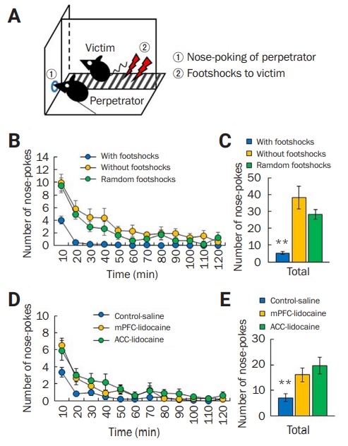

![APA format] Physiology References for the Native American Flute](https://flutopedia.com/img/JoinCVFluteNewsletter_150.jpg)