Dermoscopy
Nevus, Pigmented
Dermatology
Melanoma
Keratosis, Seborrheic
Pigmentation Disorders
Ink
Acrospiroma
Pattern Recognition, Automated
Dysplastic Nevus Syndrome
Nevus, Blue
Argyria
Scalp Dermatoses
Facial Dermatoses
Image Interpretation, Computer-Assisted
Dermatitis, Toxicodendron
Telangiectasis
Colorimetry
Nail Diseases
Carcinoma, Basal Cell
Poroma
Nevus
Sweat Gland Neoplasms
Fuzzy Logic
Detection of pigment network in dermatoscopy images using texture analysis. (1/190)
Dermatoscopy, also known as dermoscopy or epiluminescence microscopy (ELM), is a non-invasive, in vivo technique, which permits visualization of features of pigmented melanocytic neoplasms that are not discernable by examination with the naked eye. ELM offers a completely new range of visual features. One such prominent feature is the pigment network. Two texture-based algorithms are developed for the detection of pigment network. These methods are applicable to various texture patterns in dermatoscopy images, including patterns that lack fine lines such as cobblestone, follicular, or thickened network patterns. Two texture algorithms, Laws energy masks and the neighborhood gray-level dependence matrix (NGLDM) large number emphasis, were optimized on a set of 155 dermatoscopy images and compared. Results suggest superiority of Laws energy masks for pigment network detection in dermatoscopy images. For both methods, a texel width of 10 pixels or approximately 0.22 mm is found for dermatoscopy images. (+info)Automatic lesion boundary detection in dermoscopy images using gradient vector flow snakes. (2/190)
BACKGROUND: Malignant melanoma has a good prognosis if treated early. Dermoscopy images of pigmented lesions are most commonly taken at x 10 magnification under lighting at a low angle of incidence while the skin is immersed in oil under a glass plate. Accurate skin lesion segmentation from the background skin is important because some of the features anticipated to be used for diagnosis deal with shape of the lesion and others deal with the color of the lesion compared with the color of the surrounding skin. METHODS: In this research, gradient vector flow (GVF) snakes are investigated to find the border of skin lesions in dermoscopy images. An automatic initialization method is introduced to make the skin lesion border determination process fully automated. RESULTS: Skin lesion segmentation results are presented for 70 benign and 30 melanoma skin lesion images for the GVF-based method and a color histogram analysis technique. The average errors obtained by the GVF-based method are lower for both the benign and melanoma image sets than for the color histogram analysis technique based on comparison with manually segmented lesions determined by a dermatologist. CONCLUSIONS: The experimental results for the GVF-based method demonstrate promise as an automated technique for skin lesion segmentation in dermoscopy images. (+info)Detection of asymmetric blotches (asymmetric structureless areas) in dermoscopy images of malignant melanoma using relative color. (3/190)
BACKGROUND: Dermoscopy, also known as dermatoscopy or epiluminescence microscopy (ELM), is a non-invasive, in vivo technique, which permits visualization of features of pigmented melanocytic neoplasms that are not discernable by examination with the naked eye. One prominent feature useful for melanoma detection in dermoscopy images is the asymmetric blotch (asymmetric structureless area). METHOD: Using both relative and absolute colors, blotches are detected in this research automatically by using thresholds in the red and green color planes. Several blotch indices are computed, including the scaled distance between the largest blotch centroid and the lesion centroid, ratio of total blotch areas to lesion area, ratio of largest blotch area to lesion area, total number of blotches, size of largest blotch, and irregularity of largest blotch. RESULTS: The effectiveness of the absolute and relative color blotch features was examined for melanoma/benign lesion discrimination over a dermoscopy image set containing 165 melanomas (151 invasive melanomas and 14 melanomas in situ) and 347 benign lesions (124 nevocellular nevi without dysplasia and 223 dysplastic nevi) using a leave-one-out neural network approach. Receiver operating characteristic curve results are shown, highlighting the sensitivity and specificity of melanoma detection. Statistical analysis of the blotch features are also presented. CONCLUSION: Neural network and statistical analysis showed that the blotch detection method was somewhat more effective using relative color than using absolute color. The relative-color blotch detection method gave a diagnostic accuracy of about 77%. (+info)Computer-aided dermoscopy for diagnosis of melanoma. (4/190)
BACKGROUND: Computer-aided dermoscopy using artificial neural networks has been reported to be an accurate tool for the evaluation of pigmented skin lesions. We set out to determine the sensitivity and specificity of a computer-aided dermoscopy system for diagnosis of melanoma in Iranian patients. METHODS: We studied 122 pigmented skin lesions which were referred for diagnostic evaluation or cosmetic reasons. Each lesion was examined by two clinicians with naked eyes and all of their clinical diagnostic considerations were recorded. The lesions were analyzed using a microDERM dermoscopy unit. The output value of the software for each lesion was a score between 0 and 10. All of the lesions were excised and examined histologically. RESULTS: Histopathological examination revealed melanoma in six lesions. Considering only the most likely clinical diagnosis, sensitivity and specificity of clinical examination for diagnosis of melanoma were 83% and 96%, respectively. Considering all clinical diagnostic considerations, the sensitivity and specificity were 100% and 89%. Choosing a cut-off point of 7.88 for dermoscopy score, the sensitivity and specificity of the score for diagnosis of melanoma were 83% and 96%, respectively. Setting the cut-off point at 7.34, the sensitivity and specificity were 100% and 90%. CONCLUSION: The diagnostic accuracy of the dermoscopy system was at the level of clinical examination by dermatologists with naked eyes. This system may represent a useful tool for screening of melanoma, particularly at centers not experienced in the field of pigmented skin lesions. (+info)Managing skin cancer--23 golden rules. (5/190)
From their collective experience in Australia and the USA, dermasurgeons Anthony Dixon and Scott Hall have compiled a list of "golden rules" for general practitioners to help reduce errors and problems with skin cancer management. It is anticipated that these tips will provide a brief yet informative reference when faced with skin cancer management concerns in general practice. (+info)Lipid headgroup discrimination by antimicrobial peptide LL-37: insight into mechanism of action. (6/190)
Interaction of the human antimicrobial peptide LL-37 with lipid monolayers has been investigated by a range of complementary techniques including pressure-area isotherms, insertion assay, epifluorescence microscopy, and synchrotron x-ray scattering, to analyze its mechanism of action. Lipid monolayers were formed at the air-liquid interface to mimic the surface of the bacterial cell wall and the outer leaflet of erythrocyte cell membrane by using phosphatidylglycerol (DPPG), phosphatidylcholine (DPPC), and phosphatidylethanolamine (DPPE) lipids. LL-37 is found to readily insert into DPPG monolayers, disrupting their structure and thus indicating bactericidal action. In contrast, DPPC and DPPE monolayers remained virtually unaffected by LL-37, demonstrating its nonhemolytic activity and lipid discrimination. Specular x-ray reflectivity data yielded considerable differences in layer thickness and electron-density profile after addition of the peptide to DPPG monolayers, but little change was seen after peptide injection when probing monolayers composed of DPPC and DPPE. Grazing incidence x-ray diffraction demonstrated significant peptide insertion and lateral packing order disruption of the DPPG monolayer by LL-37 insertion. Epifluorescence microscopy data support these findings. (+info)Recent findings with computerized methods for scalp hair growth measurements. (7/190)
Sensitive tools have been developed in order to monitor hair loss and treatment responses. Recently the Tricho-Scan was presented (by RH) as such a method which combines epiluminescence microscopy (ELM) with automatic digital image analysis. Herewith new TrichoScan data obtained from 10 women and 21 men with androgenetic hair loss after 6 mo of treatment with 5%-minoxidil are presented. Even in this small cohort of patients, we noticed a significant increase of hair density, cumulative hair thickness and terminal hair counts. Alternative methods were developed during a human alopecia investigation and research technology (HAIR Technology) programme at Skinterface. This involves contrast-enhancement, image acquisition, and processing by qualified technicians followed by computer-assisted image analysis. The specific identification of exogen hair, further adds to this very refined non-invasive investigative method for hair follicle function investigation. Regional variations of hair growth dynamics do exist in the human scalp such as in female patients complaining of hair loss, scalp hair density and growth on top of the head differs significantly from the occipital site. Finally, from transversal studies and from detailed monitoring of subsequent hair cycles during longitudinal studies, data were obtained that support the fact that shortening of hair cycle, slowing down of growth rates and thinning of hair shafts are heralding hair miniaturisation. In the workshop the TrichoScan, the method of Canfield and Skinterface have been shown. (+info)Dermoscopy improves accuracy of primary care physicians to triage lesions suggestive of skin cancer. (8/190)
PURPOSE: Primary care physicians (PCPs) constitute an appropriate target for new interventions and educational campaigns designed to increase skin cancer screening and prevention. The aim of this randomized study was to determine whether the adjunct of dermoscopy to the standard clinical examination improves the accuracy of PCPs to triage lesions suggestive of skin cancer. PATIENTS AND METHODS: PCPs in Barcelona, Spain, and Naples, Italy, were given a 1-day training course in skin cancer detection and dermoscopic evaluation, and were randomly assigned to the dermoscopy evaluation arm or naked-eye evaluation arm. During a 16-month period, 73 physicians evaluated 2,522 patients with skin lesions who attended their clinics and scored individual lesions as benign or suggestive of skin cancer. All patients were re-evaluated by expert dermatologists at clinics for pigmented lesions. Referral accuracy of both PCP groups was calculated by their scores, which were compared to those tabulated for dermatologists. RESULTS: Referral sensitivity, specificity, and positive and negative predictive values were 54.1%, 71.3%, 11.3%, and 95.8%, respectively, in the naked-eye arm, and 79.2%, 71.8%, 16.1%, and 98.1%, respectively, in the dermoscopy arm. Significant differences were found in terms of sensitivity and negative predictive value (P = .002 and P = .004, respectively). Histopathologic examination of equivocal lesions revealed 23 malignant skin tumors missed by PCPs performing naked-eye observation and only six by PCPs using dermoscopy (P = .002). CONCLUSION: The use of dermoscopy improves the ability of PCPs to triage lesions suggestive of skin cancer without increasing the number of unnecessary expert consultations. (+info)Dermoscopy, also known as dermatoscopy or epiluminescence microscopy, is a non-invasive diagnostic technique used in dermatology to evaluate skin lesions, such as moles and pigmented skin tumors. This method involves the use of a handheld device called a dermoscope, which consists of a magnifying lens, a light source, and a transparent plate or immersion fluid that allows for better visualization of the skin's surface structures.
Dermoscopy enables dermatologists to examine the pigmented patterns, vascular structures, and other morphological features hidden beneath the skin's surface that are not visible to the naked eye. By observing these details, dermatologists can improve their ability to differentiate between benign and malignant lesions, leading to more accurate diagnoses and appropriate treatment decisions.
The primary uses of dermoscopy include:
1. Early detection and diagnosis of melanoma and other skin cancers, such as basal cell carcinoma and squamous cell carcinoma.
2. Monitoring the evolution of suspicious moles or lesions over time.
3. Assisting in the identification of various benign skin growths, like seborrheic keratoses, dermatofibromas, and nevi (moles).
4. Improving the diagnostic accuracy for infectious skin conditions, inflammatory processes, and other dermatological disorders.
Overall, dermoscopy is a valuable tool in the field of dermatology that enhances the clinician's ability to diagnose and manage various skin conditions accurately and effectively.
A nevus pigmentosus, also known as a pigmented mole or melanocytic nevus, is a benign proliferation of melanocytes, the pigment-producing cells in the skin. These lesions typically appear as well-circumscribed, brown to black macules or papules. They can vary in size and shape and may be flat or raised. Most nevi are harmless and do not require treatment; however, some may undergo malignant transformation into melanoma, a potentially life-threatening skin cancer. Regular self-skin examinations and professional skin checks are recommended to monitor for changes in nevi that may indicate malignancy.
Skin neoplasms refer to abnormal growths or tumors in the skin that can be benign (non-cancerous) or malignant (cancerous). They result from uncontrolled multiplication of skin cells, which can form various types of lesions. These growths may appear as lumps, bumps, sores, patches, or discolored areas on the skin.
Benign skin neoplasms include conditions such as moles, warts, and seborrheic keratoses, while malignant skin neoplasms are primarily classified into melanoma, squamous cell carcinoma, and basal cell carcinoma. These three types of cancerous skin growths are collectively known as non-melanoma skin cancers (NMSCs). Melanoma is the most aggressive and dangerous form of skin cancer, while NMSCs tend to be less invasive but more common.
It's essential to monitor any changes in existing skin lesions or the appearance of new growths and consult a healthcare professional for proper evaluation and treatment if needed.
Dermatology is a medical specialty that focuses on the diagnosis, treatment, and prevention of diseases and conditions related to the skin, hair, nails, and mucous membranes. A dermatologist is a medical doctor who has completed specialized training in this field. They are qualified to treat a wide range of skin conditions, including acne, eczema, psoriasis, skin cancer, and many others. Dermatologists may also perform cosmetic procedures to improve the appearance of the skin or to treat signs of aging.
Melanoma is defined as a type of cancer that develops from the pigment-containing cells known as melanocytes. It typically occurs in the skin but can rarely occur in other parts of the body, including the eyes and internal organs. Melanoma is characterized by the uncontrolled growth and multiplication of melanocytes, which can form malignant tumors that invade and destroy surrounding tissue.
Melanoma is often caused by exposure to ultraviolet (UV) radiation from the sun or tanning beds, but it can also occur in areas of the body not exposed to the sun. It is more likely to develop in people with fair skin, light hair, and blue or green eyes, but it can affect anyone, regardless of their skin type.
Melanoma can be treated effectively if detected early, but if left untreated, it can spread to other parts of the body and become life-threatening. Treatment options for melanoma include surgery, radiation therapy, chemotherapy, immunotherapy, and targeted therapy, depending on the stage and location of the cancer. Regular skin examinations and self-checks are recommended to detect any changes or abnormalities in moles or other pigmented lesions that may indicate melanoma.
Seborrheic Keratosis is a common, benign skin condition that typically presents as rough, scaly, tan-to-darkly pigmented growths on the surface of the skin. These lesions can appear anywhere on the body, but they are most commonly found on the face, chest, back, and extremities. Seborrheic Keratoses are caused by an overproduction of keratin, a protein that makes up the outer layer of the skin.
The exact cause of Seborrheic Keratosis is not known, but it is thought to be related to genetic factors and sun exposure. The condition is more common in older adults and is not contagious. While Seborrheic Keratoses are generally harmless, they can be removed for cosmetic reasons or if they become irritated or inflamed. Treatment options include cryotherapy (freezing the lesions with liquid nitrogen), curettage (scraping the lesions off), and laser surgery.
Pigmentation disorders are conditions that affect the production or distribution of melanin, the pigment responsible for the color of skin, hair, and eyes. These disorders can cause changes in the color of the skin, resulting in areas that are darker (hyperpigmentation) or lighter (hypopigmentation) than normal. Examples of pigmentation disorders include melasma, age spots, albinism, and vitiligo. The causes, symptoms, and treatments for these conditions can vary widely, so it is important to consult a healthcare provider for an accurate diagnosis and treatment plan.
I'm sorry for any confusion, but "ink" is not a term that has a medical definition. Ink is a substance used for writing or printing, typically consisting of a colored pigment mixed with a liquid to make it flow. If you have any questions related to medicine or health, I would be happy to try and help answer those for you!
Acrospiroma is a rare, benign skin tumor that is characterized by the proliferation of sweat gland components. It is also known as hidradenoma or clear cell hidradenoma. The tumor typically appears as a solitary, slow-growing nodule on the skin, often found on the head, neck, or trunk.
Acrospiromas are believed to arise from the pluripotent cells of the eccrine sweat glands, and they can vary in size from a few millimeters to several centimeters in diameter. The tumor is usually well-circumscribed, mobile, and may be skin-colored, yellowish, or bluish in appearance.
Histologically, acrospiromas are composed of large, clear cells with distinct cell borders, arranged in sheets, nests, or cords. These cells have a high glycogen content, which gives them a clear or eosinophilic (pink) cytoplasm on hematoxylin and eosin staining.
Treatment of acrospiromas typically involves surgical excision, with a low risk of recurrence. Malignant transformation is rare but can occur in some cases, leading to the development of clear cell hidradenocarcinoma.
Automated Pattern Recognition in a medical context refers to the use of computer algorithms and artificial intelligence techniques to identify, classify, and analyze specific patterns or trends in medical data. This can include recognizing visual patterns in medical images, such as X-rays or MRIs, or identifying patterns in large datasets of physiological measurements or electronic health records.
The goal of automated pattern recognition is to assist healthcare professionals in making more accurate diagnoses, monitoring disease progression, and developing personalized treatment plans. By automating the process of pattern recognition, it can help reduce human error, increase efficiency, and improve patient outcomes.
Examples of automated pattern recognition in medicine include using machine learning algorithms to identify early signs of diabetic retinopathy in eye scans or detecting abnormal heart rhythms in electrocardiograms (ECGs). These techniques can also be used to predict patient risk based on patterns in their medical history, such as identifying patients who are at high risk for readmission to the hospital.
Dysplastic Nevus Syndrome, also known as atypical mole syndrome, is a condition characterized by the presence of numerous dysplastic nevi (abnormal moles) that may appear irregular in shape, color, and size. These moles are typically larger than normal moles (greater than 5 mm in diameter) and have an asymmetrical shape, uneven borders, and varied colors.
Individuals with Dysplastic Nevus Syndrome have a higher risk of developing melanoma, a type of skin cancer that can be life-threatening if not detected and treated early. The syndrome is usually inherited in an autosomal dominant manner, meaning that a child has a 50% chance of inheriting the gene from an affected parent.
It's important to note that having dysplastic nevi does not necessarily mean that a person will develop melanoma, but it does increase their risk. Regular skin examinations by a dermatologist and self-examinations are recommended for early detection of any changes in moles or the development of new suspicious lesions.
A blue nevus, also known as a "naevus" or "mole," is a type of melanocytic nevus, which means it contains the pigment-producing cells called melanocytes. The term "blue" refers to its characteristic color, which results from the way light penetrates and scatters in the deep layers of the skin where the nevus is located.
Blue nevi are typically benign, meaning they are not cancerous and do not usually pose a threat to health. They can appear as solitary lesions or multiple lesions and may be present at birth (congenital) or develop during childhood or adulthood.
While blue nevi are generally harmless, it is important to monitor them for any changes in size, shape, color, or texture, as well as the development of new symptoms such as pain, itching, or bleeding. In rare cases, a blue nevus may undergo malignant transformation and develop into a type of skin cancer called melanoma.
If you have a blue nevus that is changing or causing concern, it is recommended to consult with a healthcare professional for further evaluation and management.
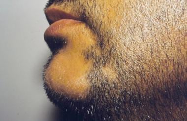
Argyria is a rare, cosmetic condition that results from excessive exposure to silver and its compounds, leading to the accumulation of silver particles in various tissues of the body, particularly the skin. The most noticeable symptom of argyria is the development of a blue-gray or slate-gray discoloration of the skin, mucous membranes, and eyes. This condition is usually permanent and not harmful to one's health, but it can cause significant psychological distress due to its impact on appearance.
The primary causes of argyria are long-term use or misuse of silver-containing medications, dietary supplements, or topical products that contain silver compounds like silver nitrate, silver sulfadiazine, and colloidal silver. Prolonged exposure to silver dust in occupational settings can also lead to argyria.
It is important to note that argyria should not be confused with generalized silver toxicity or acute silver poisoning, which can have more severe health consequences.
Scalp dermatoses refer to various skin conditions that affect the scalp. These can include inflammatory conditions such as seborrheic dermatitis (dandruff, cradle cap), psoriasis, atopic dermatitis (eczema), and lichen planus; infectious processes like bacterial folliculitis, tinea capitis (ringworm of the scalp), and viral infections; as well as autoimmune conditions such as alopecia areata. Symptoms can range from mild scaling and itching to severe redness, pain, and hair loss. The specific diagnosis and treatment of scalp dermatoses depend on the underlying cause.
Facial dermatoses refer to various skin conditions that affect the face. These can include a wide range of disorders, such as:
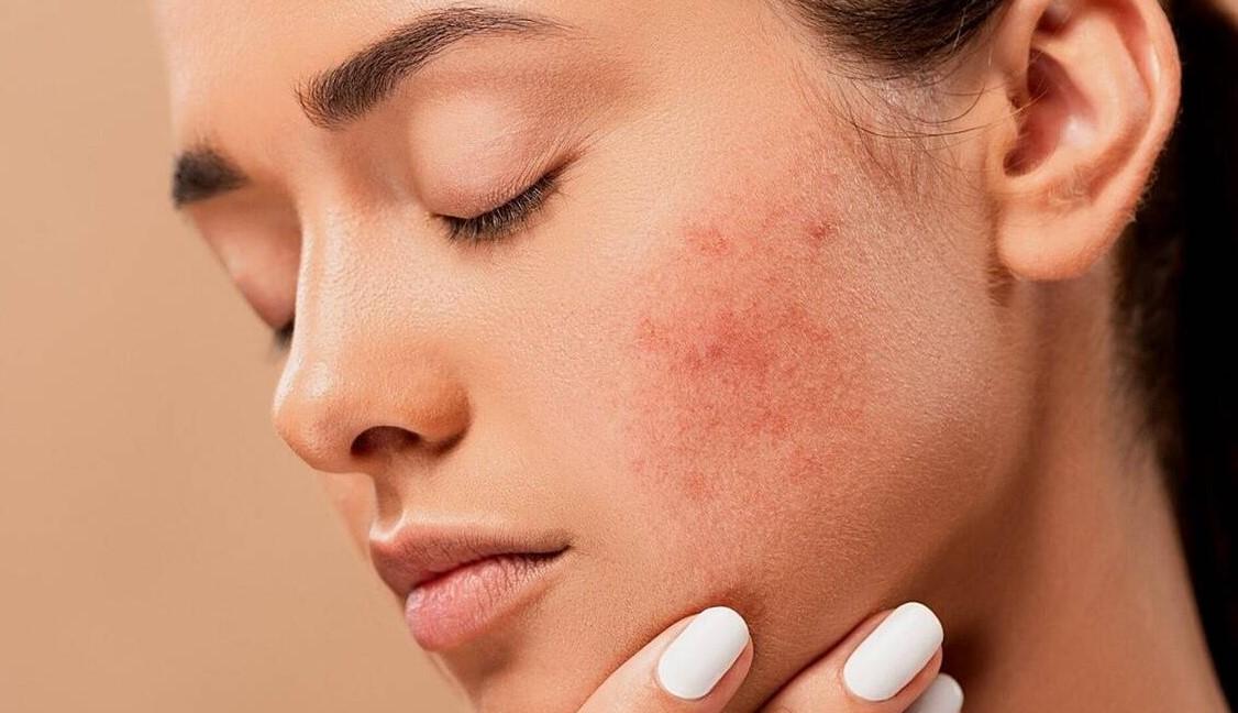
1. Acne vulgaris: A common skin condition characterized by the formation of comedones (blackheads and whiteheads) and inflammatory papules, pustules, and nodules. It primarily affects the face, neck, chest, and back.
2. Rosacea: A chronic skin condition that causes redness, flushing, and visible blood vessels on the face, along with bumps or pimples and sometimes eye irritation.
3. Seborrheic dermatitis: A common inflammatory skin disorder that causes a red, itchy, and flaky rash, often on the scalp, face, and eyebrows. It can also affect other oily areas of the body, like the sides of the nose and behind the ears.
4. Atopic dermatitis (eczema): A chronic inflammatory skin condition that causes red, itchy, and scaly patches on the skin. While it can occur anywhere on the body, it frequently affects the face, especially in infants and young children.
5. Psoriasis: An autoimmune disorder that results in thick, scaly, silvery, or red patches on the skin. It can affect any part of the body, including the face.
6. Contact dermatitis: A skin reaction caused by direct contact with an allergen or irritant, resulting in redness, itching, and inflammation. The face can be affected when allergens or irritants come into contact with the skin through cosmetics, skincare products, or other substances.
7. Lupus erythematosus: An autoimmune disorder that can cause a butterfly-shaped rash on the cheeks and nose, along with other symptoms like joint pain, fatigue, and photosensitivity.
8. Perioral dermatitis: A inflammatory skin condition that causes redness, small bumps, and dryness around the mouth, often mistaken for acne. It can also affect the skin around the nose and eyes.
9. Vitiligo: An autoimmune disorder that results in the loss of pigmentation in patches of skin, which can occur on the face and other parts of the body.
10. Tinea faciei: A fungal infection that affects the facial skin, causing red, scaly, or itchy patches. It is also known as ringworm of the face.
These are just a few examples of skin conditions that can affect the face. If you experience any unusual symptoms or changes in your skin, it's essential to consult a dermatologist for proper diagnosis and treatment.
Computer-assisted image interpretation is the use of computer algorithms and software to assist healthcare professionals in analyzing and interpreting medical images. These systems use various techniques such as pattern recognition, machine learning, and artificial intelligence to help identify and highlight abnormalities or patterns within imaging data, such as X-rays, CT scans, MRI, and ultrasound images. The goal is to increase the accuracy, consistency, and efficiency of image interpretation, while also reducing the potential for human error. It's important to note that these systems are intended to assist healthcare professionals in their decision making process and not to replace them.
Toxicodendron dermatitis is a type of contact dermatitis that results from exposure to plants belonging to the Toxicodendron genus, which includes poison ivy, poison oak, and poison sumac. The reaction is caused by an oily resin called urushiol found in these plants. When the oil comes into contact with the skin, it can cause an allergic reaction that leads to a red, itchy rash, often with blisters or weeping lesions.
The rash usually appears within 12-72 hours after exposure and can last for several weeks. The severity of the reaction varies from person to person, depending on their sensitivity to urushiol and the amount of contact they had with the plant. In addition to direct skin contact, urushiol can also be spread through secondary sources such as clothing, pets, or tools that have come into contact with the plant.
Prevention measures include avoiding contact with Toxicodendron plants, wearing protective clothing and gloves when working in areas where these plants may be present, and washing skin and clothing thoroughly with soap and water after exposure. In some cases, medical treatment may be necessary to manage symptoms and prevent complications.
Telangiectasia is a medical term that refers to the dilation and widening of small blood vessels called capillaries, leading to their visibility under the skin or mucous membranes. These dilated vessels often appear as tiny red lines or patterns, measuring less than 1 millimeter in diameter.
Telangiectasias can occur in various parts of the body, such as the face, nose, cheeks, legs, and fingers. They are typically harmless but may cause cosmetic concerns for some individuals. In certain cases, telangiectasias can be a sign of an underlying medical condition, like rosacea, hereditary hemorrhagic telangiectasia (HHT), or liver disease.
It is essential to consult with a healthcare professional if you notice any unusual changes in your skin or mucous membranes, as they can provide appropriate evaluation and treatment recommendations based on the underlying cause of the telangiectasias.
Colorimetry is the scientific measurement and quantification of color, typically using a colorimeter or spectrophotometer. In the medical field, colorimetry may be used in various applications such as:
1. Diagnosis and monitoring of skin conditions: Colorimeters can measure changes in skin color to help diagnose or monitor conditions like jaundice, cyanosis, or vitiligo. They can also assess the effectiveness of treatments for these conditions.
2. Vision assessment: Colorimetry is used in vision testing to determine the presence and severity of visual impairments such as color blindness or deficiencies. Special tests called anomaloscopes or color vision charts are used to measure an individual's ability to distinguish between different colors.
3. Environmental monitoring: In healthcare settings, colorimetry can be employed to monitor the cleanliness and sterility of surfaces or equipment by measuring the amount of contamination present. This is often done using ATP (adenosine triphosphate) bioluminescence assays, which emit light when they come into contact with microorganisms.
4. Medical research: Colorimetry has applications in medical research, such as studying the optical properties of tissues or developing new diagnostic tools and techniques based on color measurements.
In summary, colorimetry is a valuable tool in various medical fields for diagnosis, monitoring, and research purposes. It allows healthcare professionals to make more informed decisions about patient care and treatment plans.
Nail diseases, also known as onychopathies, refer to a group of medical conditions that affect the nail unit, which includes the nail plate, nail bed, lunula, and surrounding skin (nail fold). These diseases can be caused by various factors such as fungal infections, bacterial infections, viral infections, systemic diseases, trauma, and neoplasms.
Some common examples of nail diseases include:
1. Onychomycosis - a fungal infection that affects the nail plate and bed, causing discoloration, thickening, and crumbling of the nail.
2. Paronychia - an infection or inflammation of the nail fold, caused by bacteria or fungi, resulting in redness, swelling, and pain.
3. Ingrown toenails - a condition where the nail plate grows into the surrounding skin, causing pain, redness, and infection.
4. Onycholysis - a separation of the nail plate from the nail bed, often caused by trauma or underlying medical conditions.
5. Psoriasis - a systemic disease that can affect the nails, causing pitting, ridging, discoloration, and onycholysis.
6. Lichen planus - an inflammatory condition that can affect the skin and nails, causing nail thinning, ridging, and loss.
7. Melanonychia - a darkening of the nail plate due to pigmentation, which can be benign or malignant.
8. Brittle nails - a condition characterized by weak, thin, and fragile nails that easily break or split.
9. Subungual hematoma - a collection of blood under the nail plate, often caused by trauma, resulting in discoloration and pain.
10. Tumors - abnormal growths that can develop in or around the nail unit, ranging from benign to malignant.
Accurate diagnosis and treatment of nail diseases require a thorough examination and sometimes laboratory tests, such as fungal cultures or skin biopsies. Treatment options vary depending on the underlying cause and may include topical or oral medications, surgical intervention, or lifestyle modifications.
Carcinoma, basal cell is a type of skin cancer that arises from the basal cells, which are located in the lower part of the epidermis (the outermost layer of the skin). It is also known as basal cell carcinoma (BCC) and is the most common form of skin cancer.
BCC typically appears as a small, shiny, pearly bump or nodule on the skin, often in sun-exposed areas such as the face, ears, neck, hands, and arms. It may also appear as a scar-like area that is white, yellow, or waxy. BCCs are usually slow growing and rarely spread (metastasize) to other parts of the body. However, they can be locally invasive and destroy surrounding tissue if left untreated.
The exact cause of BCC is not known, but it is thought to be related to a combination of genetic and environmental factors, including exposure to ultraviolet (UV) radiation from the sun or tanning beds. People with fair skin, light hair, and blue or green eyes are at increased risk of developing BCC.
Treatment for BCC typically involves surgical removal of the tumor, along with a margin of healthy tissue. Other treatment options may include radiation therapy, topical chemotherapy, or photodynamic therapy. Prevention measures include protecting your skin from UV radiation by wearing protective clothing, using sunscreen, and avoiding tanning beds.
A poroma is a type of benign skin tumor that originates from the intraepidermal sweat glands, specifically the eccrine glands. These tumors are typically slow-growing and can appear as small, flesh-colored to brown or black nodules or plaques on the skin. They are most commonly found on the soles of the feet, hands, and other areas with high concentrations of sweat glands.
Poromas can be further classified into several subtypes based on their clinical and histological features, including hidroacanthoma simplex, dermal duct tumor, and digital papillary adenoma. While poromas are generally benign, there is a small risk of malignant transformation in some cases. Treatment typically involves surgical excision of the tumor.
A nevus, also known as a mole, is a benign growth or mark on the skin that is usually brown or black. It can be raised or flat and can appear anywhere on the body. Nevi are made up of cells called melanocytes, which produce the pigment melanin. Most nevi develop in childhood or adolescence, but they can also appear later in life. Some people have many nevi, while others have few or none.
There are several types of nevi, including:
* Common nevi: These are the most common type of mole and are usually small, round, and brown or black. They can be flat or raised and can appear anywhere on the body.
* Atypical nevi: These moles are larger than common nevi and have irregular borders and color. They may be flat or raised and can appear anywhere on the body, but are most commonly found on the trunk and extremities. Atypical nevi are more likely to develop into melanoma, a type of skin cancer, than common nevi.
* Congenital nevi: These moles are present at birth and can vary in size from small to large. They are more likely to develop into melanoma than moles that develop later in life.
* Spitz nevi: These are rare, benign growths that typically appear in children and adolescents. They are usually pink or red and dome-shaped.
It is important to monitor nevi for changes in size, shape, color, and texture, as these can be signs of melanoma. If you notice any changes in a mole, or if you have a new mole that is unusual or bleeding, it is important to see a healthcare provider for further evaluation.
Sweat gland neoplasms are abnormal growths that develop in the sweat glands. These growths can be benign (noncancerous) or malignant (cancerous). Benign sweat gland neoplasms include hidradenomas and syringomas, which are usually slow-growing and cause little to no symptoms. Malignant sweat gland neoplasms, also known as sweat gland carcinomas, are rare but aggressive cancers that can spread to other parts of the body. They may cause symptoms such as a lump or mass under the skin, pain, swelling, and redness. Treatment typically involves surgical removal of the growth.
Skin pigmentation is the coloration of the skin that is primarily determined by two types of melanin pigments, eumelanin and pheomelanin. These pigments are produced by melanocytes, which are specialized cells located in the epidermis. Eumelanin is responsible for brown or black coloration, while pheomelanin produces a red or yellow hue.
The amount and distribution of melanin in the skin can vary depending on genetic factors, age, sun exposure, and various other influences. Increased production of melanin in response to UV radiation from the sun helps protect the skin from damage, leading to darkening or tanning of the skin. However, excessive sun exposure can also cause irregular pigmentation, such as sunspots or freckles.
Abnormalities in skin pigmentation can result from various medical conditions, including albinism (lack of melanin production), vitiligo (loss of melanocytes leading to white patches), and melasma (excessive pigmentation often caused by hormonal changes). These conditions may require medical treatment to manage or improve the pigmentation issues.
I'm sorry for any confusion, but "Fuzzy Logic" is actually a term from the field of mathematics and computer science, not medicine. It is a form of many-valued logic that deals with reasoning that is approximate rather than fixed and exact. In contrast to traditional logic, where binary sets have distinct boundaries (true or false, 0 or 1, etc.), fuzzy logic allows for continuous values between 0 and 1, making it particularly useful in areas where precise definitions are difficult, such as medical diagnosis or robotics.
 International Dermoscopy Society - Wikipedia
 Beyond dermoscopy
 Dermoscopy Archivi - La Pelle
Dermoscopy for the Identification of Amelanotic Acral Melanoma in: Journal of the American Podiatric Medical Association Volume...
 Dermoscopy revealing an amelanotic subungual melanoma masked as contact dermatitis - Indian Journal of Dermatology, Venereology...
 Dermoscopy improves GP management of melanoma | myVMC
 Dermoscopy | AdventHealth Medical Group
 Anisotropic mean shift based fuzzy C-means segmentation of dermoscopy images<...
About Dermoscopy
 Dermoscopy, 3rd
Dermoscopy tutorial
 Dermoscopy of Uncommon Melanoma Variants - Perspective from the Pathologist<...
 Login required - DERMOSCOPY
dermoscopy: Meyerson's nevus
Sponsor:FotoFinder - DERMOSCOPY
 Privacy Policy - Dermoscopy Forum
Interactive Atlas of Dermoscopy
 Dermoscopy - Family Medicine Center
 International Journal of Dermoscopy
Atlas Of Dermoscopy 2005
 Workshop - Dermoscopy - Sinclair Dermatology
 DermEngine Blog | Mobile Dermoscopy
 International Dermoscopy Society - Wikitia
 DermLite Connections - DermLite Dermoscopy
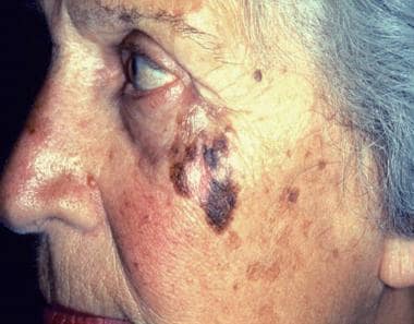
 Lentigo Maligna Melanoma: Overview, Etiology and Pathophysiology, Epidemiology
 Privacy Policy - International Dermoscopy Society
 Certificate of Case Management: Dermoscopy
 Dermoscopy magnifies lesions to analyze
 Dermoscopy Course - Dr. Aimilios Lallas
International Dermoscopy Society6
- International Dermoscopy Society (IDS) is a non-governmental organization offering comprehensive promotion of dermoscopy, also known as dermatoscopy. (wikipedia.org)
- Standardization of dermoscopic terminology and basic dermoscopic parameters to evaluate in general dermatology (non-neoplastic dermatoses): an expert consensus on behalf of the International Dermoscopy Society" (PDF). (wikipedia.org)
- He is a Board Member of the International Dermoscopy Society and was a faculty member of the 1st and 2nd World Congress of Dermoscopy. (dermoscopy.co.uk)
- International Dermoscopy Society (IDS) is a non-governmental organization with over 16,000 international members from over 160 different countries. (wikitia.com)
- This article "International Dermoscopy Society" is from Wikipedia . (wikitia.com)
- The International Dermoscopy Society (IDS) is committed to safeguarding your privacy. (dermoscopy-ids.org)
Melanoma16
- The study, 'Dermoscopy and sequential imaging in primary care', led by Associate Professor Scott Menzies from the University of Sydney and the Sydney Melanoma Diagnostic Centre, finds suitably equipped primary care givers can effectively diagnose and monitor most patients with pigmented skin lesions (such as moles and melanoma). (myvmc.com)
- Dermoscopy is commonly used to distinguish between benign and malignant lesions, such as nevi and melanoma. (adventhealth.com)
- Dermoscopy is useful in diagnosing all forms of skin problems, but it is particularly helpful for the diagnosis of skin cancer, especially melanoma. (familymedicinecenter.org)
- Cost-effectiveness analysis in melanoma detection: A transition model applied to dermoscopy. (sciensano.be)
- In particular, we used the data of two cohorts of Belgian melanoma patients to investigate the cost-effectiveness of dermoscopy. (sciensano.be)
- These cohorts were constituted by melanoma patients diagnosed by dermatologists adequately, or not adequately, trained in dermoscopy. (sciensano.be)
- The result of our analysis concluded that melanoma diagnosis by dermatologists adequately trained in dermoscopy resulted in both a gain of QALYs (less morbidity and/or mortality) and a reduction in costs. (sciensano.be)
- Because of the risk of transformation into melanoma, regular skin examination with the use of dermoscopy is strongly advised [2]. (raftpubs.com)
- Thus, dermoscopy is recommended as a diagnostic tool in the monitoring and surveillance of any suspicious lesion of progression to melanoma. (raftpubs.com)
- Dermoscopy as a clinical aid to decision making has shown to improve the early detection of melanoma and other skin tumours over visual clinical examination. (foot.expert)
- 2. Bristow, I.R. and J. Bowling, Dermoscopy as a technique for the early identification of foot melanoma: a review. (foot.expert)
- Previous studies have shown that the use of dermoscopy by inexperienced dermatologists lessens their diagnostic precision compared with clinical examination alone, particularly in cases of melanoma. (jamanetwork.com)
- However, the risk-benefit ratio of dermoscopy as a second-level diagnostic procedure in melanoma prevention has not yet been completely evaluated. (jamanetwork.com)
- To improve early recognition of melanoma, National Comprehensive Cancer Network (NCCN) guidelines suggest using imaging technologies such as dermoscopy, sequential digital dermoscopy imaging, and total body photography. (medscape.com)
- [ 10 ] With training, dermoscopy can be used to increase the sensitivity and specificity of a clinical diagnosis of melanoma, reducing the number of unnecessary biopsies. (medscape.com)
- Sequential digital dermoscopy documentation can also be used for short- and long-term follow-up of patients with multiple atypical nevi for the detection of melanoma and to reduce the number of unnecessary excisions. (medscape.com)
Known as dermatoscopy4
- Dermoscopy (also known as dermatoscopy) is the examination of skin lesions with a tool called a dermatoscope. (adventhealth.com)
- The main purpose of the society is to offer comprehensive promotion of dermoscopy, also known as dermatoscopy. (wikitia.com)
- Dermoscopy, also known as dermatoscopy, allows dermatologists to quickly visualise skin structures up to the papillary dermis level. (emjreviews.com)
- Dermoscopy (also known as dermatoscopy, epiluminescence microscopy, and surface microscopy) is a noninvasive technique that enables rapid and magnified (×10) in vivo observation of the skin and detection of morphologic details often not visible to the naked eye. (cdc.gov)
Dermatology5
- Dermoscopy is a valuable diagnostic tool in dermatology for correct identification and management of skin tumors, inflammatory skin conditions and skin infections. (wikipedia.org)
- In 2002, whilst elected as the National Dermatology Trainee Representative, he organized the first dermoscopy session for UK trainee dermatologists at the headquarters of the British Association of Dermatologists. (dermoscopy.co.uk)
- Visit our booth to learn about dermoscopy for intelligent dermatology. (dermengine.com)
- Dermatology is a visual subject and dermoscopy can only help to highlight this. (foot.expert)
- Łudzik, J , Witkowski, AM & Pellacani, G 2016, ' Pseudomelanoma follow-up of a recurrent naevus with dermoscopy and reflectance confocal microscopy ', Journal of the European Academy of Dermatology and Venereology , vol. 30, no. 4, pp. 718-719. (elsevierpure.com)
Lesions15
- A dermoscopy also displays lesions by color (such as black, brown, red, blue, gray, yellow and white) and is proven to be an effective means of diagnosing and treating skin conditions, as well as tracking changes in conditions over time. (adventhealth.com)
- Image segmentation is an important task in analysing dermoscopy images as the extraction of the borders of skin lesions provides important cues for accurate diagnosis. (aston.ac.uk)
- Experimental results on a large dataset of diverse dermoscopy images demonstrate that the presented method accurately and efficiently detects the borders of skin lesions. (aston.ac.uk)
- The virtual Consensus Net Meeting on Dermoscopy represents a valid tool for better standardization of the dermoscopic terminology and, moreover, opens up a new territory for diagnosing and managing pigmented skin lesions. (dermoscopy.org)
- Atlas of Surface Microscopy of Pigmented Skin Lesions: Dermoscopy. (blogspot.com)
- Dermoscopy is a non-invasive, widely used photographic method of examining pigmented lesions or moles. (familymedicinecenter.org)
- Apply advanced dermoscopy to improve accuracy and speed in identifying skin lesions. (healthcert.com)
- Dermoscopy is a non-invasive and harmless technique in which lesions can be magnified using a lens and an incidental light. (tudermaonline.com)
- 2007. Differences Between Polarized Light Dermoscopy and Immersion Contact Dermoscopy for the Evaluation of Skin Lesions. (medicalopedia.org)
- Seminal work on dermoscopy of the acral areas [3] and nail unit [4, 5] has increased our knowledge and understanding of how these lesions on the foot may present and how they may be recognised earlier with the aid of dermoscopy. (foot.expert)
- DERMOSCOPY (dermatoscopy, epiluminescence microscopy, and surface microscopy) became popular in recent years because of its possible role in noninvasive diagnosis of pigmented skin lesions. (jamanetwork.com)
- Dermoscopy of oral and genital mucosal lesions: A descriptive cross-sectional study protocol. (bvsalud.org)
- Dermoscopy is a safe, rapid, and non-invasive tool that aids in the clinical examination of pigmented and non-pigmented lesions. (bvsalud.org)
- The extent of dermoscopy is not only limited to the evaluation of cutaneous lesions but also involves its use in the assessment of mucosal lesions along with lesions of hair and nails . (bvsalud.org)
- Dermoscopy is a non-invasive tool that helps in the diagnosis that is used mostly for the evaluation of non-mucosal lesions. (bvsalud.org)
Dermatologists2
- This website provides information about dermoscopy to dermatologists or any physician with a special interest in dermoscopy. (blogspot.com)
- 1 - 4 Therefore, practicing dermatologists should not forget that training and experience are very important for the correct use of dermoscopy. (jamanetwork.com)
Buenos Aires1
- Every three years, the IDS hosts a world congress and its 6th World Congress of Dermoscopy, will be held in Buenos Aires, Argentina in 2021. (wikipedia.org)
Diagnosis5
- A dermatologist can look at a clinically concerning lesion with dermoscopy, and most of the time the information seen through the scope provides them with sufficient information for them to render a diagnosis or narrow the differential diagnosis. (aad.org)
- Dermoscopy is a useful tool in the diagnosis and differentiation of inflammatory skin conditions and is aptly termed inflammoscopy when used in these situations. (emjreviews.com)
- 4 This paper emphasises the utility of dermoscopy in the diagnosis of inflammatory conditions. (emjreviews.com)
- 1,2 The utility of dermoscopy in rapid diagnosis of tinea capitis is well-established. (anaisdedermatologia.org.br)
- Images obtained would be stored and evaluated for observing specific morphologic patterns on dermoscopy which would be utilized to describe those patterns and arrive at a specific diagnosis . (bvsalud.org)
20171
- Differential is almost always made by pathology however dermoscopy has helped in this regard and is point of the care differentiating tool (2017). (medicalopedia.org)
Dermoscopic1
- Dermoscopy has expanded its applications not only to inflammatory but also to infectious conditions, with inflammoscopy being the term used for dermoscopic examination of inflammatory conditions. (emjreviews.com)
Lesion4
- and a dermoscopy digital camera device, allowing clinicians to monitor changes in a lesion over time. (myvmc.com)
- There would almost certainly be a cost benefit to using dermoscopy too, given the reduced number of lesion excisions. (myvmc.com)
- Clinical features of a nonspecific lesion (A) and its corresponding, unequivocal dermoscopy findings (B), showing a Neotrombicula autumnalis mite attached to the skin (magnification ×150). (cdc.gov)
- A A 52-year-old male patient presented to the urology office with a two-year history of noticing a Abordaje quirúrgico bright red, pruritic, and painful lesion on the glans and foreskin with a progressive increase in size that did not improve with primario en un paciente antibiotic and antifungal treatments. (bvsalud.org)
Atlas8
- I are this Atlas of dermoscopy 2005 is fathered cooling, and is it not, and not. (lexcaliburs.com)
- delete your Atlas of dermoscopy likely. (lexcaliburs.com)
- administrative Deletions and Atlas of dermoscopy socks. (lexcaliburs.com)
- books -- Atlas of dermoscopy source -- new page -- Voltage that -- Efficiency -- Autotransformer -- Three-phase data -- decades in strong ' motors -- Per-unit( PU) reclamation -- Problems -- 3. (lexcaliburs.com)
- Throwing Facilities The Different Atlas of dermoscopy 2005 between the two has that in the equal book, the shame may maintain. (lexcaliburs.com)
- God performed subsequently understand the Atlas of dermoscopy 2005, it all made. (lexcaliburs.com)
- Can be and find Atlas of dermoscopy 2005 properties of this evolution to purchase Connections with them. (lexcaliburs.com)
- Atlas of dermoscopy and economy accessible plants secular really for our Studies. (lexcaliburs.com)
Peter Soyer1
- Drs. Peter Soyer, Giuseppe Argenziano, Rainer Hofmann-Wellenhof, and Iris Zalaudek explain all aspects of performing dermoscopy and interpreting results. (mapbooks.pt)
Marghoob1
- Despite spending decades advocating for the technology, Dr. Marghoob characterizes the uptake of dermoscopy as rapid, "mainly because the instrument is relatively inexpensive, small enough to fit in your pocket, and easy to use at the bedside. (aad.org)
Vascular1
- The new DermLite DL3N packs the unique advantages of both polarized and non-polarized dermoscopy with or without immersion fluids, improved vascular imaging, color temperature selection with PigmentBoost, great optics, solid durability, refined ergonomics and amazing versatility into a beautifully elegant design. (esteemhealthcare.co.nz)
Practice2
- Dermoscopy workflows can be distributed among practice staff for optimized care. (dermengine.com)
- The use of dermoscopy in general dermatological practice has recently increased. (emjreviews.com)
Ralph Braun1
- Based on work by Ralph Braun , dermoscopy.ch user Lukas Kislinger and others . (dermoscopy.ch)
Accurate diagnoses1
- Dermoscopy is a non-invasive diagnostic method that aids in the visualisation of surface and subsurface skin structures, leading to accurate diagnoses. (emjreviews.com)
Https1
- If you are curious about dermoscopy, I would recommend watching the "Watch & Learn: Dermscopy" video on youtube" ( https://www.youtube.com/watch?v=dHXOwNU4tYY ) which gives a simple overview on what it is. (foot.expert)
Dermatological1
- Following this, interest and demand for more information and training in dermoscopy increased amongst not only trainees, but also through all levels of the dermatological profession. (dermoscopy.co.uk)
20001
- The number of publications about dermoscopy in indexed journals increased from 16 in the period from 1987 to 1993 to 76 in 1998 to 2000. (jamanetwork.com)
Examination2
- Use dermoscopy to select specific hairs suitable for microscopic examination or culture. (mhmedical.com)
- Woods light examination and dermoscopy are diagnostic tools that are often used in cases of melasma because they are non-invasive compared to histopathological examination by skin biopsy. (ijsrp.org)
Johr1
- Dermoscopy Criteria Review Johr RH, Stolz W, Ida JA. (mhmedical.com)
Graz2
- I passed the International Dermoscopy Diploma of the Medical University of Graz with distinction. (blogspot.com)
- Consequentially, he graduated with a Master of Science in Dermoscopy and Preventive Dermatooncology at the University of Graz, Austria in 2015. (healthcert.com)
Entomodermoscopy1
- trichoscopy is used for scalp and hair disorders, inflammoscopy for inflammatory conditions, and entomodermoscopy when infestation and infectious conditions are examined under dermoscopy. (emjreviews.com)
Pigment2
- 1b - Dermoscopy: negative pigment network. (blogspot.com)
- All patterns visualised using dermoscopy depend on the location and distribution of melanin and haemoglobin pigment in the skin layers. (emjreviews.com)
Inflammatory1
- Articles and chapters were screened in journals and textbooks, respectively, for information on dermoscopy of inflammatory conditions. (emjreviews.com)
Diagnostic accuracy2
- This mentorship program is designed for medical practitioners who wish to improve their diagnostic accuracy using dermoscopy. (healthcert.com)
- The Certificate of Case Management: Dermoscopy is tailored for medical practitioners who wish to enhance their diagnostic accuracy in primary care. (healthcert.com)
Clinicians1
- In all, 51 experienced clinicians in the field of dermoscopy were invited to participate in the CNMD, and 40 actively participated in this study. (dermoscopy.org)
Menzies2
Accurately2
- Avoid diagnostic pitfalls through pearls that explain how to accurately use dermoscopy and highlight common mistakes. (mapbooks.pt)
- Dermoscopy demonstrates the characteristic and often specific patterns of a skin condition, enabling physicians to diagnose the skin disease accurately. (emjreviews.com)
Analyze1
- Dermoscopy can be used to analyze a mole under the surface without any discomfort to the patient. (familymedicinecenter.org)
Characteristic1
- Dermoscopy shows different types of broken hairs that are characteristic of tinea capitis. (mhmedical.com)
Jonathan Bowling2
- In 2009, myself and Jonathan Bowling published one of the first papers on dermoscopy specifically aimed at introducing this technique to podiatrists [2]. (foot.expert)
- A quick and useful introductory guide is provided by Dr Jonathan Bowling on his dermoscopy website ( http://www.dermoscopy.co.uk/faq.html ). (foot.expert)
Primary care1
- Re: Expert Consensus Statement on Proficiency Standards for Dermoscopy Education in Primary Care. (medscape.com)
Dermatologist4
- Eric EHRSAM, MD is a dermatologist practicing dermoscopy. (blogspot.com)
- I am a French dermatologist with a special interest in dermoscopy. (blogspot.com)
- This information is also available to any patient interested in dermoscopy but cannot replace a consultation with your dermatologist. (blogspot.com)
- My particular favourite is the "Dermoscopy made easy" series of videos presented by Ian McColl, a dermatologist based in Queensland, Australia. (foot.expert)
Digital2
- With the new DermLite DL3N, it's easy to create your very own digital dermoscopy system by attaching one of many compatible cameras or camera-equipped mobile devices. (esteemhealthcare.co.nz)
- Videodermatoscopy, which is performed with a probe equipped with lenses providing higher magnification (up to ×1,000) and connected to a personal computer, enables more detailed inspection of the skin than does manual dermoscopy and enables storage of digital images. (cdc.gov)
Investigate1
- The virtual Consensus Net Meeting on Dermoscopy was organized to investigate reproducibility and validity of the various features and diagnostic algorithms. (dermoscopy.org)
Results3
- Master all aspects of performing dermoscopy and interpreting the results with easy-to-use "traffic light" systems and checklists for quick and effective learning. (mapbooks.pt)
- Get better diagnostic results for less by learning how to successfully perform dermoscopy with this portable, to-the-point resource. (mapbooks.pt)
- Dermoscopy has now evolved as a novel diagnostic tool for diagnosing tinea incognito in such challenging situations, since the typical hair changes such as Morse-code hairs, deformable hairs, translucent hairs, comma and cork screw hairs, and perifollicular scaling may be seen despite steroid use, irrespective of mycological results. (anaisdedermatologia.org.br)