Dactinomycin
Gestational Trophoblastic Disease
Wilms Tumor
Leukocyte Disorders
Rhabdomyosarcoma
Trophoblastic Neoplasms
Ifosfamide
Etoposide
Sarcoma, Ewing
Antineoplastic Combined Chemotherapy Protocols
Cyclophosphamide
Neoplasms, Germ Cell and Embryonal
Combined Modality Therapy
Doxorubicin
Bleomycin
Methotrexate
Drug Administration Schedule
Neoplasm Recurrence, Local
Disease-Free Survival
Neoplasm Staging
Treatment Outcome
Effect of hepatocarcinogens on the binding of glucocorticoid-receptor complex in rat liver nuclei. (1/3633)
The effects of a number of carcinogens and hepatotoxins on the binding kinetics of the interactions of glucocorticoidcytosol receptor complex with nuclear acceptor sites in rat liver were investigated. Both the apparent sites in rat liver were investigated. Both the apparent concentration of nuclear binding sites and the Kd were significantly diminished following treatment of rats with sublethal doses of the carcinogens aflatoxin B1, diethylnitrosamine, dimethylnitrosamine, thioacetamide, 3'-methyl-4-dimethylaminoazobenzene, 4-dimethylaminoazobenzene, and 3-methylcholanthrene. Treatment with actinomycin D resulted in a slight reduction in the apparent concentration of nuclear acceptor sites but had no effect on the nuclear binding Kd. The hepatotoxic but noncarcinogenic analgesic, acetaminophen, as well as the weakly toxic aflatoxin B1 cognate, aflatoxin B2, were without effect on the kinetics or binding capacity of glucocorticoid-nuclear acceptor site interaction. These experiments suggest that chemically induced alteration of functional glucocorticoid binding sites on chromatin may be involved in the biochemical effects produced in liver by carcinogens of several chemical types. This experimental model may provide a useful approach for further elucidation of early events in carcinogenesis. (+info)The stability and fate of a spliced intron from vertebrate cells. (2/3633)
Introns constitute most of the length of typical pre-mRNAs in vertebrate cells. Thus, the turnover rate of introns may significantly influence the availability of ribonucleotides and splicing factors for further rounds of transcription and RNA splicing, respectively. Given the importance of intron turnover, it is surprising that there have been no reports on the half-life of introns from higher eukaryotic cells. Here, we determined the stability of IVS1Cbeta1, the first intron from the constant region of the mouse T-cell receptor-beta, (TCR-beta) gene. Using a tetracycline (tet)-regulated promoter, we demonstrate that spliced IVS1Cbeta1 and its pre-mRNA had half-lives of 6.0+/-1.4 min and 3.7+/-1.0 min, respectively. We also examined the half-lives of these transcripts by using actinomycin D (Act.D). Act.D significantly stabilized IVS1Cbeta1 and its pre-mRNA, suggesting that Act.D not only blocks transcription but exerts rapid and direct posttranscriptional effects in the nucleus. We observed that in vivo spliced IVS1Cbeta1 accumulated predominantly as lariat molecules that use a consensus branchpoint nucleotide. The accumulation of IVS1Cbeta1 as a lariat did not result from an intrinsic inability to be debranched, as it could be debranched in vitro, albeit somewhat less efficiently than an adenovirus intron. Subcellular-fractionation and sucrose-gradient analyses showed that most spliced IVS1Cbeta1 lariats cofractionated with pre-mRNA, but not always with mRNA in the nucleus. Some IVS1Cbeta1 also appeared to be selectively exported to the cytoplasm, whereas TCR-beta pre-mRNA remained in the nucleus. This study constitutes the first detailed analysis of the stability and fate of a spliced nuclear intron in vivo. (+info)Retinoic acid stimulates the expression of 11beta-hydroxysteroid dehydrogenase type 2 in human choriocarcinoma JEG-3 cells. (3/3633)
The syncytiotrophoblasts of the human placenta express high levels of 11beta-hydroxysteroid dehydrogenase type 2 (11beta-HSD2), the enzyme responsible for the inactivation of glucocorticoids. It has been proposed that the placental 11beta-HSD2 serves as a barrier to protect the fetus from high levels of maternal cortisol. To examine the hypothesis that nutritional signals regulate the expression of 11beta-HSD2 in placental syncytiotrophoblasts, we investigated the effects of retinoic acids (RAs), the major metabolites of vitamin A, on the expression of 11beta-HSD2 using human choriocarcinoma JEG-3 cells as a model. This trophoblast-like cell line displays a number of functional similarities to the syncytiotrophoblast. Treatment for 24 h with all-trans RA (1-1000 nM) resulted in a dose-dependent increase in 11beta-HSD2 activity with a maximal effect (increase to 3-fold) at 100 nM. The effect of all-trans RA (100 nM) was also time-dependent in that the effect was detectable at 6 h and reached its maximum by 48 h. Similar increases in 11beta-HSD2 activity were observed when the cells were treated with 9-cis RA. Results from semi-quantitative reverse transcription-polymerase chain reaction demonstrated that there was a corresponding increase in 11beta-HSD2 mRNA after RA treatment. Moreover, treatment with actinomycin D (100 ng/ml) abrogated the increase in 11beta-HSD2 mRNA induced by RA, indicating an effect on transcription. In conclusion, the present study has demonstrated for the first time that RA, at physiological concentrations, induces 11beta-HSD2 gene expression and enzyme activity in JEG-3 cells. If this occurs in vivo, the present finding suggests that high expression of 11beta-HSD2 in the human placenta may be maintained, at least in part, by dietary intake of vitamin A. (+info)Inhibition of Pichinde virus replication by actinomycin D. (4/3633)
The yields of Pichinde virus, a member of the arenavirus group, were markedly inhibited when infected BHK 21 cells were incubated in the presence of 0.4 to 4 mug/ml of actinomycin D. Maximal inhibition was observed when actinomycin D was added after the adsorption of virus to cultures; however, addition of drug as late as 12 h after infection reduced the 24 h yield by 50%. Virus antigen synthesis, as measured by complement fixation and immunodiffusion, was not dramatically reduced by actinomycin D. The expression of virus antigens on the surface of infected cells was greater on cells treated with actinomycin D than on untreated cells. Putative defective particles with a density of Pichinde virus were not detected in fluids of cultures incubated with actinomycin D and 3H-amino acids. Actinomycin D appears to inhibit Pichinde virus late in the replicative cycle. The observations raise the possibility that the drug inhibits the synthesis of proteins of the host cell membrane which are required for virus maturation. (+info)Regulation of interleukin-8 expression by reduced oxygen pressure in human glioblastoma. (5/3633)
Oxygen deprivation is an important biological feature of tumor growth. We previously showed that in glioma, anoxia increases expression of IL-8, a chemokine and angiogenic factor. Here, we analysed for the first time the biochemical mechanisms inducing the IL-8 gene upon anoxia in glioma cells, and showed that they differ from those inducing the VEGF gene. Both genes are induced in biologically and genetically heterogenous glioblastoma cell lines (LN-229, LN-Z308, U87MG, T98G), whereas, in gliosarcoma cells (D247MG), only the VEGF gene is induced. The kinetics of IL-8 and VEGF mRNA inductions differ in these cells and reoxygenation experiments showed that the induction is due to the anoxic stress per se. Furthermore, in LN-229 and LN-Z308 cell lines actinomycin D, DRB and nuclear run-on experiments showed that anoxia stimulates increased transcription of both genes. Electromobility shift assays show increased protein binding to the AP-1 site on the IL-8 promoter following anoxia treatment. Finally, in situ hybridization on glioblastoma sections shows that the in vivo expression patterns of IL-8 and VEGF genes overlap, but are not identical. Since intratumoral augmentation of IL-8 and VEGF secretion, following microenvironmental decreases in oxygen pressure, may promote angiogenesis, further definition of these pathways is essential to appropriately target them for antitumoral therapy. (+info)Nuclear foci of mammalian recombination proteins are located at single-stranded DNA regions formed after DNA damage. (6/3633)
A sensitive and rapid in situ method was developed to visualize sites of single-stranded (ss) DNA in cultured cells and in experimental test animals. Anti-bromodeoxyuridine antibody recognizes the halogenated base analog incorporated into chromosomal DNA only when substituted DNA is in the single strand form. After treatment of cells with DNA-damaging agents or gamma irradiation, ssDNA molecules form nuclear foci in a dose-dependent manner within 60 min. The mammalian recombination protein Rad51 and the replication protein A then accumulate at sites of ssDNA and form foci, suggesting that these are sites of recombinational DNA repair. (+info)Interaction of asparagine and EGF in the regulation of ornithine decarboxylase in IEC-6 cells. (7/3633)
Our laboratory has shown that asparagine (ASN) stimulates both ornithine decarboxylase (ODC) activity and gene expression in an intestinal epithelial cell line (IEC-6). The effect of ASN is specific, and other A- and N-system amino acids are almost as effective as ASN when added alone. In the present study, epidermal growth factor (EGF) was unable to increase ODC activity in cells maintained in a salt-glucose solution (Earle's balanced salt solution). However, the addition of ASN (10 mM) in the presence of EGF (30 ng/ml) increased the activity of ODC 0.5- to 4-fold over that stimulated by ASN alone. EGF also showed induction of ODC with glutamine and alpha-aminoisobutyric acid, but ODC induction was maximum with ASN and EGF. Thus the mechanism of the interaction between ASN and EGF is important for understanding the regulation of ODC under physiological conditions. Therefore, we examined the expression of the ODC gene and those for several protooncogenes under the same conditions. Increased expression of the genes for c-Jun and c-Fos but not for ODC occurred with EGF alone. The addition of ASN did not further increase the expression of the protooncogenes, but the combination of EGF and ASN further increased the expression of ODC over that of ASN alone. Western analysis showed no significant difference in the level of ODC protein in Earle's balanced salt solution, ASN, EGF, or EGF plus ASN. Addition of cycloheximide during ASN and ASN plus EGF treatment completely inhibited ODC activity without affecting the level of ODC protein. These results indicated that 1) the increased expression of protooncogenes in response to EGF is independent of increases in ODC activity and 2) potentiation between EGF and ASN on ODC activity may not be due to increased gene transcription but to posttranslational regulation and the requirement of ongoing protein synthesis involving a specific factor dependent on ASN. (+info)Stochastic and nonstochastic post-transcriptional silencing of chitinase and beta-1,3-glucanase genes involves increased RNA turnover-possible role for ribosome-independent RNA degradation. (8/3633)
Stochastic and nonstochastic post-transcriptional gene silencing (PTGS) in Nicotiana sylvestris plants carrying tobacco class I chitinase (CHN) and beta-1,3-glucanase transgenes differs in incidence, stability, and pattern of expression. Measurements with inhibitors of RNA synthesis (cordycepin, actinomycin D, and alpha-amanitin) showed that both forms of PTGS are associated with increased sequence-specific degradation of transcripts, suggesting that increased RNA turnover may be a general feature of PTGS. The protein synthesis inhibitors cycloheximide and verrucarin A did not inhibit degradation of CHN RNA targeted for PTGS, confirming that PTGS-related RNA degradation does not depend on ongoing protein synthesis. Because verrucarin A, unlike cycloheximide, dissociates mRNA from ribosomes, our results also suggest that ribosome-associated RNA degradation pathways may not be involved in CHN PTGS. (+info)Dactinomycin is an antineoplastic antibiotic, which means it is used to treat cancer. It is specifically used to treat certain types of testicular cancer, Wilms' tumor (a type of kidney cancer that occurs in children), and some gestational trophoblastic tumors (a type of tumor that can develop in the uterus after pregnancy). Dactinomycin works by interfering with the DNA in cancer cells, which prevents them from dividing and growing. It is often used in combination with other chemotherapy drugs as part of a treatment regimen.
Dactinomycin is administered intravenously (through an IV) and its use is usually limited to hospitals or specialized cancer treatment centers due to the need for careful monitoring during administration. Common side effects include nausea, vomiting, and hair loss. More serious side effects can include bone marrow suppression, which can lead to an increased risk of infection, and tissue damage at the site where the drug is injected. Dactinomycin can also cause severe allergic reactions in some people.
It's important to note that dactinomycin should only be used under the supervision of a qualified healthcare professional, as its use requires careful monitoring and management of potential side effects.
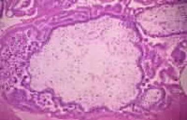
Gestational Trophoblastic Disease (GTD) is a group of rare pregnancy-related disorders that involve abnormal growth of cells inside a woman's uterus. These cells are part of the placenta, which provides nutrients to the developing fetus. GTD occurs when some of these cells grow in an uncontrolled way, forming tumors or tumor-like growths.
There are several types of GTD:
1. Hydatidiform Mole (HM): Also known as a molar pregnancy, this is the most common type of GTD. It occurs when an egg that has no genetic information is fertilized by a sperm and then divides into multiple copies. This results in a growth that resembles a cluster of grapes, rather than a developing fetus. There are two types of HMs: complete and partial. A complete HM forms when an empty egg is fertilized by two sperms, resulting in no fetal tissue. A partial HM forms when a normal egg is fertilized by two sperm or an abnormal egg with two sets of genetic material, resulting in some fetal tissue.
2. Invasive Mole: This type of GTD occurs when cells from a molar pregnancy invade the uterine wall and surrounding tissues. It can also spread to other parts of the body, such as the lungs or brain.
3. Choriocarcinoma: This is a rare form of GTD that develops from trophoblastic cells and forms a malignant tumor. It can grow rapidly and spread quickly to other organs.
4. Placental Site Trophoblastic Tumor (PSTT): This is an even rarer type of GTD that forms in the tissue where the placenta attaches to the uterus. PSTTs are usually slow-growing but can sometimes spread to other parts of the body.
5. Epithelioid Trophoblastic Tumor (ETT): This is a very rare type of GTD that forms in the tissue where the placenta attaches to the uterus. ETTs are usually slow-growing and have a good prognosis.
It's important to note that most molar pregnancies do not develop into more serious forms of GTD, but regular follow-up care is necessary to monitor for any signs of progression. Treatment options depend on the type and stage of GTD and may include surgery, chemotherapy, or radiation therapy.
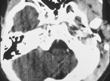
Wilms tumor, also known as nephroblastoma, is a type of kidney cancer that primarily affects children. It occurs in the cells of the developing kidneys and is named after Dr. Max Wilms, who first described this type of tumor in 1899. Wilms tumor typically develops before the age of 5, with most cases occurring in children under the age of 3.
The medical definition of Wilms tumor is:
A malignant, embryonal kidney tumor originating from the metanephric blastema, which is a mass of undifferentiated cells in the developing kidney. Wilms tumor is characterized by its rapid growth and potential for spread (metastasis) to other parts of the body, particularly the lungs and liver. The tumor usually presents as a large, firm, and irregular mass in the abdomen, and it may be associated with various symptoms such as abdominal pain, swelling, or blood in the urine.
Wilms tumor is typically treated with a combination of surgery, chemotherapy, and radiation therapy. The prognosis for children with Wilms tumor has improved significantly over the past few decades due to advances in treatment methods and early detection.
Leukocyte disorders, also known as white blood cell disorders, refer to a group of conditions that affect the production, function, or number of leukocytes (white blood cells) in the body. Leukocytes play a crucial role in protecting the body against infection and disease. Therefore, disorders that affect these cells can significantly impact an individual's immune system and overall health.
There are several types of leukocyte disorders, including:
1. Leukopenia: A condition characterized by abnormally low levels of white blood cells in the blood. This can increase the risk of infection.
2. Leukocytosis: A condition characterized by an elevated number of white blood cells in the blood. While this can be a normal response to infection or inflammation, it can also indicate an underlying medical condition such as leukemia.
3. Neutropenia: A condition characterized by abnormally low levels of neutrophils, a type of white blood cell that helps fight bacterial infections. This can increase the risk of infection.
4. Neutrophilia: A condition characterized by an elevated number of neutrophils in the blood. This can be a normal response to infection or inflammation, but it can also indicate an underlying medical condition such as an acute bacterial infection.
5. Lymphocytosis: A condition characterized by an elevated number of lymphocytes, a type of white blood cell that helps fight viral infections and cancer cells. This can be a normal response to infection or vaccination, but it can also indicate an underlying medical condition such as chronic lymphocytic leukemia.
6. Lymphopenia: A condition characterized by abnormally low levels of lymphocytes in the blood. This can increase the risk of infection and indicate an underlying medical condition such as HIV/AIDS or autoimmune disorders.
7. Monocytosis: A condition characterized by an elevated number of monocytes, a type of white blood cell that helps fight chronic infections and cancer cells. This can be a normal response to infection or inflammation, but it can also indicate an underlying medical condition such as chronic inflammatory diseases.
8. Monocytopenia: A condition characterized by abnormally low levels of monocytes in the blood. This can increase the risk of infection and indicate an underlying medical condition such as bone marrow disorders or autoimmune diseases.
These conditions can be caused by various factors, including infections, inflammation, cancer, autoimmune disorders, medications, and genetic disorders. Proper diagnosis and treatment require a thorough evaluation of the patient's medical history, physical examination, laboratory tests, and imaging studies.
Vincristine is an antineoplastic agent, specifically a vinca alkaloid. It is derived from the Madagascar periwinkle plant (Catharanthus roseus). Vincristine binds to tubulin, a protein found in microtubules, and inhibits their polymerization, which results in disruption of mitotic spindles leading to cell cycle arrest and apoptosis (programmed cell death). It is used in the treatment of various types of cancer including leukemias, lymphomas, and solid tumors. Common side effects include peripheral neuropathy, constipation, and alopecia.
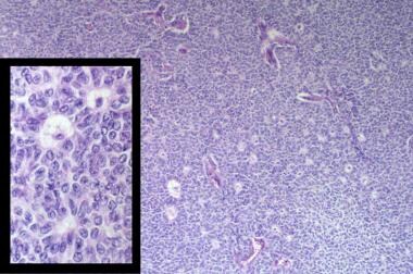
Rhabdomyosarcoma is a type of cancer that develops in the body's soft tissues, specifically in the muscle cells. It is a rare and aggressive form of sarcoma, which is a broader category of cancers that affect the connective tissues such as muscles, tendons, cartilages, bones, blood vessels, and fatty tissues.
Rhabdomyosarcomas can occur in various parts of the body, including the head, neck, arms, legs, trunk, and genitourinary system. They are more common in children than adults, with most cases diagnosed before the age of 18. The exact cause of rhabdomyosarcoma is not known, but genetic factors and exposure to radiation or certain chemicals may increase the risk.
There are several subtypes of rhabdomyosarcoma, including embryonal, alveolar, pleomorphic, and spindle cell/sclerosing. The type and stage of the cancer determine the treatment options, which may include surgery, radiation therapy, chemotherapy, or a combination of these approaches. Early diagnosis and prompt treatment are crucial for improving the prognosis and long-term survival rates.
Trophoblastic neoplasms are a group of rare tumors that originate from the trophoblast, which is the outer layer of cells that surrounds a developing embryo and helps to form the placenta during pregnancy. These tumors can be benign or malignant and are characterized by their ability to produce human chorionic gonadotropin (hCG), a hormone that is normally produced during pregnancy.
There are several types of trophoblastic neoplasms, including:
1. Hydatidiform mole: A benign growth that forms in the uterus when a fertilized egg implants but does not develop into a normal embryo. There are two types of hydatidiform moles: complete and partial. Complete moles have no fetal tissue, while partial moles have some fetal tissue.
2. Invasive mole: A malignant form of hydatidiform mole that invades the uterine wall and may spread to other parts of the body.
3. Choriocarcinoma: A rapidly growing and highly invasive malignant tumor that can arise from a hydatidiform mole, a normal pregnancy, or an ectopic pregnancy. It can spread quickly to other parts of the body, such as the lungs, liver, and brain.
4. Placental site trophoblastic tumor (PSTT): A rare type of trophoblastic neoplasm that arises from the cells that attach the placenta to the uterine wall. It is usually slow-growing but can be aggressive in some cases.
5. Epithelioid trophoblastic tumor (ETT): Another rare type of trophoblastic neoplasm that arises from the cells that form the placental villi. It is typically low-grade and has a good prognosis, but it can recur in some cases.
The treatment for trophoblastic neoplasms depends on the type and stage of the tumor. Treatment options may include surgery, chemotherapy, radiation therapy, or a combination of these approaches. Regular monitoring of hCG levels is also important to ensure that the tumor has been completely removed and to detect any recurrence early.
Pulse therapy, in the context of drug treatment, refers to a therapeutic regimen where a medication is administered in large doses for a short period of time, followed by a break or "drug-free" interval before the next dose. This cycle is then repeated at regular intervals. The goal of pulse therapy is to achieve high concentrations of the drug in the body to maximize its therapeutic effect while minimizing overall exposure and potential side effects.
This approach is often used for drugs that have a long half-life or slow clearance, as it allows for periodic "washing out" of the drug from the body. Pulse therapy can also help reduce the risk of developing drug resistance in certain conditions like rheumatoid arthritis and tuberculosis. Common examples include pulse methotrexate for rheumatoid arthritis and intermittent preventive treatment with anti-malarial drugs.
It is important to note that the use of pulse therapy should be based on a thorough understanding of the drug's pharmacokinetics, therapeutic index, and potential adverse effects. Close monitoring of patients undergoing pulse therapy is essential to ensure safety and efficacy.
Ifosfamide is an alkylating agent, which is a type of chemotherapy medication. It works by interfering with the DNA of cancer cells, preventing them from dividing and growing. Ifosfamide is used to treat various types of cancers, such as testicular cancer, small cell lung cancer, ovarian cancer, cervical cancer, and certain types of sarcomas.
The medical definition of Ifosfamide is:
Ifosfamide is a synthetic antineoplastic agent, an oxazaphosphorine derivative, with the chemical formula C6H15Cl2N2O2P. It is used in the treatment of various malignancies, including germ cell tumors, sarcomas, lymphomas, and testicular cancer. The drug is administered intravenously and exerts its cytotoxic effects through the alkylation and cross-linking of DNA, leading to the inhibition of DNA replication and transcription. Ifosfamide can cause significant myelosuppression and has been associated with urotoxicity, neurotoxicity, and secondary malignancies. Therefore, it is essential to monitor patients closely during treatment and manage any adverse effects promptly.
Etoposide is a chemotherapy medication used to treat various types of cancer, including lung cancer, testicular cancer, and certain types of leukemia. It works by inhibiting the activity of an enzyme called topoisomerase II, which is involved in DNA replication and transcription. By doing so, etoposide can interfere with the growth and multiplication of cancer cells.
Etoposide is often administered intravenously in a hospital or clinic setting, although it may also be given orally in some cases. The medication can cause a range of side effects, including nausea, vomiting, hair loss, and an increased risk of infection. It can also have more serious side effects, such as bone marrow suppression, which can lead to anemia, bleeding, and a weakened immune system.
Like all chemotherapy drugs, etoposide is not without risks and should only be used under the close supervision of a qualified healthcare provider. It is important for patients to discuss the potential benefits and risks of this medication with their doctor before starting treatment.
Ewing sarcoma is a type of cancer that originates in bones or the soft tissues surrounding them, such as muscles and tendons. It primarily affects children and adolescents, although it can occur in adults as well. The disease is characterized by small, round tumor cells that typically grow quickly and are prone to metastasize (spread) to other parts of the body, most commonly the lungs, bones, and bone marrow.
Ewing sarcoma is caused by a genetic abnormality, specifically a chromosomal translocation that results in the fusion of two genes, EWSR1 and FLI1. This gene fusion leads to the formation of an abnormal protein that disrupts normal cell growth and division processes, ultimately resulting in cancer.
Symptoms of Ewing sarcoma can vary depending on the location and size of the tumor but may include pain or swelling in the affected area, fever, fatigue, and weight loss. Diagnosis typically involves imaging studies such as X-rays, CT scans, or MRI scans to locate the tumor, followed by a biopsy to confirm the presence of cancer cells. Treatment may involve surgery, radiation therapy, chemotherapy, or a combination of these approaches, depending on the stage and location of the disease.
Antineoplastic combined chemotherapy protocols refer to a treatment plan for cancer that involves the use of more than one antineoplastic (chemotherapy) drug given in a specific sequence and schedule. The combination of drugs is used because they may work better together to destroy cancer cells compared to using a single agent alone. This approach can also help to reduce the likelihood of cancer cells becoming resistant to the treatment.
The choice of drugs, dose, duration, and frequency are determined by various factors such as the type and stage of cancer, patient's overall health, and potential side effects. Combination chemotherapy protocols can be used in various settings, including as a primary treatment, adjuvant therapy (given after surgery or radiation to kill any remaining cancer cells), neoadjuvant therapy (given before surgery or radiation to shrink the tumor), or palliative care (to alleviate symptoms and prolong survival).
It is important to note that while combined chemotherapy protocols can be effective in treating certain types of cancer, they can also cause significant side effects, including nausea, vomiting, hair loss, fatigue, and an increased risk of infection. Therefore, patients undergoing such treatment should be closely monitored and managed by a healthcare team experienced in administering chemotherapy.
Cyclophosphamide is an alkylating agent, which is a type of chemotherapy medication. It works by interfering with the DNA of cancer cells, preventing them from dividing and growing. This helps to stop the spread of cancer in the body. Cyclophosphamide is used to treat various types of cancer, including lymphoma, leukemia, multiple myeloma, and breast cancer. It can be given orally as a tablet or intravenously as an injection.
Cyclophosphamide can also have immunosuppressive effects, which means it can suppress the activity of the immune system. This makes it useful in treating certain autoimmune diseases, such as rheumatoid arthritis and lupus. However, this immunosuppression can also increase the risk of infections and other side effects.
Like all chemotherapy medications, cyclophosphamide can cause a range of side effects, including nausea, vomiting, hair loss, fatigue, and increased susceptibility to infections. It is important for patients receiving cyclophosphamide to be closely monitored by their healthcare team to manage these side effects and ensure the medication is working effectively.
Kidney neoplasms refer to abnormal growths or tumors in the kidney tissues that can be benign (non-cancerous) or malignant (cancerous). These growths can originate from various types of kidney cells, including the renal tubules, glomeruli, and the renal pelvis.
Malignant kidney neoplasms are also known as kidney cancers, with renal cell carcinoma being the most common type. Benign kidney neoplasms include renal adenomas, oncocytomas, and angiomyolipomas. While benign neoplasms are generally not life-threatening, they can still cause problems if they grow large enough to compromise kidney function or if they undergo malignant transformation.
Early detection and appropriate management of kidney neoplasms are crucial for improving patient outcomes and overall prognosis. Regular medical check-ups, imaging studies, and urinalysis can help in the early identification of these growths, allowing for timely intervention and treatment.
Neoplasms, germ cell and embryonal are types of tumors that originate from the abnormal growth of cells. Here's a brief medical definition for each:
1. Neoplasms: Neoplasms refer to abnormal tissue growths or masses, which can be benign (non-cancerous) or malignant (cancerous). They result from uncontrolled cell division and may invade surrounding tissues or spread to other parts of the body through a process called metastasis.
2. Germ Cell Tumors: These are rare tumors that develop from the germ cells, which give rise to sperm and eggs in the reproductive organs (ovaries and testes). They can be benign or malignant and may occur in both children and adults. Germ cell tumors can also arise outside of the reproductive organs, a condition known as extragonadal germ cell tumors.
3. Embryonal Tumors: These are a type of malignant neoplasm that primarily affects infants and young children. They develop from embryonic cells, which are immature cells present during fetal development. Embryonal tumors can occur in various organs, including the brain (medulloblastomas), nervous system (primitive neuroectodermal tumors or PNETs), and other areas like the kidneys and liver.
It is essential to note that these conditions require professional medical evaluation and treatment by healthcare professionals with expertise in oncology and related fields.
Combined modality therapy (CMT) is a medical treatment approach that utilizes more than one method or type of therapy simultaneously or in close succession, with the goal of enhancing the overall effectiveness of the treatment. In the context of cancer care, CMT often refers to the combination of two or more primary treatment modalities, such as surgery, radiation therapy, and systemic therapies (chemotherapy, immunotherapy, targeted therapy, etc.).
The rationale behind using combined modality therapy is that each treatment method can target cancer cells in different ways, potentially increasing the likelihood of eliminating all cancer cells and reducing the risk of recurrence. The specific combination and sequence of treatments will depend on various factors, including the type and stage of cancer, patient's overall health, and individual preferences.
For example, a common CMT approach for locally advanced rectal cancer may involve preoperative (neoadjuvant) chemoradiation therapy, followed by surgery to remove the tumor, and then postoperative (adjuvant) chemotherapy. This combined approach allows for the reduction of the tumor size before surgery, increases the likelihood of complete tumor removal, and targets any remaining microscopic cancer cells with systemic chemotherapy.
It is essential to consult with a multidisciplinary team of healthcare professionals to determine the most appropriate CMT plan for each individual patient, considering both the potential benefits and risks associated with each treatment method.
Doxorubicin is a type of chemotherapy medication known as an anthracycline. It works by interfering with the DNA in cancer cells, which prevents them from growing and multiplying. Doxorubicin is used to treat a wide variety of cancers, including leukemia, lymphoma, breast cancer, lung cancer, ovarian cancer, and many others. It may be given alone or in combination with other chemotherapy drugs.
Doxorubicin is usually administered through a vein (intravenously) and can cause side effects such as nausea, vomiting, hair loss, mouth sores, and increased risk of infection. It can also cause damage to the heart muscle, which can lead to heart failure in some cases. For this reason, doctors may monitor patients' heart function closely while they are receiving doxorubicin treatment.
It is important for patients to discuss the potential risks and benefits of doxorubicin therapy with their healthcare provider before starting treatment.
Bleomycin is a type of chemotherapeutic agent used to treat various types of cancer, including squamous cell carcinoma, testicular cancer, and lymphomas. It works by causing DNA damage in rapidly dividing cells, which can inhibit the growth and proliferation of cancer cells.
Bleomycin is an antibiotic derived from Streptomyces verticillus and is often administered intravenously or intramuscularly. While it can be effective in treating certain types of cancer, it can also have serious side effects, including lung toxicity, which can lead to pulmonary fibrosis and respiratory failure. Therefore, bleomycin should only be used under the close supervision of a healthcare professional who is experienced in administering chemotherapy drugs.
Methotrexate is a medication used in the treatment of certain types of cancer and autoimmune diseases. It is an antimetabolite that inhibits the enzyme dihydrofolate reductase, which is necessary for the synthesis of purines and pyrimidines, essential components of DNA and RNA. By blocking this enzyme, methotrexate interferes with cell division and growth, making it effective in treating rapidly dividing cells such as cancer cells.
In addition to its use in cancer treatment, methotrexate is also used to manage autoimmune diseases such as rheumatoid arthritis, psoriasis, and inflammatory bowel disease. In these conditions, methotrexate modulates the immune system and reduces inflammation.
It's important to note that methotrexate can have significant side effects and should be used under the close supervision of a healthcare provider. Regular monitoring of blood counts, liver function, and kidney function is necessary during treatment with methotrexate.
Bone neoplasms are abnormal growths or tumors that develop in the bone. They can be benign (non-cancerous) or malignant (cancerous). Benign bone neoplasms do not spread to other parts of the body and are rarely a threat to life, although they may cause problems if they grow large enough to press on surrounding tissues or cause fractures. Malignant bone neoplasms, on the other hand, can invade and destroy nearby tissue and may spread (metastasize) to other parts of the body.
There are many different types of bone neoplasms, including:
1. Osteochondroma - a benign tumor that develops from cartilage and bone
2. Enchondroma - a benign tumor that forms in the cartilage that lines the inside of the bones
3. Chondrosarcoma - a malignant tumor that develops from cartilage
4. Osteosarcoma - a malignant tumor that develops from bone cells
5. Ewing sarcoma - a malignant tumor that develops in the bones or soft tissues around the bones
6. Giant cell tumor of bone - a benign or occasionally malignant tumor that develops from bone tissue
7. Fibrosarcoma - a malignant tumor that develops from fibrous tissue in the bone
The symptoms of bone neoplasms vary depending on the type, size, and location of the tumor. They may include pain, swelling, stiffness, fractures, or limited mobility. Treatment options depend on the type and stage of the tumor but may include surgery, radiation therapy, chemotherapy, or a combination of these treatments.
A "Drug Administration Schedule" refers to the plan for when and how a medication should be given to a patient. It includes details such as the dose, frequency (how often it should be taken), route (how it should be administered, such as orally, intravenously, etc.), and duration (how long it should be taken) of the medication. This schedule is often created and prescribed by healthcare professionals, such as doctors or pharmacists, to ensure that the medication is taken safely and effectively. It may also include instructions for missed doses or changes in the dosage.
Local neoplasm recurrence is the return or regrowth of a tumor in the same location where it was originally removed or treated. This means that cancer cells have survived the initial treatment and started to grow again in the same area. It's essential to monitor and detect any local recurrence as early as possible, as it can affect the prognosis and may require additional treatment.
Disease-free survival (DFS) is a term used in medical research and clinical practice, particularly in the field of oncology. It refers to the length of time after primary treatment for a cancer during which no evidence of the disease can be found. This means that the patient shows no signs or symptoms of the cancer, and any imaging studies or other tests do not reveal any tumors or other indications of the disease.
DFS is often used as an important endpoint in clinical trials to evaluate the effectiveness of different treatments for cancer. By measuring the length of time until the cancer recurs or a new cancer develops, researchers can get a better sense of how well a particular treatment is working and whether it is improving patient outcomes.
It's important to note that DFS is not the same as overall survival (OS), which refers to the length of time from primary treatment until death from any cause. While DFS can provide valuable information about the effectiveness of cancer treatments, it does not necessarily reflect the impact of those treatments on patients' overall survival.
Neoplasm staging is a systematic process used in medicine to describe the extent of spread of a cancer, including the size and location of the original (primary) tumor and whether it has metastasized (spread) to other parts of the body. The most widely accepted system for this purpose is the TNM classification system developed by the American Joint Committee on Cancer (AJCC) and the Union for International Cancer Control (UICC).
In this system, T stands for tumor, and it describes the size and extent of the primary tumor. N stands for nodes, and it indicates whether the cancer has spread to nearby lymph nodes. M stands for metastasis, and it shows whether the cancer has spread to distant parts of the body.
Each letter is followed by a number that provides more details about the extent of the disease. For example, a T1N0M0 cancer means that the primary tumor is small and has not spread to nearby lymph nodes or distant sites. The higher the numbers, the more advanced the cancer.
Staging helps doctors determine the most appropriate treatment for each patient and estimate the patient's prognosis. It is an essential tool for communication among members of the healthcare team and for comparing outcomes of treatments in clinical trials.
Treatment outcome is a term used to describe the result or effect of medical treatment on a patient's health status. It can be measured in various ways, such as through symptoms improvement, disease remission, reduced disability, improved quality of life, or survival rates. The treatment outcome helps healthcare providers evaluate the effectiveness of a particular treatment plan and make informed decisions about future care. It is also used in clinical research to compare the efficacy of different treatments and improve patient care.
Medical survival rate is a statistical measure used to determine the percentage of patients who are still alive for a specific period of time after their diagnosis or treatment for a certain condition or disease. It is often expressed as a five-year survival rate, which refers to the proportion of people who are alive five years after their diagnosis. Survival rates can be affected by many factors, including the stage of the disease at diagnosis, the patient's age and overall health, the effectiveness of treatment, and other health conditions that the patient may have. It is important to note that survival rates are statistical estimates and do not necessarily predict an individual patient's prognosis.
 Dactinomycin
Immunosuppressive drug
Epithelioid sarcoma
Robert T. Francoeur
Gestational trophoblastic disease
Anagen effluvium
Immature teratoma
GT198
1940 in science
Phenoxazine
Intercalation (biochemistry)
Polypeptide antibiotic
Index of biochemistry articles
Chemotherapy
July 1917
ATC code L01
Osteosarcoma
Cyclic peptide
Clear-cell sarcoma of the kidney
WHO Model List of Essential Medicines
WHO Model List of Essential Medicines for Children
Female infertility
Index of oncology articles
Streptomyces isolates
List of drugs: D-Dd
Intergroup Rhabdomyosarcoma Study Group
Fertility preservation
Extravasation (intravenous)
List of MeSH codes (D04)
List of MeSH codes (D12.644)
Dactinomycin - Wikipedia
 Dactinomycin: MedlinePlus Drug Information
 dactinomycin | Taber's Medical Dictionary
 TINOWEL 0.5(Dactinomycin) - VEA Impex
 Anti Cancer Dactinomycin Injection Manufacturer,Exporter
 Gestational Trophoblastic Neoplasia Medication: Antineoplastics, Chemotherapy modulating agent
 dactinomycin: Uses, Taking, Side Effects, Warnings - Medicine.com
 WYETH v. LEVINE
Table 2 - Analysis of MarketScan Data for Immunosuppressive Conditions and Hospitalizations for Acute Respiratory Illness,...
Drug Eruptions: Practice Essentials, Background, Pathophysiology
 DailyMed - METHOTREXATE- methotrexate sodium injection, powder, lyophilized, for solution
Journal of Medical Internet Research - The Anatomy of the SARS-CoV-2 Biomedical Literature: Introducing the CovidX Network...
 Dactinomycin-induced veno-occlusive disease in rats. The hepatoprotective action of amifostine. Evaluation in a light and...
 Cosmegen - Uses, Side Effects, Interactions - MedBroadcast.com
 Melanoma - HealthWell Foundation
Drugs beginning with the letter C - MedBroadcast.com
 Chemotherapy in Children | CureSearch
 CanMED: HCPCS
 Elsevier's 2023 Intravenous Medications - Elsevier E-Book on VitalSource, 39th Edition - 9780323933377
 Abhinav Adhikari, Ph.D. | Harvard Catalyst Profiles | Harvard Catalyst
CanMED: HCPCS
 Rhabdomyosarcoma - Pediatrics - Merck Manuals Professional Edition
 Types of chemotherapy Information | Mount Sinai - New York
 Placental Site Trophoblastic Tumor & Chemo questions - Cancer Survivors Network
 WHOCC - ATC/DDD Index
 Human Metabolome Database: 1H NMR Spectrum (1D, 300 MHz, D2O, predicted) (HMDB0015105)
 Floxuridine
- FUdR
Summary Report | CureHunter
Floxuridine
- FUdR
Summary Report | CureHunter
Provenge (sipuleucel-T) dosing, indications, interactions, adverse effects, and more
Doxil, Lipodox (doxorubicin liposomal) dosing, indications, interactions, adverse effects, and more
 Ghent University Academic Bibliography
Doxorubicin1
- Patients with stage III or IV disease received regimen M (vincristine, dactinomycin, and doxorubicin, alternating with 4 cycles of cyclophosphamide [Cytoxan]/etoposide [Toposar], plus radiotherapy). (ahdbonline.com)
Chemotherapy4
- Dactinomycin, also known as actinomycin D, is a chemotherapy medication used to treat a number of types of cancer. (wikipedia.org)
- Dactinomycin injection must be given in a hospital or medical facility under the supervision of a doctor who is experienced in giving chemotherapy medications for cancer. (medlineplus.gov)
- Dactinomycin is a type of antibiotic that is only used in cancer chemotherapy. (medlineplus.gov)
- It is important to realize that patients with high risk molar pregnancy receive treatment with combination chemotherapy consisting of methotrexate, dactinomycin and chlorambucil. (nethealthbook.com)
Pregnant while receiving da1
- If you become pregnant while receiving dactinomycin, call your doctor. (medlineplus.gov)
Allergic to dactinomycin1
- tell your doctor and pharmacist if you are allergic to dactinomycin, any other medications, or any of the ingredients in dactinomycin injection. (medlineplus.gov)
Actinomycin1
- The researchers applied a wide array of DNA-specific drugs, including actinomycin D, also known as dactinomycin, to pea tissue. (frontiersin.org)
Antibiotic1
- Dactinomycin is in the cytotoxic antibiotic family of medications. (wikipedia.org)
Pregnancy2
- If you are able to get pregnant, a pregnancy test will be done to show that you are NOT pregnant before starting dactinomycin. (medicine.com)
- Use birth control that you can trust to prevent pregnancy while taking dactinomycin and for 6 months after stopping dactinomycin. (medicine.com)
Medication1
- Dactinomycin often causes nausea and vomiting, but it is important to keep using this medication even if you feel ill. (medbroadcast.com)
Harm1
- Dactinomycin may harm the fetus. (medlineplus.gov)
Powder2
- Dactinomycin comes as a powder to be mixed with liquid to be injected intravenously (into a vein) by a doctor or nurse in a medical facility. (medlineplus.gov)
- Each vial contains lyophilized, amorphous yellow dactinomycin powder 500 µg and mannitol 20 mg. (medbroadcast.com)
Liver2
- Very bad blood vessel problems in the liver have happened with dactinomycin. (medicine.com)
- Amifostine appears to act protectively to liver changes caused by dactinomycin. (viamedica.pl)
Dose4
- Do not breast-feed while you take dactinomycin and for 2 weeks after your last dose. (medicine.com)
- If you are a man and your sex partner gets pregnant while you take dactinomycin or within 3 months after your last dose, call your doctor right away. (medicine.com)
- If you get pregnant while taking dactinomycin or within 6 months after your last dose, call your doctor right away. (medicine.com)
- The recommended dose and dosing schedule of dactinomycin varies according to the specific type of cancer being treated, the response to therapy, and the other medications or treatments being used. (medbroadcast.com)
Cancer6
- Dactinomycin is used in combination with other medications, surgery, and/or radiation therapy to treat Wilms' tumor (a type of kidney cancer that occurs in children) and rhabdomyosarcoma (cancer that forms in muscles) in children. (medlineplus.gov)
- Dactinomycin is also used in combination with other medications to treat certain types of testicular cancer and Ewing's sarcoma (a type of cancer in bones or muscles). (medlineplus.gov)
- Dactinomycin is also sometimes used to treat a type of cancer of the ovaries (a cancer that begins in the female reproductive organs where eggs are formed). (medlineplus.gov)
- Dactinomycin is used to treat cancer. (medicine.com)
- Dactinomycin causes the death of cancer cells by interfering with their growth and reproduction. (medbroadcast.com)
- As well as interfering with the genetic material DNA of cancer cells, dactinomycin can interfere with some of your normal cells. (medbroadcast.com)
Side effects3
- Dactinomycin may cause side effects. (medlineplus.gov)
- Worse side effects from radiation treatment have happened with dactinomycin. (medicine.com)
- What are the side effects of dactinomycin that I need to call my doctor about immediately? (medicine.com)
Ordered by your doctor1
- Use dactinomycin as ordered by your doctor. (medicine.com)
Treat certain types1
- Dactinomycin may also be used to treat certain types of cancerous tumors that are located in a specific area of the body. (medlineplus.gov)
Breast-feed1
- You should not become pregnant or breast-feed while you are receiving dactinomycin. (medlineplus.gov)
Receive1
- Your doctor will probably not want you to receive dactinomycin injection. (medlineplus.gov)
Specific1
- Dactinomycin may also be injected by a doctor directly into a specific the part of the body or the organ to treat the area where a tumor is located. (medlineplus.gov)
Combination1
- Dactinomycin is also used alone or in combination with other medications to treat gestational trophoblastic tumors (a type of tumor that forms inside a woman's uterus while she is pregnant). (medlineplus.gov)
Doctor1
- Talk to your doctor about the risks of receiving dactinomycin injection. (medlineplus.gov)
Medical1
- Dactinomycin was approved for medical use in the United States in 1964. (wikipedia.org)
Work1
- Use of some vaccines with dactinomycin may either raise the chance of an infection or make the vaccine not work as well. (medicine.com)
Doxorubicin1
- Although most secondary malignant neoplasms reported (eg, bone tumors, breast and thyroid cancers) have occurred in irradiated areas, certain chemotherapeutic agents, including doxorubicin, dactinomycin, and vincristine, may contribute to an increased risk for secondary malignancies. (medscape.com)
Vincristine and dactinomycin1
- Reports have documented hepatic toxicity with the combination of vincristine and dactinomycin even in the absence of radiation therapy (which many early reports suggested was the major etiologic factor in liver damage). (medscape.com)
Cosmegen1
- Dactinomycin is available as a solution or lyophilized powder for injection in vials of varying concentrations under the commercial name Cosmegen and must be given by the intravenous route, avoiding tissue extravasation. (nih.gov)
Topotecan1
- Coronary heart disease textbook of pediatric dcm usually entails combinations of dactinomycin, vincristine, and topotecan. (elastizell.com)
Injection3
- tell your doctor and pharmacist if you are allergic to dactinomycin, any other medications, or any of the ingredients in dactinomycin injection. (medlineplus.gov)
- Your doctor will probably not want you to receive dactinomycin injection. (medlineplus.gov)
- Talk to your doctor about the risks of receiving dactinomycin injection. (medlineplus.gov)
Hepatic1
- In large studies, between 1% and 5% of children with cancer treated with regimens including dactinomycin developed acute hepatic injury and thrombocytopenia suggestive of HTS. (nih.gov)
Acute2
Wilms2
- Dactinomycin is used in combination with other medications, surgery, and/or radiation therapy to treat Wilms' tumor (a type of kidney cancer that occurs in children) and rhabdomyosarcoma (cancer that forms in muscles) in children. (medlineplus.gov)
- Dactinomycin was formally approved for use in the United States in 1964 and continues to be a valuable agent in the therapy of solid tumors of childhood (Wilms tumor, rhabdomyosarcoma) and in choriocarcinoma in adult women. (nih.gov)
Generic1
- Different insurance companies currently manufacture generic dactinomycin products, including merck & co., teva pharmaceuticals, and mylan laboratories. (sbolot.org)
Medications2
- Dactinomycin is also used in combination with other medications to treat certain types of testicular cancer and Ewing's sarcoma (a type of cancer in bones or muscles). (medlineplus.gov)
- Dactinomycin is also used alone or in combination with other medications to treat gestational trophoblastic tumors (a type of tumor that forms inside a woman's uterus while she is pregnant). (medlineplus.gov)
Disease1
- The fact that patients who received less dactinomycin than others (ie, those with relatively low-stage disease) had a low incidence of 2.8% suggests a dose-related toxicity for dactinomycin. (medscape.com)
Drugs1
- USES: Dactinomycin is used alone or with other anti- cancer drugs to treat cancer . (oceanpharmaglobal.com)
Agents1
- In many instances, it is difficult to attribute the liver test abnormalities to dactinomycin, because of the exposure to other potentially hepatotoxic agents. (nih.gov)
Side1
- Dactinomycin may cause side effects. (medlineplus.gov)
Children1
- The syndrome is more common in younger children and with higher doses of dactinomycin. (nih.gov)
Area1
- Dactinomycin may also be injected by a doctor directly into a specific the part of the body or the organ to treat the area where a tumor is located. (medlineplus.gov)
Risk1
- Dactinomycin may increase the risk that you will develop other cancers. (medlineplus.gov)