Cryoelectron Microscopy
Cryoultramicrotomy
Microscopy, Electron
Models, Molecular
Atadenovirus
Negative Staining
Image Processing, Computer-Assisted
Protein Conformation
Models, Structural
Electron Microscope Tomography
Encephalomyelitis, Western Equine
Virion
Protein Structure, Quaternary
Crystallography, X-Ray
Tomography
Virus Assembly
Microscopy
Rhinovirus
Crystallization
Protein Structure, Tertiary
Imaging, Three-Dimensional
X-Ray Diffraction
Cryopreservation
Immunoglobulin Fab Fragments
Binding Sites
Microscopy, Electron, Scanning
Molecular Sequence Data
Protein Structure, Secondary
Amino Acid Sequence
Macromolecular Substances
Microscopy, Confocal
Receptors, Virus
Protein Subunits
Dimerization
Protein Binding
Microscopy, Fluorescence
Endoplasmic Reticulum, Rough
Sequence Homology, Amino Acid
Escherichia coli
Microscopy, Atomic Force
Microscopy, Electron, Transmission
Microscopy, Immunoelectron
Sindbis Virus
Cryo-electron microscopy structure of an SH3 amyloid fibril and model of the molecular packing. (1/1790)
Amyloid fibrils are assemblies of misfolded proteins and are associated with pathological conditions such as Alzheimer's disease and the spongiform encephalopathies. In the amyloid diseases, a diverse group of normally soluble proteins self-assemble to form insoluble fibrils. X-ray fibre diffraction studies have shown that the protofilament cores of fibrils formed from the various proteins all contain a cross-beta-scaffold, with beta-strands perpendicular and beta-sheets parallel to the fibre axis. We have determined the threedimensional structure of an amyloid fibril, formed by the SH3 domain of phosphatidylinositol-3'-kinase, using cryo-electron microscopy and image processing at 25 A resolution. The structure is a double helix of two protofilament pairs wound around a hollow core, with a helical crossover repeat of approximately 600 A and an axial subunit repeat of approximately 27 A. The native SH3 domain is too compact to fit into the fibril density, and must unfold to adopt a longer, thinner shape in the amyloid form. The 20x40-A protofilaments can only accommodate one pair of flat beta-sheets stacked against each other, with very little inter-strand twist. We propose a model for the polypeptide packing as a basis for understanding the structure of amyloid fibrils in general. (+info)MENT, a heterochromatin protein that mediates higher order chromatin folding, is a new serpin family member. (2/1790)
Terminal cell differentiation is correlated with the extensive sequestering of previously active genes into compact transcriptionally inert heterochromatin. In vertebrate blood cells, these changes can be traced to the accumulation of a developmentally regulated heterochromatin protein, MENT. Cryoelectron microscopy of chicken granulocyte chromatin, which is highly enriched with MENT, reveals exceptionally compact polynucleosomes, which maintain a level of higher order folding above that imposed by linker histones. The amino acid sequence of MENT reveals a close structural relationship with serpins, a large family of proteins known for their ability to undergo dramatic conformational transitions. Conservation of the "hinge region" consensus in MENT indicates that this ability is retained by the protein. MENT is distinguished from the other serpins by being a basic protein, containing several positively charged surface clusters, which are likely to be involved in ionic interactions with DNA. One of the positively charged domains bears a significant similarity to the chromatin binding region of nuclear lamina proteins and with the A.T-rich DNA-binding motif, which may account for the targeting of MENT to peripheral heterochromatin. MENT ectopically expressed in a mammalian cell line is transported into nuclei and is associated with intranuclear foci of condensed chromatin. (+info)Characterization of two related Drosophila gamma-tubulin complexes that differ in their ability to nucleate microtubules. (3/1790)
gamma-tubulin exists in two related complexes in Drosophila embryo extracts (Moritz, M., Y. Zheng, B.M. Alberts, and K. Oegema. 1998. J. Cell Biol. 142:1- 12). Here, we report the purification and characterization of both complexes that we name gamma-tubulin small complex (gammaTuSC; approximately 280,000 D) and Drosophila gammaTuRC ( approximately 2,200,000 D). In addition to gamma-tubulin, the gammaTuSC contains Dgrip84 and Dgrip91, two proteins homologous to the Spc97/98p protein family. The gammaTuSC is a structural subunit of the gammaTuRC, a larger complex containing about six additional polypeptides. Like the gammaTuRC isolated from Xenopus egg extracts (Zheng, Y., M.L. Wong, B. Alberts, and T. Mitchison. 1995. Nature. 378:578-583), the Drosophila gammaTuRC can nucleate microtubules in vitro and has an open ring structure with a diameter of 25 nm. Cryo-electron microscopy reveals a modular structure with approximately 13 radially arranged structural repeats. The gammaTuSC also nucleates microtubules, but much less efficiently than the gammaTuRC, suggesting that assembly into a larger complex enhances nucleating activity. Analysis of the nucleotide content of the gammaTuSC reveals that gamma-tubulin binds preferentially to GDP over GTP, rendering gamma-tubulin an unusual member of the tubulin superfamily. (+info)Cryoelectron microscopy of a nucleating model bile in vitreous ice: formation of primordial vesicles. (4/1790)
Because gallstones form so frequently in human bile, pathophysiologically relevant supersaturated model biles are commonly employed to study cholesterol crystal formation. We used cryo-transmission electron microscopy, complemented by polarizing light microscopy, to investigate early stages of cholesterol nucleation in model bile. In the system studied, the proposed microscopic sequence involves the evolution of small unilamellar to multilamellar vesicles to lamellar liquid crystals and finally to cholesterol crystals. Small aliquots of a concentrated (total lipid concentration = 29.2 g/dl) model bile containing 8.5% cholesterol, 22.9% egg yolk lecithin, and 68.6% taurocholate (all mole %) were vitrified at 2 min to 20 days after fourfold dilution to induce supersaturation. Mixed micelles together with a category of vesicles denoted primordial, small unilamellar vesicles of two distinct morphologies (sphere/ellipsoid and cylinder/arachoid), large unilamellar vesicles, multilamellar vesicles, and cholesterol monohydrate crystals were imaged. No evidence of aggregation/fusion of small unilamellar vesicles to form multilamellar vesicles was detected. Low numbers of multilamellar vesicles were present, some of which were sufficiently large to be identified as liquid crystals by polarizing light microscopy. Dimensions, surface areas, and volumes of spherical/ellipsoidal and cylindrical/arachoidal vesicles were quantified. Early stages in the separation of vesicles from micelles, referred to as primordial vesicles, were imaged 23-31 min after dilution. Observed structures such as enlarged micelles in primordial vesicle interiors, segments of bilayer, and faceted edges at primordial vesicle peripheries are probably early stages of small unilamellar vesicle assembly. A decrease in the mean surface area of spherical/ellipsoidal vesicles was correlated with the increased production of cholesterol crystals at 10-20 days after supersaturation by dilution, supporting the role of small unilamellar vesicles as key players in cholesterol nucleation and as cholesterol donors to crystals. This is the first visualization of an intermediate structure that has been temporally linked to the development of small unilamellar vesicles in the separation of vesicles from micelles in a model bile and suggests a time-resolved system for further investigation. (+info)Native display of complete foreign protein domains on the surface of hepatitis B virus capsids. (5/1790)
The nucleocapsid of hepatitis B virus (HBV), or HBcAg, is a highly symmetric structure formed by multiple dimers of a single core protein that contains potent T helper epitopes in its 183-aa sequence. Both factors make HBcAg an unusually strong immunogen and an attractive candidate as a carrier for foreign epitopes. The immunodominant c/e1 epitope on the capsid has been suggested as a superior location to convey high immunogenicity to a heterologous sequence. Because of its central position, however, any c/e1 insert disrupts the core protein's primary sequence; hence, only peptides, or rather small protein fragments seemed to be compatible with particle formation. According to recent structural data, the epitope is located at the tips of prominent surface spikes formed by the very stable dimer interfaces. We therefore reasoned that much larger inserts might be tolerated, provided the individual parts of a corresponding fusion protein could fold independently. Using the green fluorescent protein (GFP) as a model insert, we show that the chimeric protein efficiently forms fluorescent particles; hence, all of its structurally important parts must be properly folded. We also demonstrate that the GFP domains are surface-exposed and that the chimeric particles elicit a potent humoral response against native GFP. Hence, proteins of at least up to 238 aa can be natively displayed on the surface of HBV core particles. Such chimeras may not only be useful as vaccines but may also open the way for high resolution structural analyses of nonassembling proteins by electron microscopy. (+info)Flexibility of the major antigenic loop of foot-and-mouth disease virus bound to a Fab fragment of a neutralising antibody: structure and neutralisation. (6/1790)
The interaction of foot-and-mouth disease virus (FMDV) serotype C (clone C-S8c1) with a strongly neutralising monoclonal antibody (MAb) 4C4 has been studied by combining data from cryoelectron microscopy and x-ray crystallography. The MAb 4C4 binds to the exposed flexible GH-loop of viral protein 1 (VP1), which appears to retain its flexibility, allowing movement of the bound Fab. This is in striking contrast to MAb SD6, which binds to the same GH-loop of VP1 but exhibits no movement of the bound Fab when observed under identical conditions. However, MAbs 4C4 and SD6 have very similar neutralisation characteristics. The known atomic structure of FMDV C-S8c1 and that of the 4C4 Fab cocrystallised with a synthetic peptide corresponding to the GH-loop of VP1 were fitted to the cryoelectron microscope density map. The best fit of the 4C4 Fab is compatible only with monovalent binding of the MAb in agreement with the neutralisation data on 4C4 MAbs, Fab2s, and Fabs. The position of the bound GH-loop is related to other known positions of this loop by a hinge rotation about the base of the loop. The 4C4 Fab appears to interact almost exclusively with the G-H loop of VP1, making no other contacts with the viral capsid. (+info)Visualization of tegument-capsid interactions and DNA in intact herpes simplex virus type 1 virions. (7/1790)
Herpes simplex virus type 1 virions were examined by electron cryomicroscopy, allowing the three-dimensional structure of the infectious particle to be visualized for the first time. The capsid shell is identical to that of B-capsids purified from the host cell nucleus, with the exception of the penton channel, which is closed. The double-stranded DNA genome is organized as regularly spaced ( approximately 26 A) concentric layers inside the capsid. This pattern suggests a spool model for DNA packaging, similar to that for some bacteriophages. The bulk of the tegument is not icosahedrally ordered. However, a small portion appears as filamentous structures around the pentons, interacting extensively with the capsid. Their locations and interactions suggest possible roles for the tegument proteins in regulating DNA transport through the penton channel and binding to cellular transport proteins during viral infection. (+info)Effect of buffer conditions on the position of tRNA on the 70 S ribosome as visualized by cryoelectron microscopy. (8/1790)
The effect of buffer conditions on the binding position of tRNA on the Escherichia coli 70 S ribosome have been studied by means of three-dimensional (3D) cryoelectron microscopy. Either deacylated tRNAfMet or fMet-tRNAfMet were bound to the 70 S ribosomes, which were programmed with a 46-nucleotide mRNA having AUG codon in the middle, under two different buffer conditions (conventional buffer: containing Tris and higher Mg2+ concentration [10-15 mM]; and polyamine buffer: containing Hepes, lower Mg2+ concentration [6 mM], and polyamines). Difference maps, obtained by subtracting 3D maps of naked control ribosome in the corresponding buffer from the 3D maps of tRNA.ribosome complexes, reveal the distinct locations of tRNA on the ribosome. The position of deacylated tRNAfMet depends on the buffer condition used, whereas that of fMet-tRNAfMet remains the same in both buffer conditions. The acylated tRNA binds in the classical P site, whereas deacylated tRNA binds mostly in an intermediate P/E position under the conventional buffer condition and mostly in the position corresponding to the classical P site, i. e. in the P/P state, under the polyamine buffer conditions. (+info)Cryo-electron microscopy (Cryo-EM) is a type of electron microscopy where the sample is studied at cryogenic temperatures, typically liquid nitrogen temperatures. This technique is used to investigate the structure and shape of biological molecules and complexes, viruses, and other nanoscale particles.
In Cryo-EM, the sample is rapidly frozen to preserve its natural structure and then imaged using a beam of electrons. The images are collected at different angles and then computationally combined to generate a 3D reconstruction of the sample. This technique allows researchers to visualize biological structures in their native environment with near-atomic resolution, providing valuable insights into their function and behavior.
Cryo-EM has become an increasingly popular tool in structural biology due to its ability to image large and complex structures that are difficult or impossible to crystallize for X-ray crystallography. It has been used to determine the structures of many important biological molecules, including membrane proteins, ribosomes, viruses, and protein complexes involved in various cellular processes.
"Freezing" is a term used in the medical field to describe a phenomenon that can occur in certain neurological conditions, most notably in Parkinson's disease. It refers to a sudden and temporary inability to move or initiate movement, often triggered by environmental factors such as narrow spaces, turning, or approaching a destination. This can increase the risk of falls and make daily activities challenging for affected individuals.
Freezing is also known as "freezing of gait" (FOG) when it specifically affects a person's ability to walk. During FOG episodes, the person may feel like their feet are glued to the ground, making it difficult to take steps forward. This can be very distressing and debilitating for those affected.
It is important to note that "freezing" has different meanings in different medical contexts, such as in the field of orthopedics, where it may refer to a loss of joint motion due to stiffness or inflammation. Always consult with a healthcare professional for accurate information tailored to your specific situation.
Cryoultramicrotomy is a specialized microscopy technique used in the field of pathology and biology. It involves cutting extremely thin sections (typically less than 100 nanometers thick) of biological samples that have been frozen and hardened at very low temperatures, often using liquid nitrogen or helium.
The process begins by embedding the sample in a suitable medium, such as a cryoprotectant or a low-temperature wax, to prevent ice crystal formation during freezing. The embedded sample is then mounted on a specimen holder and cooled to a temperature below its glass transition point, typically around -150°C to -196°C.
Once the sample is frozen and hardened, it is cut using an ultramicrotome, a precision instrument that uses a diamond knife to slice the sample into thin sections. These sections are then collected on a grid or other support and can be stained with various dyes or stains to enhance contrast and visualization under an electron microscope.
Cryoultramicrotomy is particularly useful for studying the ultrastructure of biological samples, such as cells, tissues, and organelles, that may be sensitive to heat or chemical fixation methods commonly used in traditional histology techniques. It allows researchers to visualize details at the molecular level, providing valuable insights into cellular processes and disease mechanisms.
Electron microscopy (EM) is a type of microscopy that uses a beam of electrons to create an image of the sample being examined, resulting in much higher magnification and resolution than light microscopy. There are several types of electron microscopy, including transmission electron microscopy (TEM), scanning electron microscopy (SEM), and reflection electron microscopy (REM).
In TEM, a beam of electrons is transmitted through a thin slice of the sample, and the electrons that pass through the sample are focused to form an image. This technique can provide detailed information about the internal structure of cells, viruses, and other biological specimens, as well as the composition and structure of materials at the atomic level.
In SEM, a beam of electrons is scanned across the surface of the sample, and the electrons that are scattered back from the surface are detected to create an image. This technique can provide information about the topography and composition of surfaces, as well as the structure of materials at the microscopic level.
REM is a variation of SEM in which the beam of electrons is reflected off the surface of the sample, rather than scattered back from it. This technique can provide information about the surface chemistry and composition of materials.
Electron microscopy has a wide range of applications in biology, medicine, and materials science, including the study of cellular structure and function, disease diagnosis, and the development of new materials and technologies.
Molecular models are three-dimensional representations of molecular structures that are used in the field of molecular biology and chemistry to visualize and understand the spatial arrangement of atoms and bonds within a molecule. These models can be physical or computer-generated and allow researchers to study the shape, size, and behavior of molecules, which is crucial for understanding their function and interactions with other molecules.
Physical molecular models are often made up of balls (representing atoms) connected by rods or sticks (representing bonds). These models can be constructed manually using materials such as plastic or wooden balls and rods, or they can be created using 3D printing technology.
Computer-generated molecular models, on the other hand, are created using specialized software that allows researchers to visualize and manipulate molecular structures in three dimensions. These models can be used to simulate molecular interactions, predict molecular behavior, and design new drugs or chemicals with specific properties. Overall, molecular models play a critical role in advancing our understanding of molecular structures and their functions.
Atadenovirus is a genus of viruses in the family *Parvoviridae*, which infect a wide range of animals including reptiles, birds, and mammals. These viruses are non-enveloped, meaning they do not have a lipid membrane, and have a single-stranded DNA genome. Atadenoviruses can cause various diseases in their hosts, depending on the specific species of the virus and the animal it infects. Some atadenoviruses have been associated with gastrointestinal illnesses, respiratory infections, and liver disease in animals. However, more research is needed to fully understand the impact of atadenovirus infections in various animal populations.
Negative staining is a histological or microscopy technique used to enhance the contrast of transparent or translucent specimens, such as bacteria and viruses. This technique involves applying a thin layer of a dense, dark-staining material (such as a heavy metal salt) onto the surface of the sample. The stain does not penetrate the specimen but rather forms a thin layer around it, creating a "negative" image where the specimen appears lighter against the dark background. This method is particularly useful for visualizing the shape and structure of small or delicate biological samples that would be difficult to see using other staining techniques.
A capsid is the protein shell that encloses and protects the genetic material of a virus. It is composed of multiple copies of one or more proteins that are arranged in a specific structure, which can vary in shape and symmetry depending on the type of virus. The capsid plays a crucial role in the viral life cycle, including protecting the viral genome from host cell defenses, mediating attachment to and entry into host cells, and assisting with the assembly of new virus particles during replication.
Computer-assisted image processing is a medical term that refers to the use of computer systems and specialized software to improve, analyze, and interpret medical images obtained through various imaging techniques such as X-ray, CT (computed tomography), MRI (magnetic resonance imaging), ultrasound, and others.
The process typically involves several steps, including image acquisition, enhancement, segmentation, restoration, and analysis. Image processing algorithms can be used to enhance the quality of medical images by adjusting contrast, brightness, and sharpness, as well as removing noise and artifacts that may interfere with accurate diagnosis. Segmentation techniques can be used to isolate specific regions or structures of interest within an image, allowing for more detailed analysis.
Computer-assisted image processing has numerous applications in medical imaging, including detection and characterization of lesions, tumors, and other abnormalities; assessment of organ function and morphology; and guidance of interventional procedures such as biopsies and surgeries. By automating and standardizing image analysis tasks, computer-assisted image processing can help to improve diagnostic accuracy, efficiency, and consistency, while reducing the potential for human error.
Protein conformation refers to the specific three-dimensional shape that a protein molecule assumes due to the spatial arrangement of its constituent amino acid residues and their associated chemical groups. This complex structure is determined by several factors, including covalent bonds (disulfide bridges), hydrogen bonds, van der Waals forces, and ionic bonds, which help stabilize the protein's unique conformation.
Protein conformations can be broadly classified into two categories: primary, secondary, tertiary, and quaternary structures. The primary structure represents the linear sequence of amino acids in a polypeptide chain. The secondary structure arises from local interactions between adjacent amino acid residues, leading to the formation of recurring motifs such as α-helices and β-sheets. Tertiary structure refers to the overall three-dimensional folding pattern of a single polypeptide chain, while quaternary structure describes the spatial arrangement of multiple folded polypeptide chains (subunits) that interact to form a functional protein complex.
Understanding protein conformation is crucial for elucidating protein function, as the specific three-dimensional shape of a protein directly influences its ability to interact with other molecules, such as ligands, nucleic acids, or other proteins. Any alterations in protein conformation due to genetic mutations, environmental factors, or chemical modifications can lead to loss of function, misfolding, aggregation, and disease states like neurodegenerative disorders and cancer.
Structural models in medicine and biology are theoretical or physical representations used to explain the arrangement, organization, and relationship of various components or parts of a living organism or its systems. These models can be conceptual, graphical, mathematical, or computational and are used to understand complex biological structures and processes, such as molecular interactions, cell signaling pathways, organ system functions, and whole-body physiology. Structural models help researchers and healthcare professionals form hypotheses, design experiments, interpret data, and develop interventions for various medical conditions and diseases.
Capsid proteins are the structural proteins that make up the capsid, which is the protective shell of a virus. The capsid encloses the viral genome and helps to protect it from degradation and detection by the host's immune system. Capsid proteins are typically arranged in a symmetrical pattern and can self-assemble into the capsid structure when exposed to the viral genome.
The specific arrangement and composition of capsid proteins vary between different types of viruses, and they play important roles in the virus's life cycle, including recognition and binding to host cells, entry into the cell, and release of the viral genome into the host cytoplasm. Capsid proteins can also serve as targets for antiviral therapies and vaccines.
Electron microscope tomography (EMT) is a 3D imaging technique used in electron microscopy. It involves collecting a series of images of a sample at different tilt angles, and then using computational algorithms to reconstruct the 3D structure of the sample from these images.
In EMT, a sample is prepared and placed in an electron microscope, where it is exposed to a beam of electrons. The electrons interact with the atoms in the sample, producing contrast that allows the features of the sample to be visualized. By tilting the sample and collecting images at multiple angles, a range of perspectives can be obtained, which are then used to create a 3D reconstruction of the sample.
EMT is a powerful tool for studying the ultrastructure of cells and tissues, as it allows researchers to visualize structures that may not be visible using other imaging techniques. It has been used to study a wide range of biological systems, including viruses, bacteria, organelles, and cells.
EMT is a complex technique that requires specialized equipment and expertise to perform. However, it can provide valuable insights into the structure and function of biological systems, making it an important tool in the field of biology and medicine.
Western equine encephalomyelitis (WEE) is a viral disease that affects the central nervous system of horses and, less commonly, humans. The medical definition of WEE is as follows:
Western equine encephalomyelitis is an inflammation of the brain and spinal cord (encephalomyelitis) caused by the Western equine encephalitis virus (WEEV), a member of the family Togaviridae, genus Alphavirus. The virus is primarily transmitted to horses and other animals through the bite of infected mosquitoes, most commonly Culex tarsalis.
Horses are the primary amplifying host for WEEV, meaning that they can develop high levels of the virus in their bloodstream, which makes them attractive targets for mosquitoes. Humans and other animals can become incidentally infected when bitten by an infectious mosquito.
In humans, WEE is often asymptomatic or may cause mild flu-like symptoms such as fever, headache, and muscle aches. However, in severe cases, the virus can invade the central nervous system, causing encephalitis (inflammation of the brain) or meningitis (inflammation of the membranes surrounding the brain and spinal cord). These neurological manifestations can lead to symptoms such as seizures, coma, and permanent neurological damage or death.
There is no specific treatment for WEE, and management primarily focuses on supportive care, such as addressing fever, dehydration, and other complications. Prevention measures include avoiding mosquito bites through the use of insect repellent, wearing protective clothing, and reducing mosquito breeding sites around homes and communities. Vaccines are available for horses to protect them from WEEV infection, but no human vaccine is currently available.
A virion is the complete, infectious form of a virus outside its host cell. It consists of the viral genome (DNA or RNA) enclosed within a protein coat called the capsid, which is often surrounded by a lipid membrane called the envelope. The envelope may contain viral proteins and glycoproteins that aid in attachment to and entry into host cells during infection. The term "virion" emphasizes the infectious nature of the virus particle, as opposed to non-infectious components like individual capsid proteins or naked viral genome.
Quaternary protein structure refers to the arrangement and interaction of multiple folded protein molecules in a multi-subunit complex. These subunits can be identical or different forms of the same protein or distinctly different proteins that associate to form a functional complex. The quaternary structure is held together by non-covalent interactions, such as hydrogen bonds, ionic bonds, and van der Waals forces. Understanding quaternary structure is crucial for comprehending the function, regulation, and assembly of many protein complexes involved in various cellular processes.
X-ray crystallography is a technique used in structural biology to determine the three-dimensional arrangement of atoms in a crystal lattice. In this method, a beam of X-rays is directed at a crystal and diffracts, or spreads out, into a pattern of spots called reflections. The intensity and angle of each reflection are measured and used to create an electron density map, which reveals the position and type of atoms in the crystal. This information can be used to determine the molecular structure of a compound, including its shape, size, and chemical bonds. X-ray crystallography is a powerful tool for understanding the structure and function of biological macromolecules such as proteins and nucleic acids.
Tomography is a medical imaging technique used to produce cross-sectional images or slices of specific areas of the body. This technique uses various forms of radiation (X-rays, gamma rays) or sound waves (ultrasound) to create detailed images of the internal structures, such as organs, bones, and tissues. Common types of tomography include Computerized Tomography (CT), Positron Emission Tomography (PET), and Magnetic Resonance Imaging (MRI). The primary advantage of tomography is its ability to provide clear and detailed images of internal structures, allowing healthcare professionals to accurately diagnose and monitor a wide range of medical conditions.
Virus assembly, also known as virion assembly, is the final stage in the virus life cycle where individual viral components come together to form a complete viral particle or virion. This process typically involves the self-assembly of viral capsid proteins around the viral genome (DNA or RNA) and, in enveloped viruses, the acquisition of a lipid bilayer membrane containing viral glycoproteins. The specific mechanisms and regulation of virus assembly vary among different viral families, but it is often directed by interactions between viral structural proteins and genomic nucleic acid.
Microscopy is a technical field in medicine that involves the use of microscopes to observe structures and phenomena that are too small to be seen by the naked eye. It allows for the examination of samples such as tissues, cells, and microorganisms at high magnifications, enabling the detection and analysis of various medical conditions, including infections, diseases, and cellular abnormalities.
There are several types of microscopy used in medicine, including:
1. Light Microscopy: This is the most common type of microscopy, which uses visible light to illuminate and magnify samples. It can be used to examine a wide range of biological specimens, such as tissue sections, blood smears, and bacteria.
2. Electron Microscopy: This type of microscopy uses a beam of electrons instead of light to produce highly detailed images of samples. It is often used in research settings to study the ultrastructure of cells and tissues.
3. Fluorescence Microscopy: This technique involves labeling specific molecules within a sample with fluorescent dyes, allowing for their visualization under a microscope. It can be used to study protein interactions, gene expression, and cell signaling pathways.
4. Confocal Microscopy: This type of microscopy uses a laser beam to scan a sample point by point, producing high-resolution images with reduced background noise. It is often used in medical research to study the structure and function of cells and tissues.
5. Scanning Probe Microscopy: This technique involves scanning a sample with a physical probe, allowing for the measurement of topography, mechanical properties, and other characteristics at the nanoscale. It can be used in medical research to study the structure and function of individual molecules and cells.
Rhinovirus is a type of virus that belongs to the Picornaviridae family. It's one of the most common causes of the common cold in humans, responsible for around 10-40% of all adult cases and up to 80% of cases in children. The virus replicates in the upper respiratory tract, leading to symptoms such as nasal congestion, sneezing, sore throat, and cough.
Rhinovirus infections are typically mild and self-limiting, but they can be more severe or even life-threatening in people with weakened immune systems, such as those with HIV/AIDS or who are undergoing cancer treatment. There is no vaccine available to prevent rhinovirus infections, and treatment is generally supportive, focusing on relieving symptoms rather than targeting the virus itself.
The virus can be transmitted through respiratory droplets or direct contact with contaminated surfaces, and it's highly contagious. It can survive on surfaces for several hours, making hand hygiene and environmental disinfection important measures to prevent its spread.
Crystallization is a process in which a substance transitions from a liquid or dissolved state to a solid state, forming a crystal lattice. In the medical context, crystallization can refer to the formation of crystals within the body, which can occur under certain conditions such as changes in pH, temperature, or concentration of solutes. These crystals can deposit in various tissues and organs, leading to the formation of crystal-induced diseases or disorders.
For example, in patients with gout, uric acid crystals can accumulate in joints, causing inflammation, pain, and swelling. Similarly, in nephrolithiasis (kidney stones), minerals in the urine can crystallize and form stones that can obstruct the urinary tract. Crystallization can also occur in other medical contexts, such as in the formation of dental calculus or plaque, and in the development of cataracts in the eye.
Tertiary protein structure refers to the three-dimensional arrangement of all the elements (polypeptide chains) of a single protein molecule. It is the highest level of structural organization and results from interactions between various side chains (R groups) of the amino acids that make up the protein. These interactions, which include hydrogen bonds, ionic bonds, van der Waals forces, and disulfide bridges, give the protein its unique shape and stability, which in turn determines its function. The tertiary structure of a protein can be stabilized by various factors such as temperature, pH, and the presence of certain ions. Any changes in these factors can lead to denaturation, where the protein loses its tertiary structure and thus its function.
Three-dimensional (3D) imaging in medicine refers to the use of technologies and techniques that generate a 3D representation of internal body structures, organs, or tissues. This is achieved by acquiring and processing data from various imaging modalities such as X-ray computed tomography (CT), magnetic resonance imaging (MRI), ultrasound, or confocal microscopy. The resulting 3D images offer a more detailed visualization of the anatomy and pathology compared to traditional 2D imaging techniques, allowing for improved diagnostic accuracy, surgical planning, and minimally invasive interventions.
In 3D imaging, specialized software is used to reconstruct the acquired data into a volumetric model, which can be manipulated and viewed from different angles and perspectives. This enables healthcare professionals to better understand complex anatomical relationships, detect abnormalities, assess disease progression, and monitor treatment response. Common applications of 3D imaging include neuroimaging, orthopedic surgery planning, cancer staging, dental and maxillofacial reconstruction, and interventional radiology procedures.
X-ray diffraction (XRD) is not strictly a medical definition, but it is a technique commonly used in the field of medical research and diagnostics. XRD is a form of analytical spectroscopy that uses the phenomenon of X-ray diffraction to investigate the crystallographic structure of materials. When a beam of X-rays strikes a crystal, it is scattered in specific directions and with specific intensities that are determined by the arrangement of atoms within the crystal. By measuring these diffraction patterns, researchers can determine the crystal structures of various materials, including biological macromolecules such as proteins and viruses.
In the medical field, XRD is often used to study the structure of drugs and drug candidates, as well as to analyze the composition and structure of tissues and other biological samples. For example, XRD can be used to investigate the crystal structures of calcium phosphate minerals in bone tissue, which can provide insights into the mechanisms of bone formation and disease. Additionally, XRD is sometimes used in the development of new medical imaging techniques, such as phase-contrast X-ray imaging, which has the potential to improve the resolution and contrast of traditional X-ray images.
Cryopreservation is a medical procedure that involves the preservation of cells, tissues, or organs by cooling them to very low temperatures, typically below -150°C. This is usually achieved using liquid nitrogen. The low temperature slows down or stops biological activity, including chemical reactions and cellular metabolism, which helps to prevent damage and decay.
The cells, tissues, or organs that are being cryopreserved must be treated with a cryoprotectant solution before cooling to prevent the formation of ice crystals, which can cause significant damage. Once cooled, the samples are stored in specialized containers or tanks until they are needed for use.
Cryopreservation is commonly used in assisted reproductive technologies, such as the preservation of sperm, eggs, and embryos for fertility treatments. It is also used in research, including the storage of cell lines and stem cells, and in clinical settings, such as the preservation of skin grafts and corneas for transplantation.
Immunoglobulin (Ig) Fab fragments are the antigen-binding portions of an antibody that result from the digestion of the whole antibody molecule by enzymes such as papain. An antibody, also known as an immunoglobulin, is a Y-shaped protein produced by the immune system to identify and neutralize foreign substances like bacteria, viruses, or toxins. The antibody has two identical antigen-binding sites, located at the tips of the two shorter arms, which can bind specifically to a target antigen.
Fab fragments are formed when an antibody is cleaved by papain, resulting in two Fab fragments and one Fc fragment. Each Fab fragment contains one antigen-binding site, composed of a variable region (Fv) and a constant region (C). The Fv region is responsible for the specificity and affinity of the antigen binding, while the C region contributes to the effector functions of the antibody.
Fab fragments are often used in various medical applications, such as immunodiagnostics and targeted therapies, due to their ability to bind specifically to target antigens without triggering an immune response or other effector functions associated with the Fc region.
In the context of medical and biological sciences, a "binding site" refers to a specific location on a protein, molecule, or cell where another molecule can attach or bind. This binding interaction can lead to various functional changes in the original protein or molecule. The other molecule that binds to the binding site is often referred to as a ligand, which can be a small molecule, ion, or even another protein.
The binding between a ligand and its target binding site can be specific and selective, meaning that only certain ligands can bind to particular binding sites with high affinity. This specificity plays a crucial role in various biological processes, such as signal transduction, enzyme catalysis, or drug action.
In the case of drug development, understanding the location and properties of binding sites on target proteins is essential for designing drugs that can selectively bind to these sites and modulate protein function. This knowledge can help create more effective and safer therapeutic options for various diseases.
Molecular conformation, also known as spatial arrangement or configuration, refers to the specific three-dimensional shape and orientation of atoms that make up a molecule. It describes the precise manner in which bonds between atoms are arranged around a molecular framework, taking into account factors such as bond lengths, bond angles, and torsional angles.
Conformational isomers, or conformers, are different spatial arrangements of the same molecule that can interconvert without breaking chemical bonds. These isomers may have varying energies, stability, and reactivity, which can significantly impact a molecule's biological activity and function. Understanding molecular conformation is crucial in fields such as drug design, where small changes in conformation can lead to substantial differences in how a drug interacts with its target.
Scanning electron microscopy (SEM) is a type of electron microscopy that uses a focused beam of electrons to scan the surface of a sample and produce a high-resolution image. In SEM, a beam of electrons is scanned across the surface of a specimen, and secondary electrons are emitted from the sample due to interactions between the electrons and the atoms in the sample. These secondary electrons are then detected by a detector and used to create an image of the sample's surface topography. SEM can provide detailed images of the surface of a wide range of materials, including metals, polymers, ceramics, and biological samples. It is commonly used in materials science, biology, and electronics for the examination and analysis of surfaces at the micro- and nanoscale.
Biophysical phenomena refer to the observable events and processes that occur in living organisms, which can be explained and studied using the principles and methods of physics. These phenomena can include a wide range of biological processes at various levels of organization, from molecular interactions to whole-organism behaviors. Examples of biophysical phenomena include the mechanics of muscle contraction, the electrical activity of neurons, the transport of molecules across cell membranes, and the optical properties of biological tissues. By applying physical theories and techniques to the study of living systems, biophysicists seek to better understand the fundamental principles that govern life and to develop new approaches for diagnosing and treating diseases.
Molecular sequence data refers to the specific arrangement of molecules, most commonly nucleotides in DNA or RNA, or amino acids in proteins, that make up a biological macromolecule. This data is generated through laboratory techniques such as sequencing, and provides information about the exact order of the constituent molecules. This data is crucial in various fields of biology, including genetics, evolution, and molecular biology, allowing for comparisons between different organisms, identification of genetic variations, and studies of gene function and regulation.
Secondary protein structure refers to the local spatial arrangement of amino acid chains in a protein, typically described as regular repeating patterns held together by hydrogen bonds. The two most common types of secondary structures are the alpha-helix (α-helix) and the beta-pleated sheet (β-sheet). In an α-helix, the polypeptide chain twists around itself in a helical shape, with each backbone atom forming a hydrogen bond with the fourth amino acid residue along the chain. This forms a rigid rod-like structure that is resistant to bending or twisting forces. In β-sheets, adjacent segments of the polypeptide chain run parallel or antiparallel to each other and are connected by hydrogen bonds, forming a pleated sheet-like arrangement. These secondary structures provide the foundation for the formation of tertiary and quaternary protein structures, which determine the overall three-dimensional shape and function of the protein.
An amino acid sequence is the specific order of amino acids in a protein or peptide molecule, formed by the linking of the amino group (-NH2) of one amino acid to the carboxyl group (-COOH) of another amino acid through a peptide bond. The sequence is determined by the genetic code and is unique to each type of protein or peptide. It plays a crucial role in determining the three-dimensional structure and function of proteins.
Biophysics is a interdisciplinary field that combines the principles and methods of physics with those of biology to study biological systems and phenomena. It involves the use of physical theories, models, and techniques to understand and explain the properties, functions, and behaviors of living organisms and their constituents, such as cells, proteins, and DNA.
Biophysics can be applied to various areas of biology, including molecular biology, cell biology, neuroscience, and physiology. It can help elucidate the mechanisms of biological processes at the molecular and cellular levels, such as protein folding, ion transport, enzyme kinetics, gene expression, and signal transduction. Biophysical methods can also be used to develop diagnostic and therapeutic tools for medical applications, such as medical imaging, drug delivery, and gene therapy.
Examples of biophysical techniques include X-ray crystallography, nuclear magnetic resonance (NMR) spectroscopy, electron microscopy, fluorescence microscopy, atomic force microscopy, and computational modeling. These methods allow researchers to probe the structure, dynamics, and interactions of biological molecules and systems with high precision and resolution, providing insights into their functions and behaviors.
Macromolecular substances, also known as macromolecules, are large, complex molecules made up of repeating subunits called monomers. These substances are formed through polymerization, a process in which many small molecules combine to form a larger one. Macromolecular substances can be naturally occurring, such as proteins, DNA, and carbohydrates, or synthetic, such as plastics and synthetic fibers.
In the context of medicine, macromolecular substances are often used in the development of drugs and medical devices. For example, some drugs are designed to bind to specific macromolecules in the body, such as proteins or DNA, in order to alter their function and produce a therapeutic effect. Additionally, macromolecular substances may be used in the creation of medical implants, such as artificial joints and heart valves, due to their strength and durability.
It is important for healthcare professionals to have an understanding of macromolecular substances and how they function in the body, as this knowledge can inform the development and use of medical treatments.
Confocal microscopy is a powerful imaging technique used in medical and biological research to obtain high-resolution, contrast-rich images of thick samples. This super-resolution technology provides detailed visualization of cellular structures and processes at various depths within a specimen.
In confocal microscopy, a laser beam focused through a pinhole illuminates a small spot within the sample. The emitted fluorescence or reflected light from this spot is then collected by a detector, passing through a second pinhole that ensures only light from the focal plane reaches the detector. This process eliminates out-of-focus light, resulting in sharp images with improved contrast compared to conventional widefield microscopy.
By scanning the laser beam across the sample in a raster pattern and collecting fluorescence at each point, confocal microscopy generates optical sections of the specimen. These sections can be combined to create three-dimensional reconstructions, allowing researchers to study cellular architecture and interactions within complex tissues.
Confocal microscopy has numerous applications in medical research, including studying protein localization, tracking intracellular dynamics, analyzing cell morphology, and investigating disease mechanisms at the cellular level. Additionally, it is widely used in clinical settings for diagnostic purposes, such as analyzing skin lesions or detecting pathogens in patient samples.
Virus receptors are specific molecules (commonly proteins) on the surface of host cells that viruses bind to in order to enter and infect those cells. This interaction between the virus and its receptor is a critical step in the infection process. Different types of viruses have different receptor requirements, and identifying these receptors can provide important insights into the biology of the virus and potential targets for antiviral therapies.
A protein subunit refers to a distinct and independently folding polypeptide chain that makes up a larger protein complex. Proteins are often composed of multiple subunits, which can be identical or different, that come together to form the functional unit of the protein. These subunits can interact with each other through non-covalent interactions such as hydrogen bonds, ionic bonds, and van der Waals forces, as well as covalent bonds like disulfide bridges. The arrangement and interaction of these subunits contribute to the overall structure and function of the protein.
Dimerization is a process in which two molecules, usually proteins or similar structures, bind together to form a larger complex. This can occur through various mechanisms, such as the formation of disulfide bonds, hydrogen bonding, or other non-covalent interactions. Dimerization can play important roles in cell signaling, enzyme function, and the regulation of gene expression.
In the context of medical research and therapy, dimerization is often studied in relation to specific proteins that are involved in diseases such as cancer. For example, some drugs have been developed to target and inhibit the dimerization of certain proteins, with the goal of disrupting their function and slowing or stopping the progression of the disease.
Protein binding, in the context of medical and biological sciences, refers to the interaction between a protein and another molecule (known as the ligand) that results in a stable complex. This process is often reversible and can be influenced by various factors such as pH, temperature, and concentration of the involved molecules.
In clinical chemistry, protein binding is particularly important when it comes to drugs, as many of them bind to proteins (especially albumin) in the bloodstream. The degree of protein binding can affect a drug's distribution, metabolism, and excretion, which in turn influence its therapeutic effectiveness and potential side effects.
Protein-bound drugs may be less available for interaction with their target tissues, as only the unbound or "free" fraction of the drug is active. Therefore, understanding protein binding can help optimize dosing regimens and minimize adverse reactions.
Fluorescence microscopy is a type of microscopy that uses fluorescent dyes or proteins to highlight and visualize specific components within a sample. In this technique, the sample is illuminated with high-energy light, typically ultraviolet (UV) or blue light, which excites the fluorescent molecules causing them to emit lower-energy, longer-wavelength light, usually visible light in the form of various colors. This emitted light is then collected by the microscope and detected to produce an image.
Fluorescence microscopy has several advantages over traditional brightfield microscopy, including the ability to visualize specific structures or molecules within a complex sample, increased sensitivity, and the potential for quantitative analysis. It is widely used in various fields of biology and medicine, such as cell biology, neuroscience, and pathology, to study the structure, function, and interactions of cells and proteins.
There are several types of fluorescence microscopy techniques, including widefield fluorescence microscopy, confocal microscopy, two-photon microscopy, and total internal reflection fluorescence (TIRF) microscopy, each with its own strengths and limitations. These techniques can provide valuable insights into the behavior of cells and proteins in health and disease.
The rough endoplasmic reticulum (RER) is a type of organelle found in eukaryotic cells, which are characterized by the presence of ribosomes on their cytoplasmic surface. These ribosomes give the RER a "rough" appearance and are responsible for the synthesis of proteins that are destined to be exported from the cell or targeted to various organelles within the cell.
The RER is involved in several important cellular processes, including:
1. Protein folding and modification: Once proteins are synthesized by ribosomes on the RER, they are transported into the lumen of the RER where they undergo folding and modifications such as glycosylation.
2. Quality control: The RER plays a crucial role in ensuring that only properly folded and modified proteins are transported to their final destinations within the cell or exported from the cell. Misfolded or improperly modified proteins are retained within the RER and targeted for degradation.
3. Transport: Proteins that are synthesized on the RER are packaged into vesicles and transported to the Golgi apparatus, where they undergo further modifications and sorting before being transported to their final destinations.
Overall, the rough endoplasmic reticulum is a critical organelle for protein synthesis, folding, modification, and transport in eukaryotic cells.
Sequence homology, amino acid, refers to the similarity in the order of amino acids in a protein or a portion of a protein between two or more species. This similarity can be used to infer evolutionary relationships and functional similarities between proteins. The higher the degree of sequence homology, the more likely it is that the proteins are related and have similar functions. Sequence homology can be determined through various methods such as pairwise alignment or multiple sequence alignment, which compare the sequences and calculate a score based on the number and type of matching amino acids.
'Escherichia coli' (E. coli) is a type of gram-negative, facultatively anaerobic, rod-shaped bacterium that commonly inhabits the intestinal tract of humans and warm-blooded animals. It is a member of the family Enterobacteriaceae and one of the most well-studied prokaryotic model organisms in molecular biology.
While most E. coli strains are harmless and even beneficial to their hosts, some serotypes can cause various forms of gastrointestinal and extraintestinal illnesses in humans and animals. These pathogenic strains possess virulence factors that enable them to colonize and damage host tissues, leading to diseases such as diarrhea, urinary tract infections, pneumonia, and sepsis.
E. coli is a versatile organism with remarkable genetic diversity, which allows it to adapt to various environmental niches. It can be found in water, soil, food, and various man-made environments, making it an essential indicator of fecal contamination and a common cause of foodborne illnesses. The study of E. coli has contributed significantly to our understanding of fundamental biological processes, including DNA replication, gene regulation, and protein synthesis.
Atomic Force Microscopy (AFM) is a type of microscopy that allows visualization and measurement of surfaces at the atomic level. It works by using a sharp probe, called a tip, that is mounted on a flexible cantilever. The tip is brought very close to the surface of the sample and as the sample is scanned, the forces between the tip and the sample cause the cantilever to deflect. This deflection is measured and used to generate a topographic map of the surface with extremely high resolution, often on the order of fractions of a nanometer. AFM can be used to study both conductive and non-conductive samples, and can operate in various environments, including air and liquid. It has applications in fields such as materials science, biology, and chemistry.
Recombinant proteins are artificially created proteins produced through the use of recombinant DNA technology. This process involves combining DNA molecules from different sources to create a new set of genes that encode for a specific protein. The resulting recombinant protein can then be expressed, purified, and used for various applications in research, medicine, and industry.
Recombinant proteins are widely used in biomedical research to study protein function, structure, and interactions. They are also used in the development of diagnostic tests, vaccines, and therapeutic drugs. For example, recombinant insulin is a common treatment for diabetes, while recombinant human growth hormone is used to treat growth disorders.
The production of recombinant proteins typically involves the use of host cells, such as bacteria, yeast, or mammalian cells, which are engineered to express the desired protein. The host cells are transformed with a plasmid vector containing the gene of interest, along with regulatory elements that control its expression. Once the host cells are cultured and the protein is expressed, it can be purified using various chromatography techniques.
Overall, recombinant proteins have revolutionized many areas of biology and medicine, enabling researchers to study and manipulate proteins in ways that were previously impossible.
A cell line is a culture of cells that are grown in a laboratory for use in research. These cells are usually taken from a single cell or group of cells, and they are able to divide and grow continuously in the lab. Cell lines can come from many different sources, including animals, plants, and humans. They are often used in scientific research to study cellular processes, disease mechanisms, and to test new drugs or treatments. Some common types of human cell lines include HeLa cells (which come from a cancer patient named Henrietta Lacks), HEK293 cells (which come from embryonic kidney cells), and HUVEC cells (which come from umbilical vein endothelial cells). It is important to note that cell lines are not the same as primary cells, which are cells that are taken directly from a living organism and have not been grown in the lab.
Transmission electron microscopy (TEM) is a type of microscopy in which an electron beam is transmitted through a ultra-thin specimen, interacting with it as it passes through. An image is formed from the interaction of the electrons with the specimen; the image is then magnified and visualized on a fluorescent screen or recorded on an electronic detector (or photographic film in older models).
TEM can provide high-resolution, high-magnification images that can reveal the internal structure of specimens including cells, viruses, and even molecules. It is widely used in biological and materials science research to investigate the ultrastructure of cells, tissues and materials. In medicine, TEM is used for diagnostic purposes in fields such as virology and bacteriology.
It's important to note that preparing a sample for TEM is a complex process, requiring specialized techniques to create thin (50-100 nm) specimens. These include cutting ultrathin sections of embedded samples using an ultramicrotome, staining with heavy metal salts, and positive staining or negative staining methods.
Immunoelectron microscopy (IEM) is a specialized type of electron microscopy that combines the principles of immunochemistry and electron microscopy to detect and localize specific antigens within cells or tissues at the ultrastructural level. This technique allows for the visualization and identification of specific proteins, viruses, or other antigenic structures with a high degree of resolution and specificity.
In IEM, samples are first fixed, embedded, and sectioned to prepare them for electron microscopy. The sections are then treated with specific antibodies that have been labeled with electron-dense markers, such as gold particles or ferritin. These labeled antibodies bind to the target antigens in the sample, allowing for their visualization under an electron microscope.
There are several different methods of IEM, including pre-embedding and post-embedding techniques. Pre-embedding involves labeling the antigens before embedding the sample in resin, while post-embedding involves labeling the antigens after embedding. Post-embedding techniques are generally more commonly used because they allow for better preservation of ultrastructure and higher resolution.
IEM is a valuable tool in many areas of research, including virology, bacteriology, immunology, and cell biology. It can be used to study the structure and function of viruses, bacteria, and other microorganisms, as well as the distribution and localization of specific proteins and antigens within cells and tissues.
Sindbis virus is an alphavirus that belongs to the Togaviridae family. It's named after the location where it was first isolated, in Sindbis, Egypt, in 1952. This virus is primarily transmitted by mosquitoes and can infect a wide range of animals, including birds and humans. In humans, Sindbis virus infection often causes a mild flu-like illness characterized by fever, rash, and joint pain. However, some people may develop more severe symptoms, such as neurological disorders, although this is relatively rare. There is no specific treatment for Sindbis virus infection, and management typically involves supportive care to alleviate symptoms.
 Fibril structure of amyloid-β(1-42) by cryo-electron microscopy
 Mature HIV-1 capsid structure by cryo-electron microscopy and all-atom molecular dynamics | Nature
 Transformative High Resolution Cryo-Electron Microscopy (CryoEM)
 The Nobel Prize in Physics 2017 - Scientific Background: The development of cryo-electron microscopy
 Workshop III: Cryo-Electron Microscopy and Beyond
 World record resolution in cryo electron microscopy
 Cryo-Electron Microscopy at eBIC for Industry - - Diamond Light Source
Cryo-Electron Microscopy - EMBL Imaging Centre
 V-CEM | Vanderbilt Cryo Electron Microscopy Facility
 Collaborative Supplements for Cryo-Electron Microscopy Technology Transfer (Admin Supp) | Government Grant
 Ball-and-chain inactivation of ion channels visualized by cryo-electron microscopy
 Cryo-electron Microscopy Specimen Preparation By Means Of a Focused Ion Beam | Protocol (Translated to Japanese)
 Krios G4: IMP enters next level of cryo-Electron Microscopy | Research Institute of Molecular Pathology (IMP)
 Visualising nascent chain dynamics at the ribosome exit tunnel by cryo-electron microscopy | bioRxiv
 Podcast: Combining Cryo-electron Microscopy with Supercomputer Simulation - High-Performance Computing News Analysis | insideHPC
Tailoring surface acoustic wave atomisation for cryo-electron microscopy sample preparation<...
 Cryo-Electron Microscopy Core | School of Medicine | School of Medicine | Case Western Reserve University
 35. A New Support Film for Cryo Electron Microscopy Protein Structure Analysis Based on Covalently Functionalized Graphene •...
 User Fee Schedule |
Cryo-Electron Microscopy Facility |
...
User Fee Schedule |
Cryo-Electron Microscopy Facility |
...
 Cryo-Electron Microscopy Analysis of Coronavirus Spike Protein May Point Way to Effective Vaccine Strategies - BioQuick News
 Cryo-electron microscopy structures of the N501Y SARS-CoV-2 spike protein in complex with ACE2 and 2 potent neutralizing...
 Frontiers in Cryo-Electron Microscopy | EK19 - System Checker - Keystone Symposia
Cryo-Electron Microscopy | Virus Biophysics
 Image processing for cryo electron microscopy
 Cryo Electron Microscopy Visualizes Coronavirus Structure
 Cryo-electron microscopy EPN BAG - Dataset - B2FIND
 High-resolution cryo-electron microscopy structure of the Trypanosoma brucei ribosome - The Advanced Science Research Center
![CNC] Observation of sensitive samples : Cryo-electron Microscopy - GDR Imabio](data:image/png;base64,/9j/4AAQSkZJRgABAQAAAQABAAD/2wCEAAkGBwgHBgkIBwgKCgkLDRYPDQwMDRsUFRAWIB0iIiAdHx8kKDQsJCYxJx8fLT0tMTU3Ojo6Iys/RD84QzQ5OjcBCgoKDQwNGg8PGjclHyU3Nzc3Nzc3Nzc3Nzc3Nzc3KzcrKzc3Nys3Nzc3Nzc3Nzc3NysrNzc3Nzc3NzcrNzc3N//AABEIABAAEAMBIgACEQEDEQH/xAAWAAADAAAAAAAAAAAAAAAAAAADBQf/xAAkEAACAQMCBgMAAAAAAAAAAAABAgMEETEAIQUSEyJRsTJhkf/EABUBAQEAAAAAAAAAAAAAAAAAAAMC/8QAFxEBAAMAAAAAAAAAAAAAAAAAAQACIf/aAAwDAQACEQMRAD8AsvE3K0jqpfqSKUjCfIsRtb3oPDKh2LU8gYslyCbkgXw19/sXyPNidLZeGVkEkssU8gbtvIqXugyLA83jG/btnRqSKp6iKlU8hLFnbkYADO5Pr9FtWGQlS0//2Q==) CNC] Observation of sensitive samples : Cryo-electron Microscopy - GDR Imabio
CNC] Observation of sensitive samples : Cryo-electron Microscopy - GDR Imabio
Direct Visualization of HIV-1 with Correlative Live-Cell Microscopy and Cryo-Electron Tomography - Wellcome Centre for Human...
 Bacteria's hidden weapon: toxins locked inside a capsule secured by a cork | Max-Planck-Gesellschaft
Determined by cryo-electron m3
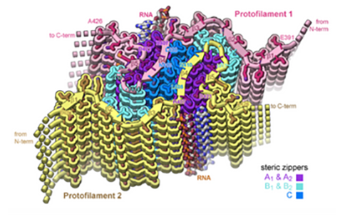
- We present the structure of an Aβ(1-42) fibril composed of two intertwined protofilaments determined by cryo-electron microscopy (cryo-EM) to 4.0-angstrom resolution, complemented by solid-state nuclear magnetic resonance experiments. (nih.gov)
- Scholars@Duke publication: Prefusion structure of trimeric HIV-1 envelope glycoprotein determined by cryo-electron microscopy. (duke.edu)
- Prefusion structure of trimeric HIV-1 envelope glycoprotein determined by cryo-electron microscopy. (duke.edu)
CryoEM4
- The program is broadening access to high-resolution cryoelectron microscopy (cryoEM) and tomography (cryoET) for biomedical researchers by creating national service centers, and cultivating a skilled workforce through the development and implementation of cryoEM tra ining material . (nih.gov)
- Did you miss one of the free monthly webinars on cryoEM current practices and strategies hosted by the National Centers for Cryoelectron Microscopy? (nih.gov)
- Protein adsorption at the air-water interface is a serious problem in cryogenic electron microscopy (cryoEM) as it restricts particle orientations in the vitrified ice-film and promotes protein denaturation. (fu-berlin.de)
- Congratulations to PhD candidate Christopher Kirchhoff for attending the competitive, two-week course on cryo electron microscopy (cryoEM) at the Cold Spring Harbor Laboratory (CSHL). (rochester.edu)
Crystallography5
- Cryogenic electron microscopy (cryo-EM) has revolutionized biology and life science as a powerful alternative to X-ray crystallography or NMR spectroscopy for macromolecular structure determination. (ucla.edu)
- In 2015, Bridget Carragher and colleagues at the Scripps National Resource for Automated Molecular Microscopy used techniques she and Clint Potter developed to determine the first cryo-EM structure with a resolution finer than 3 Å, thereby elevating CryoTEM as a tool comparable to and potentially superior to traditional x-ray crystallography techniques. (wikipedia.org)
- The work in the Gouaux Lab is concentrated on developing molecular mechanisms for the function of receptors and transporters at chemical synapses by utilizing cryo-electron microscopy, x-ray crystallography, and electrophysiology. (ohsu.edu)
- Cryoelectron microscopy and x-ray crystallography analyses of NoV VLPs identified the shell (S) and protruding domains (subdomains P1-1, P1-2, and P2) ( 18 ). (cdc.gov)
- Through combining cryo-electron microscopy at different pHs, X-ray crystallography and biochemical and structural analysis, we showed that NendoU can shift between open and closed forms, which probably correspond to active and inactive states, respectively. (lu.se)
Cryo-Electron T3
- Here we report the cryo-electron microscopy structure of a tubular HIV-1 capsid-protein assembly at 8 Å resolution and the three-dimensional structure of a native HIV-1 core by cryo-electron tomography. (nature.com)
- The Cryo-Electron Microscopy Service Platform at EMBL Heidelberg is now offering remote service to all interested users, including the COVID-19-related projects (single-particle analysis and cryo-electron tomography). (embl.org)
- We employ state-of-the-art techniques, such as cryo-transmission electron microscopy and cryo-electron tomography, small angle X-ray scattering, dynamic and static light scattering, high-sensitivity differential scanning and isothermal titration calorimetry, NMR and measurements of the electrophoretic mobility. (lu.se)
Electron-microscopy facility2
- Here, a brief overview is provided of the plans for a UK national three-dimensional electron-microscopy facility for integrated structural biology to enable internationally leading research on the machinery of life. (ox.ac.uk)
- The team has direct access to several platforms and facilities, including a well-equipped electron microscopy facility, protein crystallization, mass-spectrometry and several instruments for the biophysical characterization of proteins. (cnrs.fr)
Cryogenic electron2
- Cryogenic Electron Microscopy (Cryo-EM) is an approach that allows the observation of hydrated biological specimens in their native environment at cryogenic temperatures in TEM. (case.edu)
- Transmission electron cryomicroscopy (CryoTEM), commonly known as cryo-EM, is a form of cryogenic electron microscopy, more specifically a type of transmission electron microscopy (TEM) where the sample is studied at cryogenic temperatures (generally liquid-nitrogen temperatures). (wikipedia.org)
Structures10
- The cryo-electron-microscopy structures enable modelling by large-scale molecular dynamics simulation, resulting in all-atom models for the hexamer-of-hexamer and pentamer-of-hexamer elements as well as for the entire capsid. (nature.com)
- One method applied for that is cryo electron microscopy (cryo-EM), which can be used to make three-dimensional structures of biomolecules visible. (phys.org)
- In transmission electron microscopy (TEM), a high-energy electron beam is used to examine the structures of molecules, down to the level of atomic details. (case.edu)
- Cryo-electron microscopy structures of the N501Y SARS-CoV-2. (ubc.ca)
- Cryo electron microscopy (cryo EM) is a major structural biology method for studying macromolecular complexes and cellular structures in their native states. (embo.org)
- Researchers used cryo electron microscopy to visualize these structures. (syntecoptics.com)
- Cryo-electron microscopy (EM) is an optimal method to study protein structures that cannot be easily deciphered using other techniques. (researchsquare.com)
- In recent years, major advances in cryo-electron microscopy (cryo-EM) have enabled the routine determination of complex biomolecular structures at atomistic resolution. (bvsalud.org)
- In the last years there has been an explosion of the cryo electron microscopy single particle technique to get high-resolution structures of protein complexes. (lu.se)
- In addition to low-resolution models of the intact T3SS core from cryo-electron microscopy, several high-resolution structures of monomeric T3SS components have been determined. (lu.se)
Optical microscopy2
Particle3
- EMBL Heidelberg hosts a cryo-electron microscopy (cryo-EM) service platform, available for use by external scientists with both single particle and tomography projects. (embl.org)
- Structural determination of human Na1.4 and Na1.7 using single particle cryo-electron microscopy. (princeton.edu)
- The limiting factor for the structural determination of Na channels using single particle cryo-electron microscopy (cryo-EM) resides in the generation of sufficient high-quality recombinant proteins. (princeton.edu)
Protein2
- We present a 2.9-Å resolution cryo-electron microscopy (cryo-EM) structure of the complex between the ACE2 receptor and N501Y spike protein ectodomains that shows Y501 inserted into a cavity at the binding interface near Y41 of ACE2. (ubc.ca)
- Using cryo-electron microscopy, a technology with extremely high resolution, researchers have now, for the first time, managed to produce an image of an aquaporin with a drug candidate bound to the protein. (lu.se)
Facility2
Researchers1
- We provide researchers from Europe and beyond with a synergistic portfolio of imaging services including cryo-EM, super-resolution and intravital microscopy to enable new ground-breaking research that crosses the scales of biology. (embl.org)
Frozen-hydrated2
- The biological material is spread on an electron microscopy grid and is preserved in a frozen-hydrated state by rapid freezing, usually in liquid ethane near liquid nitrogen temperature. (wikipedia.org)
- In particular during cryo-transfer and (re-)mounting of frozen hydrated samples, which often in electron microscopy (EM), have a very large surface area compared to its volume, can be very challenging when samples have to be exchanged or remounted several times. (emc-proceedings.com)
Infrastructure3
- arranged in Lund by National Microscopy Infrastructure (NMI) Sweden ( nmisweden.se ). (lu.se)
- September 12th National Microscopy Infrastructure (NMI) Sweden ( nmisweden.se ) organized a clearing/expansion sample preparation symposium. (lu.se)
- September 13th National Microscopy Infrastructure (NMI) Sweden ( nmisweden.se ) organized a hands-on course - see link . (lu.se)
ACE21
- Further, we conducted a structural analysis of a mink variant S glycoprotein and American mink ACE2 (mvACE2) using cryo-electron microscopy (cryo-EM), revealing four distinct conformations. (cdc.gov)
Glycoprotein1
- The article is titled "Cryo-Electron Microscopy Structure of a Coronavirus Spike Glycoprotein Trimer. (bioquicknews.com)
Structural biology2
- NCCAT is housed at the Simons Electron Microscopy Center at the New York Structural Biology Center in New York City, New York. (cryoemcenters.org)
- To address this problem, we developed the metadynamic electron microscopy metainference (MEMMI) method , which incorporates metadynamics, an enhanced conformational sampling approach, into the metainference method of integrative structural biology . (bvsalud.org)
Transmission1
- To address this issue, the preparation of a graphene-based modified support film for coverage of conventional holey carbon transmission electron microscopy (TEM) grids is presented. (fu-berlin.de)
Structure4
- We use cryo electron microscopy (cryo-EM) and image reconstruction to examine the virion structure at subnanometer resolution. (lu.se)
- Groups led by Stefan Raunser of the Max Planck Institute for Molecular Physiology in Dortmund, Germany, and John Whitney of McMaster University in Canada have now jointly elucidated the high-resolution 3D structure of such an effector from Pseudomonas protegens using cryo-electron microscopy. (mpg.de)
- Determination of the Structure and Dynamics of the Fuzzy Coat of an Amyloid Fibril of IAPP Using Cryo-Electron Microscopy. (bvsalud.org)
- Sirohi et al described the structure of mature Zika virus based on cryoelectron microscopy. (medscape.com)
High-resolution1
- High-resolution cryo-electron microscopy and supercomputing have now made it possible to analyze, in detail, the infection mechanisms of coronaviruses. (bioquicknews.com)
Grid1
- For the application of cryo electron microscopy grid preparation, however, what is required is the transfer of very little fluid onto the grid in a well-defined manner. (monash.edu)
Congress2
- The 16th European Microscopy Congress, Lyon, France. (emc-proceedings.com)
- The official web site of the 16th European Microscopy Congress. (emc-proceedings.com)
Molecular1
- Cellular cryo electron microscopy helped visualize a molecular pore complex that spans both membranes of the double-membrane vesicle and would allow the export of RNA to the cytosol. (syntecoptics.com)
Technique1
- They used a cutting-edge technique called cryo-electron microscopy and combined it with supercomputer analysis. (insidehpc.com)
Chemistry1
- Holger Stark from the Max Planck Institute for Biophysical Chemistry in Göttingen and his team have broken a crucial resolution barrier in cryo electron microscopy. (phys.org)
Lund University1
- As part of MicLUs work, a survey to investigate the needs and capabilities regarding microscopy as well as image processing and analysis at Lund University has been created. (lu.se)
Techniques1
- The direct imaging of the ball-and-chain mechanism, using electron-microscopy techniques, can also provide a new angle to design drugs that target it to improve ion channel function. (medicalxpress.com)
Seminar1
- One-day Introductory seminar series (Online mode) on Cryo-Electron Microscopy for Structural Bio. (indiabioscience.org)
Super-resolution1
- Application of super-resolution and correlative double sampling in cryo-electron microscopy. (ox.ac.uk)
Life2
Image1
- Here we have used cryo-electron microscopy (cryo-EM) and image processing to determine the lateral and radial arrangement of Gag in in vivo and in vitro assembled Rous sarcoma virus (RSV) particles and to compare these features with those of HIV-1. (mpg.de)
Resolution1
- With low-temperature electron microscopy (cryo-EM), which bounces electrons instead of light off objects to make atomic-resolution images of them, the scientists obtained pictures of the MthK channel when it was switched open by calcium and switched closed. (medicalxpress.com)
Complex1
- Over 1.4 million 'freeze frames' of the human preinitiation complex, or PIC, were obtained with cryo-electron microscopy. (insidehpc.com)
Study1
- Microscopy is an important tool for scientists in the study of cells, tissues, and organs. (nih.gov)