Cranial Sinuses
Maxillary Sinus
Paranasal Sinuses
Cavernous Sinus
Carotid Sinus
Frontal Sinus
Paranasal Sinus Diseases
Sphenoid Sinus
Coronary Sinus
Sinus Thrombosis, Intracranial
Sick Sinus Syndrome
Ethmoid Sinus
Tachycardia, Sinus
Maxillary Sinus Neoplasms
Transverse Sinuses
Superior Sagittal Sinus
Sinoatrial Node
Multiple dural arteriovenous shunts in a 5-year-old boy. (1/170)
We describe a rare case of multiple dural arteriovenous shunts (DAVSs) in a 5-year-old boy. MR imaging performed at 1 year of age showed only a dilated anterior part of the superior sagittal sinus; however, angiography at 5 years of age revealed an infantile-type DAVS there and two other DAVSs of the adult type. The pathophysiological evolution of DAVSs in children and their treatment strategies are discussed. (+info)Variant arteriovenous fistula of the superior sagittal sinus--case report. (2/170)
A 57-year-old male presented with a rare variant of dural arteriovenous fistula, located in the wall of an unobstructed superior sagittal sinus. Drainage occurred through a cortical vein no longer connected to its parent sinus, which filled up a cluster of transmedullary running veins, one of which was the presumed site of hemorrhage. Arterial blood was supplied via the external carotid artery branches. This type of fistula seriously increases the risk of hemorrhage in the patient and therefore requires complete obliteration. Attempts to embolize the fistula failed. The draining vein was isolated and coagulated resulting in permanent occlusion of the fistula. The fistula probably developed through a process of thrombophlebitis and revascularization via arterioles of the vein rather than previous occlusion of the sinus. (+info)Cavernous sinus and inferior petrosal sinus flow signal on three-dimensional time-of-flight MR angiography. (3/170)
BACKGROUND AND PURPOSE: Venous flow signal in the cavernous sinus and inferior petrosal sinus has been shown on MR angiograms in patients with carotid cavernous fistula (CCF). We, however, identified flow signal in some patients without symptoms and signs of CCF. This review was performed to determine the frequency of such normal venous flow depiction at MR angiography. METHODS: Twenty-five 3D time-of-flight (TOF) MR angiograms obtained on two different imaging units (scanners A and B) were reviewed with attention to presence of venous flow signal in the cavernous sinus or inferior petrosal sinus or both. Twenty-five additional MR angiograms were reviewed in patients who had also had cerebral arteriography to document absence of CCF where venous MR angiographic signal was detected, as well as to gain insight into venous flow patterns that might contribute to MR angiographic venous flow signal. Differences in scanning technique parameters were reviewed. RESULTS: Nine (36%) of the 25 MR angiograms obtained on scanner A but only one (4%) of the 25 obtained on scanner B showed flow signal in the cavernous or inferior petrosal sinus or both in the absence of signs of CCF. On review of 25 patients who had both MR angiography and arteriography, three patients with venous signal at MR angiography failed to exhibit CCF at arteriography. CONCLUSION: Identification of normal cavernous sinus or inferior petrosal sinus venous signal on 3D TOF MR angiograms may occur frequently, and is probably dependent on technical factors that vary among scanners. The exact factors most responsible, however, were not elucidated by this preliminary review. (+info)Regions of interest in the venous sinuses as input functions for quantitative PET. (4/170)
As clinical PET becomes increasingly available, quantitative methods that are feasible in busy clinical settings are becoming necessary. We investigated the use of intracranial blood pools as sources of an input function for quantitative PET. METHODS: We studied 25 patients after the intravenous injection of [18F]6-fluoro-L-m-tyrosine and compared sampled blood time-activity curves with those obtained in small regions of interest (ROIs) defined in the blood pools visible in the PET images. Because of the comparatively large dimensions of the blood pool at the confluence of the superior sagittal, straight and transverse sinuses, a venous ROI input function was chosen for further analysis. We applied simple corrections to the ROI-derived time-activity curves, deriving expressions for partial volume, spillover and partition of tracer between plasma and red blood cells. The results of graphic and compartmental analysis using both sampled [Cs(t)] and ROI [Cr(t)] venous input functions for each patient were compared. We also used an analytic approach to examine possible differences between venous and arterial input functions in the cerebral circulation. RESULTS: Cr(t) peaked significantly earlier and higher than Cs(t) in this patient population, although the total integral under the curves did not differ significantly. We report some apparent differences in the results of modeling using the two input functions; however, neither the graphically determined influx constant, Ki, nor the model parameter that reflects presynaptic dopaminergic metabolism, k3, differed significantly between the two methods. The analytic results suggest that the venous ROI input function may be closer to the arterial supply of radiotracer to the brain than arterialized venous blood, at least in some patient populations. CONCLUSION: We present a simple method of obtaining an input function for PET that is applicable to a wide range of tracers and quantitative methods and is feasible for diagnostic PET imaging. (+info)Skull metastasis of Ewing's sarcoma--three case reports. (5/170)
Three cases of skull metastasis of Ewing's sarcoma were treated. The metastatic lesion was located at the midline of the skull above the superior sagittal sinus in all cases. Surgery was performed in two patients with solitary skull lesions involving short segments of the superior sagittal sinus without remarkable systemic metastasis, resulting in good outcome. The third patient had extensive, multiple tumors involving the superior sagittal sinus which could not be excised, and died due to intracranial hypertension. The surgical indication for skull metastasis of Ewing's sarcoma depends on the location and length of the involved superior sagittal sinus, and general condition. (+info)Absent vestibulo-ocular reflexes and acute supratentorial lesions. (6/170)
Loss of vestibulo-ocular reflexes occurred in two patients with acute supratentorial lesions who received therapeutic doses of anticonvulsant drugs. There was no clinical or angiographic evidence of focal brain-stem damage. Absence of vestibulo-ocular reflexes is attributed to a combination of acute cerebral damage and anticonvulsant drugs. The loss of these reflexes in patients with acute cerebral lesions cannot be interpreted as evidence of irreversible brain-stem injury. (+info)Scalp vein detected using internal carotid angiography that did not result in venous sinus compromise. (7/170)
We present an unusual case of a scalp vein detected by using angiography of the internal carotid artery. The vein arose from the superior sagittal sinus and drained into the deep posterior cervical vein via the parietal emissary vein. This scalp vein may be a collateral pathway for venous sinuses; however, the patient had no evidence of venous sinus occlusive disease or intracranial hypertension. (+info)Sigmoid sinus thrombosis after mild closed head injury in an infant: diagnosis by magnetic resonance imaging in the acute phase--case report. (8/170)
Intracranial sinus thrombosis following a mild closed head injury without a skull fracture or intracranial hematoma is extremely rare. A 23-month-old girl presented with vomiting and gait ataxia 1 day after occipital trauma. Computed tomography revealed a slightly increased density area in the region of the left sigmoid sinus. T1-weighted magnetic resonance (MR) imaging demonstrated an isointense area in the left sigmoid sinus and T2-weighted imaging showed a hyperintense area reflecting the characteristics of oxyhemoglobin. MR angiography and cerebral angiography indicated occlusion of the left sigmoid sinus. After 4 days of conservative treatment, her symptoms subsided completely. Follow-up MR angiography and cerebral angiography showed recanalization of the sigmoid sinus. The MR images and MR angiograms were useful for both early diagnosis and follow-up. Treatment should reflect the severity of individual cases, and early diagnosis will help achieve a good outcome. (+info)Cranial sinuses are a part of the venous system in the human head. They are air-filled spaces located within the skull and are named according to their location. The cranial sinuses include:
1. Superior sagittal sinus: It runs along the top of the brain, inside the skull, and drains blood from the scalp and the veins of the brain.
2. Inferior sagittal sinus: It runs along the bottom of the brain and drains into the straight sinus.
3. Straight sinus: It is located at the back of the brain and receives blood from the inferior sagittal sinus and great cerebral vein.
4. Occipital sinuses: They are located at the back of the head and drain blood from the scalp and skull.
5. Cavernous sinuses: They are located on each side of the brain, near the temple, and receive blood from the eye and surrounding areas.
6. Sphenoparietal sinus: It is a small sinus that drains blood from the front part of the brain into the cavernous sinus.
7. Petrosquamosal sinuses: They are located near the ear and drain blood from the scalp and skull.
The cranial sinuses play an essential role in draining blood from the brain and protecting it from injury.
The maxillary sinuses, also known as the antrums of Highmore, are the largest of the four pairs of paranasal sinuses located in the maxilla bones. They are air-filled cavities that surround the nasolacrimal duct and are situated superior to the upper teeth and lateral to the nasal cavity. Each maxillary sinus is lined with a mucous membrane, which helps to warm, humidify, and filter the air we breathe. Inflammation or infection of the maxillary sinuses can result in conditions such as sinusitis, leading to symptoms like facial pain, headaches, and nasal congestion.
Paranasal sinuses are air-filled cavities in the skull that surround the nasal cavity. There are four pairs of paranasal sinuses, including the maxillary, frontal, ethmoid, and sphenoid sinuses. These sinuses help to warm, humidify, and filter the air we breathe. They also contribute to our voice resonance and provide a slight cushioning effect for the skull. The openings of the paranasal sinuses lead directly into the nasal cavity, allowing mucus produced in the sinuses to drain into the nose. Infections or inflammation of the paranasal sinuses can result in conditions such as sinusitis.
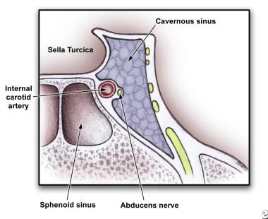
The cavernous sinus is a venous structure located in the middle cranial fossa, which is a depression in the skull that houses several important nerves and blood vessels. The cavernous sinus is situated on either side of the sphenoid bone, near the base of the skull, and it contains several important structures:
* The internal carotid artery, which supplies oxygenated blood to the brain
* The abducens nerve (cranial nerve VI), which controls lateral movement of the eye
* The oculomotor nerve (cranial nerve III), which controls most of the muscles that move the eye
* The trochlear nerve (cranial nerve IV), which controls one of the muscles that moves the eye
* The ophthalmic and maxillary divisions of the trigeminal nerve (cranial nerve V), which transmit sensory information from the face and head
The cavernous sinus is an important structure because it serves as a conduit for several critical nerves and blood vessels. However, it is also vulnerable to various pathological conditions such as thrombosis (blood clots), infection, tumors, or aneurysms, which can lead to serious neurological deficits or even death.
The Sinus of Valsalva are three pouch-like dilations or outpouchings located at the upper part (root) of the aorta, just above the aortic valve. They are named after Antonio Maria Valsalva, an Italian anatomist and physician. These sinuses are divided into three parts:
1. Right Sinus of Valsalva: It is located to the right of the ascending aorta and usually gives rise to the right coronary artery.
2. Left Sinus of Valsalva: It is situated to the left of the ascending aorta and typically gives rise to the left coronary artery.
3. Non-coronary Sinus of Valsalva: This sinus is located in between the right and left coronary sinuses, and it does not give rise to any coronary arteries.
These sinuses play a crucial role during the cardiac cycle, particularly during ventricular contraction (systole). The pressure difference between the aorta and the ventricles causes the aortic valve cusps to be pushed into these sinuses, preventing the backflow of blood from the aorta into the ventricles.
Anatomical variations in the size and shape of the Sinuses of Valsalva can occur, and certain conditions like congenital heart diseases (e.g., aortic valve stenosis or bicuspid aortic valve) may affect their structure and function. Additionally, aneurysms or ruptures of the sinuses can lead to severe complications, such as cardiac tamponade, endocarditis, or stroke.
The carotid sinus is a small, dilated area located at the bifurcation (or fork) of the common carotid artery into the internal and external carotid arteries. It is a baroreceptor region, which means it contains specialized sensory nerve endings that can detect changes in blood pressure. When the blood pressure increases, the walls of the carotid sinus stretch, activating these nerve endings and sending signals to the brain. The brain then responds by reducing the heart rate and relaxing the blood vessels, which helps to lower the blood pressure back to normal.
The carotid sinus is an important part of the body's autonomic nervous system, which regulates various involuntary functions such as heart rate, blood pressure, and digestion. It plays a crucial role in maintaining cardiovascular homeostasis and preventing excessive increases in blood pressure that could potentially damage vital organs.
A frontal sinus is a paired, air-filled paranasal sinus located in the frontal bone of the skull, above the eyes and behind the forehead. It is one of the four pairs of sinuses found in the human head. The frontal sinuses are lined with mucous membrane and are interconnected with the nasal cavity through small openings called ostia. They help to warm, humidify, and filter the air we breathe, and contribute to the resonance of our voice. Variations in size, shape, and asymmetry of frontal sinuses are common among individuals.
Paranasal sinus diseases refer to a group of medical conditions that affect the paranasal sinuses, which are air-filled cavities located within the skull near the nasal cavity. These sinuses include the maxillary, frontal, ethmoid, and sphenoid sinuses.
Paranasal sinus diseases can be caused by a variety of factors, including viral, bacterial, or fungal infections, allergies, structural abnormalities, or autoimmune disorders. Some common paranasal sinus diseases include:
1. Sinusitis: Inflammation or infection of the sinuses, which can cause symptoms such as nasal congestion, thick nasal discharge, facial pain or pressure, and reduced sense of smell.
2. Nasal polyps: Soft, benign growths that develop in the lining of the nasal passages or sinuses, which can obstruct airflow and cause difficulty breathing through the nose.
3. Sinonasal tumors: Abnormal growths that can be benign or malignant, which can cause symptoms such as nasal congestion, facial pain, and bleeding from the nose.
4. Sinus cysts: Fluid-filled sacs that form in the sinuses, which can cause symptoms similar to those of sinusitis.
5. Fungal sinusitis: Infection of the sinuses with fungi, which can cause symptoms such as nasal congestion, facial pain, and thick, discolored mucus.
Treatment for paranasal sinus diseases depends on the underlying cause and severity of the condition. Treatment options may include medications, such as antibiotics, antihistamines, or corticosteroids, as well as surgical intervention in more severe cases.
The sphenoid sinuses are air-filled spaces located within the sphenoid bone, which is one of the bones that make up the skull base. These sinuses are located deep inside the skull, behind the eyes and nasal cavity. They are paired and separated by a thin bony septum, and each one opens into the corresponding nasal cavity through a small opening called the sphenoethmoidal recess. The sphenoid sinuses vary greatly in size and shape between individuals. They develop during childhood and continue to grow until early adulthood. The function of the sphenoid sinuses, like other paranasal sinuses, is not entirely clear, but they may contribute to reducing the weight of the skull, resonating voice during speech, and insulating the brain from trauma.
The coronary sinus is a large vein that receives blood from the heart's muscle tissue. It is located on the posterior side of the heart and is a part of the cardiovascular system. The coronary sinus collects oxygen-depleted blood from the myocardium (the heart muscle) and drains it into the right atrium, where it will then be pumped to the lungs for oxygenation.
The coronary sinus is an essential structure in medical procedures such as cardiac catheterization and electrophysiological studies. It is also a common site for the implantation of pacemakers and other cardiac devices.
Intracranial sinus thrombosis is a medical condition characterized by the formation of a blood clot (thrombus) within the intracranial venous sinuses, which are responsible for draining blood from the brain. The condition can lead to various neurological symptoms and complications, such as increased intracranial pressure, headaches, seizures, visual disturbances, and altered consciousness. Intracranial sinus thrombosis may result from various factors, including hypercoagulable states, infections, trauma, and malignancies. Immediate medical attention is necessary for proper diagnosis and treatment to prevent potential long-term neurological damage or even death.
Sick Sinus Syndrome (SSS) is a term used to describe a group of abnormal heart rhythm disturbances that originates in the sinoatrial node (the natural pacemaker of the heart). This syndrome is characterized by impaired functioning of the sinoatrial node, resulting in various abnormalities such as sinus bradycardia (abnormally slow heart rate), sinus arrest (complete cessation of sinus node activity), and/or sinoatrial exit block (failure of the electrical impulse to leave the sinus node and spread to the atria).
People with SSS may experience symptoms such as palpitations, dizziness, fatigue, shortness of breath, or syncope (fainting) due to inadequate blood supply to the brain caused by slow heart rate. The diagnosis of SSS is typically made based on the patient's symptoms and the results of an electrocardiogram (ECG), Holter monitoring, or event recorder that shows evidence of abnormal sinus node function. Treatment options for SSS may include lifestyle modifications, medications, or implantation of a pacemaker to regulate the heart rate.
The ethmoid sinuses are a pair of air-filled spaces located in the ethmoid bone, which is a part of the skull that forms the upper portion of the nasal cavity and the inner eye socket. These sinuses are divided into anterior and posterior groups and are present in adults, but not at birth. They continue to grow and develop until early adulthood.
The ethmoid sinuses are lined with mucous membrane, which helps to warm, humidify, and filter the air we breathe. They are surrounded by a network of blood vessels and nerves, making them susceptible to inflammation and infection. Inflammation of the ethmoid sinuses can lead to conditions such as sinusitis, which can cause symptoms such as nasal congestion, headache, and facial pain.
Paranasal sinus neoplasms refer to abnormal growths or tumors that develop within the paranasal sinuses, which are air-filled cavities located inside the skull near the nasal cavity. These tumors can be benign (noncancerous) or malignant (cancerous), and they can arise from various types of tissue within the sinuses, such as the lining of the sinuses (mucosa), bone, or other soft tissues.
Paranasal sinus neoplasms can cause a variety of symptoms, including nasal congestion, nosebleeds, facial pain or numbness, and visual disturbances. The diagnosis of these tumors typically involves a combination of imaging studies (such as CT or MRI scans) and biopsy to determine the type and extent of the tumor. Treatment options may include surgery, radiation therapy, chemotherapy, or a combination of these approaches, depending on the specific type and stage of the neoplasm.
Sinus tachycardia is a type of rapid heart rate, characterized by an abnormally fast sinus rhythm, with a rate greater than 100 beats per minute in adults. The sinoatrial node (SA node), which is the natural pacemaker of the heart, generates these impulses regularly and at an increased rate.
Sinus tachycardia is usually a physiological response to various stimuli or conditions, such as physical exertion, strong emotions, fever, anxiety, pain, or certain medications. It can also be caused by hormonal imbalances, anemia, hyperthyroidism, or other medical disorders.
In most cases, sinus tachycardia is not harmful and resolves once the underlying cause is addressed. However, if it occurs persistently or is associated with symptoms like palpitations, shortness of breath, dizziness, or chest discomfort, further evaluation by a healthcare professional is recommended to rule out any underlying heart conditions or other medical issues.
Maxillary sinus neoplasms refer to abnormal growths or tumors that develop in the maxillary sinuses, which are located in the upper part of your cheekbones, below your eyes. These growths can be benign (non-cancerous) or malignant (cancerous).
Benign neoplasms may include conditions such as an osteoma (a benign bone tumor), a papilloma (a benign growth of the lining of the sinus), or a fibrous dysplasia (a condition where bone is replaced by fibrous tissue).
Malignant neoplasms, on the other hand, can be primary (originating in the maxillary sinuses) or secondary (spreading to the maxillary sinuses from another site in the body). Common types of malignant tumors that arise in the maxillary sinus include squamous cell carcinoma, adenocarcinoma, and mucoepidermoid carcinoma.
Symptoms of maxillary sinus neoplasms may include nasal congestion, nosebleeds, facial pain or numbness, vision changes, and difficulty swallowing or speaking. Treatment options depend on the type, size, and location of the tumor but may include surgery, radiation therapy, chemotherapy, or a combination of these approaches.
A pilonidal sinus is a small hole or tunnel in the skin that usually develops in the cleft at the top of the buttocks. It can be painful and may become infected, causing symptoms such as redness, swelling, pain, and pus discharge. The condition often affects young adults and is more common in men than women.
The term "pilonidal" comes from the Latin words "pilus," meaning hair, and "nidus," meaning nest. This refers to the fact that the sinus often contains hairs that have become embedded in the skin. The exact cause of pilonidal sinuses is not known, but they are thought to develop as a result of ingrown hairs or chronic irritation in the affected area.
Treatment for pilonidal sinuses typically involves surgical removal of the sinus and any associated hair follicles. In some cases, this may be done using a minor procedure that can be performed in a doctor's office. More complex cases may require hospitalization and a more extensive surgical procedure. After surgery, patients will need to take steps to prevent the sinus from recurring, such as keeping the area clean and avoiding prolonged periods of sitting or driving.
The transverse sinuses are a pair of venous channels located within the skull. They are part of the intracranial venous system and are responsible for draining blood from the brain. The transverse sinuses run horizontally along the upper portion of the inner skull, starting at the occipital bone (at the back of the head) and extending to the temporal bones (on the sides of the head).
These sinuses receive blood from the superior sagittal sinus, straight sinus, and the occipital sinus. After passing through the transverse sinuses, the blood is then drained into the sigmoid sinuses, which in turn drain into the internal jugular veins. The transverse sinuses are an essential component of the cerebral venous system, ensuring proper blood flow and drainage from the brain.
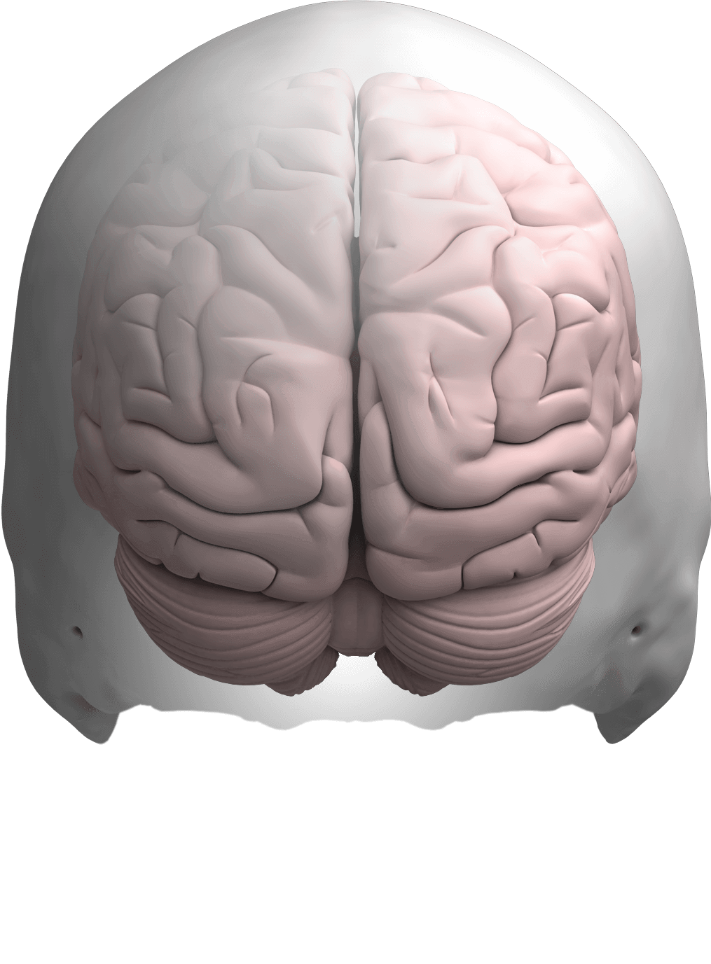
The Superior Sagittal Sinus is a medical term that refers to a venous sinus (a channel for blood flow) located in the superior part (highest portion) of the sagittal suture, which is the line along the top of the skull where the two parietal bones join in the middle. It runs from front to back, starting at the frontal bone and ending at the occipital bone, and it receives blood from veins that drain the cerebral hemispheres (the right and left halves of the brain).
The Superior Sagittal Sinus is an important structure in the circulatory system of the brain as it plays a critical role in draining venous blood from the cranial cavity. It also contains valveless venous channels that allow for the flow of cerebrospinal fluid (CSF) between the intracranial and extracranial compartments.
It is worth noting that any damage to this structure, such as through trauma or infection, can lead to serious neurological complications, including increased intracranial pressure, seizures, and even death.
The sinoatrial (SA) node, also known as the sinus node, is the primary pacemaker of the heart. It is a small bundle of specialized cardiac conduction tissue located in the upper part of the right atrium, near the entrance of the superior vena cava. The SA node generates electrical impulses that initiate each heartbeat, causing the atria to contract and pump blood into the ventricles. This process is called sinus rhythm.
The SA node's electrical activity is regulated by the autonomic nervous system, which can adjust the heart rate in response to changes in the body's needs, such as during exercise or rest. The SA node's rate of firing determines the heart rate, with a normal resting heart rate ranging from 60 to 100 beats per minute.
If the SA node fails to function properly or its electrical impulses are blocked, other secondary pacemakers in the heart may take over, resulting in abnormal heart rhythms called arrhythmias.
 Paranthropus robustus
Congenital dermal sinus
Groove for sigmoid sinus
Sphenoid wing meningioma
Whale vocalization
Falx cerebri
Carnotaurus
Great cerebral vein
Occipital vein
Craniometaphyseal dysplasia
Osteoma
Phycomycosis
Jugular foramen
Dura mater
Danger triangle of the face
Junggarsuchus
Diprotodon
Raccoon eyes
Epidural abscess
Ceratosaurus
Headache
Cerebrospinal venous system
List of MeSH codes (A07)
Skull fracture
Fovea
Carotid sinus nerve
Pterygoid plexus
Mastoid cells
Emissary veins
Cranial cavity
 Intracranial Sinus Thrombosis (Cranial Sinus Thromboses): Symptoms, Diagnosis and Treatment - Symptoma
 Base of skull, cranial fossa and cranial sinuses | Anatomia Collection: anatomical plates 1522-1867
Carotid and Cranial Nerve Reconstruction after Removal of Cavernous Sinus Lesions<...
 Petrosal sinus sampling: technique and rationale
 Skull Base Anatomy: Overview, Anterior Skull Base, Middle Skull Base
Paranthropus robustus - Wikipedia
Endovascular management of dural arteriovenous fistulas of the transverse and sigmoid sinus in 150 patients
 Funded Research - American Osteopathic Association
Cavernous Sinus Thrombosis: Practice Essentials, Background, Pathophysiology
Diprotodon - Wikipedia
 Head CT scan: MedlinePlus Medical Encyclopedia
 WTS database | WHO FCTC
 Clinical Characteristics of Recurrent Nasopharyngeal Carcinoma in High-Incidence Area
Brain Meningioma Imaging: Practice Essentials, Radiography, Computed Tomography
 SciELO - Radiologia Brasileira, Volume: 34, Issue: 4, Published: 2001
 Early evidence of trepanation along the Yellow River Basin in Neolithic China | Archaeological and Anthropological Sciences
 3b. 4. The Veins of the Brain - Collection at Bartleby.com
 One-Hour Contact with the Earth's Surface (Grounding) Improves Inflammation and Blood Flow-A Randomized, Double-Blind, Pilot...
Differences in hematocrit of blood samples obtained from two venipuncture sites in sharks in: American Journal of Veterinary...
 Ptosis Correction - StatPearls - NCBI Bookshelf
 Sinus Headache or Migraine: Differences, Symptoms, Relief
Plus it
Glossary - Vetneuro.com
 paranasal sinus - Ontology Report - Rat Genome Database
 LOINC 37857-0 XR Sinuses Caldwell
Top 10 Chemo Cooling For Mens Head And Neck - Home Previews
 Iliac vein. Medical search
Anterior crani7
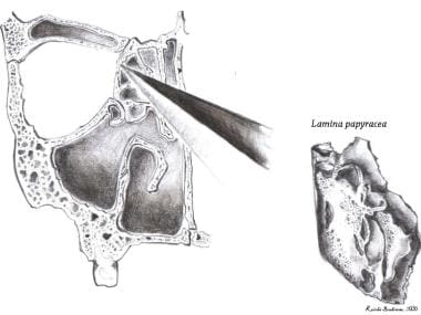
- Anterior cranial fossa and body of the sphenoid. (medscape.com)
- The ethmoid bone forms the central part of the floor, which is the deepest area of the anterior cranial fossa. (medscape.com)
- The most important anatomic structures below the anterior cranial fossa are the orbits and the paranasal sinuses. (medscape.com)
- Computed tomography (CT) and magnetic resonance (MR) imaging revealed a lesion primarily involving the upper nasal cavity extending intracranially through the cribriform plates into the anterior cranial fossa. (surgicalneurologyint.com)
- Lymphoma exclusively involving the nasal sinus or the anterior cranial fossa is rare in Western populations. (surgicalneurologyint.com)
- The tumor eroded from the nasal cavity into the skull base, and extended intracranially through the cribriform plates into the anterior cranial fossa. (surgicalneurologyint.com)
- The roof is formed by the frontal bone and the lesser wing of the sphenoid bone, and communicates anteriorly with the frontal sinus and posteriorly with the anterior cranial fossa. (tidsskriftet.no)
Nerves7
- The clinical consequences of these changes include headaches that are often but not always orthostatic, nausea, occasional emesis, neck and interscapular pain, cochleovestibular manifestations, cranial nerve palsies, and several other manifestations attributed to pressure upon or stretching of the cranial nerves or brain or brainstem structures. (medscape.com)
- 1- 3 However, the pituitary gland can be involved by a wide range of other pathological processes arising both within the gland itself and in the surrounding structures: the skull in the region of the sella turcica, dura mater, blood vessels, cranial nerves, and brain. (bmj.com)
- Which of the following nerves is not a branch of cranial nerve VII? (medsterz.com)
- a thick meningeal layer and a much thinner translucent layer surrounding the cranial nerves 5,6 . (radiopaedia.org)
- The cavernous sinus transmits multiple cranial nerves to the superior orbital fissure and foramen rotundum . (radiopaedia.org)
- Most of the patients usually have external ophthalmoplegia , which results from the venous congestion of eye tissues, inflammation of body organ including extra-ocular muscle and/or cranial nerves III, IV and VI [7]. (symptoma.com)
- As the sinus lies proximal to few cranial nerves, neurological disturbances may be experienced. (symptoma.com)
Thrombosis10
- Between 2001 and 2003, 10 consecutive patients (six men and four women, age range 54-79 years) who had presented with transverse and/or sigmoid sinus DAVFs with or without sinus thrombosis underwent self-expanding stent placement and balloon angioplasty. (nih.gov)
- Cerebral venous sinus thrombosis as a complication is fortunately less common, and superficial siderosis and bibrachial amyotrophy are rare. (medscape.com)
- Infective sinus thrombosis. (nih.gov)
- I'd like to welcome you to today's COCA Call: Johnson and Johnson Janssen COVID-19 Vaccine and Cerebral Venous Sinus Thrombosis with Thrombocytopenia -- Update for Clinicians on Early Detection and Treatment. (cdc.gov)
- Today I'll be discussing some background on the CVST situation and then move into a description of the reports of cerebral venous sinus thrombosis with thrombocytopenia following the Janssen COVID-19 vaccine. (cdc.gov)
- Case report: Cerebrovascular revascularization in a case of cranial venous sinus thrombosis. (sveta-anna.eu)
- Cavernous sinus thrombosis (CST) is an uncommon clinical condition caused by blood clot formation that obstructs the cavernous cavity . (symptoma.com)
- Causes Most cases of septic cavernous sinus thrombosis (CST) are due to an acute infection in an otherwise healthy individual. (symptoma.com)
- A cranial computed tomographic (CT) scan showed thrombosis of the superior sagittal sinus associated with 3 cerebral hematomas (left frontal and bilateral parieto-occipital) and diffuse cerebral edema with signs of increased intracranial pressure ( Figure ). (cdc.gov)
- Thrombosis of the superior sagittal sinus was caused by a platelet-fibrin thrombus. (cdc.gov)
Middle crani3
- The petro-occipital fissure subdivides the middle cranial fossa into 1 central component and 2 lateral components. (medscape.com)
- The dural venous sinus found on the floor of the middle cranial fossa is the? (medsterz.com)
- The lateral wall of the cavernous sinus is primarily formed by the continuation of the meningeal layer of the dura, flowing medially up from the floor of the middle cranial fossa, over the cavernous sinus, to the clinoid processes before forming the diaphragma sella. (radiopaedia.org)
Posterior12
- The skull base can be subdivided into 3 regions: the anterior, middle, and posterior cranial fossae. (medscape.com)
- The anterior limit of the anterior skull base is the posterior wall of the frontal sinus. (medscape.com)
- The anterior clinoid processes and the planum sphenoidale, which forms the roof of the sphenoid sinus, mark the posterior limit. (medscape.com)
- The posterior wall is thin and adjacent to the superior sagittal sinus and frontal lobe dura. (medscape.com)
- 1. [Dermal sinus and dermoid cyst revealed by abscess formation in posterior fossa. (nih.gov)
- 2. Posterior fossa abscesses secondary to dermal sinus associated with dermoid cyst in children. (nih.gov)
- 5. Dermoid cyst of the posterior fossa associated with congenital dermal sinus in a child. (nih.gov)
- 8. Posterior fossa dermoid cyst with sinus tract and meningitis in a toddler. (nih.gov)
- 9. Infected Intradural Dermoid Cyst with Complete Dermal Sinus of Posterior Fossa. (nih.gov)
- 14. [A case of dermoid cyst in the posterior fossa with dermal sinus]. (nih.gov)
- Additionally, there was involvement of the sphenoid sinus and right cavernous sinus with extension into the posterior fossa along the course of the trigeminal nerve and encasement with narrowing of the right carotid artery. (karger.com)
- The internal carotid artery enters the posterior inferior aspect of the sinus and bends upon itself as the carotid siphon ( cavernous segment - C4 ). (radiopaedia.org)
Frontal sinus3
- You have two large frontal sinus cavities. (tylenol.com)
- The Frontal Sinus, Perpendicular Lamina and Vomer are fitted with Folders-Flaps which can be opened to view the Lateral Nose wall and Sphenoidal Sinus. (buyamag.com)
- Frontal Bone has a hinged flap into the Frontal Sinus. (buyamag.com)
Trigeminal nerve3
- Maxillary sinus overpressurization can compress the maxillary branch of the trigeminal nerve, causing hyperesthesia over the cheek. (msdmanuals.com)
- The superior orbital fissure continues in a craniolateral direction from the inferior orbital fissure and contains cranial nerve III (oculomotor nerve), cranial nerve IV (trochlear nerve), branches of cranial nerve V (trigeminal nerve), cranial nerve VI (abducens nerve), sympathetic fibres from the cavernous plexus and superior ophthalmic vein and inferior ophthalmic vein. (tidsskriftet.no)
- The TVS consists of the trigeminal nerve, spinal trigeminal nucleus, and cranial vessels. (eyewiki.org)
Cavernous10
- Many have advocated radical surgical resection (i.e. exenteration of the cavernous sinus with carotid sacrifice and en bloc resection) with administration of amphotericin B. We present a case of mucormycosis involving the paranasal sinuses and cranial base in a pediatric patient who experienced long-term survival with a more limited resection. (karger.com)
- Major cranial sinuses include a postero-superior group (such as superior sagittal, inferior sagittal, straight, transverse, and occipital) and an antero-inferior group (such as cavernous, petrosal, and basilar plexus). (nih.gov)
- The cavernous sinuses are paired dural venous sinuses . (radiopaedia.org)
- The cavernous sinus is located on either side of the pituitary fossa and body of the sphenoid bone . (radiopaedia.org)
- From here, the meningeal layer passes downwards to surround the pituitary gland, thus forming the medial wall of the cavernous sinus. (radiopaedia.org)
- In contrast, the floor of the cavernous sinus is formed by the endosteal layer of the meninges (actually just periosteum) that covers the sphenoid bone and passes medially across the midline below the pituitary gland, separated from the aforementioned meningeal layer by the intercavernous sinus 5 . (radiopaedia.org)
- Depending on relative pressures the superior ophthalmic veins either drain to or from the cavernous sinus. (radiopaedia.org)
- Additionally, the cavernous sinuses connect to each other via the intercavernous sinuses . (radiopaedia.org)
- Fatty deposits may be present within the cavernous sinus, especially in obese patients or in those who are taking corticosteroids 3 . (radiopaedia.org)
- Although vision loss may be rare since the orbital nerve is located externally to cavernous sinus , it can develop through other pathophysiological mechanisms such as internal carotid artery occlusion (ICA), ophthalmic (central) retinal arteries obstructions, orbital congestion and arteritis [7]. (symptoma.com)
Occipital8
- Eight fistulas involved the transverse sinus, three the sigmoid sinus, and one the torcular and occipital sinuses. (nih.gov)
- 4. Cerebellar dermoid cyst with occipital dermal sinus. (nih.gov)
- 6. Occipital dermoid cyst associated with dermal sinus complicated with meningitis: A case report. (nih.gov)
- 7. Cerebellar abscesses secondary to occipital dermoid cyst with dermal sinus: case report. (nih.gov)
- 10. [Cerebellar abscesses secondary to infection of an occipital dermal sinus]. (nih.gov)
- 19. Report of eight cases of occipital dermal sinus: an update, and MRI findings. (nih.gov)
- 20. Unusual cause of cerebellar abscess: occipital dermal sinus and dermoid cyst. (nih.gov)
- A) Noncontrast cranial computed tomographic (CT) scan of a 26-year-old immunocompetent man with influenza, showing diffuse cerebral edema (Ed) and bilateral parieto-occipital hematoma (H). B) Cranial CT scan with. (cdc.gov)
Spinal11
- HEADACHE secondary to low cerebrospinal fluid (CSF) pressure occurs after diagnostic lumbar puncture, myelography, cranial or spinal injury, or spinal anesthesia. (asahq.org)
- She had no history of chronic headache, meningitis, cranial or spinal trauma, or paranasal sinus infection. (asahq.org)
- The cranial dura mater commonly referred to as the cranial durum, is a strong, fibrous membrane that acts as the outermost layer of protection surrounding the brain as well as the spinal cord inside the skull cavity. (keydifference.in)
- Understanding the distinctions between the cranial dura and the spinal dura is vital for a variety of reasons in the fields of neurology, medicine, and neurosurgery. (keydifference.in)
- Recognizing the distinct features of the spinal and cranial dura is crucial for accurate diagnosis in clinical practice and treatment plans. (keydifference.in)
- Surgery specialists operating within the cranial and spinal regions require a clear understanding of the differences between dura mater to carry out procedures in a safe and efficient manner whether it's cranial surgery like craniotomies or spinal surgeries such as laminectomies. (keydifference.in)
- Distinctive pathological conditions may affect the cranial or spinal dura. (keydifference.in)
- The cranial dura is home to structures similar to the dural sinuses that are involved with CSF absorption, and the spinal dura is a part as well in the CSF circulatory system and regulates pressure. (keydifference.in)
- Healthcare professionals and neurologists need to recognize the involvement of the spinal dura and the cranial nerve when evaluating symptoms of neurological conditions. (keydifference.in)
- Educating patients about the difference between cranial as well as spinal dura is a great way of increasing their understanding of surgical and medical issues which can improve their decision-making abilities and adherence to treatment plans. (keydifference.in)
- A thorough knowledge of the difference between spinal and cranial dura is vital in the field of clinical practices, surgery and diagnostics, research, and patient care in the fields of neurology as well as neurosurgery. (keydifference.in)
Transverse Sinuses1
- Near the lower back of the CRANIUM, the superior sagittal sinus deviates to one side (usually the right) and continues on as one of the TRANSVERSE SINUSES. (wakehealth.edu)
CRANIUM2
- Color is used to highlight the sinuses and Semicular Canals, and to Trace Cranial Nerve Tracts and the Arteries and Meningeal Sinuses within the Cranium. (buyamag.com)
- A demonstration of palpation of the cranium and maxillary sinuses and auscultation of cranial bruits is included. (nih.gov)
Cerebral3
- Considering that the skull is a rigid noncollapsible container, loss of CSF volume is typically compensated by subdural fluid collections and by increase in intracranial venous blood which, in turn, causes pachymeningeal thickening, enlarged pituitary, and engorgement of cerebral venous sinuses on magnetic resonance imaging (MRI). (medscape.com)
- These images are a random sampling from a Bing search on the term "Cerebral Sinus. (fpnotebook.com)
- Anatomic measurements of cerebral venous sinuses in idiopathic intracranial hypertension patients. (cornell.edu)
Ethmoid2
- The Perpendicular plate of the Ethmoid and Vomer is hinged to expose the Sphenoid Sinus. (buyamag.com)
- The Nasal Cavity and Ethmoid Sinus Cancer (Clinical) calculator is created by QxMD. (qxmd.com)
Intra cranial1
- Invasive fungal sinusitis occurring in diabetics and immunocompromised patients is notorious for its insidious onset, rapid intra cranial spread and tissue destruction. (weeksmd.com)
Paranasal sinus1
- A neomorphic ossification of the nasal cartilages and the structure of paranasal sinus system of the glyptodont Neosclerocalyptus Paula Couto 1957 (Mammalia, Xenarthra). (palaeo-electronica.org)
Cavities3
- Your sinuses (also called paranasal sinuses) are hollow spaces or air-filled cavities in the skull and the bones around your nose. (tylenol.com)
- Sinuses can present a range of conditions requiring relief when the cavities around the nasal passages become inflamed or obstructed. (tylenol.com)
- We use a detailed physical preparation combined with high-resolution computed tomography to provide an expanded description of this braincase that includes details of the neurocranium and its dermal roof, pneumatic recesses and sinuses, cranial endocast, and inner ear cavities. (amnh.org)
Meningeal1
- The cranial sutures are shown in color, as are the meningeal vessels and venous sinuses. (anatomywarehouse.com)
Nasal passages2
- Your sinuses play an important role in keeping your body healthy and free of bacteria and invaders like dust, germs, and dirt by creating a thin layer of mucus that's constantly draining through your nasal passages. (tylenol.com)
- The sinuses produce the helpful mucus that lubricates your nasal passages and the sinuses themselves. (tylenol.com)
Carotid1
- The nerve supplying the carotid artery and sinus is a branch of cranial nerve? (medsterz.com)
Anatomical3
- Other remarkable anatomical features are the presence of an expanded paranasal sinuses system that involves the nasal, frontal, parietal and squamosal bones, and the wide separation between the maxillo-atrioturbinates and ethmoturbinates. (palaeo-electronica.org)
- The cranial dura mammer and spine dura mater form both crucial elements of the central nerve system's defense structures, but they show distinct anatomical as well as functional distinctions. (keydifference.in)
- In ordinary recreational scuba diving, many anatomical parts can be involved in disorders of cranial regions: ears and eyes are involved but also sinuses. (daneurope.org)
Osteopathy2
- Personally speaking, I had a chronic sinus problem that lasted 10 years before I discovered cranial osteopathy, and I have seen many students, including one of my instructors, who always experienced bleeding from their sinuses until they visited a cranial osteopath. (deeperblue.com)
- Right: Cranial Osteopathy to help her sinuses drain properly. (shiftingperspectives.co.uk)
Maxillary4
- The nerve is composed of three divisions: ophthalmic, maxillary, and mandibular, which provide sensory innervation to structures of the face, sinuses, and portions of the cranial vault. (curehunter.com)
- The "Head>Sinuses" System is defined as the paranasal sinuses, which include the frontal, sphenoid, maxillary and ethmoidal sinuses. (loinc.org)
- Additional opening Folders are located at the Maxillary Sinus and the right half of the Mandible, so that the Dental Roots of the Premolars and Molars of the Lower Jaw can also be viewed. (buyamag.com)
- Additional flaps are located at the maxillary sinus and the right half of the mandible, so that the dental roots of the premolars and molars of the lower jaw can also be viewed. (buyamag.com)
Palsy2
- An isolated fourth cranial nerve palsy usually can be diagnosed using the 3-step test. (medscape.com)
- Tumor, aneurysm, multiple sclerosis , or iatrogenic injury may present with isolated fourth nerve palsy that may evolve over time to include other cranial nerve palsies or neurologic symptoms. (medscape.com)
Dural sinus1
- The goal of this study was to evaluate the clinical and angiography results in 10 patients with transverse-sigmoid dural arteriovenous fistulas (DAVFs) treated using sinus angioplasty and dural sinus stent insertion. (nih.gov)
Structures2
- The skull base forms the floor of the cranial cavity and separates the brain from other facial structures. (medscape.com)
- The skull model also features the numbering of the cranial bones, bone components, fissures, foramina and other structures. (anatomywarehouse.com)
Complications3
- 13. Cranial dermal sinus: presentation, complications and management. (nih.gov)
- Imaging tests are not recommended for children with uncomplicated acute bacterial sinusitis, although children with suspected orbital or CNS complications should undergo CT scanning of the paranasal sinuses. (medscape.com)
- A surgical means of sinus drainage should be used when appropriate medical therapy has failed to control the infection and prolonged or slowly resolving symptoms result or when complications of sinusitis occur. (medscape.com)
Nerve palsies2
- Several studies reported the incidence and etiology of acquired cranial nerve palsies in adult and pediatric patients. (medscape.com)
- A series of high-definition magnetic resonance imaging (MRI) studies by Yang et al have identified 2 etiologies of congenital trochlear nerve palsies, with the most common being congenital cranial dysinnervation syndrome. (medscape.com)
Symptoms2
- Dr Goadsby breaks down the cranial autonomic symptoms in migraines and outlines how to avoid over-diagnosis of sinus headaches in patients presenting with these symptoms. (neurodiem.nl)
- It can affect the ear (causing ear pain, hearing loss, and/or vestibular symptoms) or the sinuses (causing pain and congestion). (msdmanuals.com)
Sphenoid bone1
- Located in the butterfly-shaped sphenoid bone, the two sphenoid sinuses are the deepest in the nasal cavity and can be found behind the eye near the optic nerve and the pituitary gland on the side of the skull. (tylenol.com)
Brain1
- A head computed tomography (CT) scan uses many x-rays to create pictures of the head, including the skull, brain, eye sockets, and sinuses. (medlineplus.gov)
Clinical1
- In this series, sinus stent insertion resulted in a cure or significant clinical improvement in all patients harboring a DAVF, with no severe or permanent complication. (nih.gov)
Veins1
- The foramen cecum sits between the frontal crest and the prominent crista galli and is a site of communication between the draining veins of the nasal cavity and the superior sagittal sinus. (medscape.com)
Lateral ventricle1
- 11. [Dermoid cyst of the lateral ventricle associated with ethmoidal dermal sinus. (nih.gov)
Idiopathic1
- Impaired drainage of vein of Labbé following venous sinus stenting for idiopathic intracranial hypertension. (cornell.edu)
Drainage2
- Drainage of the involved sinus can be achieved both medically and surgically. (medscape.com)
- The goal of sinus surgery is to restore ventilation and drainage while preserving mucosa. (medtronic.com)
Nose2
- The nose and sinuses are lined with a layer of mucous-producing cells. (tylenol.com)
- Jahrsdoerfer R, Ejercito V, Johns M, Cantreller W, Sydney D: Aspergillosis of the nose and the paranasal sinuses. (karger.com)
Skull base2
- The frontal bone houses the supraorbital foramina, which, along with the frontal sinuses, form 2 important surgical landmarks during approaches involving the anterior skull base. (medscape.com)
- Our enabling technologies help you perform sinus and skull base surgery using powered ENT instruments, surgical navigation systems, and balloon sinus surgery tools. (medtronic.com)
Dermal2
Intracranial1
- The fourth cranial nerve exits dorsally and has the longest intracranial course. (medscape.com)
Headache1
- We recommend TYLENOL® Sinus + Headache for powerful, nasal congestion, sinus pressure and pain relief. (tylenol.com)
Sinusitis1
- If you find you have severe congestion and your doctor has ruled out sinusitis or any other condition, I always recommend people visit a cranial osteopath to see if that can help. (deeperblue.com)
Lungs2
- Most often found in the cranial sinuses and lungs, it is likely that cranial sinuses are the definitive site for adult parasites to grow and thrive. (nmlc.org)
- The air spaces that need to be equalized when we freedive are our ears, sinuses, mask, and lungs. (deeperblue.com)
Congenital1
- 17. [Vertebral canal abscess as a complication of congenital sacral sinus in a two year old girl]. (nih.gov)
Sigmoid2
- Stent placement for transverse and/or sigmoid sinus DAVFs is a promising technique whose viability should be confirmed in larger series with longer follow-up periods. (nih.gov)
- On the Right Half: the Temporal Bone is opened to reveal the Sigmoid Sinus, Facial Nerve Canal and the Semicircular Ducts. (buyamag.com)
Infection1
- happens when your sinuses become inflamed due to an allergy or infection. (tylenol.com)
Congestion6
- In this article, we will discuss what sinuses are, where the four types of sinuses are located, how they function, common sinus conditions, and ways to relieve congestion. (tylenol.com)
- When you have nasal congestion or if your sinuses are obstructed, the reduced airflow can affect the quality of your voice resulting in hyponasal speech (or not enough nasal resonance). (tylenol.com)
- There are a few ways to help relieve sinus pressure and nasal congestion-from home remedies to over-the-counter (OTC) medicines. (tylenol.com)
- Divers experience mild pressure to severe pain, with a feeling of congestion in the involved sinus compartments during ascent or descent and sometimes epistaxis. (msdmanuals.com)
- When your sinuses are free from blockage or congestion, they will equalize at the same time as your ears, so we don't need to think about doing anything specific to equalize them. (deeperblue.com)
- If there is congestion (such as when you have a cold, for example) however, the air spaces in the sinuses can get blocked, preventing the passage of air. (deeperblue.com)
Compartments1
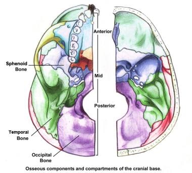
- Osseous components and compartments of the cranial base. (medscape.com)
Surgical3
- Sinus puncture and irrigation techniques allow for a surgical means of removal of thick purulent sinus secretions. (medscape.com)
- Alleyne C, Vishteh A, Speltzler R, Detweiler P: Long-term survival of a patient with invasive cranial base rhinocerebral mucormycosis treated with combined endovascular, surgical, and medical therapies: Case report. (karger.com)
- Treatment, when required, may involve decongestants, analgesics, and sometimes oral corticosteroids or surgical repair of serious inner or middle ear or sinus injuries. (msdmanuals.com)
Innervates1
- The fourth cranial nerve innervates the superior oblique muscle, which intorts, depresses, and abducts the globe. (medscape.com)