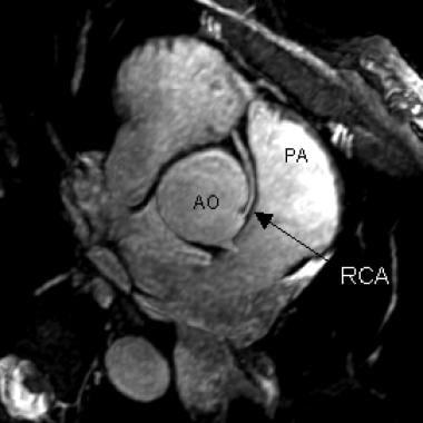
Coronary Vessel Anomalies
Coronary Angiography
Coronary Artery Disease
Angioplasty, Balloon, Coronary
Pericardium
Coronary Disease
Coronary Artery Bypass
Blood Vessels
Myocardial Infarction
Coronary Restenosis
Dogs
Ebstein Anomaly
Coronary Aneurysm
Myocardium
Coronary Thrombosis
Stents
Endothelium, Vascular
Nitroglycerin
Vasodilation
Treatment Outcome
Risk Factors
Vascular Resistance
Myocardial Ischemia
Prospective Studies
Coronary Occlusion
Ultrasonography, Interventional
Electrocardiography
Tomography, X-Ray Computed
Predictive Value of Tests
Follow-Up Studies
Percutaneous Coronary Intervention
Adenosine
Swine
Hemodynamics
Blood Flow Velocity
Magnetic Resonance Angiography
Vasoconstriction
Abnormalities, Multiple
Neovascularization, Physiologic
Coronary Care Units
Acetylcholine
Sensitivity and Specificity
Retrospective Studies
Models, Cardiovascular
Quail
Coturnix
Nitric Oxide
Reproducibility of Results
Severity of Illness Index
Biological Markers
Exercise Test
Pelger-Huet Anomaly
Tunica Media
Tunica Intima
Feasibility Studies
Sirolimus
Myocardial Revascularization
Coronary Artery Bypass, Off-Pump
Collateral Circulation
Cardiac Catheterization
Heart Ventricles
Chick Embryo
Urogenital Abnormalities
Paracrine Communication
Image Processing, Computer-Assisted
Angina Pectoris
NG-Nitroarginine Methyl Ester
Risk Assessment
Platelet Aggregation Inhibitors
Immunohistochemistry
Vascular Endothelial Growth Factor A
Heart Defects, Congenital
Echocardiography
Drug-Eluting Stents
Gene Expression Regulation, Developmental
Morphogenesis
Nitric Oxide Synthase
Multidetector Computed Tomography
Prognosis
Pregnancy
Ultrasonography, Prenatal
Dipyridamole
Cardiovascular Agents
Myocardial Reperfusion
Cohort Studies
Cell Differentiation
Arterioles
Fractional Flow Reserve, Myocardial
Chi-Square Distribution
Vascular Malformations
Mammary Arteries
Signal Transduction
Vasomotor System
Stem Cells
Incidence
Fetal Diseases
Ticlopidine
Multivariate Analysis
Mucocutaneous Lymph Node Syndrome
Ventricular Function, Left
Disease Models, Animal
Hyperemia
Case-Control Studies
Constriction, Pathologic
Postoperative Complications
Angina Pectoris, Variant
Aspirin
Tomography, Emission-Computed, Single-Photon
Radial Artery
Myocardial Bridging
Anus, Imperforate
Vascular Calcification
Registries
Tomography, Spiral Computed
Myocardial Perfusion Imaging
Ergonovine
Logistic Models
Arteriosclerosis
Dilatation, Pathologic
Endothelial Cells
Anomalous origin of the left coronary artery from the pulmonary artery: natural history and normal pregnancies. (1/601)
Two female patients are described with anomalous origin of the left coronary artery arising from the pulmonary artery who sustained an anterolateral myocardial infarction in infancy. Neither patient received surgical treatment although both have lived to middle age with minimal cardiovascular problems and have had uncomplicated pregnancies. Good exercise tolerance and long term survival may be possible even without surgery for patients with this anomaly. (+info)Short left coronary artery trunk as a risk factor in the development of coronary atherosclerosis. Pathological study. (2/601)
The relation between the length of the main left coronary artery and the degree of atherosclerosis in its branches was studied by postmortem examination in 204 subjects aged 20 to 90 years. The findings suggest that in cases with a short main left coronary artery the atherosclerotic lesions in the anterior descending and circumflex branches appear earlier, progress faster at higher levels of severity, and lead more frequently to myocardial infarction, than in cases with a long left coronary artery trunk. In cases over the age of 50 years, where disease is expected to have developed, it was shown that the degree of atherosclerosis in the left anterior descending and circumflex branches was inversely related to the length of the main left coronary artery. The correlation coefficients were -0-527 and -0-428, respectively, and in either case a test for zero correlations was significant (P less than 0-001). The possible changes in the haemodynamic and mechanical conditions associated with the variations of the anatomical pattern of the coronary arteries and their influence in the development of atherosclerosis are discussed. It is suggested that the length of the main left coronary artery is a congenital anatomical and possibly hereditary factor influencing the rate of development of atherosclerosis in the branches of the main left coronary artery. (+info)Coronary artery disease with single coronary artery. (3/601)
The authors have reviewed the literature in search of the coexistence of single coronary artery with significant coronary artery disease. Two cases of single right coronary artery are described. In both, the anomalies were unsuspected and diagnosed roentgenographically in life. Both patients had angina pectoris, positive graded-exercise stress tests, and hemodynamically significant obstruction or occlusion to the coronary arteries. In neither case was the stenosis proximal or amenable to bypass surgery. (+info)Evolution of risk factors influencing early mortality of the arterial switch operation. (4/601)
OBJECTIVES: The present study was undertaken to determine the independent risk factors for early mortality in the current era after arterial switch operation (ASO). BACKGROUND: Prior reports on factors affecting outcome of the ASO demonstrated that abnormal coronary arterial patterns were associated with increased risk of early mortality. As diagnostic, surgical and perioperative management techniques continue to evolve, the risk factors for the ASO may have changed. METHODS: All patients who underwent the ASO at Children's Hospital, Boston between January 1, 1992 and December 31, 1996 were included. Hospital charts, echocardiographic and cardiac catheterization data and operative reports of all patients were reviewed. Demographics and preoperative, intraoperative and postoperative variables were recorded. RESULTS: Of the 223 patients included in the study (median age at ASO = 6 days and median weight = 3.5 kg), 26 patients had aortic arch obstruction or interruption, 12 had Taussig-Bing anomaly, 12 had multiple ventricular septal defects, 8 had right ventricular hypoplasia and 6 were premature. There were 16 early deaths (7%), with 3 deaths in the 109 patients considered "low risk" (2.7%). Coronary artery pattern was not associated with an increased risk of death. Compared with usual coronary anatomy pattern, however, inverted coronary patterns and single right coronary patterns were associated with increased incidence of delayed sternal closure (p = 0.003) and longer duration of mechanical ventilation (p = 0.008). In a multivariate logistic regression model using only preoperative variables, aortic arch repair at a separate procedure before ASO and smaller birth weight were independent predictors of early mortality. In a second model that included both pre- and intraoperative variables, circulatory arrest time and right ventricular hypoplasia were independent predictors of early death. CONCLUSIONS: The ASO can be performed in the current era without excess early mortality related to uncommon coronary artery patterns. Aortic arch repair before ASO, right ventricular hypoplasia, lower birth weight and longer intraoperative support continue to be independent risk factors for early mortality after the ASO. (+info)Unusual congenital coronary anomaly and myocardial ischaemia. (5/601)
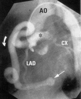
Angiography was used to diagnose a rare congenital coronary anomaly with myocardial ischaemia in a woman with typical angina. All three coronary arteries arose from a solitary coronary ostium in the right aortic sinus; the left anterior descending coronary artery followed a septal course, the circumflex coronary artery ran behind the ascending aorta, and the right coronary artery followed a normal course. No significant coronary lumen narrowing was found. Transoesophageal echocardiography confirmed the anomalous origin and course of the aberrant coronary arteries. An exercise test reproduced angina, and ECG changes and myocardial perfusion study showed an anterior reversible defect. In contrast to previous reports, myocardial ischaemia was associated with the septal (intramuscular) course of the left anterior descending coronary artery; there was no other significant coronary artery disease. (+info)New signs characteristic of myocardial bridging demonstrated by intracoronary ultrasound and Doppler. (6/601)
BACKGROUND: Large discrepancies exist concerning the incidence of myocardial bridging. This has been reported to be 0.5%-2.5% following coronary angiography but 15%-85% following autopsy. The purpose of the study was to use intravascular ultrasound and intracoronary Doppler to study the morphology and flow characteristics of myocardial bridging in order to find feasible parameters of this syndrome. METHODS AND RESULTS: Intravascular ultrasound was performed in 62/69 patients in whom typical angiographic 'milking effects' were present. In 48 patients, intracoronary Doppler was performed. A specific, echolucent 'half moon' phenomenon surrounding the myocardial bridge was found in all the patients. The thickness of the half moon area was 0.47 +/- 0.19 mm in diastole and 0.52 +/- 0.23 mm in systole. There was systolic compression of the myocardial bridge with a lumen reduction during systole of 36.4 +/- 8.8%. Using intracoronary Doppler, a characteristic early diastolic 'finger tip' phenomenon was observed in 42 (87%) of the patients. All patients showed no or reduced antegrade systolic flow. Coronary flow velocity reserve was 2.03 +/- 0. 54. After intracoronary nitroglycerin injection, retrograde systolic flow occurred in 37 (77%) of the 48 patients, with a velocity of -22. 2 +/- 13.2 cm. s(-1). Intravascular ultrasound revealed atherosclerotic involvement of the proximal segment in 61 (88%) of the 69 patients, with an area stenosis of 42 +/- 13%. No plaques were found in the bridge or distal segments in the 62 patients in whom it was possible to introduce the ultrasound catheter throughout the bridging segment. CONCLUSION: Myocardial bridging is characterized by the following morphological and functional signs: a specific, echolucent half moon phenomenon over the bridge segment, which exists throughout the cardiac cycle; systolic compression of the bridge segment of the coronary artery; accelerated flow velocity at early diastole (finger-tip phenomenon); no or reduced systolic antegrade flow; decreased diastolic/systolic velocity ratio; retrograde flow in the proximal segment, which is provoked and enhanced by nitroglycerin injection. (+info)A 72 year old woman with ALCAPA. (7/601)
ALCAPA syndrome (anomalous origin of the left coronary artery from the pulmonary artery), which causes the left coronary artery to grow with an anomalous origin from the pulmonary artery, is a rare disease which may result in myocardial infarction, congestive heart failure, and sometimes death during the early infantile period. A 72 year old woman with ALCAPA syndrome is presented. The asymptomatic patient presented with a cardiac murmur which was discovered during a routine check up for a gynaecological intervention. Coronary cineangiography established the diagnosis. Although surgical correction is the usual treatment for such cases, medical treatment was preferred for this patient because she was asymptomatic without clinical signs of heart failure. (+info)Mice lacking the vascular endothelial growth factor-B gene (Vegfb) have smaller hearts, dysfunctional coronary vasculature, and impaired recovery from cardiac ischemia. (8/601)
Vascular endothelial growth factor-B (VEGF-B) is closely related to VEGF-A, an effector of blood vessel growth during development and disease and a strong candidate for angiogenic therapies. To further study the in vivo function of VEGF-B, we have generated Vegfb knockout mice (Vegfb(-/-)). Unlike Vegfa knockout mice, which die during embryogenesis, Vegfb(-/-) mice are healthy and fertile. Despite appearing overtly normal, Vegfb(-/-) hearts are reduced in size and display vascular dysfunction after coronary occlusion and impaired recovery from experimentally induced myocardial ischemia. These findings reveal a role for VEGF-B in the development or function of coronary vasculature and suggest potential clinical use in therapeutic angiogenesis. (+info)Coronary vessel anomalies refer to abnormalities in the structure, origin, or course of the coronary arteries or veins. These vessels are responsible for delivering oxygenated blood to the heart muscle. Some common types of coronary vessel anomalies include:
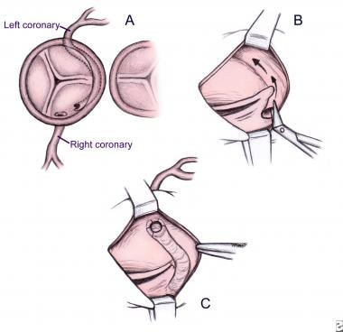
1. Anomalous Origin of the Coronary Artery (AOCA): This occurs when one or both of the coronary arteries originate from an abnormal location in the aorta. The left coronary artery may arise from the right sinus of Valsalva, while the right coronary artery may arise from the left sinus of Valsalva. This can lead to ischemia (reduced blood flow) and potentially life-threatening complications such as sudden cardiac death.
2. Coronary Artery Fistula: A fistula is an abnormal connection between a coronary artery and another chamber or vessel in the heart. Blood flows directly from the high-pressure coronary artery into a low-pressure chamber, bypassing the capillaries and leading to a steal phenomenon where oxygenated blood is diverted away from the heart muscle.
3. Coronary Artery Aneurysm: An aneurysm is a localized dilation or bulging of the coronary artery wall. This can lead to complications such as thrombosis (blood clot formation), embolism (blockage caused by a clot that travels to another location), or rupture, which can be life-threatening.
4. Myocardial Bridge: In this condition, a segment of the coronary artery passes between the muscle fibers of the heart, instead of running along its surface. This can cause compression of the artery during systole (contraction) and lead to ischemia.
5. Kawasaki Disease: Although not strictly an anomaly, Kawasaki disease is a pediatric illness that can result in coronary artery aneurysms and other complications if left untreated.
Coronary vessel anomalies may be asymptomatic or present with symptoms such as chest pain, shortness of breath, palpitations, or syncope (fainting). Diagnosis typically involves imaging techniques such as coronary angiography, computed tomography (CT) angiography, or magnetic resonance angiography. Treatment depends on the specific anomaly and may involve medications, percutaneous interventions, or surgical correction.
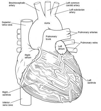
Coronary vessels refer to the network of blood vessels that supply oxygenated blood and nutrients to the heart muscle, also known as the myocardium. The two main coronary arteries are the left main coronary artery and the right coronary artery.
The left main coronary artery branches off into the left anterior descending artery (LAD) and the left circumflex artery (LCx). The LAD supplies blood to the front of the heart, while the LCx supplies blood to the side and back of the heart.
The right coronary artery supplies blood to the right lower part of the heart, including the right atrium and ventricle, as well as the back of the heart.
Coronary vessel disease (CVD) occurs when these vessels become narrowed or blocked due to the buildup of plaque, leading to reduced blood flow to the heart muscle. This can result in chest pain, shortness of breath, or a heart attack.
Coronary angiography is a medical procedure that uses X-ray imaging to visualize the coronary arteries, which supply blood to the heart muscle. During the procedure, a thin, flexible catheter is inserted into an artery in the arm or groin and threaded through the blood vessels to the heart. A contrast dye is then injected through the catheter, and X-ray images are taken as the dye flows through the coronary arteries. These images can help doctors diagnose and treat various heart conditions, such as blockages or narrowing of the arteries, that can lead to chest pain or heart attacks. It is also known as coronary arteriography or cardiac catheterization.
Coronary artery disease (CAD) is a medical condition in which the coronary arteries, which supply oxygen-rich blood to the heart muscle, become narrowed or blocked due to the buildup of cholesterol, fatty deposits, and other substances, known as plaque. Over time, this buildup can cause the arteries to harden and narrow (a process called atherosclerosis), reducing blood flow to the heart muscle.
The reduction in blood flow can lead to various symptoms and complications, including:
1. Angina (chest pain or discomfort) - This occurs when the heart muscle doesn't receive enough oxygen-rich blood, causing pain, pressure, or discomfort in the chest, arms, neck, jaw, or back.
2. Shortness of breath - When the heart isn't receiving adequate blood flow, it can't pump blood efficiently to meet the body's demands, leading to shortness of breath during physical activities or at rest.
3. Heart attack - If a piece of plaque ruptures or breaks off in a coronary artery, a blood clot can form and block the artery, causing a heart attack (myocardial infarction). This can damage or destroy part of the heart muscle.
4. Heart failure - Chronic reduced blood flow to the heart muscle can weaken it over time, leading to heart failure, a condition in which the heart can't pump blood efficiently to meet the body's needs.
5. Arrhythmias - Reduced blood flow and damage to the heart muscle can lead to abnormal heart rhythms (arrhythmias), which can be life-threatening if not treated promptly.
Coronary artery disease is typically diagnosed through a combination of medical history, physical examination, and diagnostic tests such as electrocardiograms (ECGs), stress testing, cardiac catheterization, and imaging studies like coronary computed tomography angiography (CCTA). Treatment options for CAD include lifestyle modifications, medications, medical procedures, and surgery.
Coronary circulation refers to the circulation of blood in the coronary vessels, which supply oxygenated blood to the heart muscle (myocardium) and drain deoxygenated blood from it. The coronary circulation system includes two main coronary arteries - the left main coronary artery and the right coronary artery - that branch off from the aorta just above the aortic valve. These arteries further divide into smaller branches, which supply blood to different regions of the heart muscle.
The left main coronary artery divides into two branches: the left anterior descending (LAD) artery and the left circumflex (LCx) artery. The LAD supplies blood to the front and sides of the heart, while the LCx supplies blood to the back and sides of the heart. The right coronary artery supplies blood to the lower part of the heart, including the right ventricle and the bottom portion of the left ventricle.
The veins that drain the heart muscle include the great cardiac vein, the middle cardiac vein, and the small cardiac vein, which merge to form the coronary sinus. The coronary sinus empties into the right atrium, allowing deoxygenated blood to enter the right side of the heart and be pumped to the lungs for oxygenation.
Coronary circulation is essential for maintaining the health and function of the heart muscle, as it provides the necessary oxygen and nutrients required for proper contraction and relaxation of the myocardium. Any disruption or blockage in the coronary circulation system can lead to serious consequences, such as angina, heart attack, or even death.
Coronary balloon angioplasty is a minimally invasive medical procedure used to widen narrowed or obstructed coronary arteries (the blood vessels that supply oxygen-rich blood to the heart muscle) and improve blood flow to the heart. This procedure is typically performed in conjunction with the insertion of a stent, a small mesh tube that helps keep the artery open.
During coronary balloon angioplasty, a thin, flexible catheter with a deflated balloon at its tip is inserted into a blood vessel, usually through a small incision in the groin or arm. The catheter is then guided to the narrowed or obstructed section of the coronary artery. Once in position, the balloon is inflated to compress the plaque against the artery wall and widen the lumen (the inner space) of the artery. This helps restore blood flow to the heart muscle.
The procedure is typically performed under local anesthesia and conscious sedation to minimize discomfort. Coronary balloon angioplasty is a relatively safe and effective treatment for many people with coronary artery disease, although complications such as bleeding, infection, or re-narrowing of the artery (restenosis) can occur in some cases.
Coronary stenosis is a medical condition that refers to the narrowing of the coronary arteries, which supply oxygen-rich blood to the heart muscle. This narrowing is typically caused by the buildup of plaque, made up of fat, cholesterol, and other substances, on the inner walls of the arteries. Over time, as the plaque hardens and calcifies, it can cause the artery to become narrowed or blocked, reducing blood flow to the heart muscle.
Coronary stenosis can lead to various symptoms and complications, including chest pain (angina), shortness of breath, irregular heart rhythms (arrhythmias), and heart attacks. Treatment options for coronary stenosis may include lifestyle changes, medications, medical procedures such as angioplasty or bypass surgery, or a combination of these approaches. Regular check-ups and diagnostic tests, such as stress testing or coronary angiography, can help detect and monitor coronary stenosis over time.
The pericardium is the double-walled sac that surrounds the heart. It has an outer fibrous layer and an inner serous layer, which further divides into two parts: the parietal layer lining the fibrous pericardium and the visceral layer (epicardium) closely adhering to the heart surface.
The space between these two layers is filled with a small amount of lubricating serous fluid, allowing for smooth movement of the heart within the pericardial cavity. The pericardium provides protection, support, and helps maintain the heart's normal position within the chest while reducing friction during heart contractions.
Coronary artery disease, often simply referred to as coronary disease, is a condition in which the blood vessels that supply oxygen-rich blood to the heart become narrowed or blocked due to the buildup of fatty deposits called plaques. This can lead to chest pain (angina), shortness of breath, or in severe cases, a heart attack.
The medical definition of coronary artery disease is:
A condition characterized by the accumulation of atheromatous plaques in the walls of the coronary arteries, leading to decreased blood flow and oxygen supply to the myocardium (heart muscle). This can result in symptoms such as angina pectoris, shortness of breath, or arrhythmias, and may ultimately lead to myocardial infarction (heart attack) or heart failure.
Risk factors for coronary artery disease include age, smoking, high blood pressure, high cholesterol, diabetes, obesity, physical inactivity, and a family history of the condition. Lifestyle changes such as quitting smoking, exercising regularly, eating a healthy diet, and managing stress can help reduce the risk of developing coronary artery disease. Medical treatments may include medications to control blood pressure, cholesterol levels, or irregular heart rhythms, as well as procedures such as angioplasty or bypass surgery to improve blood flow to the heart.
Coronary artery bypass surgery, also known as coronary artery bypass grafting (CABG), is a surgical procedure used to improve blood flow to the heart in patients with severe coronary artery disease. This condition occurs when the coronary arteries, which supply oxygen-rich blood to the heart muscle, become narrowed or blocked due to the buildup of fatty deposits, called plaques.
During CABG surgery, a healthy blood vessel from another part of the body is grafted, or attached, to the coronary artery, creating a new pathway for oxygen-rich blood to flow around the blocked or narrowed portion of the artery and reach the heart muscle. This bypass helps to restore normal blood flow and reduce the risk of angina (chest pain), shortness of breath, and other symptoms associated with coronary artery disease.
There are different types of CABG surgery, including traditional on-pump CABG, off-pump CABG, and minimally invasive CABG. The choice of procedure depends on various factors, such as the patient's overall health, the number and location of blocked arteries, and the presence of other medical conditions.
It is important to note that while CABG surgery can significantly improve symptoms and quality of life in patients with severe coronary artery disease, it does not cure the underlying condition. Lifestyle modifications, such as regular exercise, a healthy diet, smoking cessation, and medication therapy, are essential for long-term management and prevention of further progression of the disease.
Blood vessels are the part of the circulatory system that transport blood throughout the body. They form a network of tubes that carry blood to and from the heart, lungs, and other organs. The main types of blood vessels are arteries, veins, and capillaries. Arteries carry oxygenated blood away from the heart to the rest of the body, while veins return deoxygenated blood back to the heart. Capillaries connect arteries and veins and facilitate the exchange of oxygen, nutrients, and waste materials between the blood and the body's tissues.
Myocardial infarction (MI), also known as a heart attack, is a medical condition characterized by the death of a segment of heart muscle (myocardium) due to the interruption of its blood supply. This interruption is most commonly caused by the blockage of a coronary artery by a blood clot formed on the top of an atherosclerotic plaque, which is a buildup of cholesterol and other substances in the inner lining of the artery.
The lack of oxygen and nutrients supply to the heart muscle tissue results in damage or death of the cardiac cells, causing the affected area to become necrotic. The extent and severity of the MI depend on the size of the affected area, the duration of the occlusion, and the presence of collateral circulation.
Symptoms of a myocardial infarction may include chest pain or discomfort, shortness of breath, nausea, lightheadedness, and sweating. Immediate medical attention is necessary to restore blood flow to the affected area and prevent further damage to the heart muscle. Treatment options for MI include medications, such as thrombolytics, antiplatelet agents, and pain relievers, as well as procedures such as percutaneous coronary intervention (PCI) or coronary artery bypass grafting (CABG).
Coronary restenosis is the re-narrowing or re-occlusion of a coronary artery after a previous successful procedure to open or widen the artery, such as angioplasty or stenting. This narrowing is usually caused by the excessive growth of scar tissue or smooth muscle cells in the artery lining, which can occur spontaneously or as a response to the initial procedure. Restenosis can lead to recurrent symptoms of coronary artery disease, such as chest pain or shortness of breath, and may require additional medical intervention.
In medical terms, the heart is a muscular organ located in the thoracic cavity that functions as a pump to circulate blood throughout the body. It's responsible for delivering oxygen and nutrients to the tissues and removing carbon dioxide and other wastes. The human heart is divided into four chambers: two atria on the top and two ventricles on the bottom. The right side of the heart receives deoxygenated blood from the body and pumps it to the lungs, while the left side receives oxygenated blood from the lungs and pumps it out to the rest of the body. The heart's rhythmic contractions and relaxations are regulated by a complex electrical conduction system.
Coronary vasospasm refers to a sudden constriction (narrowing) of the coronary arteries, which supply oxygenated blood to the heart muscle. This constriction can reduce or block blood flow, leading to symptoms such as chest pain (angina) or, in severe cases, a heart attack (myocardial infarction). Coronary vasospasm can occur spontaneously or be triggered by various factors, including stress, smoking, and certain medications. It is also associated with conditions such as coronary artery disease and variant angina. Prolonged or recurrent vasospasms can cause damage to the heart muscle and increase the risk of cardiovascular events.
I believe there might be a misunderstanding in your question. "Dogs" is not a medical term or condition. It is the common name for a domesticated carnivore of the family Canidae, specifically the genus Canis, which includes wolves, foxes, and other extant and extinct species of mammals. Dogs are often kept as pets and companions, and they have been bred in a wide variety of forms and sizes for different purposes, such as hunting, herding, guarding, assisting police and military forces, and providing companionship and emotional support.
If you meant to ask about a specific medical condition or term related to dogs, please provide more context so I can give you an accurate answer.
Ebstein anomaly is a congenital heart defect that affects the tricuspid valve, which is the valve between the right atrium and right ventricle of the heart. In Ebstein anomaly, the tricuspid valve is abnormally formed and positioned, causing it to leak blood back into the right atrium. This can lead to various symptoms such as shortness of breath, fatigue, and cyanosis (bluish discoloration of the skin). Treatment for Ebstein anomaly may include medication, surgery, or a combination of both. It is important to note that the severity of the condition can vary widely among individuals, and some people with Ebstein anomaly may require more intensive treatment than others.
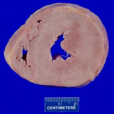
A coronary aneurysm is a localized dilation or bulging of a portion of the wall of a coronary artery, which supplies blood to the muscle tissue of the heart. It's similar to a bubble or balloon-like structure that forms within the artery wall due to weakness in the arterial wall, leading to abnormal enlargement or widening.
Coronary aneurysms can vary in size and may be classified as true or false aneurysms based on their structure. True aneurysms involve all three layers of the artery wall, while false aneurysms (also known as pseudoaneurysms) only have one or two layers involved, with the remaining layer disrupted.
These aneurysms can lead to complications such as blood clots forming inside the aneurysm sac, which can then dislodge and cause blockages in smaller coronary arteries (embolism). Additionally, coronary aneurysms may rupture, leading to severe internal bleeding and potentially life-threatening situations.
Coronary aneurysms are often asymptomatic but can present with symptoms such as chest pain, shortness of breath, or palpitations, especially if the aneurysm causes a significant narrowing (stenosis) in the affected artery. They can be diagnosed through imaging techniques like coronary angiography, computed tomography (CT), or magnetic resonance imaging (MRI). Treatment options include medications to manage symptoms and prevent complications, as well as surgical interventions such as stenting or bypass grafting to repair or reroute the affected artery.
The myocardium is the middle layer of the heart wall, composed of specialized cardiac muscle cells that are responsible for pumping blood throughout the body. It forms the thickest part of the heart wall and is divided into two sections: the left ventricle, which pumps oxygenated blood to the rest of the body, and the right ventricle, which pumps deoxygenated blood to the lungs.
The myocardium contains several types of cells, including cardiac muscle fibers, connective tissue, nerves, and blood vessels. The muscle fibers are arranged in a highly organized pattern that allows them to contract in a coordinated manner, generating the force necessary to pump blood through the heart and circulatory system.
Damage to the myocardium can occur due to various factors such as ischemia (reduced blood flow), infection, inflammation, or genetic disorders. This damage can lead to several cardiac conditions, including heart failure, arrhythmias, and cardiomyopathy.
Coronary thrombosis is a medical condition that refers to the formation of a blood clot (thrombus) inside a coronary artery, which supplies oxygenated blood to the heart muscle. The development of a thrombus can partially or completely obstruct blood flow, leading to insufficient oxygen supply to the heart muscle. This can cause chest pain (angina) or a heart attack (myocardial infarction), depending on the severity and duration of the blockage.
Coronary thrombosis often results from the rupture of an atherosclerotic plaque, a buildup of cholesterol, fat, calcium, and other substances in the inner lining (endothelium) of the coronary artery. The ruptured plaque exposes the underlying tissue to the bloodstream, triggering the coagulation cascade and resulting in the formation of a thrombus.
Immediate medical attention is crucial for managing coronary thrombosis, as timely treatment can help restore blood flow, prevent further damage to the heart muscle, and reduce the risk of complications such as heart failure or life-threatening arrhythmias. Treatment options may include medications, such as antiplatelet agents, anticoagulants, and thrombolytic drugs, or interventional procedures like angioplasty and stenting to open the blocked artery. In some cases, surgical intervention, such as coronary artery bypass grafting (CABG), may be necessary.
A stent is a small mesh tube that's used to treat narrow or weak arteries. Arteries are blood vessels that carry blood away from your heart to other parts of your body. A stent is placed in an artery as part of a procedure called angioplasty. Angioplasty restores blood flow through narrowed or blocked arteries by inflating a tiny balloon inside the blocked artery to widen it.
The stent is then inserted into the widened artery to keep it open. The stent is usually made of metal, but some are coated with medication that is slowly and continuously released to help prevent the formation of scar tissue in the artery. This can reduce the chance of the artery narrowing again.
Stents are also used in other parts of the body, such as the neck (carotid artery) and kidneys (renal artery), to help maintain blood flow and prevent blockages. They can also be used in the urinary system to treat conditions like ureteropelvic junction obstruction or narrowing of the urethra.
The endothelium is a thin layer of simple squamous epithelial cells that lines the interior surface of blood vessels, lymphatic vessels, and heart chambers. The vascular endothelium, specifically, refers to the endothelial cells that line the blood vessels. These cells play a crucial role in maintaining vascular homeostasis by regulating vasomotor tone, coagulation, platelet activation, inflammation, and permeability of the vessel wall. They also contribute to the growth and repair of the vascular system and are involved in various pathological processes such as atherosclerosis, hypertension, and diabetes.
Nitroglycerin, also known as glyceryl trinitrate, is a medication used primarily for the treatment of angina pectoris (chest pain due to coronary artery disease) and hypertensive emergencies (severe high blood pressure). It belongs to a class of drugs called nitrates or organic nitrites.
Nitroglycerin works by relaxing and dilating the smooth muscle in blood vessels, which leads to decreased workload on the heart and increased oxygen delivery to the myocardium (heart muscle). This results in reduced symptoms of angina and improved cardiac function during hypertensive emergencies.
The drug is available in various forms, including sublingual tablets, sprays, transdermal patches, ointments, and intravenous solutions. The choice of formulation depends on the specific clinical situation and patient needs. Common side effects of nitroglycerin include headache, dizziness, and hypotension (low blood pressure).
Vasodilator agents are pharmacological substances that cause the relaxation or widening of blood vessels by relaxing the smooth muscle in the vessel walls. This results in an increase in the diameter of the blood vessels, which decreases vascular resistance and ultimately reduces blood pressure. Vasodilators can be further classified based on their site of action:
1. Systemic vasodilators: These agents cause a generalized relaxation of the smooth muscle in the walls of both arteries and veins, resulting in a decrease in peripheral vascular resistance and preload (the volume of blood returning to the heart). Examples include nitroglycerin, hydralazine, and calcium channel blockers.
2. Arterial vasodilators: These agents primarily affect the smooth muscle in arterial vessel walls, leading to a reduction in afterload (the pressure against which the heart pumps blood). Examples include angiotensin-converting enzyme (ACE) inhibitors, angiotensin receptor blockers (ARBs), and direct vasodilators like sodium nitroprusside.
3. Venous vasodilators: These agents primarily affect the smooth muscle in venous vessel walls, increasing venous capacitance and reducing preload. Examples include nitroglycerin and other organic nitrates.
Vasodilator agents are used to treat various cardiovascular conditions such as hypertension, heart failure, angina, and pulmonary arterial hypertension. It is essential to monitor their use carefully, as excessive vasodilation can lead to orthostatic hypotension, reflex tachycardia, or fluid retention.
Vasodilation is the widening or increase in diameter of blood vessels, particularly the involuntary relaxation of the smooth muscle in the tunica media (middle layer) of the arteriole walls. This results in an increase in blood flow and a decrease in vascular resistance. Vasodilation can occur due to various physiological and pathophysiological stimuli, such as local metabolic demands, neural signals, or pharmacological agents. It plays a crucial role in regulating blood pressure, tissue perfusion, and thermoregulation.
Treatment outcome is a term used to describe the result or effect of medical treatment on a patient's health status. It can be measured in various ways, such as through symptoms improvement, disease remission, reduced disability, improved quality of life, or survival rates. The treatment outcome helps healthcare providers evaluate the effectiveness of a particular treatment plan and make informed decisions about future care. It is also used in clinical research to compare the efficacy of different treatments and improve patient care.
Medical Definition:
"Risk factors" are any attribute, characteristic or exposure of an individual that increases the likelihood of developing a disease or injury. They can be divided into modifiable and non-modifiable risk factors. Modifiable risk factors are those that can be changed through lifestyle choices or medical treatment, while non-modifiable risk factors are inherent traits such as age, gender, or genetic predisposition. Examples of modifiable risk factors include smoking, alcohol consumption, physical inactivity, and unhealthy diet, while non-modifiable risk factors include age, sex, and family history. It is important to note that having a risk factor does not guarantee that a person will develop the disease, but rather indicates an increased susceptibility.
In the field of medicine, "time factors" refer to the duration of symptoms or time elapsed since the onset of a medical condition, which can have significant implications for diagnosis and treatment. Understanding time factors is crucial in determining the progression of a disease, evaluating the effectiveness of treatments, and making critical decisions regarding patient care.
For example, in stroke management, "time is brain," meaning that rapid intervention within a specific time frame (usually within 4.5 hours) is essential to administering tissue plasminogen activator (tPA), a clot-busting drug that can minimize brain damage and improve patient outcomes. Similarly, in trauma care, the "golden hour" concept emphasizes the importance of providing definitive care within the first 60 minutes after injury to increase survival rates and reduce morbidity.
Time factors also play a role in monitoring the progression of chronic conditions like diabetes or heart disease, where regular follow-ups and assessments help determine appropriate treatment adjustments and prevent complications. In infectious diseases, time factors are crucial for initiating antibiotic therapy and identifying potential outbreaks to control their spread.
Overall, "time factors" encompass the significance of recognizing and acting promptly in various medical scenarios to optimize patient outcomes and provide effective care.
Vascular resistance is a measure of the opposition to blood flow within a vessel or a group of vessels, typically expressed in units of mmHg/(mL/min) or sometimes as dynes*sec/cm^5. It is determined by the diameter and length of the vessels, as well as the viscosity of the blood flowing through them. In general, a decrease in vessel diameter, an increase in vessel length, or an increase in blood viscosity will result in an increase in vascular resistance, while an increase in vessel diameter, a decrease in vessel length, or a decrease in blood viscosity will result in a decrease in vascular resistance. Vascular resistance is an important concept in the study of circulation and cardiovascular physiology because it plays a key role in determining blood pressure and blood flow within the body.
The vasa vasorum are small blood vessels that supply larger blood vessels, such as the arteries and veins, with oxygen and nutrients. They are located in the outer layers (the adventitia and media) of these larger vessels and form a network of vessels that surround and penetrate the walls of the larger vessels. The vasa vasorum are particularly important in supplying blood to the thicker walls of larger arteries, such as the aorta, where diffusion from the lumen may not be sufficient to meet the metabolic needs of the vessel wall.
Myocardial ischemia is a condition in which the blood supply to the heart muscle (myocardium) is reduced or blocked, leading to insufficient oxygen delivery and potential damage to the heart tissue. This reduction in blood flow typically results from the buildup of fatty deposits, called plaques, in the coronary arteries that supply the heart with oxygen-rich blood. The plaques can rupture or become unstable, causing the formation of blood clots that obstruct the artery and limit blood flow.
Myocardial ischemia may manifest as chest pain (angina pectoris), shortness of breath, fatigue, or irregular heartbeats (arrhythmias). In severe cases, it can lead to myocardial infarction (heart attack) if the oxygen supply is significantly reduced or cut off completely, causing permanent damage or death of the heart muscle. Early diagnosis and treatment of myocardial ischemia are crucial for preventing further complications and improving patient outcomes.
Prospective studies, also known as longitudinal studies, are a type of cohort study in which data is collected forward in time, following a group of individuals who share a common characteristic or exposure over a period of time. The researchers clearly define the study population and exposure of interest at the beginning of the study and follow up with the participants to determine the outcomes that develop over time. This type of study design allows for the investigation of causal relationships between exposures and outcomes, as well as the identification of risk factors and the estimation of disease incidence rates. Prospective studies are particularly useful in epidemiology and medical research when studying diseases with long latency periods or rare outcomes.
Coronary occlusion is the medical term used to describe a complete blockage in one or more of the coronary arteries, which supply oxygenated blood to the heart muscle. This blockage is usually caused by the buildup of fatty deposits, called plaques, inside the artery walls, a condition known as atherosclerosis. Over time, these plaques can rupture, leading to the formation of blood clots that completely obstruct the flow of blood through the coronary artery.
Coronary occlusion can lead to serious complications, such as a heart attack (myocardial infarction), angina (chest pain), or even sudden cardiac death, depending on the severity and duration of the blockage. Immediate medical attention is required in case of coronary occlusion to restore blood flow to the affected areas of the heart and prevent further damage. Treatment options may include medications, minimally invasive procedures like angioplasty and stenting, or surgical interventions such as coronary artery bypass grafting (CABG).
Interventional ultrasonography is a medical procedure that involves the use of real-time ultrasound imaging to guide minimally invasive diagnostic and therapeutic interventions. This technique combines the advantages of ultrasound, such as its non-ionizing nature (no radiation exposure), relatively low cost, and portability, with the ability to perform precise and targeted procedures.
In interventional ultrasonography, a specialized physician called an interventional radiologist or an interventional sonographer uses high-frequency sound waves to create detailed images of internal organs and tissues. These images help guide the placement of needles, catheters, or other instruments used during the procedure. Common interventions include biopsies (tissue sampling), fluid drainage, tumor ablation, and targeted drug delivery.
The real-time visualization provided by ultrasonography allows for increased accuracy and safety during these procedures, minimizing complications and reducing recovery time compared to traditional surgical approaches. Additionally, interventional ultrasonography can be performed on an outpatient basis, further contributing to its appeal as a less invasive alternative in many clinical scenarios.
Electrocardiography (ECG or EKG) is a medical procedure that records the electrical activity of the heart. It provides a graphic representation of the electrical changes that occur during each heartbeat. The resulting tracing, called an electrocardiogram, can reveal information about the heart's rate and rhythm, as well as any damage to its cells or abnormalities in its conduction system.
During an ECG, small electrodes are placed on the skin of the chest, arms, and legs. These electrodes detect the electrical signals produced by the heart and transmit them to a machine that amplifies and records them. The procedure is non-invasive, painless, and quick, usually taking only a few minutes.
ECGs are commonly used to diagnose and monitor various heart conditions, including arrhythmias, coronary artery disease, heart attacks, and electrolyte imbalances. They can also be used to evaluate the effectiveness of certain medications or treatments.
X-ray computed tomography (CT or CAT scan) is a medical imaging method that uses computer-processed combinations of many X-ray images taken from different angles to produce cross-sectional (tomographic) images (virtual "slices") of the body. These cross-sectional images can then be used to display detailed internal views of organs, bones, and soft tissues in the body.
The term "computed tomography" is used instead of "CT scan" or "CAT scan" because the machines take a series of X-ray measurements from different angles around the body and then use a computer to process these data to create detailed images of internal structures within the body.
CT scanning is a noninvasive, painless medical test that helps physicians diagnose and treat medical conditions. CT imaging provides detailed information about many types of tissue including lung, bone, soft tissue and blood vessels. CT examinations can be performed on every part of the body for a variety of reasons including diagnosis, surgical planning, and monitoring of therapeutic responses.
In computed tomography (CT), an X-ray source and detector rotate around the patient, measuring the X-ray attenuation at many different angles. A computer uses this data to construct a cross-sectional image by the process of reconstruction. This technique is called "tomography". The term "computed" refers to the use of a computer to reconstruct the images.
CT has become an important tool in medical imaging and diagnosis, allowing radiologists and other physicians to view detailed internal images of the body. It can help identify many different medical conditions including cancer, heart disease, lung nodules, liver tumors, and internal injuries from trauma. CT is also commonly used for guiding biopsies and other minimally invasive procedures.
In summary, X-ray computed tomography (CT or CAT scan) is a medical imaging technique that uses computer-processed combinations of many X-ray images taken from different angles to produce cross-sectional images of the body. It provides detailed internal views of organs, bones, and soft tissues in the body, allowing physicians to diagnose and treat medical conditions.
The Predictive Value of Tests, specifically the Positive Predictive Value (PPV) and Negative Predictive Value (NPV), are measures used in diagnostic tests to determine the probability that a positive or negative test result is correct.
Positive Predictive Value (PPV) is the proportion of patients with a positive test result who actually have the disease. It is calculated as the number of true positives divided by the total number of positive results (true positives + false positives). A higher PPV indicates that a positive test result is more likely to be a true positive, and therefore the disease is more likely to be present.
Negative Predictive Value (NPV) is the proportion of patients with a negative test result who do not have the disease. It is calculated as the number of true negatives divided by the total number of negative results (true negatives + false negatives). A higher NPV indicates that a negative test result is more likely to be a true negative, and therefore the disease is less likely to be present.
The predictive value of tests depends on the prevalence of the disease in the population being tested, as well as the sensitivity and specificity of the test. A test with high sensitivity and specificity will generally have higher predictive values than a test with low sensitivity and specificity. However, even a highly sensitive and specific test can have low predictive values if the prevalence of the disease is low in the population being tested.
Follow-up studies are a type of longitudinal research that involve repeated observations or measurements of the same variables over a period of time, in order to understand their long-term effects or outcomes. In medical context, follow-up studies are often used to evaluate the safety and efficacy of medical treatments, interventions, or procedures.
In a typical follow-up study, a group of individuals (called a cohort) who have received a particular treatment or intervention are identified and then followed over time through periodic assessments or data collection. The data collected may include information on clinical outcomes, adverse events, changes in symptoms or functional status, and other relevant measures.
The results of follow-up studies can provide important insights into the long-term benefits and risks of medical interventions, as well as help to identify factors that may influence treatment effectiveness or patient outcomes. However, it is important to note that follow-up studies can be subject to various biases and limitations, such as loss to follow-up, recall bias, and changes in clinical practice over time, which must be carefully considered when interpreting the results.
Percutaneous Coronary Intervention (PCI), also known as coronary angioplasty, is a non-surgical procedure that opens up clogged coronary arteries to improve blood flow to the heart. It involves inserting a thin, flexible catheter into an artery in the groin or wrist and guiding it to the blocked artery in the heart. A small balloon is then inflated to widen the narrowed or blocked artery, and sometimes a stent (a tiny mesh tube) is placed to keep the artery open. This procedure helps to restore and maintain blood flow to the heart muscle, reducing symptoms of angina and improving overall cardiac function.
Adenosine is a purine nucleoside that is composed of a sugar (ribose) and the base adenine. It plays several important roles in the body, including serving as a precursor for the synthesis of other molecules such as ATP, NAD+, and RNA.
In the medical context, adenosine is perhaps best known for its use as a pharmaceutical agent to treat certain cardiac arrhythmias. When administered intravenously, it can help restore normal sinus rhythm in patients with paroxysmal supraventricular tachycardia (PSVT) by slowing conduction through the atrioventricular node and interrupting the reentry circuit responsible for the arrhythmia.
Adenosine can also be used as a diagnostic tool to help differentiate between narrow-complex tachycardias of supraventricular origin and those that originate from below the ventricles (such as ventricular tachycardia). This is because adenosine will typically terminate PSVT but not affect the rhythm of VT.
It's worth noting that adenosine has a very short half-life, lasting only a few seconds in the bloodstream. This means that its effects are rapidly reversible and generally well-tolerated, although some patients may experience transient symptoms such as flushing, chest pain, or shortness of breath.
"Swine" is a common term used to refer to even-toed ungulates of the family Suidae, including domestic pigs and wild boars. However, in a medical context, "swine" often appears in the phrase "swine flu," which is a strain of influenza virus that typically infects pigs but can also cause illness in humans. The 2009 H1N1 pandemic was caused by a new strain of swine-origin influenza A virus, which was commonly referred to as "swine flu." It's important to note that this virus is not transmitted through eating cooked pork products; it spreads from person to person, mainly through respiratory droplets produced when an infected person coughs or sneezes.
Calcinosis is a medical condition characterized by the abnormal deposit of calcium salts in various tissues of the body, commonly under the skin or in the muscles and tendons. These calcium deposits can form hard lumps or nodules that can cause pain, inflammation, and restricted mobility. Calcinosis can occur as a complication of other medical conditions, such as autoimmune disorders, kidney disease, and hypercalcemia (high levels of calcium in the blood). In some cases, the cause of calcinosis may be unknown. Treatment for calcinosis depends on the underlying cause and may include medications to manage calcium levels, physical therapy, and surgical removal of large deposits.
Lymphatic vessels are thin-walled, valved structures that collect and transport lymph, a fluid derived from the interstitial fluid surrounding the cells, throughout the lymphatic system. They play a crucial role in immune function and maintaining fluid balance in the body. The primary function of lymphatic vessels is to return excess interstitial fluid, proteins, waste products, and immune cells to the bloodstream via the subclavian veins near the heart.
There are two types of lymphatic vessels:
1. Lymphatic capillaries: These are the smallest lymphatic vessels, found in most body tissues except for the central nervous system (CNS). They have blind ends and are highly permeable to allow the entry of interstitial fluid, proteins, and other large molecules.
2. Larger lymphatic vessels: These include precollecting vessels, collecting vessels, and lymphatic trunks. Precollecting vessels have valves that prevent backflow of lymph and merge to form larger collecting vessels. Collecting vessels contain smooth muscle in their walls, which helps to propel the lymph forward. They also have valves at regular intervals to ensure unidirectional flow towards the heart. Lymphatic trunks are large vessels that collect lymph from various regions of the body and eventually drain into the two main lymphatic ducts: the thoracic duct and the right lymphatic duct.
Overall, lymphatic vessels play a vital role in maintaining fluid balance, immune surveillance, and waste removal in the human body.
Hemodynamics is the study of how blood flows through the cardiovascular system, including the heart and the vascular network. It examines various factors that affect blood flow, such as blood volume, viscosity, vessel length and diameter, and pressure differences between different parts of the circulatory system. Hemodynamics also considers the impact of various physiological and pathological conditions on these variables, and how they in turn influence the function of vital organs and systems in the body. It is a critical area of study in fields such as cardiology, anesthesiology, and critical care medicine.
Blood flow velocity is the speed at which blood travels through a specific part of the vascular system. It is typically measured in units of distance per time, such as centimeters per second (cm/s) or meters per second (m/s). Blood flow velocity can be affected by various factors, including cardiac output, vessel diameter, and viscosity of the blood. Measuring blood flow velocity is important in diagnosing and monitoring various medical conditions, such as heart disease, stroke, and peripheral vascular disease.
Magnetic Resonance Angiography (MRA) is a non-invasive medical imaging technique that uses magnetic fields and radio waves to create detailed images of the blood vessels or arteries within the body. It is a type of Magnetic Resonance Imaging (MRI) that focuses specifically on the circulatory system.
MRA can be used to diagnose and evaluate various conditions related to the blood vessels, such as aneurysms, stenosis (narrowing of the vessel), or the presence of plaques or tumors. It can also be used to plan for surgeries or other treatments related to the vascular system. The procedure does not use radiation and is generally considered safe, although people with certain implants like pacemakers may not be able to have an MRA due to safety concerns.
Vasoconstriction is a medical term that refers to the narrowing of blood vessels due to the contraction of the smooth muscle in their walls. This process decreases the diameter of the lumen (the inner space of the blood vessel) and reduces blood flow through the affected vessels. Vasoconstriction can occur throughout the body, but it is most noticeable in the arterioles and precapillary sphincters, which control the amount of blood that flows into the capillary network.
The autonomic nervous system, specifically the sympathetic division, plays a significant role in regulating vasoconstriction through the release of neurotransmitters like norepinephrine (noradrenaline). Various hormones and chemical mediators, such as angiotensin II, endothelin-1, and serotonin, can also induce vasoconstriction.
Vasoconstriction is a vital physiological response that helps maintain blood pressure and regulate blood flow distribution in the body. However, excessive or prolonged vasoconstriction may contribute to several pathological conditions, including hypertension, stroke, and peripheral vascular diseases.
'Abnormalities, Multiple' is a broad term that refers to the presence of two or more structural or functional anomalies in an individual. These abnormalities can be present at birth (congenital) or can develop later in life (acquired). They can affect various organs and systems of the body and can vary greatly in severity and impact on a person's health and well-being.
Multiple abnormalities can occur due to genetic factors, environmental influences, or a combination of both. Chromosomal abnormalities, gene mutations, exposure to teratogens (substances that cause birth defects), and maternal infections during pregnancy are some of the common causes of multiple congenital abnormalities.
Examples of multiple congenital abnormalities include Down syndrome, Turner syndrome, and VATER/VACTERL association. Acquired multiple abnormalities can result from conditions such as trauma, infection, degenerative diseases, or cancer.
The medical evaluation and management of individuals with multiple abnormalities depend on the specific abnormalities present and their impact on the individual's health and functioning. A multidisciplinary team of healthcare professionals is often involved in the care of these individuals to address their complex needs.
Arteries are blood vessels that carry oxygenated blood away from the heart to the rest of the body. They have thick, muscular walls that can withstand the high pressure of blood being pumped out of the heart. Arteries branch off into smaller vessels called arterioles, which further divide into a vast network of tiny capillaries where the exchange of oxygen, nutrients, and waste occurs between the blood and the body's cells. After passing through the capillary network, deoxygenated blood collects in venules, then merges into veins, which return the blood back to the heart.
Physiologic neovascularization is the natural and controlled formation of new blood vessels in the body, which occurs as a part of normal growth and development, as well as in response to tissue repair and wound healing. This process involves the activation of endothelial cells, which line the interior surface of blood vessels, and their migration, proliferation, and tube formation to create new capillaries. Physiologic neovascularization is tightly regulated by a balance of pro-angiogenic and anti-angiogenic factors, ensuring that it occurs only when and where it is needed. It plays crucial roles in various physiological processes, such as embryonic development, tissue regeneration, and wound healing.
Coronary Care Units (CCUs) are specialized hospital wards that provide intensive care to patients with severe, life-threatening heart conditions. These units are equipped with advanced monitoring and treatment technologies to continuously monitor a patient's cardiac function and provide immediate medical interventions when necessary. Common conditions treated in CCUs include acute myocardial infarction (heart attack), unstable angina, cardiac arrhythmias, and heart failure. The primary goal of a CCU is to stabilize the patient's condition, prevent further complications, and facilitate recovery.
Retinal vessels refer to the blood vessels that are located in the retina, which is the light-sensitive tissue that lines the inner surface of the eye. The retina contains two types of blood vessels: arteries and veins.
The central retinal artery supplies oxygenated blood to the inner layers of the retina, while the central retinal vein drains deoxygenated blood from the retina. These vessels can be visualized during a routine eye examination using an ophthalmoscope, which allows healthcare professionals to assess their health and any potential abnormalities.
Retinal vessels are essential for maintaining the health and function of the retina, and any damage or changes to these vessels can affect vision and lead to various eye conditions such as diabetic retinopathy, retinal vein occlusion, and hypertensive retinopathy.
Acetylcholine is a neurotransmitter, a type of chemical messenger that transmits signals across a chemical synapse from one neuron (nerve cell) to another "target" neuron, muscle cell, or gland cell. It is involved in both peripheral and central nervous system functions.
In the peripheral nervous system, acetylcholine acts as a neurotransmitter at the neuromuscular junction, where it transmits signals from motor neurons to activate muscles. Acetylcholine also acts as a neurotransmitter in the autonomic nervous system, where it is involved in both the sympathetic and parasympathetic systems.
In the central nervous system, acetylcholine plays a role in learning, memory, attention, and arousal. Disruptions in cholinergic neurotransmission have been implicated in several neurological disorders, including Alzheimer's disease, Parkinson's disease, and myasthenia gravis.
Acetylcholine is synthesized from choline and acetyl-CoA by the enzyme choline acetyltransferase and is stored in vesicles at the presynaptic terminal of the neuron. When a nerve impulse arrives, the vesicles fuse with the presynaptic membrane, releasing acetylcholine into the synapse. The acetylcholine then binds to receptors on the postsynaptic membrane, triggering a response in the target cell. Acetylcholine is subsequently degraded by the enzyme acetylcholinesterase, which terminates its action and allows for signal transduction to be repeated.
Sensitivity and specificity are statistical measures used to describe the performance of a diagnostic test or screening tool in identifying true positive and true negative results.
* Sensitivity refers to the proportion of people who have a particular condition (true positives) who are correctly identified by the test. It is also known as the "true positive rate" or "recall." A highly sensitive test will identify most or all of the people with the condition, but may also produce more false positives.
* Specificity refers to the proportion of people who do not have a particular condition (true negatives) who are correctly identified by the test. It is also known as the "true negative rate." A highly specific test will identify most or all of the people without the condition, but may also produce more false negatives.
In medical testing, both sensitivity and specificity are important considerations when evaluating a diagnostic test. High sensitivity is desirable for screening tests that aim to identify as many cases of a condition as possible, while high specificity is desirable for confirmatory tests that aim to rule out the condition in people who do not have it.
It's worth noting that sensitivity and specificity are often influenced by factors such as the prevalence of the condition in the population being tested, the threshold used to define a positive result, and the reliability and validity of the test itself. Therefore, it's important to consider these factors when interpreting the results of a diagnostic test.
Retrospective studies, also known as retrospective research or looking back studies, are a type of observational study that examines data from the past to draw conclusions about possible causal relationships between risk factors and outcomes. In these studies, researchers analyze existing records, medical charts, or previously collected data to test a hypothesis or answer a specific research question.
Retrospective studies can be useful for generating hypotheses and identifying trends, but they have limitations compared to prospective studies, which follow participants forward in time from exposure to outcome. Retrospective studies are subject to biases such as recall bias, selection bias, and information bias, which can affect the validity of the results. Therefore, retrospective studies should be interpreted with caution and used primarily to generate hypotheses for further testing in prospective studies.
Cardiovascular models are simplified representations or simulations of the human cardiovascular system used in medical research, education, and training. These models can be physical, computational, or mathematical and are designed to replicate various aspects of the heart, blood vessels, and blood flow. They can help researchers study the structure and function of the cardiovascular system, test new treatments and interventions, and train healthcare professionals in diagnostic and therapeutic techniques.
Physical cardiovascular models may include artificial hearts, blood vessels, or circulation systems made from materials such as plastic, rubber, or silicone. These models can be used to study the mechanics of heart valves, the effects of different surgical procedures, or the impact of various medical devices on blood flow.
Computational and mathematical cardiovascular models use algorithms and equations to simulate the behavior of the cardiovascular system. These models may range from simple representations of a single heart chamber to complex simulations of the entire circulatory system. They can be used to study the electrical activity of the heart, the biomechanics of blood flow, or the distribution of drugs in the body.
Overall, cardiovascular models play an essential role in advancing our understanding of the human body and improving patient care.
Congenital abnormalities, also known as birth defects, are structural or functional anomalies that are present at birth. These abnormalities can develop at any point during fetal development, and they can affect any part of the body. They can be caused by genetic factors, environmental influences, or a combination of both.
Congenital abnormalities can range from mild to severe and may include structural defects such as heart defects, neural tube defects, and cleft lip and palate, as well as functional defects such as intellectual disabilities and sensory impairments. Some congenital abnormalities may be visible at birth, while others may not become apparent until later in life.
In some cases, congenital abnormalities may be detected through prenatal testing, such as ultrasound or amniocentesis. In other cases, they may not be diagnosed until after the baby is born. Treatment for congenital abnormalities varies depending on the type and severity of the defect, and may include surgery, therapy, medication, or a combination of these approaches.
I believe there may be some confusion in your question. "Quail" is typically used to refer to a group of small birds that belong to the family Phasianidae and the subfamily Perdicinae. There is no established medical definition for "quail."
However, if you're referring to the verb "to quail," it means to shrink back, draw back, or cower, often due to fear or intimidation. In a medical context, this term could be used metaphorically to describe a patient's psychological response to a threatening situation, such as receiving a difficult diagnosis. But again, "quail" itself is not a medical term.
"Coturnix" is a genus of birds that includes several species of quails. The most common species is the Common Quail (Coturnix coturnix), which is also known as the European Quail or the Eurasian Quail. This small ground-dwelling bird is found throughout Europe, Asia, and parts of Africa, and it is known for its distinctive call and its migratory habits. Other species in the genus Coturnix include the Rain Quail (Coturnix coromandelica), the Stubble Quail (Coturnix pectoralis), and the Harlequin Quail (Coturnix delegorguei). These birds are all similar in appearance and behavior, with small, round bodies, short wings, and strong legs that are adapted for running and scratching in leaf litter. They are also known for their cryptic coloration, which helps them blend in with their surroundings and avoid predators. Quails are popular game birds and are also kept as pets and for ornamental purposes in some parts of the world.
Blood pressure is the force exerted by circulating blood on the walls of the blood vessels. It is measured in millimeters of mercury (mmHg) and is given as two figures:
1. Systolic pressure: This is the pressure when the heart pushes blood out into the arteries.
2. Diastolic pressure: This is the pressure when the heart rests between beats, allowing it to fill with blood.
Normal blood pressure for adults is typically around 120/80 mmHg, although this can vary slightly depending on age, sex, and other factors. High blood pressure (hypertension) is generally considered to be a reading of 130/80 mmHg or higher, while low blood pressure (hypotension) is usually defined as a reading below 90/60 mmHg. It's important to note that blood pressure can fluctuate throughout the day and may be affected by factors such as stress, physical activity, and medication use.
Nitric oxide (NO) is a molecule made up of one nitrogen atom and one oxygen atom. In the body, it is a crucial signaling molecule involved in various physiological processes such as vasodilation, immune response, neurotransmission, and inhibition of platelet aggregation. It is produced naturally by the enzyme nitric oxide synthase (NOS) from the amino acid L-arginine. Inhaled nitric oxide is used medically to treat pulmonary hypertension in newborns and adults, as it helps to relax and widen blood vessels, improving oxygenation and blood flow.
A smooth muscle within the vascular system refers to the involuntary, innervated muscle that is found in the walls of blood vessels. These muscles are responsible for controlling the diameter of the blood vessels, which in turn regulates blood flow and blood pressure. They are called "smooth" muscles because their individual muscle cells do not have the striations, or cross-striped patterns, that are observed in skeletal and cardiac muscle cells. Smooth muscle in the vascular system is controlled by the autonomic nervous system and by hormones, and can contract or relax slowly over a period of time.
Reproducibility of results in a medical context refers to the ability to obtain consistent and comparable findings when a particular experiment or study is repeated, either by the same researcher or by different researchers, following the same experimental protocol. It is an essential principle in scientific research that helps to ensure the validity and reliability of research findings.
In medical research, reproducibility of results is crucial for establishing the effectiveness and safety of new treatments, interventions, or diagnostic tools. It involves conducting well-designed studies with adequate sample sizes, appropriate statistical analyses, and transparent reporting of methods and findings to allow other researchers to replicate the study and confirm or refute the results.
The lack of reproducibility in medical research has become a significant concern in recent years, as several high-profile studies have failed to produce consistent findings when replicated by other researchers. This has led to increased scrutiny of research practices and a call for greater transparency, rigor, and standardization in the conduct and reporting of medical research.
A Severity of Illness Index is a measurement tool used in healthcare to assess the severity of a patient's condition and the risk of mortality or other adverse outcomes. These indices typically take into account various physiological and clinical variables, such as vital signs, laboratory values, and co-morbidities, to generate a score that reflects the patient's overall illness severity.
Examples of Severity of Illness Indices include the Acute Physiology and Chronic Health Evaluation (APACHE) system, the Simplified Acute Physiology Score (SAPS), and the Mortality Probability Model (MPM). These indices are often used in critical care settings to guide clinical decision-making, inform prognosis, and compare outcomes across different patient populations.
It is important to note that while these indices can provide valuable information about a patient's condition, they should not be used as the sole basis for clinical decision-making. Rather, they should be considered in conjunction with other factors, such as the patient's overall clinical presentation, treatment preferences, and goals of care.
A biological marker, often referred to as a biomarker, is a measurable indicator that reflects the presence or severity of a disease state, or a response to a therapeutic intervention. Biomarkers can be found in various materials such as blood, tissues, or bodily fluids, and they can take many forms, including molecular, histologic, radiographic, or physiological measurements.
In the context of medical research and clinical practice, biomarkers are used for a variety of purposes, such as:
1. Diagnosis: Biomarkers can help diagnose a disease by indicating the presence or absence of a particular condition. For example, prostate-specific antigen (PSA) is a biomarker used to detect prostate cancer.
2. Monitoring: Biomarkers can be used to monitor the progression or regression of a disease over time. For instance, hemoglobin A1c (HbA1c) levels are monitored in diabetes patients to assess long-term blood glucose control.
3. Predicting: Biomarkers can help predict the likelihood of developing a particular disease or the risk of a negative outcome. For example, the presence of certain genetic mutations can indicate an increased risk for breast cancer.
4. Response to treatment: Biomarkers can be used to evaluate the effectiveness of a specific treatment by measuring changes in the biomarker levels before and after the intervention. This is particularly useful in personalized medicine, where treatments are tailored to individual patients based on their unique biomarker profiles.
It's important to note that for a biomarker to be considered clinically valid and useful, it must undergo rigorous validation through well-designed studies, including demonstrating sensitivity, specificity, reproducibility, and clinical relevance.
An exercise test, also known as a stress test or an exercise stress test, is a medical procedure used to evaluate the heart's function and response to physical exertion. It typically involves walking on a treadmill or pedaling a stationary bike while being monitored for changes in heart rate, blood pressure, electrocardiogram (ECG), and sometimes other variables such as oxygen consumption or gas exchange.
During the test, the patient's symptoms, such as chest pain or shortness of breath, are also closely monitored. The exercise test can help diagnose coronary artery disease, assess the severity of heart-related symptoms, and evaluate the effectiveness of treatments for heart conditions. It may also be used to determine a person's safe level of physical activity and fitness.
There are different types of exercise tests, including treadmill stress testing, stationary bike stress testing, nuclear stress testing, and stress echocardiography. The specific type of test used depends on the patient's medical history, symptoms, and overall health status.
Contrast media are substances that are administered to a patient in order to improve the visibility of internal body structures or processes in medical imaging techniques such as X-rays, CT scans, MRI scans, and ultrasounds. These media can be introduced into the body through various routes, including oral, rectal, or intravenous administration.
Contrast media work by altering the appearance of bodily structures in imaging studies. For example, when a patient undergoes an X-ray examination, contrast media can be used to highlight specific organs, tissues, or blood vessels, making them more visible on the resulting images. In CT and MRI scans, contrast media can help to enhance the differences between normal and abnormal tissues, allowing for more accurate diagnosis and treatment planning.
There are several types of contrast media available, each with its own specific properties and uses. Some common examples include barium sulfate, which is used as a contrast medium in X-ray studies of the gastrointestinal tract, and iodinated contrast media, which are commonly used in CT scans to highlight blood vessels and other structures.
While contrast media are generally considered safe, they can sometimes cause adverse reactions, ranging from mild symptoms such as nausea or hives to more serious complications such as anaphylaxis or kidney damage. As a result, it is important for healthcare providers to carefully evaluate each patient's medical history and individual risk factors before administering contrast media.
Pelger-Huet Anomaly is a genetic disorder characterized by abnormalities in the shape and segmentation of granulocytes (a type of white blood cell involved in the immune response). In this condition, the granulocytes, specifically neutrophils, have a characteristic "double-nucleated" or "bilobed" appearance instead of the typical multi-lobed or segmented shape.
This anomaly can be classified into two types: complete and partial. In the complete form, all granulocytes are affected, while in the partial form, only a percentage of them show abnormalities. It's important to note that people with Pelger-Huet Anomaly usually have no symptoms and a normal life expectancy. However, in some cases, it can be associated with other genetic disorders or conditions, such as Kabuki syndrome or congenital heart defects.
It is essential to differentiate this benign condition from other disorders that may present similar abnormalities in granulocytes, like myelodysplastic syndromes or leukemia. A thorough clinical evaluation and laboratory tests are necessary for an accurate diagnosis and appropriate management.
Veins are blood vessels that carry deoxygenated blood from the tissues back to the heart. They have a lower pressure than arteries and contain valves to prevent the backflow of blood. Veins have a thin, flexible wall with a larger lumen compared to arteries, allowing them to accommodate more blood volume. The color of veins is often blue or green due to the absorption characteristics of light and the reduced oxygen content in the blood they carry.
Regional blood flow (RBF) refers to the rate at which blood flows through a specific region or organ in the body, typically expressed in milliliters per minute per 100 grams of tissue (ml/min/100g). It is an essential physiological parameter that reflects the delivery of oxygen and nutrients to tissues while removing waste products. RBF can be affected by various factors such as metabolic demands, neural regulation, hormonal influences, and changes in blood pressure or vascular resistance. Measuring RBF is crucial for understanding organ function, diagnosing diseases, and evaluating the effectiveness of treatments.
Perfusion, in medical terms, refers to the process of circulating blood through the body's organs and tissues to deliver oxygen and nutrients and remove waste products. It is a measure of the delivery of adequate blood flow to specific areas or tissues in the body. Perfusion can be assessed using various methods, including imaging techniques like computed tomography (CT) scans, magnetic resonance imaging (MRI), and perfusion scintigraphy.
Perfusion is critical for maintaining proper organ function and overall health. When perfusion is impaired or inadequate, it can lead to tissue hypoxia, acidosis, and cell death, which can result in organ dysfunction or failure. Conditions that can affect perfusion include cardiovascular disease, shock, trauma, and certain surgical procedures.
The tunica media is the middle layer of the wall of a blood vessel or hollow organ in the body. It is primarily composed of smooth muscle cells and elastic fibers, which allow the vessel or organ to expand and contract. This layer helps regulate the diameter of the lumen (the inner space) of the vessel or organ, thereby controlling the flow of fluids such as blood or lymph through it. The tunica media plays a crucial role in maintaining proper organ function and blood pressure regulation.
Tunica intima, also known as the intima layer, is the innermost layer of a blood vessel, including arteries and veins. It is in direct contact with the flowing blood and is composed of simple squamous endothelial cells that form a continuous, non-keratinized, stratified epithelium. These cells play a crucial role in maintaining vascular homeostasis by regulating the passage of molecules and immune cells between the blood and the vessel wall, as well as contributing to the maintenance of blood fluidity and preventing coagulation.
The tunica intima is supported by a thin layer of connective tissue called the basement membrane, which provides structural stability and anchorage for the endothelial cells. Beneath the basement membrane lies a loose network of elastic fibers and collagen, known as the internal elastic lamina, that separates the tunica intima from the middle layer, or tunica media.
In summary, the tunica intima is the innermost layer of blood vessels, primarily composed of endothelial cells and a basement membrane, which regulates various functions to maintain vascular homeostasis.
A feasibility study is a preliminary investigation or analysis conducted to determine the viability of a proposed project, program, or product. In the medical field, feasibility studies are often conducted before implementing new treatments, procedures, equipment, or facilities. These studies help to assess the practicality and effectiveness of the proposed intervention, as well as its potential benefits and risks.
Feasibility studies in healthcare typically involve several steps:
1. Problem identification: Clearly define the problem that the proposed project, program, or product aims to address.
2. Objectives setting: Establish specific, measurable, achievable, relevant, and time-bound (SMART) objectives for the study.
3. Literature review: Conduct a thorough review of existing research and best practices related to the proposed intervention.
4. Methodology development: Design a methodology for data collection and analysis that will help answer the research questions and achieve the study's objectives.
5. Resource assessment: Evaluate the availability and adequacy of resources, including personnel, time, and finances, required to carry out the proposed intervention.
6. Risk assessment: Identify potential risks and challenges associated with the implementation of the proposed intervention and develop strategies to mitigate them.
7. Cost-benefit analysis: Estimate the costs and benefits of the proposed intervention, including direct and indirect costs, as well as short-term and long-term benefits.
8. Stakeholder engagement: Engage relevant stakeholders, such as patients, healthcare providers, administrators, and policymakers, to gather their input and support for the proposed intervention.
9. Decision-making: Based on the findings of the feasibility study, make an informed decision about whether or not to proceed with the proposed project, program, or product.
Feasibility studies are essential in healthcare as they help ensure that resources are allocated efficiently and effectively, and that interventions are evidence-based, safe, and beneficial for patients.
Sirolimus is a medication that belongs to a class of drugs called immunosuppressants. It is also known as rapamycin. Sirolimus works by inhibiting the mammalian target of rapamycin (mTOR), which is a protein that plays a key role in cell growth and division.
Sirolimus is primarily used to prevent rejection of transplanted organs, such as kidneys, livers, and hearts. It works by suppressing the activity of the immune system, which can help to reduce the risk of the body rejecting the transplanted organ. Sirolimus is often used in combination with other immunosuppressive drugs, such as corticosteroids and calcineurin inhibitors.
Sirolimus is also being studied for its potential therapeutic benefits in a variety of other conditions, including cancer, tuberous sclerosis complex, and lymphangioleiomyomatosis. However, more research is needed to fully understand the safety and efficacy of sirolimus in these contexts.
It's important to note that sirolimus can have significant side effects, including increased risk of infections, mouth sores, high blood pressure, and kidney damage. Therefore, it should only be used under the close supervision of a healthcare provider.
A syndrome, in medical terms, is a set of symptoms that collectively indicate or characterize a disease, disorder, or underlying pathological process. It's essentially a collection of signs and/or symptoms that frequently occur together and can suggest a particular cause or condition, even though the exact physiological mechanisms might not be fully understood.
For example, Down syndrome is characterized by specific physical features, cognitive delays, and other developmental issues resulting from an extra copy of chromosome 21. Similarly, metabolic syndromes like diabetes mellitus type 2 involve a group of risk factors such as obesity, high blood pressure, high blood sugar, and abnormal cholesterol or triglyceride levels that collectively increase the risk of heart disease, stroke, and diabetes.
It's important to note that a syndrome is not a specific diagnosis; rather, it's a pattern of symptoms that can help guide further diagnostic evaluation and management.
Myocardial revascularization is a medical term that refers to the restoration of blood flow to the heart muscle (myocardium), typically through a surgical or interventional procedure. This is often performed in patients with coronary artery disease, where the buildup of plaque in the coronary arteries restricts blood flow to the heart muscle, causing symptoms such as chest pain (angina) or shortness of breath, and increasing the risk of a heart attack (myocardial infarction).
There are two main types of myocardial revascularization:
1. Coronary artery bypass grafting (CABG): This is a surgical procedure in which a healthy blood vessel from another part of the body is used to create a detour around the blocked or narrowed coronary artery, allowing blood to flow more freely to the heart muscle.
2. Percutaneous coronary intervention (PCI), also known as angioplasty and stenting: This is a minimally invasive procedure in which a thin catheter is inserted into an artery in the groin or arm and threaded up to the blocked or narrowed coronary artery. A balloon is then inflated to widen the artery, and a stent may be placed to keep it open.
Both procedures aim to improve symptoms, reduce the risk of heart attack, and prolong survival in appropriately selected patients with coronary artery disease.
Coronary artery bypass, off-pump refers to a surgical procedure used to treat coronary artery disease (CAD), which is the narrowing or blockage of the coronary arteries due to the buildup of fatty deposits called plaques. This procedure is also known as off-pump coronary artery bypass (OPCAB) or beating heart bypass surgery.
In a coronary artery bypass, off-pump procedure, the surgeon creates a new pathway for blood to flow around the blocked or narrowed portion of the coronary artery using a healthy blood vessel from another part of the body, such as the chest wall (internal mammary artery) or the leg (saphenous vein). This allows oxygen-rich blood to bypass the blockage and reach the heart muscle directly.
The key difference between on-pump and off-pump coronary artery bypass surgery is that in an off-pump procedure, the heart continues to beat during the operation, and no heart-lung machine (cardiopulmonary bypass) is used. This approach has several potential advantages over on-pump CABG, including reduced risks of bleeding, stroke, and kidney failure. However, it may not be suitable for all patients, particularly those with complex or extensive coronary artery disease.
Overall, coronary artery bypass, off-pump surgery is a safe and effective treatment option for many patients with CAD, and can help improve symptoms, quality of life, and long-term outcomes.
Unstable angina is a term used in cardiology to describe chest pain or discomfort that occurs suddenly and unexpectedly, often at rest or with minimal physical exertion. It is caused by an insufficient supply of oxygen-rich blood to the heart muscle due to reduced blood flow, typically as a result of partial or complete blockage of the coronary arteries.
Unlike stable angina, which tends to occur predictably during physical activity and can be relieved with rest or nitroglycerin, unstable angina is more severe, unpredictable, and may not respond to traditional treatments. It is considered a medical emergency because it can be a sign of an impending heart attack or other serious cardiac event.
Unstable angina is often treated in the hospital with medications such as nitroglycerin, beta blockers, calcium channel blockers, and antiplatelet agents to improve blood flow to the heart and prevent further complications. In some cases, more invasive treatments such as coronary angioplasty or bypass surgery may be necessary to restore blood flow to the affected areas of the heart.
Collateral circulation refers to the alternate blood supply routes that bypass an obstructed or narrowed vessel and reconnect with the main vascular system. These collateral vessels can develop over time as a result of the body's natural adaptation to chronic ischemia (reduced blood flow) caused by various conditions such as atherosclerosis, thromboembolism, or vasculitis.
The development of collateral circulation helps maintain adequate blood flow and oxygenation to affected tissues, minimizing the risk of tissue damage and necrosis. In some cases, well-developed collateral circulations can help compensate for significant blockages in major vessels, reducing symptoms and potentially preventing the need for invasive interventions like revascularization procedures. However, the extent and effectiveness of collateral circulation vary from person to person and depend on factors such as age, overall health status, and the presence of comorbidities.
Cardiac catheterization is a medical procedure used to diagnose and treat cardiovascular conditions. In this procedure, a thin, flexible tube called a catheter is inserted into a blood vessel in the arm or leg and threaded up to the heart. The catheter can be used to perform various diagnostic tests, such as measuring the pressure inside the heart chambers and assessing the function of the heart valves.
Cardiac catheterization can also be used to treat certain cardiovascular conditions, such as narrowed or blocked arteries. In these cases, a balloon or stent may be inserted through the catheter to open up the blood vessel and improve blood flow. This procedure is known as angioplasty or percutaneous coronary intervention (PCI).
Cardiac catheterization is typically performed in a hospital cardiac catheterization laboratory by a team of healthcare professionals, including cardiologists, radiologists, and nurses. The procedure may be done under local anesthesia with sedation or general anesthesia, depending on the individual patient's needs and preferences.
Overall, cardiac catheterization is a valuable tool in the diagnosis and treatment of various heart conditions, and it can help improve symptoms, reduce complications, and prolong life for many patients.
The heart ventricles are the two lower chambers of the heart that receive blood from the atria and pump it to the lungs or the rest of the body. The right ventricle pumps deoxygenated blood to the lungs, while the left ventricle pumps oxygenated blood to the rest of the body. Both ventricles have thick, muscular walls to generate the pressure necessary to pump blood through the circulatory system.
A chick embryo refers to the developing organism that arises from a fertilized chicken egg. It is often used as a model system in biological research, particularly during the stages of development when many of its organs and systems are forming and can be easily observed and manipulated. The study of chick embryos has contributed significantly to our understanding of various aspects of developmental biology, including gastrulation, neurulation, organogenesis, and pattern formation. Researchers may use various techniques to observe and manipulate the chick embryo, such as surgical alterations, cell labeling, and exposure to drugs or other agents.
Urogenital abnormalities refer to structural or functional anomalies that affect the urinary and genital systems. These two systems are closely linked during embryonic development, and sometimes they may not develop properly, leading to various types of congenital defects. Urogenital abnormalities can range from minor issues like a bifid scrotum (a condition where the scrotum is split into two parts) to more severe problems such as bladder exstrophy (where the bladder develops outside the body).
These conditions may affect urination, reproduction, and sexual function. They can also increase the risk of infections and other complications. Urogenital abnormalities can be diagnosed through physical examination, imaging tests, or genetic testing. Treatment options depend on the specific condition but may include surgery, medication, or lifestyle changes.
Paracrine communication is a form of cell-to-cell communication in which a cell releases a signaling molecule, known as a paracrine factor, that acts on nearby cells within the local microenvironment. This type of communication allows for the coordination and regulation of various cellular processes, including growth, differentiation, and survival.
Paracrine factors can be released from a cell through various mechanisms, such as exocytosis or diffusion through the extracellular matrix. Once released, these factors bind to specific receptors on the surface of nearby cells, triggering intracellular signaling pathways that lead to changes in gene expression and cell behavior.
Paracrine communication is an important mechanism for maintaining tissue homeostasis and coordinating responses to injury or disease. For example, during wound healing, paracrine signals released by immune cells can recruit other cells to the site of injury and stimulate their proliferation and differentiation to promote tissue repair.
It's worth noting that paracrine communication should be distinguished from autocrine signaling, where a cell releases a signaling molecule that binds back to its own receptors, and endocrine signaling, where a hormone is released into the bloodstream and travels to distant target cells.
Computer-assisted image processing is a medical term that refers to the use of computer systems and specialized software to improve, analyze, and interpret medical images obtained through various imaging techniques such as X-ray, CT (computed tomography), MRI (magnetic resonance imaging), ultrasound, and others.
The process typically involves several steps, including image acquisition, enhancement, segmentation, restoration, and analysis. Image processing algorithms can be used to enhance the quality of medical images by adjusting contrast, brightness, and sharpness, as well as removing noise and artifacts that may interfere with accurate diagnosis. Segmentation techniques can be used to isolate specific regions or structures of interest within an image, allowing for more detailed analysis.
Computer-assisted image processing has numerous applications in medical imaging, including detection and characterization of lesions, tumors, and other abnormalities; assessment of organ function and morphology; and guidance of interventional procedures such as biopsies and surgeries. By automating and standardizing image analysis tasks, computer-assisted image processing can help to improve diagnostic accuracy, efficiency, and consistency, while reducing the potential for human error.
Angina pectoris is a medical term that describes chest pain or discomfort caused by an inadequate supply of oxygen-rich blood to the heart muscle. This condition often occurs due to coronary artery disease, where the coronary arteries become narrowed or blocked by the buildup of cholesterol, fatty deposits, and other substances, known as plaques. These blockages can reduce blood flow to the heart, causing ischemia (lack of oxygen) and leading to angina symptoms.
There are two primary types of angina: stable and unstable. Stable angina is predictable and usually occurs during physical exertion or emotional stress when the heart needs more oxygen-rich blood. The pain typically subsides with rest or after taking prescribed nitroglycerin medication, which helps widen the blood vessels and improve blood flow to the heart.
Unstable angina, on the other hand, is more severe and unpredictable. It can occur at rest, during sleep, or with minimal physical activity and may not be relieved by rest or nitroglycerin. Unstable angina is considered a medical emergency, as it could indicate an imminent heart attack.
Symptoms of angina pectoris include chest pain, pressure, tightness, or heaviness that typically radiates to the left arm, neck, jaw, or back. Shortness of breath, nausea, sweating, and fatigue may also accompany angina symptoms. Immediate medical attention is necessary if you experience chest pain or discomfort, especially if it's new, severe, or persistent, as it could be a sign of a more serious condition like a heart attack.
Myocardial contraction refers to the rhythmic and forceful shortening of heart muscle cells (myocytes) in the myocardium, which is the muscular wall of the heart. This process is initiated by electrical signals generated by the sinoatrial node, causing a wave of depolarization that spreads throughout the heart.
During myocardial contraction, calcium ions flow into the myocytes, triggering the interaction between actin and myosin filaments, which are the contractile proteins in the muscle cells. This interaction causes the myofilaments to slide past each other, resulting in the shortening of the sarcomeres (the functional units of muscle contraction) and ultimately leading to the contraction of the heart muscle.
Myocardial contraction is essential for pumping blood throughout the body and maintaining adequate circulation to vital organs. Any impairment in myocardial contractility can lead to various cardiac disorders, such as heart failure, cardiomyopathy, and arrhythmias.
NG-Nitroarginine Methyl Ester (L-NAME) is not a medication, but rather a research chemical used in scientific studies. It is an inhibitor of nitric oxide synthase, an enzyme that synthesizes nitric oxide, a molecule involved in the relaxation of blood vessels.
Therefore, L-NAME is often used in experiments to investigate the role of nitric oxide in various physiological and pathophysiological processes. It is important to note that the use of L-NAME in humans is not approved for therapeutic purposes due to its potential side effects, which can include hypertension, decreased renal function, and decreased cerebral blood flow.
Risk assessment in the medical context refers to the process of identifying, evaluating, and prioritizing risks to patients, healthcare workers, or the community related to healthcare delivery. It involves determining the likelihood and potential impact of adverse events or hazards, such as infectious diseases, medication errors, or medical devices failures, and implementing measures to mitigate or manage those risks. The goal of risk assessment is to promote safe and high-quality care by identifying areas for improvement and taking action to minimize harm.
Platelet aggregation inhibitors are a class of medications that prevent platelets (small blood cells involved in clotting) from sticking together and forming a clot. These drugs work by interfering with the ability of platelets to adhere to each other and to the damaged vessel wall, thereby reducing the risk of thrombosis (blood clot formation).
Platelet aggregation inhibitors are often prescribed for people who have an increased risk of developing blood clots due to various medical conditions such as atrial fibrillation, coronary artery disease, peripheral artery disease, stroke, or a history of heart attack. They may also be used in patients undergoing certain medical procedures, such as angioplasty and stenting, to prevent blood clot formation in the stents.
Examples of platelet aggregation inhibitors include:
1. Aspirin: A nonsteroidal anti-inflammatory drug (NSAID) that irreversibly inhibits the enzyme cyclooxygenase, which is involved in platelet activation and aggregation.
2. Clopidogrel (Plavix): A P2Y12 receptor antagonist that selectively blocks ADP-induced platelet activation and aggregation.
3. Prasugrel (Effient): A third-generation thienopyridine P2Y12 receptor antagonist, similar to clopidogrel but with faster onset and greater potency.
4. Ticagrelor (Brilinta): A direct-acting P2Y12 receptor antagonist that does not require metabolic activation and has a reversible binding profile.
5. Dipyridamole (Persantine): An antiplatelet agent that inhibits platelet aggregation by increasing cyclic adenosine monophosphate (cAMP) levels in platelets, which leads to decreased platelet reactivity.
6. Iloprost (Ventavis): A prostacyclin analogue that inhibits platelet aggregation and causes vasodilation, often used in the treatment of pulmonary arterial hypertension.
7. Cilostazol (Pletal): A phosphodiesterase III inhibitor that increases cAMP levels in platelets, leading to decreased platelet activation and aggregation, as well as vasodilation.
8. Ticlopidine (Ticlid): An older P2Y12 receptor antagonist with a slower onset of action and more frequent side effects compared to clopidogrel or prasugrel.
Immunohistochemistry (IHC) is a technique used in pathology and laboratory medicine to identify specific proteins or antigens in tissue sections. It combines the principles of immunology and histology to detect the presence and location of these target molecules within cells and tissues. This technique utilizes antibodies that are specific to the protein or antigen of interest, which are then tagged with a detection system such as a chromogen or fluorophore. The stained tissue sections can be examined under a microscope, allowing for the visualization and analysis of the distribution and expression patterns of the target molecule in the context of the tissue architecture. Immunohistochemistry is widely used in diagnostic pathology to help identify various diseases, including cancer, infectious diseases, and immune-mediated disorders.
Microcirculation is the circulation of blood in the smallest blood vessels, including arterioles, venules, and capillaries. It's responsible for the delivery of oxygen and nutrients to the tissues and the removal of waste products. The microcirculation plays a crucial role in maintaining tissue homeostasis and is regulated by various physiological mechanisms such as autonomic nervous system activity, local metabolic factors, and hormones.
Impairment of microcirculation can lead to tissue hypoxia, inflammation, and organ dysfunction, which are common features in several diseases, including diabetes, hypertension, sepsis, and ischemia-reperfusion injury. Therefore, understanding the structure and function of the microcirculation is essential for developing new therapeutic strategies to treat these conditions.
Vascular Endothelial Growth Factor A (VEGFA) is a specific isoform of the vascular endothelial growth factor (VEGF) family. It is a well-characterized signaling protein that plays a crucial role in angiogenesis, the process of new blood vessel formation from pre-existing vessels. VEGFA stimulates the proliferation and migration of endothelial cells, which line the interior surface of blood vessels, thereby contributing to the growth and development of new vasculature. This protein is essential for physiological processes such as embryonic development and wound healing, but it has also been implicated in various pathological conditions, including cancer, age-related macular degeneration, and diabetic retinopathy. The regulation of VEGFA expression and activity is critical to maintaining proper vascular function and homeostasis.
Congenital heart defects (CHDs) are structural abnormalities in the heart that are present at birth. They can affect any part of the heart's structure, including the walls of the heart, the valves inside the heart, and the major blood vessels that lead to and from the heart.
Congenital heart defects can range from mild to severe and can cause various symptoms depending on the type and severity of the defect. Some common symptoms of CHDs include cyanosis (a bluish tint to the skin, lips, and fingernails), shortness of breath, fatigue, poor feeding, and slow growth in infants and children.
There are many different types of congenital heart defects, including:
1. Septal defects: These are holes in the walls that separate the four chambers of the heart. The two most common septal defects are atrial septal defect (ASD) and ventricular septal defect (VSD).
2. Valve abnormalities: These include narrowed or leaky valves, which can affect blood flow through the heart.
3. Obstruction defects: These occur when blood flow is blocked or restricted due to narrowing or absence of a part of the heart's structure. Examples include pulmonary stenosis and coarctation of the aorta.
4. Cyanotic heart defects: These cause a lack of oxygen in the blood, leading to cyanosis. Examples include tetralogy of Fallot and transposition of the great arteries.
The causes of congenital heart defects are not fully understood, but genetic factors and environmental influences during pregnancy may play a role. Some CHDs can be detected before birth through prenatal testing, while others may not be diagnosed until after birth or later in childhood. Treatment for CHDs may include medication, surgery, or other interventions to improve blood flow and oxygenation of the body's tissues.
Echocardiography is a medical procedure that uses sound waves to produce detailed images of the heart's structure, function, and motion. It is a non-invasive test that can help diagnose various heart conditions, such as valve problems, heart muscle damage, blood clots, and congenital heart defects.
During an echocardiogram, a transducer (a device that sends and receives sound waves) is placed on the chest or passed through the esophagus to obtain images of the heart. The sound waves produced by the transducer bounce off the heart structures and return to the transducer, which then converts them into electrical signals that are processed to create images of the heart.
There are several types of echocardiograms, including:
* Transthoracic echocardiography (TTE): This is the most common type of echocardiogram and involves placing the transducer on the chest.
* Transesophageal echocardiography (TEE): This type of echocardiogram involves passing a specialized transducer through the esophagus to obtain images of the heart from a closer proximity.
* Stress echocardiography: This type of echocardiogram is performed during exercise or medication-induced stress to assess how the heart functions under stress.
* Doppler echocardiography: This type of echocardiogram uses sound waves to measure blood flow and velocity in the heart and blood vessels.
Echocardiography is a valuable tool for diagnosing and managing various heart conditions, as it provides detailed information about the structure and function of the heart. It is generally safe, non-invasive, and painless, making it a popular choice for doctors and patients alike.
Drug-eluting stents (DES) are medical devices used in the treatment of coronary artery disease. They are small, flexible tubes that are coated with a medication that is slowly released (eluted) over time to prevent the formation of scar tissue and reduce the risk of renarrowing (restenosis) of the artery after it has been treated with angioplasty and stenting.
The stent is typically placed in a narrowed or blocked coronary artery during a percutaneous coronary intervention (PCI) procedure, such as angioplasty, to open up the blood vessel and improve blood flow to the heart muscle. The medication on the DES helps to prevent the growth of smooth muscle cells and the formation of scar tissue in the artery, which can cause restenosis and require additional treatments.
The most commonly used medications on DES are sirolimus, paclitaxel, zotarolimus, and everolimus. These drugs work by inhibiting the growth of smooth muscle cells and reducing inflammation in the artery. While DES have been shown to reduce the risk of restenosis compared to bare-metal stents, they also carry a small increased risk of late stent thrombosis (blood clots forming in the stent), which can lead to serious complications such as heart attack or stroke. Therefore, patients who receive DES are typically prescribed long-term antiplatelet therapy to reduce this risk.
Chest pain is a discomfort or pain that you feel in the chest area. The pain can be sharp, dull, burning, crushing, heaviness, or tightness. It may be accompanied by other symptoms such as shortness of breath, sweating, nausea, dizziness, or pain that radiates to the arm, neck, jaw, or back.
Chest pain can have many possible causes, including heart-related conditions such as angina or a heart attack, lung conditions such as pneumonia or pleurisy, gastrointestinal problems such as acid reflux or gastritis, musculoskeletal issues such as costochondritis or muscle strain, and anxiety or panic attacks.
It is important to seek immediate medical attention if you experience chest pain that is severe, persistent, or accompanied by other concerning symptoms, as it may be a sign of a serious medical condition. A healthcare professional can evaluate your symptoms, perform tests, and provide appropriate treatment.
Developmental gene expression regulation refers to the processes that control the activation or repression of specific genes during embryonic and fetal development. These regulatory mechanisms ensure that genes are expressed at the right time, in the right cells, and at appropriate levels to guide proper growth, differentiation, and morphogenesis of an organism.
Developmental gene expression regulation is a complex and dynamic process involving various molecular players, such as transcription factors, chromatin modifiers, non-coding RNAs, and signaling molecules. These regulators can interact with cis-regulatory elements, like enhancers and promoters, to fine-tune the spatiotemporal patterns of gene expression during development.
Dysregulation of developmental gene expression can lead to various congenital disorders and developmental abnormalities. Therefore, understanding the principles and mechanisms governing developmental gene expression regulation is crucial for uncovering the etiology of developmental diseases and devising potential therapeutic strategies.
Morphogenesis is a term used in developmental biology and refers to the process by which cells give rise to tissues and organs with specific shapes, structures, and patterns during embryonic development. This process involves complex interactions between genes, cells, and the extracellular environment that result in the coordinated movement and differentiation of cells into specialized functional units.
Morphogenesis is a dynamic and highly regulated process that involves several mechanisms, including cell proliferation, death, migration, adhesion, and differentiation. These processes are controlled by genetic programs and signaling pathways that respond to environmental cues and regulate the behavior of individual cells within a developing tissue or organ.
The study of morphogenesis is important for understanding how complex biological structures form during development and how these processes can go awry in disease states such as cancer, birth defects, and degenerative disorders.
Nitric Oxide Synthase (NOS) is a group of enzymes that catalyze the production of nitric oxide (NO) from L-arginine. There are three distinct isoforms of NOS, each with different expression patterns and functions:
1. Neuronal Nitric Oxide Synthase (nNOS or NOS1): This isoform is primarily expressed in the nervous system and plays a role in neurotransmission, synaptic plasticity, and learning and memory processes.
2. Inducible Nitric Oxide Synthase (iNOS or NOS2): This isoform is induced by various stimuli such as cytokines, lipopolysaccharides, and hypoxia in a variety of cells including immune cells, endothelial cells, and smooth muscle cells. iNOS produces large amounts of NO, which functions as a potent effector molecule in the immune response, particularly in the defense against microbial pathogens.
3. Endothelial Nitric Oxide Synthase (eNOS or NOS3): This isoform is constitutively expressed in endothelial cells and produces low levels of NO that play a crucial role in maintaining vascular homeostasis by regulating vasodilation, inhibiting platelet aggregation, and preventing smooth muscle cell proliferation.
Overall, NOS plays an essential role in various physiological processes, including neurotransmission, immune response, cardiovascular function, and respiratory regulation. Dysregulation of NOS activity has been implicated in several pathological conditions such as hypertension, atherosclerosis, neurodegenerative diseases, and inflammatory disorders.
Tooth abnormalities refer to any variations or irregularities in the size, shape, number, structure, or development of teeth that deviate from the typical or normal anatomy. These abnormalities can occur in primary (deciduous) or permanent teeth and can be caused by genetic factors, environmental influences, systemic diseases, or localized dental conditions during tooth formation.
Some examples of tooth abnormalities include:
1. Microdontia - teeth that are smaller than normal in size.
2. Macrodontia - teeth that are larger than normal in size.
3. Peg-shaped teeth - teeth with a narrow, conical shape.
4. Talon cusps - additional cusps or points on the biting surface of a tooth.
5. Dens invaginatus - an abnormal development where the tooth crown has an extra fold or pouch that can trap bacteria and cause dental problems.
6. Taurodontism - teeth with large pulp chambers and short roots.
7. Supernumerary teeth - having more teeth than the typical number (20 primary and 32 permanent teeth).
8. Hypodontia - missing one or more teeth due to a failure of development.
9. Germination - two adjacent teeth fused together, usually occurring in the front teeth.
10. Fusion - two separate teeth that have grown together during development.
Tooth abnormalities may not always require treatment unless they cause functional, aesthetic, or dental health issues. A dentist can diagnose and manage tooth abnormalities through various treatments, such as fillings, extractions, orthodontic care, or restorative procedures.
Multidetector computed tomography (MDCT) is a type of computed tomography (CT) scan that uses multiple rows of detectors to acquire several slices of images simultaneously, thereby reducing the total time required for the scan and improving the spatial resolution. This technology allows for faster scanning of moving organs, such as the heart, and provides high-resolution images with detailed information about various body structures, including bones, soft tissues, and blood vessels. MDCT has numerous applications in diagnostic imaging, interventional procedures, and cancer staging and treatment follow-up.
Prognosis is a medical term that refers to the prediction of the likely outcome or course of a disease, including the chances of recovery or recurrence, based on the patient's symptoms, medical history, physical examination, and diagnostic tests. It is an important aspect of clinical decision-making and patient communication, as it helps doctors and patients make informed decisions about treatment options, set realistic expectations, and plan for future care.
Prognosis can be expressed in various ways, such as percentages, categories (e.g., good, fair, poor), or survival rates, depending on the nature of the disease and the available evidence. However, it is important to note that prognosis is not an exact science and may vary depending on individual factors, such as age, overall health status, and response to treatment. Therefore, it should be used as a guide rather than a definitive forecast.
Recurrence, in a medical context, refers to the return of symptoms or signs of a disease after a period of improvement or remission. It indicates that the condition has not been fully eradicated and may require further treatment. Recurrence is often used to describe situations where a disease such as cancer comes back after initial treatment, but it can also apply to other medical conditions. The likelihood of recurrence varies depending on the type of disease and individual patient factors.
Pregnancy is a physiological state or condition where a fertilized egg (zygote) successfully implants and grows in the uterus of a woman, leading to the development of an embryo and finally a fetus. This process typically spans approximately 40 weeks, divided into three trimesters, and culminates in childbirth. Throughout this period, numerous hormonal and physical changes occur to support the growing offspring, including uterine enlargement, breast development, and various maternal adaptations to ensure the fetus's optimal growth and well-being.
Prenatal ultrasonography, also known as obstetric ultrasound, is a medical diagnostic procedure that uses high-frequency sound waves to create images of the developing fetus, placenta, and amniotic fluid inside the uterus. It is a non-invasive and painless test that is widely used during pregnancy to monitor the growth and development of the fetus, detect any potential abnormalities or complications, and determine the due date.
During the procedure, a transducer (a small handheld device) is placed on the mother's abdomen and moved around to capture images from different angles. The sound waves travel through the mother's body and bounce back off the fetus, producing echoes that are then converted into electrical signals and displayed as images on a screen.
Prenatal ultrasonography can be performed at various stages of pregnancy, including early pregnancy to confirm the pregnancy and detect the number of fetuses, mid-pregnancy to assess the growth and development of the fetus, and late pregnancy to evaluate the position of the fetus and determine if it is head down or breech. It can also be used to guide invasive procedures such as amniocentesis or chorionic villus sampling.
Overall, prenatal ultrasonography is a valuable tool in modern obstetrics that helps ensure the health and well-being of both the mother and the developing fetus.
Atherosclerotic plaque is a deposit of fatty (cholesterol and fat) substances, calcium, and other substances in the inner lining of an artery. This plaque buildup causes the artery to narrow and harden, reducing blood flow through the artery, which can lead to serious cardiovascular conditions such as coronary artery disease, angina, heart attack, or stroke. The process of atherosclerosis develops gradually over decades and can start in childhood.
Regeneration in a medical context refers to the process of renewal, restoration, and growth that replaces damaged or missing cells, tissues, organs, or even whole limbs in some organisms. This complex biological process involves various cellular and molecular mechanisms, such as cell proliferation, differentiation, and migration, which work together to restore the structural and functional integrity of the affected area.
In human medicine, regeneration has attracted significant interest due to its potential therapeutic applications in treating various conditions, including degenerative diseases, trauma, and congenital disorders. Researchers are actively studying the underlying mechanisms of regeneration in various model organisms to develop novel strategies for promoting tissue repair and regeneration in humans.
Examples of regeneration in human medicine include liver regeneration after partial hepatectomy, where the remaining liver lobes can grow back to their original size within weeks, and skin wound healing, where keratinocytes migrate and proliferate to close the wound and restore the epidermal layer. However, the regenerative capacity of humans is limited compared to some other organisms, such as planarians and axolotls, which can regenerate entire body parts or even their central nervous system.
A newborn infant is a baby who is within the first 28 days of life. This period is also referred to as the neonatal period. Newborns require specialized care and attention due to their immature bodily systems and increased vulnerability to various health issues. They are closely monitored for signs of well-being, growth, and development during this critical time.
The Sinus of Valsalva are three pouch-like dilations or outpouchings located at the upper part (root) of the aorta, just above the aortic valve. They are named after Antonio Maria Valsalva, an Italian anatomist and physician. These sinuses are divided into three parts:
1. Right Sinus of Valsalva: It is located to the right of the ascending aorta and usually gives rise to the right coronary artery.
2. Left Sinus of Valsalva: It is situated to the left of the ascending aorta and typically gives rise to the left coronary artery.
3. Non-coronary Sinus of Valsalva: This sinus is located in between the right and left coronary sinuses, and it does not give rise to any coronary arteries.
These sinuses play a crucial role during the cardiac cycle, particularly during ventricular contraction (systole). The pressure difference between the aorta and the ventricles causes the aortic valve cusps to be pushed into these sinuses, preventing the backflow of blood from the aorta into the ventricles.
Anatomical variations in the size and shape of the Sinuses of Valsalva can occur, and certain conditions like congenital heart diseases (e.g., aortic valve stenosis or bicuspid aortic valve) may affect their structure and function. Additionally, aneurysms or ruptures of the sinuses can lead to severe complications, such as cardiac tamponade, endocarditis, or stroke.
Dipyridamole is a medication that belongs to a class of drugs called antiplatelet agents. It works by preventing platelets in your blood from sticking together to form clots. Dipyridamole is often used in combination with aspirin to prevent stroke and other complications in people who have had a heart valve replacement or a type of irregular heartbeat called atrial fibrillation.
Dipyridamole can also be used as a stress agent in myocardial perfusion imaging studies, which are tests used to evaluate blood flow to the heart. When used for this purpose, dipyridamole is given intravenously and works by dilating the blood vessels in the heart, allowing more blood to flow through them and making it easier to detect areas of reduced blood flow.
The most common side effects of dipyridamole include headache, dizziness, and gastrointestinal symptoms such as diarrhea, nausea, and vomiting. In rare cases, dipyridamole can cause more serious side effects, such as allergic reactions, abnormal heart rhythms, or low blood pressure. It is important to take dipyridamole exactly as directed by your healthcare provider and to report any unusual symptoms or side effects promptly.
Cardiovascular agents are a class of medications that are used to treat various conditions related to the cardiovascular system, which includes the heart and blood vessels. These agents can be further divided into several subcategories based on their specific mechanisms of action and therapeutic effects. Here are some examples:
1. Antiarrhythmics: These drugs are used to treat abnormal heart rhythms or arrhythmias. They work by stabilizing the electrical activity of the heart and preventing irregular impulses from spreading through the heart muscle.
2. Antihypertensives: These medications are used to lower high blood pressure, also known as hypertension. There are several classes of antihypertensive drugs, including diuretics, beta-blockers, calcium channel blockers, and angiotensin-converting enzyme (ACE) inhibitors.
3. Anticoagulants: These drugs are used to prevent blood clots from forming or growing larger. They work by interfering with the coagulation cascade, which is a series of chemical reactions that lead to the formation of a blood clot.
4. Antiplatelet agents: These medications are used to prevent platelets in the blood from sticking together and forming clots. They work by inhibiting the aggregation of platelets, which are small cells in the blood that help form clots.
5. Lipid-lowering agents: These drugs are used to lower cholesterol and other fats in the blood. They work by reducing the production or absorption of cholesterol in the body or increasing the removal of cholesterol from the bloodstream. Examples include statins, bile acid sequestrants, and PCSK9 inhibitors.
6. Vasodilators: These medications are used to widen blood vessels and improve blood flow. They work by relaxing the smooth muscle in the walls of blood vessels, causing them to dilate or widen. Examples include nitrates, calcium channel blockers, and ACE inhibitors.
7. Inotropes: These drugs are used to increase the force of heart contractions. They work by increasing the sensitivity of heart muscle cells to calcium ions, which are necessary for muscle contraction.
These are just a few examples of cardiovascular medications that are used to treat various conditions related to the heart and blood vessels. It is important to note that these medications can have side effects and should be taken under the guidance of a healthcare provider.
Myocardial reperfusion is the restoration of blood flow to the heart muscle (myocardium), usually after a period of ischemia or reduced oxygen supply, such as during a myocardial infarction (heart attack). This can be achieved through various medical interventions, including thrombolytic therapy, percutaneous coronary intervention (PCI), or coronary artery bypass surgery (CABG). The goal of myocardial reperfusion is to salvage the jeopardized myocardium, preserve cardiac function, and reduce the risk of complications like heart failure or arrhythmias. However, it's important to note that while reperfusion is crucial for treating ischemic heart disease, it can also lead to additional injury to the heart muscle, known as reperfusion injury.
A cohort study is a type of observational study in which a group of individuals who share a common characteristic or exposure are followed up over time to determine the incidence of a specific outcome or outcomes. The cohort, or group, is defined based on the exposure status (e.g., exposed vs. unexposed) and then monitored prospectively to assess for the development of new health events or conditions.
Cohort studies can be either prospective or retrospective in design. In a prospective cohort study, participants are enrolled and followed forward in time from the beginning of the study. In contrast, in a retrospective cohort study, researchers identify a cohort that has already been assembled through medical records, insurance claims, or other sources and then look back in time to assess exposure status and health outcomes.
Cohort studies are useful for establishing causality between an exposure and an outcome because they allow researchers to observe the temporal relationship between the two. They can also provide information on the incidence of a disease or condition in different populations, which can be used to inform public health policy and interventions. However, cohort studies can be expensive and time-consuming to conduct, and they may be subject to bias if participants are not representative of the population or if there is loss to follow-up.
Cell differentiation is the process by which a less specialized cell, or stem cell, becomes a more specialized cell type with specific functions and structures. This process involves changes in gene expression, which are regulated by various intracellular signaling pathways and transcription factors. Differentiation results in the development of distinct cell types that make up tissues and organs in multicellular organisms. It is a crucial aspect of embryonic development, tissue repair, and maintenance of homeostasis in the body.
Arterioles are small branches of arteries that play a crucial role in regulating blood flow and blood pressure within the body's circulatory system. They are the smallest type of blood vessels that have muscular walls, which allow them to contract or dilate in response to various physiological signals.
Arterioles receive blood from upstream arteries and deliver it to downstream capillaries, where the exchange of oxygen, nutrients, and waste products occurs between the blood and surrounding tissues. The contraction of arteriolar muscles can reduce the diameter of these vessels, causing increased resistance to blood flow and leading to a rise in blood pressure upstream. Conversely, dilation of arterioles reduces resistance and allows for greater blood flow at a lower pressure.
The regulation of arteriolar tone is primarily controlled by the autonomic nervous system, local metabolic factors, and various hormones. This fine-tuning of arteriolar diameter enables the body to maintain adequate blood perfusion to vital organs while also controlling overall blood pressure and distribution.
Fractional Flow Reserve (Myocardial) is a medical term used to describe the ratio of maximum blood flow through a stenosed (narrowed) coronary artery to the maximum flow that could be achieved if the artery were completely normal. It is a pressure-based index, which is measured during cardiac catheterization using a special wire that can measure pressure differences across a stenosis.
The FFR value ranges from 0 (no flow) to 1 (normal flow). An FFR value less than or equal to 0.80 is generally considered indicative of functionally significant coronary artery disease, which may benefit from revascularization (such as angioplasty or bypass surgery).
FFR is used in clinical practice to help guide decisions regarding the management of patients with coronary artery disease and has been shown to improve patient outcomes.
The Chi-square distribution is a continuous probability distribution that is often used in statistical hypothesis testing. It is the distribution of a sum of squares of k independent standard normal random variables. The resulting quantity follows a chi-square distribution with k degrees of freedom, denoted as χ²(k).
The probability density function (pdf) of the Chi-square distribution with k degrees of freedom is given by:
f(x; k) = (1/ (2^(k/2) * Γ(k/2))) \* x^((k/2)-1) \* e^(-x/2), for x > 0 and 0, otherwise.
Where Γ(k/2) is the gamma function evaluated at k/2. The mean and variance of a Chi-square distribution with k degrees of freedom are k and 2k, respectively.
The Chi-square distribution has various applications in statistical inference, including testing goodness-of-fit, homogeneity of variances, and independence in contingency tables.
Vascular malformations are abnormalities in the development and growth of blood vessels and lymphatic vessels that can occur anywhere in the body. They can be present at birth or develop later in life, and they can affect both the form and function of the affected tissues and organs. Vascular malformations can involve arteries, veins, capillaries, and/or lymphatic vessels, and they can range from simple, localized lesions to complex, multifocal disorders.
Vascular malformations are typically classified based on their location, size, flow characteristics, and the type of blood or lymphatic vessels involved. Some common types of vascular malformations include:
1. Capillary malformations (CMs): These are characterized by abnormal dilated capillaries that can cause red or pink discoloration of the skin, typically on the face or neck.
2. Venous malformations (VMs): These involve abnormal veins that can cause swelling, pain, and disfigurement in the affected area.
3. Lymphatic malformations (LMs): These involve abnormal lymphatic vessels that can cause swelling, infection, and other complications.
4. Arteriovenous malformations (AVMs): These involve a tangled mass of arteries and veins that can cause high-flow lesions, bleeding, and other serious complications.
5. Combined vascular malformations: These involve a combination of different types of blood or lymphatic vessels, such as capillary-lymphatic-venous malformations (CLVMs) or arteriovenous-lymphatic malformations (AVLMs).
The exact cause of vascular malformations is not fully understood, but they are believed to result from genetic mutations that affect the development and growth of blood vessels and lymphatic vessels. Treatment options for vascular malformations depend on the type, size, location, and severity of the lesion, as well as the patient's age and overall health. Treatment may include medication, compression garments, sclerotherapy, surgery, or a combination of these approaches.
The mammary arteries are a set of blood vessels that supply oxygenated blood to the mammary glands, which are the structures in female breasts responsible for milk production during lactation. The largest mammary artery, also known as the internal thoracic or internal mammary artery, originates from the subclavian artery and descends along the inner side of the chest wall. It then branches into several smaller arteries that supply blood to the breast tissue. These include the anterior and posterior intercostal arteries, lateral thoracic artery, and pectoral branches. The mammary arteries are crucial in maintaining the health and function of the breast tissue, and any damage or blockage to these vessels can lead to various breast-related conditions or diseases.
Signal transduction is the process by which a cell converts an extracellular signal, such as a hormone or neurotransmitter, into an intracellular response. This involves a series of molecular events that transmit the signal from the cell surface to the interior of the cell, ultimately resulting in changes in gene expression, protein activity, or metabolism.
The process typically begins with the binding of the extracellular signal to a receptor located on the cell membrane. This binding event activates the receptor, which then triggers a cascade of intracellular signaling molecules, such as second messengers, protein kinases, and ion channels. These molecules amplify and propagate the signal, ultimately leading to the activation or inhibition of specific cellular responses.
Signal transduction pathways are highly regulated and can be modulated by various factors, including other signaling molecules, post-translational modifications, and feedback mechanisms. Dysregulation of these pathways has been implicated in a variety of diseases, including cancer, diabetes, and neurological disorders.
The vasomotor system is a part of the autonomic nervous system that controls the diameter of blood vessels, particularly the smooth muscle in the walls of arterioles and precapillary sphincters. It regulates blood flow to different parts of the body by constricting or dilating these vessels. The vasomotor center located in the medulla oblongata of the brainstem controls the system, receiving input from various sensory receptors and modulating the sympathetic and parasympathetic nervous systems' activity. Vasoconstriction decreases blood flow, while vasodilation increases it.
Heart rate is the number of heartbeats per unit of time, often expressed as beats per minute (bpm). It can vary significantly depending on factors such as age, physical fitness, emotions, and overall health status. A resting heart rate between 60-100 bpm is generally considered normal for adults, but athletes and individuals with high levels of physical fitness may have a resting heart rate below 60 bpm due to their enhanced cardiovascular efficiency. Monitoring heart rate can provide valuable insights into an individual's health status, exercise intensity, and response to various treatments or interventions.
According to the National Institutes of Health (NIH), stem cells are "initial cells" or "precursor cells" that have the ability to differentiate into many different cell types in the body. They can also divide without limit to replenish other cells for as long as the person or animal is still alive.
There are two main types of stem cells: embryonic stem cells, which come from human embryos, and adult stem cells, which are found in various tissues throughout the body. Embryonic stem cells have the ability to differentiate into all cell types in the body, while adult stem cells have more limited differentiation potential.
Stem cells play an essential role in the development and repair of various tissues and organs in the body. They are currently being studied for their potential use in the treatment of a wide range of diseases and conditions, including cancer, diabetes, heart disease, and neurological disorders. However, more research is needed to fully understand the properties and capabilities of these cells before they can be used safely and effectively in clinical settings.
In epidemiology, the incidence of a disease is defined as the number of new cases of that disease within a specific population over a certain period of time. It is typically expressed as a rate, with the number of new cases in the numerator and the size of the population at risk in the denominator. Incidence provides information about the risk of developing a disease during a given time period and can be used to compare disease rates between different populations or to monitor trends in disease occurrence over time.
Fetal diseases are medical conditions or abnormalities that affect a fetus during pregnancy. These diseases can be caused by genetic factors, environmental influences, or a combination of both. They can range from mild to severe and may impact various organ systems in the developing fetus. Examples of fetal diseases include congenital heart defects, neural tube defects, chromosomal abnormalities such as Down syndrome, and infectious diseases such as toxoplasmosis or rubella. Fetal diseases can be diagnosed through prenatal testing, including ultrasound, amniocentesis, and chorionic villus sampling. Treatment options may include medication, surgery, or delivery of the fetus, depending on the nature and severity of the disease.
Ticlopidine is defined as a platelet aggregation inhibitor drug, which works by preventing certain types of blood cells (platelets) from sticking together to form clots. It is used to reduce the risk of stroke and heart attack in patients who have already had a stroke or have peripheral arterial disease.
Ticlopidine is a thienopyridine derivative that selectively inhibits platelet activation and aggregation by blocking the ADP (adenosine diphosphate) receptor on the platelet surface. This action prevents the formation of platelet plugs, which can lead to the development of blood clots in the arteries.
Ticlopidine is available in oral form as tablets and is typically taken twice daily. Common side effects include diarrhea, skin rash, and itching. More serious side effects, such as neutropenia (low white blood cell count), thrombotic thrombocytopenic purpura (TTP), and aplastic anemia, are rare but can be life-threatening.
Due to the risk of serious side effects, ticlopidine is usually reserved for use in patients who cannot tolerate or have failed other antiplatelet therapies, such as aspirin or clopidogrel. It is important to monitor patients taking ticlopidine closely for signs of adverse reactions and to follow the prescribing instructions carefully.
Multivariate analysis is a statistical method used to examine the relationship between multiple independent variables and a dependent variable. It allows for the simultaneous examination of the effects of two or more independent variables on an outcome, while controlling for the effects of other variables in the model. This technique can be used to identify patterns, associations, and interactions among multiple variables, and is commonly used in medical research to understand complex health outcomes and disease processes. Examples of multivariate analysis methods include multiple regression, factor analysis, cluster analysis, and discriminant analysis.
Mucocutaneous Lymph Node Syndrome is also known as Kawasaki Disease. It is a type of vasculitis that primarily affects young children, usually those under the age of 5. The disease is named after Dr. Tomisaku Kawasaki, who first described it in Japan in 1967.
The condition is characterized by inflammation of the mucous membranes (mucosa), skin (cutaneous), and lymph nodes. The symptoms typically include fever, rash, red eyes, swollen lips and tongue, strawberry tongue, and swollen lymph nodes in the neck. In addition, children with Kawasaki disease may also experience joint pain, diarrhea, vomiting, and abdominal pain.
In severe cases, Kawasaki disease can lead to complications such as coronary artery aneurysms, which can increase the risk of heart attacks and other cardiovascular problems. The exact cause of Kawasaki disease is unknown, but it is thought to be triggered by an infection or other environmental factor in genetically susceptible children. Treatment typically involves administering high doses of intravenous immunoglobulin (IVIG) and aspirin to reduce inflammation and prevent complications.
Left ventricular function refers to the ability of the left ventricle (the heart's lower-left chamber) to contract and relax, thereby filling with and ejecting blood. The left ventricle is responsible for pumping oxygenated blood to the rest of the body. Its function is evaluated by measuring several parameters, including:
1. Ejection fraction (EF): This is the percentage of blood that is pumped out of the left ventricle with each heartbeat. A normal ejection fraction ranges from 55% to 70%.
2. Stroke volume (SV): The amount of blood pumped by the left ventricle in one contraction. A typical SV is about 70 mL/beat.
3. Cardiac output (CO): The total volume of blood that the left ventricle pumps per minute, calculated as the product of stroke volume and heart rate. Normal CO ranges from 4 to 8 L/minute.
Assessment of left ventricular function is crucial in diagnosing and monitoring various cardiovascular conditions such as heart failure, coronary artery disease, valvular heart diseases, and cardiomyopathies.
Animal disease models are specialized animals, typically rodents such as mice or rats, that have been genetically engineered or exposed to certain conditions to develop symptoms and physiological changes similar to those seen in human diseases. These models are used in medical research to study the pathophysiology of diseases, identify potential therapeutic targets, test drug efficacy and safety, and understand disease mechanisms.
The genetic modifications can include knockout or knock-in mutations, transgenic expression of specific genes, or RNA interference techniques. The animals may also be exposed to environmental factors such as chemicals, radiation, or infectious agents to induce the disease state.
Examples of animal disease models include:
1. Mouse models of cancer: Genetically engineered mice that develop various types of tumors, allowing researchers to study cancer initiation, progression, and metastasis.
2. Alzheimer's disease models: Transgenic mice expressing mutant human genes associated with Alzheimer's disease, which exhibit amyloid plaque formation and cognitive decline.
3. Diabetes models: Obese and diabetic mouse strains like the NOD (non-obese diabetic) or db/db mice, used to study the development of type 1 and type 2 diabetes, respectively.
4. Cardiovascular disease models: Atherosclerosis-prone mice, such as ApoE-deficient or LDLR-deficient mice, that develop plaque buildup in their arteries when fed a high-fat diet.
5. Inflammatory bowel disease models: Mice with genetic mutations affecting intestinal barrier function and immune response, such as IL-10 knockout or SAMP1/YitFc mice, which develop colitis.
Animal disease models are essential tools in preclinical research, but it is important to recognize their limitations. Differences between species can affect the translatability of results from animal studies to human patients. Therefore, researchers must carefully consider the choice of model and interpret findings cautiously when applying them to human diseases.
Hyperemia is a medical term that refers to an increased flow or accumulation of blood in certain capillaries or vessels within an organ or tissue, resulting in its redness and warmth. This can occur due to various reasons such as physical exertion, emotional excitement, local injury, or specific medical conditions.
There are two types of hyperemia: active and passive. Active hyperemia is a physiological response where the blood flow increases as a result of the metabolic demands of the organ or tissue. For example, during exercise, muscles require more oxygen and nutrients, leading to an increase in blood flow. Passive hyperemia, on the other hand, occurs when there is a blockage in the venous outflow, causing the blood to accumulate in the affected area. This can result from conditions like thrombosis or vasoconstriction.
It's important to note that while hyperemia itself is not a disease, it can be a symptom of various underlying medical conditions and should be evaluated by a healthcare professional if it persists or is accompanied by other symptoms.
A case-control study is an observational research design used to identify risk factors or causes of a disease or health outcome. In this type of study, individuals with the disease or condition (cases) are compared with similar individuals who do not have the disease or condition (controls). The exposure history or other characteristics of interest are then compared between the two groups to determine if there is an association between the exposure and the disease.
Case-control studies are often used when it is not feasible or ethical to conduct a randomized controlled trial, as they can provide valuable insights into potential causes of diseases or health outcomes in a relatively short period of time and at a lower cost than other study designs. However, because case-control studies rely on retrospective data collection, they are subject to biases such as recall bias and selection bias, which can affect the validity of the results. Therefore, it is important to carefully design and conduct case-control studies to minimize these potential sources of bias.
Pathological constriction refers to an abnormal narrowing or tightening of a body passage or organ, which can interfere with the normal flow of blood, air, or other substances through the area. This constriction can occur due to various reasons such as inflammation, scarring, or abnormal growths, and can affect different parts of the body, including blood vessels, airways, intestines, and ureters. Pathological constriction can lead to a range of symptoms and complications depending on its location and severity, and may require medical intervention to correct.
Postoperative complications refer to any unfavorable condition or event that occurs during the recovery period after a surgical procedure. These complications can vary in severity and may include, but are not limited to:
1. Infection: This can occur at the site of the incision or inside the body, such as pneumonia or urinary tract infection.
2. Bleeding: Excessive bleeding (hemorrhage) can lead to a drop in blood pressure and may require further surgical intervention.
3. Blood clots: These can form in the deep veins of the legs (deep vein thrombosis) and can potentially travel to the lungs (pulmonary embolism).
4. Wound dehiscence: This is when the surgical wound opens up, which can lead to infection and further complications.
5. Pulmonary issues: These include atelectasis (collapsed lung), pneumonia, or respiratory failure.
6. Cardiovascular problems: These include abnormal heart rhythms (arrhythmias), heart attack, or stroke.
7. Renal failure: This can occur due to various reasons such as dehydration, blood loss, or the use of certain medications.
8. Pain management issues: Inadequate pain control can lead to increased stress, anxiety, and decreased mobility.
9. Nausea and vomiting: These can be caused by anesthesia, opioid pain medication, or other factors.
10. Delirium: This is a state of confusion and disorientation that can occur in the elderly or those with certain medical conditions.
Prompt identification and management of these complications are crucial to ensure the best possible outcome for the patient.
Angina pectoris, variant (also known as Prinzmetal's angina or vasospastic angina) is a type of chest pain that results from reduced blood flow to the heart muscle due to spasms in the coronary arteries. These spasms cause the arteries to narrow, temporarily reducing the supply of oxygen-rich blood to the heart. This can lead to symptoms such as chest pain, shortness of breath, and fatigue.
Variant angina is typically more severe than other forms of angina and can occur at rest or with minimal physical exertion. It is often treated with medications that help relax the coronary arteries and prevent spasms, such as calcium channel blockers and nitrates. In some cases, additional treatments such as angioplasty or bypass surgery may be necessary to improve blood flow to the heart.
It's important to note that chest pain can have many different causes, so it is essential to seek medical attention if you experience any symptoms of angina or other types of chest pain. A healthcare professional can help determine the cause of your symptoms and develop an appropriate treatment plan.
Aspirin is the common name for acetylsalicylic acid, which is a medication used to relieve pain, reduce inflammation, and lower fever. It works by inhibiting the activity of an enzyme called cyclooxygenase (COX), which is involved in the production of prostaglandins, hormone-like substances that cause inflammation and pain. Aspirin also has an antiplatelet effect, which means it can help prevent blood clots from forming. This makes it useful for preventing heart attacks and strokes.
Aspirin is available over-the-counter in various forms, including tablets, capsules, and chewable tablets. It is also available in prescription strengths for certain medical conditions. As with any medication, aspirin should be taken as directed by a healthcare provider, and its use should be avoided in children and teenagers with viral infections due to the risk of Reye's syndrome, a rare but serious condition that can affect the liver and brain.
The aorta is the largest artery in the human body, which originates from the left ventricle of the heart and carries oxygenated blood to the rest of the body. It can be divided into several parts, including the ascending aorta, aortic arch, and descending aorta. The ascending aorta gives rise to the coronary arteries that supply blood to the heart muscle. The aortic arch gives rise to the brachiocephalic, left common carotid, and left subclavian arteries, which supply blood to the head, neck, and upper extremities. The descending aorta travels through the thorax and abdomen, giving rise to various intercostal, visceral, and renal arteries that supply blood to the chest wall, organs, and kidneys.
The saphenous vein is a term used in anatomical description to refer to the great or small saphenous veins, which are superficial veins located in the lower extremities of the human body.
The great saphenous vein (GSV) is the longest vein in the body and originates from the medial aspect of the foot, ascending along the medial side of the leg and thigh, and drains into the femoral vein at the saphenofemoral junction, located in the upper third of the thigh.
The small saphenous vein (SSV) is a shorter vein that originates from the lateral aspect of the foot, ascends along the posterior calf, and drains into the popliteal vein at the saphenopopliteal junction, located in the popliteal fossa.
These veins are often used as conduits for coronary artery bypass grafting (CABG) surgery due to their consistent anatomy and length.
Emission-Computed Tomography, Single-Photon (SPECT) is a type of nuclear medicine imaging procedure that generates detailed, three-dimensional images of the distribution of radioactive pharmaceuticals within the body. It uses gamma rays emitted by a radiopharmaceutical that is introduced into the patient's body, and a specialized gamma camera to detect these gamma rays and create tomographic images. The data obtained from the SPECT imaging can be used to diagnose various medical conditions, evaluate organ function, and guide treatment decisions. It is commonly used to image the heart, brain, and bones, among other organs and systems.
Thrombosis is the formation of a blood clot (thrombus) inside a blood vessel, obstructing the flow of blood through the circulatory system. When a clot forms in an artery, it can cut off the supply of oxygen and nutrients to the tissues served by that artery, leading to damage or tissue death. If a thrombus forms in the heart, it can cause a heart attack. If a thrombus breaks off and travels through the bloodstream, it can lodge in a smaller vessel, causing blockage and potentially leading to damage in the organ that the vessel supplies. This is known as an embolism.
Thrombosis can occur due to various factors such as injury to the blood vessel wall, abnormalities in blood flow, or changes in the composition of the blood. Certain medical conditions, medications, and lifestyle factors can increase the risk of thrombosis. Treatment typically involves anticoagulant or thrombolytic therapy to dissolve or prevent further growth of the clot, as well as addressing any underlying causes.
The radial artery is a key blood vessel in the human body, specifically a part of the peripheral arterial system. Originating from the brachial artery in the upper arm, the radial artery travels down the arm and crosses over the wrist, where it can be palpated easily. It then continues into the hand, dividing into several branches to supply blood to the hand's tissues and digits.
The radial artery is often used for taking pulse readings due to its easy accessibility at the wrist. Additionally, in medical procedures such as coronary angiography or bypass surgery, the radial artery can be utilized as a site for catheter insertion. This allows healthcare professionals to examine the heart's blood vessels and assess cardiovascular health.
Myocardial bridging is a congenital cardiovascular anomaly where a segment of a major epicardial coronary artery, usually the left anterior descending (LAD) coronary artery, passes between the muscle fibers of the heart (myocardium) instead of running over the surface. This results in the coronary artery being compressed or "bridged" by the contracting myocardium during systole (contraction phase of the heart cycle), which can lead to a decrease in blood flow to the affected area of the heart muscle.
Myocardial bridging is usually asymptomatic and discovered incidentally during coronary angiography or autopsy. However, in some cases, it may cause symptoms such as angina (chest pain), shortness of breath, or arrhythmias (irregular heartbeats) due to the compromised blood flow. The severity of the symptoms depends on the length and depth of the myocardial bridge and the degree of compression during systole.
While myocardial bridging is a benign condition in most cases, it can increase the risk of ischemia (reduced blood supply to the heart muscle) and adverse cardiac events such as myocardial infarction (heart attack) or sudden cardiac death in rare instances. Treatment options for symptomatic myocardial bridging include medications, lifestyle modifications, and invasive procedures such as percutaneous coronary intervention or coronary artery bypass grafting.
Imperforate anus is a congenital condition in which the opening of the anus is absent or abnormally closed or narrowed, preventing the normal passage of stool. This results in a blockage in the digestive tract and can lead to serious health complications if not treated promptly.
The anus is the external opening of the rectum, which is the lower end of the digestive tract. During fetal development, the rectum and anus normally connect through a canal called the anal canal or the recto-anal canal. In imperforate anus, this canal may be completely closed or narrowed, or it may not form properly.
Imperforate anus can occur as an isolated condition or as part of a genetic syndrome or other congenital abnormalities. The exact cause is not fully understood, but it is believed to result from a combination of genetic and environmental factors.
Treatment for imperforate anus typically involves surgery to create an opening in the anus and restore normal bowel function. In some cases, additional procedures may be necessary to correct related abnormalities or complications. The prognosis for individuals with imperforate anus depends on the severity of the condition and any associated abnormalities. With prompt and appropriate treatment, most people with imperforate anus can lead normal lives.
Vascular calcification is a pathological process characterized by the deposition of calcium phosphate crystals in the blood vessels, particularly in the tunica intima (the innermost layer) of the arterial wall. This condition can lead to the stiffening and hardening of the arteries, which can impair their ability to expand and contract with each beat of the heart. Vascular calcification is often associated with various cardiovascular diseases such as atherosclerosis, diabetes, chronic kidney disease, and aging. It can contribute to an increased risk of cardiovascular events such as myocardial infarction, stroke, and peripheral artery disease.
A registry in the context of medicine is a collection or database of standardized information about individuals who share a certain condition or attribute, such as a disease, treatment, exposure, or demographic group. These registries are used for various purposes, including:
* Monitoring and tracking the natural history of diseases and conditions
* Evaluating the safety and effectiveness of medical treatments and interventions
* Conducting research and generating hypotheses for further study
* Providing information to patients, clinicians, and researchers
* Informing public health policy and decision-making
Registries can be established for a wide range of purposes, including disease-specific registries (such as cancer or diabetes registries), procedure-specific registries (such as joint replacement or cardiac surgery registries), and population-based registries (such as birth defects or cancer registries). Data collected in registries may include demographic information, clinical data, laboratory results, treatment details, and outcomes.
Registries can be maintained by a variety of organizations, including hospitals, clinics, academic medical centers, professional societies, government agencies, and industry. Participation in registries is often voluntary, although some registries may require informed consent from participants. Data collected in registries are typically de-identified to protect the privacy of individuals.
Spiral Computed Tomography (CT), also known as Helical CT, is a type of computed tomography scan in which the X-ray tube and detector rotate around the patient in a spiral path, capturing data as the table moves the patient through the scanner. This continuous spiral motion allows for faster and more detailed volumetric imaging of internal organs and structures, reducing the need for multiple slices and providing improved image reconstruction. It is commonly used to diagnose and monitor various medical conditions, including cancer, heart disease, and trauma injuries.
Myocardial perfusion imaging (MPI) is a non-invasive nuclear medicine test used to assess the blood flow to the heart muscle (myocardium). It typically involves the injection of a radioactive tracer, such as thallium-201 or technetium-99m sestamibi, into a vein. The tracer is taken up by healthy heart muscle in proportion to blood flow. A special camera then takes images of the distribution of the tracer within the heart, providing information about areas of reduced or blocked blood flow (ischemia) or scarred tissue (infarction). MPI can help diagnose coronary artery disease, assess the effectiveness of treatments, and determine prognosis.
Ergonovine is a medication that belongs to a class of drugs called ergot alkaloids. It is derived from the ergot fungus and is used in medical settings as a uterotonic agent, which means it causes the uterus to contract. Ergonovine is often used after childbirth to help the uterus return to its normal size and reduce bleeding.
Ergonovine works by binding to specific receptors in the smooth muscle of the uterus, causing it to contract. It has a potent effect on the uterus and can also cause vasoconstriction (narrowing of blood vessels) in other parts of the body. This is why ergonovine is sometimes used to treat severe bleeding caused by conditions such as uterine fibroids or ectopic pregnancy.
Like other ergot alkaloids, ergonovine can have serious side effects if not used carefully. It should be administered under the close supervision of a healthcare provider and should not be used in women with certain medical conditions, such as high blood pressure or heart disease. Ergonovine can also interact with other medications, so it's important to inform your healthcare provider of all medications you are taking before receiving this drug.
Cineangiography is a medical imaging technique used to visualize the blood flow in the heart and cardiovascular system. It involves the injection of a contrast agent into the bloodstream while X-ray images are taken in quick succession, creating a movie-like sequence that shows the movement of the contrast through the blood vessels and chambers of the heart. This technique is often used to diagnose and evaluate various heart conditions, such as coronary artery disease, valvular heart disease, and congenital heart defects.
The procedure typically involves threading a catheter through a blood vessel in the arm or leg and guiding it to the heart. Once in place, the contrast agent is injected, and X-ray images are taken using a specialized X-ray machine called a fluoroscope. The images captured during cineangiography can help doctors identify areas of narrowing or blockage in the coronary arteries, abnormalities in heart valves, and other cardiovascular problems.
Cineangiography is an invasive procedure that carries some risks, such as bleeding, infection, and reactions to the contrast agent. However, it can provide valuable information for diagnosing and treating heart conditions, and may be recommended when other diagnostic tests have been inconclusive.
Logistic models, specifically logistic regression models, are a type of statistical analysis used in medical and epidemiological research to identify the relationship between the risk of a certain health outcome or disease (dependent variable) and one or more independent variables, such as demographic factors, exposure variables, or other clinical measurements.
In contrast to linear regression models, logistic regression models are used when the dependent variable is binary or dichotomous in nature, meaning it can only take on two values, such as "disease present" or "disease absent." The model uses a logistic function to estimate the probability of the outcome based on the independent variables.
Logistic regression models are useful for identifying risk factors and estimating the strength of associations between exposures and health outcomes, adjusting for potential confounders, and predicting the probability of an outcome given certain values of the independent variables. They can also be used to develop clinical prediction rules or scores that can aid in decision-making and patient care.
Arteriosclerosis is a general term that describes the hardening and stiffening of the artery walls. It's a progressive condition that can occur as a result of aging, or it may be associated with certain risk factors such as high blood pressure, high cholesterol, diabetes, smoking, and a sedentary lifestyle.
The process of arteriosclerosis involves the buildup of plaque, made up of fat, cholesterol, calcium, and other substances, in the inner lining of the artery walls. Over time, this buildup can cause the artery walls to thicken and harden, reducing the flow of oxygen-rich blood to the body's organs and tissues.
Arteriosclerosis can affect any of the body's arteries, but it is most commonly found in the coronary arteries that supply blood to the heart, the cerebral arteries that supply blood to the brain, and the peripheral arteries that supply blood to the limbs. When arteriosclerosis affects the coronary arteries, it can lead to heart disease, angina, or heart attack. When it affects the cerebral arteries, it can lead to stroke or transient ischemic attack (TIA). When it affects the peripheral arteries, it can cause pain, numbness, or weakness in the limbs, and in severe cases, gangrene and amputation.
Pathologic dilatation refers to an abnormal and excessive widening or enlargement of a body cavity or organ, which can result from various medical conditions. This abnormal dilation can occur in different parts of the body, including the blood vessels, digestive tract, airways, or heart chambers.
In the context of the cardiovascular system, pathologic dilatation may indicate a weakening or thinning of the heart muscle, leading to an enlarged chamber that can no longer pump blood efficiently. This condition is often associated with various heart diseases, such as cardiomyopathy, valvular heart disease, or long-standing high blood pressure.
In the gastrointestinal tract, pathologic dilatation may occur due to mechanical obstruction, neuromuscular disorders, or inflammatory conditions that affect the normal motility of the intestines. Examples include megacolon in Hirschsprung's disease, toxic megacolon in ulcerative colitis, or volvulus (twisting) of the bowel.
Pathologic dilatation can lead to various complications, such as reduced organ function, impaired circulation, and increased risk of infection or perforation. Treatment depends on the underlying cause and may involve medications, surgery, or other interventions to address the root problem and prevent further enlargement.
An acute disease is a medical condition that has a rapid onset, develops quickly, and tends to be short in duration. Acute diseases can range from minor illnesses such as a common cold or flu, to more severe conditions such as pneumonia, meningitis, or a heart attack. These types of diseases often have clear symptoms that are easy to identify, and they may require immediate medical attention or treatment.
Acute diseases are typically caused by an external agent or factor, such as a bacterial or viral infection, a toxin, or an injury. They can also be the result of a sudden worsening of an existing chronic condition. In general, acute diseases are distinct from chronic diseases, which are long-term medical conditions that develop slowly over time and may require ongoing management and treatment.
Examples of acute diseases include:
* Acute bronchitis: a sudden inflammation of the airways in the lungs, often caused by a viral infection.
* Appendicitis: an inflammation of the appendix that can cause severe pain and requires surgical removal.
* Gastroenteritis: an inflammation of the stomach and intestines, often caused by a viral or bacterial infection.
* Migraine headaches: intense headaches that can last for hours or days, and are often accompanied by nausea, vomiting, and sensitivity to light and sound.
* Myocardial infarction (heart attack): a sudden blockage of blood flow to the heart muscle, often caused by a buildup of plaque in the coronary arteries.
* Pneumonia: an infection of the lungs that can cause coughing, chest pain, and difficulty breathing.
* Sinusitis: an inflammation of the sinuses, often caused by a viral or bacterial infection.
It's important to note that while some acute diseases may resolve on their own with rest and supportive care, others may require medical intervention or treatment to prevent complications and promote recovery. If you are experiencing symptoms of an acute disease, it is always best to seek medical attention to ensure proper diagnosis and treatment.
Endothelial cells are the type of cells that line the inner surface of blood vessels, lymphatic vessels, and heart chambers. They play a crucial role in maintaining vascular homeostasis by controlling vasomotor tone, coagulation, platelet activation, and inflammation. Endothelial cells also regulate the transport of molecules between the blood and surrounding tissues, and contribute to the maintenance of the structural integrity of the vasculature. They are flat, elongated cells with a unique morphology that allows them to form a continuous, nonthrombogenic lining inside the vessels. Endothelial cells can be isolated from various tissues and cultured in vitro for research purposes.
Graft occlusion in the context of vascular surgery refers to the complete or partial blockage of a blood vessel that has been surgically replaced or repaired with a graft. The graft can be made from either synthetic materials or autologous tissue (taken from another part of the patient's body).
Graft occlusion can occur due to various reasons, including:
1. Thrombosis: Formation of a blood clot within the graft, which can obstruct blood flow.
2. Intimal hyperplasia: Overgrowth of the inner lining (intima) of the graft or the adjacent native vessel, causing narrowing of the lumen and reducing blood flow.
3. Atherosclerosis: Deposition of cholesterol and other substances in the walls of the graft, leading to hardening and narrowing of the vessel.
4. Infection: Bacterial or fungal infection of the graft can cause inflammation, weakening, and ultimately occlusion of the graft.
5. Mechanical factors: Kinking, twisting, or compression of the graft can lead to obstruction of blood flow.
Graft occlusion is a significant complication following vascular surgery, as it can result in reduced perfusion to downstream tissues and organs, leading to ischemia (lack of oxygen supply) and potential tissue damage or loss.
Arteries20
- 1% of the clinical population, and characterized by an intramyocardial course of coronary arteries within the muscle fibers. (wikipedia.org)
- Coronary arteriovenous fistulas are anomalies at the termination consisting of an anomalous connection of coronary arteries to coronary veins, veins of the pulmonary or systemic circulations, or to any cardiac cavity. (wikipedia.org)
- IVUS consists of cross-sectional imaging of coronary arteries in a catheterization laboratory by advancing a thin probe inside the vascular lumen, obtaining precise in-vivo information about degree of area stenosis in different arterial segments, providing a solid basis for treatment strategies. (wikipedia.org)
- Ano- malous aortic origin of coronary arteries. (scirp.org)
- Anomalies of the coronary arteries. (scirp.org)
- Rapid identification of the course of anomalous coronary arteries in adults: The 'dot and eye' method. (scirp.org)
- Coronary computed tomography angiography (CCTA) is a noninvasive method to image the coronary arteries. (medscape.com)
- The administration of sublingual nitroglycerin dilates the coronary arteries and increases side branch visualization. (medscape.com)
- This is supplied by blood carried by two blood vessels known as the left and right coronary arteries. (myheart.org.uk)
- A CT scan that looks at the coronary arteries. (medicalnewstoday.com)
- Arteries are blood vessels that carry oxygen-rich blood to your heart and other parts of your body. (cdc.gov)
- Blood Pressure is the force of blood on the inside walls of blood vessels, measured by analyzing both the systolic blood pressure, the pressure when the heart pushes blood out into the arteries (systole), and the diastolic blood pressure, when the heart is at rest (diastole). (cdc.gov)
- Coronary heart disease (CHD) is a disease in which a waxy substance called plaque builds up inside the coronary arteries. (cdc.gov)
- EKG is appropriate for gate aortic dissection, aneurysm CTA, pulmonary embolus studies in men over age 45 and women over age 55, as well as analyze and report the coronary arteries. (diagnosticimaging.com)
- Intravascular ultrasound is a test that uses sound waves to see inside the coronary arteries, the blood vessels that supply the heart. (iuhealth.org)
- In cases of the pathological course of RCA between aorta and main pulmonary arteries with anomalous RCA originated from left coronary sinus, myocardial ischemia and associated chest pain may cause sudden cardiac death related to compression of the coronary artery (7). (hvt-journal.com)
- We perform detailed anatomic, functional, and physiologic imaging of the coronary arteries, myocardium, cardiac chambers, valves, aorta, pulmonary arteries, and pericardium using cardiac magnetic resonance imaging (MRI) and computed tomography (CT) in adults and pediatric patients suffering from a broad range of congenital and acquired cardiac diseases. (ohsu.edu)
- Arteries are blood vessels that transport blood away from the heart, and veins transport the blood back to the heart. (medscape.com)
- The right and left coronary arteries branch from the ascending aorta and, through their branches (anterior and posterior interventricular, marginal and circumflex arteries), supply the heart muscle (myocardial) tissue. (medscape.com)
- They continued to survey each man periodically and monitored hospital records for heart attacks, coronary artery surgery and heart failure due to blocked arteries. (ucsf.edu)
Angiography7
- Compared to CMR, coronary computed tomographic angiography (CCTA) provides more precise assessment of coronary anatomy, course and degree of stenosis, but its clinical use for screening is strongly limited by its cost, the need for ionizing radiation, intravenous contrast and, in many cases, drugs administration. (wikipedia.org)
- Coronary artery anomalies in 126,595 patients undergoing coronary angiography. (scirp.org)
- Coronary artery anomalies: Assessment with free-breathing three-dimensional coronary MR angiography. (scirp.org)
- We performed coronary angiography by using the Judkins technique from right femoral artery. (hindawi.com)
- Coronary angiography showed proximal thin channels and distal reanastomosis. (hindawi.com)
- There are new recommendations for the use of coronary computed tomographic angiography (CCTA) from the Society of Cardiovascular Computed Tomography (SCCT). (diagnosticimaging.com)
- CTA is appropriate for coronary artery evaluation before non-coronary cardiac surgery as an equivalent alternative to invasive angiography in patients with low-to-intermediate probability of CAD and younger patients with primarily non-degenerative valvular conditions. (diagnosticimaging.com)
Aneurysm1
- Multidetector computed tomography (MDCT) revealed aortic root aneurysm and right coronary artery (RCA) origin anomaly with related myocardial infarction. (hvt-journal.com)
Pulmonary artery11
- Myocardial scarring after repair of anomalous origin of the left coronary artery from pulmonary artery. (nel.edu)
- Prognosis of patients with anomalous origin of the left coronary artery from pulmonary artery has dramatically improved as a result of both, early diagnosis and improvements in surgical techniques. (nel.edu)
- Authors present 6 cases of anomalous origin of the left coronary artery from pulmonary artery observed at Martin Univesity Hospital and Pediatric Cardiology Clinic over the last eight-year period. (nel.edu)
- Jurko A, Mistinova Polakova J, Jurko A, Jurko T, Minarik M, Tonhajzerova I. Myocardial scarring after repair of anomalous origin of the left coronary artery from pulmonary artery. (nel.edu)
- In association with anomalous coronary artery arising from the pulmonary artery, milrinone may precipitate a reduction in pulmonary vascular resistance. (medscape.com)
- In addition to all the varied techniques of coronary arterial bypass, anomalous origin of coronary artery from pulmonary artery may require Takeuchi tunnel repair techniques or the Jatene button relocation technique. (medscape.com)
- Farouk et al published a discussion of operative technique and review of the literature on anomalous left coronary artery arising from the right pulmonary artery. (medscape.com)
- Anomalous left coronary artery from the pulmonary artery (ALCAPA) is a heart defect. (medlineplus.gov)
- The left coronary artery (LCA), which carries blood to the heart muscle, begins from the pulmonary artery instead of the aorta. (medlineplus.gov)
- The pulmonary artery is the major blood vessel that takes oxygen-poor blood from the heart to the lungs to pick up oxygen. (medlineplus.gov)
- 7,10,11 It has been suggested that a scissor-like mechanism where the coronary artery is compressed between the aorta and pulmonary artery is the culprit for ischaemia. (icrjournal.com)
Aortic11
- However, other cardiovascular abnormalities such as anomalies of coronary vessels, arrhythmogenic dysplasia of right ventricle (ADRV), mitral valve prolapse, myocarditis, coronary vessel bridge, Marfan Syndrome, bicuspid aortic valve, pulmonary thrombo-embolism and channelopathies also significantly contribute to cardiovascular risk in athletes. (escardio.org)
- The abnormal origin of the right coronary artery from the left aortic sinus coursing between the aorta and the pulmonary trunk is a rare congenital anomaly. (scirp.org)
- We describe, in a 61 year old man, with coexistent aortic stenosis, the anomalous origin of posterior descending artery (PDA) from a stenotic left anterior descending (LAD) artery, as its continuation across the left ventricular apex, in the presence of a normally arising and atretic proximal right coronary artery. (biomedcentral.com)
- The patient underwent mechanical aortic valve replacement and triple coronary artery bypass grafting and made an uneventful recovery. (biomedcentral.com)
- A review of literature of the anomalies of right coronary artery and, in particular, of its anomalous origin from LAD and its coexistence with aortic stenosis, is presented. (biomedcentral.com)
- Aortic root angiogram showed mild aortic regurgitation and a small and atretic normally arising proximal right coronary artery and a normally arising left coronary artery (Fig 1 ). (biomedcentral.com)
- Aortic root angiogram showing normally situated left and right coronary ostia, normal left main stem and small, atretic right coronary artery. (biomedcentral.com)
- The slitlike anomalous origin of the left coronary artery from the right aortic sinus of Valsalva is demonstrated, as is the intramural course of the coronary artery. (medscape.com)
- C) Tacking sutures are used to secure the intima of the new coronary ostium and to reinforce the adjacent commissure of the aortic valve. (medscape.com)
- Although controversy exists regarding the diagnosis and treatment of anomalous aortic origin of a coronary artery, a study of 50 patients who underwent surgical repair demonstrated no operative mortality and good medium-term (median follow-up, 5.7 y) results (47/50 free of cardiac symptoms, and no patient with sudden death). (medscape.com)
- Therefore, the interarterial course instead acts as the surrogate for the crucial anatomical high-risk feature of the intramural course (coursing of the proximal vessel within the tunica media of the aortic wall). (icrjournal.com)
Malformation1
- In this malformation a part of epicardial coronary artery is divided into many long and thin channels. (hindawi.com)
Congestive Heart F1
- A coronary fistula creates a left-to-right shunt eventually leading to ventricular overload and congestive heart failure. (alliedacademies.org)
Stenosis4
- Woven pattern was confirmed at RCA with no apparent coronary stenosis. (hindawi.com)
- Imbalance between the myocardial oxygen need and the availability of oxygen, and consequently myocardial ischaemia may also be caused, in the absence of an acute coronary stenosis (plaque rupture), by tachycardia and bradycardia, coronary spasm, hypotension, anaemia, respiratory insufficiency or other severe disease. (unboundmedicine.com)
- Elements of the fixed component are best compared to the concept of stenosis in coronary artery disease, with flow restriction due to decreasing cross-sectional area. (icrjournal.com)
- Concerning the coronary vessels, they were saved, showing no signs of fibrosis or stenosis. (scitechnol.com)
Abnormalities5
- CAFs commonly result from congenital abnormalities but may be acquired via iatrogenic mechanisms, affecting approximately 0.002% of the general population and accounting for only 0.1% of coronary anomalies [ 1 ]. (alliedacademies.org)
- This field of surgery involves treatment of various problems in the chest cavity, including lung cancer, coronary artery disease, vessel and heart valve abnormalities, and congenital anomalies. (fusionhcs.com)
- This involves injecting a contrast dye into the blood vessels to detect blood vessel abnormalities via X-ray imaging . (medicalnewstoday.com)
- Beta-adrenergic blockers may be used in selected cases of coronary abnormalities to reduce myocardial oxygen consumption and reduce predisposition to ischemia. (medscape.com)
- Surgical management of coronary abnormalities varies and often requires the combined expertise of both adult and congenital cardiac surgical specialists. (medscape.com)
Ostium3
- There is one previous case report of continuation of LAD as PDA across the left ventricular apex in a patient with single left coronary coronary artery with an absent right coronary ostium. (biomedcentral.com)
- Selective right coronary angiogram showing a normally situated coronary ostium, a small atretic RCA giving off SA nodal, right atrial and right ventricular branches and petering out thereafter. (biomedcentral.com)
- Along with the intramural course, the vessel presents with a slit-like ostium, an acute take-off angle, an elliptic vessel shape and proximal narrowing (after the vessel leaves the intramural part). (icrjournal.com)
Left coronary6
- Stress test to STEMI: Utility of coronary CTA in the diagnosis and management of anomalous right coronary artery from the left coronary cusp. (rush.edu)
- Previously published cases about this subject have shown that this anomaly may affect both right and left coronary artery (LAD). (hindawi.com)
- The selective left coronary angiogram showed a normal left main stem (Fig 3 ), ostial and mid vessel stenotic disease in left anterior descending artery (LAD) and a normal circumflex artery (Fig 4 ). (biomedcentral.com)
- Selective left coronary angiogram demonstrating normal left main stem and circumflex and continuation of LAD as PDA. (biomedcentral.com)
- Operative repair of anomalous left coronary artery (LCA) from the right sinus of Valsalva. (medscape.com)
- Coronary artery output anomalies are benign anomalies when they do not affect hemodynamics, and the most common, occurrence of RCA from left coronary sinus is seen (5). (hvt-journal.com)
Disease21
- Some cases are congenital/idiopathic, but most are secondary to atherosclerosis or Kawasaki disease (an immuno-inflammatory disease especially targeting coronary vessels wall). (wikipedia.org)
- According to the various accessible medical records, hypertrophic cardiomyopathy (HCM), congenital heart defects, premature coronary artery disease are the most frequent causes of cardiovascular events in athletes (2,3). (escardio.org)
- The family history of cardiovascular diseases is considered positive in athletes when close relatives had experienced a premature heart attack or sudden death (below 55 years of age in males and 65 years in females), or suffered from cardiomyopathy, Marfan syndrome, long QT syndrome, Brugada syndrome, severe arrhythmias, coronary artery disease, or other disabling cardiovascular diseases. (escardio.org)
- Structural heart disease known as a non-coronary anomaly of the heart, therefore not disturbing the blood vessels in the heart. (medgadget.com)
- Haramati LB, Glickstein JS, Issenberg H Haramati N, Crooke GA. MR imaging and CT of vascular anomalies and connections in patients with congenital heart disease: Significance in surgical planning. (scirp.org)
- Woven coronary artery (WCA) disease is an extremely rare congenital anomaly with unexplained etiology [ 1 ]. (hindawi.com)
- This anomaly may be accepted as a benign disease. (hindawi.com)
- When the need for regeneration arises, for example in the setting of coronary artery disease, a reactivation of embryonic processes ensues, redeploying many of the same molecular regulators. (ox.ac.uk)
- Overview of Congenital Cardiovascular Anomalies Congenital heart disease is the most common congenital anomaly, occurring in almost 1% of live births ( 1). (msdmanuals.com)
- This case demonstrates a rare congenital coronary artery anomaly found incidentally with a presentation of atypical angina and three-vessel coronary artery disease (CAD). (skinscanapp.com)
- Coronary artery ectasia (CAE) or aneurismal coronary artery disease (CAD) is dilatation of an arterial segment to a diameter at least 1.5 times that of the adjacent normal coronary artery. (skinscanapp.com)
- Coronary stenting (STENT) and left internal mammary artery bypass grafting of the LAD (LIMA-LAD) are other options that have been successfully used for single-vessel LAD disease. (skinscanapp.com)
- The optimal mode of revascularization for patients with isolated single-vessel LAD disease is unclear. (skinscanapp.com)
- Angina usually is a symptom of coronary heart disease (CHD). (cdc.gov)
- Surgeons use CABG to treat people who have severe coronary heart disease (CHD). (cdc.gov)
- A 48-year-old woman with a family history of coronary artery disease, chest pain induced by exertion, and dyspnea presented to the cardiology clinic in October 2018. (hvt-journal.com)
- Anomalous coronary artery originating from the opposite sinus of valsalva (ACAOS) is a rare inherited cardiac disease with a low prevalence of 0.26% in the general population. (icrjournal.com)
- Coronary artery blockage, Congenital heart defect (CHD) or congenital heart anomaly is a type of genetic disease that occurs in inborn baby in which is a defect in the structure of the heart and great vessels at the time of birth. (hilarispublisher.com)
- Congenital heart disease (CHD) is the most frequent type of birth defect and a leading cause of perinatal death due to congenital anomalies. (e-ultrasonography.org)
- CVDs are a group of disorders of the heart and blood vessels and include coronary heart disease, cerebrovascular disease, rheumatic heart disease and other conditions. (who.int)
- Cardiovascular diseases (CVDs) are disorders of the heart and blood vessels and include coronary heart disease, cerebrovascular disease, rheumatic heart disease and other conditions. (who.int)
Stent3
- Herein we report a case with right woven coronary artery managed with drug-eluted stent implantation without complication. (hindawi.com)
- Right coronary artery and WCA after first distal stent implantation. (hindawi.com)
- CCTA is appropriate in symptomatic patients with intra-coronary stent diameters great than or equal to 3.0 mm, implementing measures to improve stent imaging accuracy, such as heart-rate control, iterative, sharp kernel, and mono-energetic reconstruction. (diagnosticimaging.com)
Benign1
- Most coronary artery anomalies are benign, incidental findings that do not require treatment. (medicalnewstoday.com)
Percutaneous coronary int3
- Herein, we report a case of WCA in right coronary artery (RCA) successfully managed with percutaneous coronary intervention (PCI) after abnormal myocardial perfusion scintigraphy. (hindawi.com)
- RCA after percutaneous coronary intervention and distal normal blood flow. (hindawi.com)
- Percutaneous Coronary Intervention in Anomalous Right Coronary Artery: Ready to Implement in Clinica. (icrjournal.com)
Arteriovenous2
- anomalies at the termination (such as coronary arteriovenous fistulas). (wikipedia.org)
- A giant left circumflex coronary artery-right atrium arteriovenous fistula detected by multislice spiral computed tomography. (scirp.org)
Proximal2
- To the best of our knowledge, origin of PDA as a continuation of LAD across the left ventricular apex in the presence of a normally arising but atretic proximal right coronary artery has never been described in literature before. (biomedcentral.com)
- The selective right coronary angiogram demonstrated the atretic right coronary artery (RCA) supplying the SA nodal, right atrial and the proximal right ventricular branches and petering out thereafter (Fig 2 ). (biomedcentral.com)
Rare congenital anomaly1
- Woven coronary artery (WCA) is a very rare congenital anomaly which can affect both RCA and LAD and may lead to acute coronary syndromes in some circumstances [ 4 ]. (hindawi.com)
Ostial1
- Addressing coronary ostial obstruction or intramural coronary courses often requires unroofing (as is shown in the image below), unique resection, and patching strategies. (medscape.com)
Blood vessel4
- The developing blood vessel to the heart muscle does not attach correctly. (medlineplus.gov)
- The aorta is the major blood vessel that takes oxygen-rich blood from the heart to the rest of the body. (medlineplus.gov)
- HDlive and the HDlive Flow silhouette rendering mode improve depth perception and the resolution of anatomic cardiac details and blood vessel walls compared to standard two-dimensional ultrasonography. (e-ultrasonography.org)
- HDlive and the HDlive Flow silhouette rendering mode improve depth perception and the resolution of anatomic cardiac details and blood vessel walls. (e-ultrasonography.org)
Vascular anomalies1
- Coronary vascular anomalies occur in ~1% of the population and lead to myocardial ischemia, infarction and heart failure. (pcom.edu)
Blocked coronary artery1
- During CABG, a healthy artery or vein from the body is connected, or grafted, to the blocked coronary artery. (cdc.gov)
Distal1
- There is normal blood flow at the distal RCA segment of the anomaly. (hindawi.com)
Circumflex5
- The most commonly affected vessel is the right coronary artery while origination from the left circumflex is rare. (alliedacademies.org)
- Coronary angiogram showing a dilated left circumflex (Cx) artery with first obtuse marginal branch (OM1). (alliedacademies.org)
- Cardiac multi-slice CT scan showing the communication (white arrow) between a largely dilated circumflex artery and the right atrium via the coronary sinus. (alliedacademies.org)
- Circumflex artery was a normal sized vessel with a normal sized obtuse marginal branch. (biomedcentral.com)
- LMCA was then divided into the left anterior coronary artery, circumflex coronary artery and ramus intermedius coronary artery branches. (hvt-journal.com)
Acute4
- Acute inferior myocardial infarction and coronary spasm in a patient with an anomalous origin of the right coronary artery from the left sinus of Valsalva. (scirp.org)
- Woven coronary artery is relatively rare and can be complicated in both acute and chronic phases. (hindawi.com)
- Acute myocardial infarction results from a blockage in one or more of the blood vessels leading to the heart. (cdc.gov)
- Acute coronary syndrome results from an abrupt reduction or cessation of blood flow locally within a coronary artery. (unboundmedicine.com)
Dissection1
- The differential diagnosis should include recanalized thrombus, spontaneous coronary artery dissection, and bridging collaterals [ 3 , 4 ]. (hindawi.com)
Atypical1
- it is now evident that this anomaly can be associated with atypical chest pain, myocardial ischemia, and sudden death. (scirp.org)
Diagnosis2
- Anomalous coronary origin (ACO) is a well-described cause of cardiac symptoms and SCD, but the diagnosis is usually missed by conventional non-invasive investigations designed to identify myocardial ischaemia. (ox.ac.uk)
- Diagnosis of coronary artery anomalies has increased with the use of advanced MDCT technology (4). (hvt-journal.com)
Heart and the blood vessels2
- Diseases relating to the heart and the blood vessels or the circulation. (cdc.gov)
- However, this puts a keen responsibility on the heart and the blood vessels to get blood to the lowest part of the body, the legs. (checkbiotech.org)
Origins of coronary1
- Identification of molecular signals and cell origins of coronary vascular smooth muscle and endothelial cells during coronary vasculogenesis are key to understanding these anomalies. (pcom.edu)
Balloon angioplasty1
- In adult patients, most of coronary artery aneurysms and coronary artery ectasias are caused by atherosclerosis or vessel wall injury after a coronary intervention (balloon angioplasty, stenting, or atherectomy). (skinscanapp.com)
Arterial3
- An intraoperative TEE showed a markedly dilated coronary sinus with brisk arterial flow ( Figure 3 ). (alliedacademies.org)
- Intra-operative TEE showing a markedly dilated coronary sinus with brisk arterial flow at color Doppler. (alliedacademies.org)
- He said: "With ageing also comes impaired blood circulation to the heart and brain, resulting from narrowing of arterial blood vessels, deposition of fat in the walls of blood vessels, blood clots and rupture of fragile vessels. (safebeat.org)
Ischemia2
- However, only in a minority of cases ischemia in the context of coronary artery anomalies is reproducible by stress or imaging testing and is mainly associated with particular conditions such as intense (maximal) exercise, which may lead to confusing results and misdiagnosis by techniques such as treadmill test or nuclear testing. (wikipedia.org)
- An infant with symptoms of coronary artery ischemia or injury requires intensive care management and therapies. (medscape.com)
Circulation2
- Utilizing data relevant to basal conditions existing in the major blood vessels of the human coronary circulation, it is found (in the absence of any persistent flow anomalies) that the shear stress at the wall is at least one to two orders of magnitude lower than values reported to be damaging to vascular endothelium. (silverchair.com)
- Coronary circulation is the circulation to the heart organ itself. (medscape.com)
Great vessels2
- Given the large difference in pressure between the two circulatory systems, this hypothesis seems to be unlikely, especially in absence of pulmonary hypertension or pathologically enlarged great vessels. (icrjournal.com)
- Genome-Wide Association Studies of Conotruncal Heart Defects with Normally Related Great Vessels in the United States. (cdc.gov)
Cardiac Catheterization1
- In cases of coronary fistula, cardiac catheterization with coronary embolization using coils and devices has been an effective therapy in many instances. (medscape.com)
Anatomic1
- Coronary artery fistulae are rare anatomic anomalies that may be congenital or acquired. (alliedacademies.org)
Main coronary2
- Thereafter these channels merge again in order to form the main coronary lumen after twisting along anomalous artery axis [ 2 ]. (hindawi.com)
- In the obtained images, RCA and left main coronary artery (LMCA) originated from the right sinus with two different ostiums. (hvt-journal.com)
Embryonic3
- PE-specific deletion of Tbx5 impairs epicardium and coronary vessel formation in embryonic mouse hearts and reduces expression of Reln mRNA that encodes the Reelin extracellular matrix glycoprotein. (pcom.edu)
- Reelin is expressed in the PE and epicardium as well as nascent and mature coronary vessels of embryonic mouse hearts. (pcom.edu)
- Thus, an understanding of the mechanisms of embryonic coronary vasculogenesis and angiogenesis may prove invaluable in developing novel strategies for cardiovascular regeneration and therapeutic coronary angiogenesis. (ox.ac.uk)
Right atrium5
- The following case study discusses a symptomatic patient who was diagnosed with a fistula between the LCX and the right atrium, through the coronary sinus. (alliedacademies.org)
- There appears to be a fistulous communication with the right atrium or coronary sinus. (alliedacademies.org)
- Sagittal and coronal images confirmed the communication of the LCX with the right atrium via the coronary sinus ( Figures 2A and B ). (alliedacademies.org)
- Oxygen-poor systemic blood reaches the right atrium via 3 major venous structures: the superior vena cava, inferior vena cava, and coronary sinus. (medscape.com)
- HN - 2008 BX - Granulosa Cells, Cumulus MH - Coronary Sinus UI - D054326 MN - A07.231.908.194.500 MS - A short vein that collects about two thirds of the venous blood from the MYOCARDIUM and drains into the RIGHT ATRIUM. (bvsalud.org)
Lipidiques2
- Notre objectif était de décrire le profil clinico-biologique chez une population de diabé tique type 2 et d'étudier la relation entre l'équilibre glycémique et les anomalies lipidiques avec les complications micro et macroangiopathiques. (bvsalud.org)
- 50 ans porteurs des deux anomalies lipidiques. (who.int)
Sinus Valsalva1
- N. Söylemez, R. Demirbağ, T. Hazırolan and O. AkpınarP, "Anomalous Origin of the Right Coronary Artery from the Left Sinus Valsalva with Coronary Ectasia," International Journal of Clinical Medicine , Vol. 2 No. 3, 2011, pp. 269-271. (scirp.org)
Sudden cardi4
- Anomalous coronary origin: the challenge in preventing exercise-related sudden cardiac death. (ox.ac.uk)
- Taylor AJ, Rogan KM, Virmani R. Sudden cardiac death associated with isolated congenital coronary artery ano- malies. (scirp.org)
- Doctors may recommend surgery for people with high risk anomalies to fix the anomaly and help prevent a heart attack or sudden cardiac death. (medicalnewstoday.com)
- Despite a low prevalence, coronary artery anomalies are the second most common cause of sudden cardiac death in young patients (3). (hvt-journal.com)
Prevalence1
- Coronary artery anomalies are congenital pathologies with a low prevalence of 1-2% in the general population (2). (hvt-journal.com)