Choroid Plexus
Choroid
Choroid Plexus Neoplasms
Papilloma, Choroid Plexus
Choroid Neoplasms
Choroid Diseases
Cerebral Ventricle Neoplasms
Cerebrospinal Fluid
Uvea
Ependyma
Pigment Epithelium of Eye
Sclera
Gyrate Atrophy
Ornithine-Oxo-Acid Transaminase
Eye
Meninges
Ciliary Body
Retina
Blood-Brain Barrier
Retinal Pigment Epithelium
Cysts
Fluorescein Angiography
Prealbumin
Choroidal Neovascularization
Cerebral Ventricles
Hydrocephalus
Ependymoma
Brain Diseases
Iris
Brain
Fundus Oculi
Vitreous Body
Fourth Ventricle
Lateral Ventricles
Epithelium
Retinal Detachment
Arachnoid
Sturge-Weber Syndrome
Macular Degeneration
Ganglia, Parasympathetic
Coloboma
Immunohistochemistry
Fluorescein
Visna-maedi virus
Indocyanine Green
Fetal Diseases
Corrosion Casting
Myopia
Encyclopedias as Topic
MedlinePlus
Microvascular loops and networks as prognostic indicators in choroidal and ciliary body melanomas. (1/1300)
BACKGROUND: Malignant melanoma of the ciliary body and choroid of the eye is a tumor that disseminates frequently, and 50% of the diagnosed patients die within 10 years. We investigated the hypothesis that, by histopathologic analysis of the arrangement of microvessels (i.e., small blood vessels) in loops and networks, we might be able to differentiate better those patients with a favorable prognosis from those with a poor prognosis. METHODS: We conducted a population-based, retrospective cohort study of melanoma-specific and all-cause mortality for 167 consecutive patients who had an eye surgically removed because of malignant choroidal or ciliary body melanoma during the period from 1972 through 1981. Microvascular loops and networks were evaluated independently by two pathologists who were unaware of patient outcome. RESULTS: Microvascular patterns could be assessed in 134 (80%) of 167 melanoma specimens. The 10-year probability of melanoma-specific survival was worse if microvascular loops (0.45 versus 0.83; two-sided P<.0001) and networks (0.41 versus 0.72, two-sided P<.0001) were present. In multivariate Cox regression analysis of melanoma-specific survival, the hazard ratios were 1.66 (95% confidence interval [CI] = 1.19-2.30) for the presence of loops and networks as a combined three-category variable, 2.36 (95% CI = 1.37-4.05) for the presence of epithelioid cells, 1.11 (95% CI = 1.03-1.19) for the largest basal tumor diameter (evaluated as a continuous variable), and 2.14 (95% CI = 1.25-3.67) for ciliary body involvement. CONCLUSIONS: Patients with malignant uveal melanoma who have a favorable prognosis can be distinguished from those with a poor prognosis by histopathologic analysis of microvascular patterns in uveal melanoma tumor specimens. (+info)Increase in the advanced glycation end product pentosidine in Bruch's membrane with age. (2/1300)
PURPOSE: To determine whether there is an age-related increase of pentosidine in human Bruch's membranes and to localize pentosidine and carboxymethyllysine (CML), two well-characterized, advanced glycation end products (AGEs) in aged human Bruch's membranes and choroid in vivo. METHODS: Human Bruch's membrane samples were isolated from the retinal pigment epithelium (RPE) and choroid and subjected to reversed-phase high-performance liquid chromatography to determine pentosidine content. A polyclonal anti-pentosidine antibody and a monoclonal antibody specific for carboxymethyllysine were used to localize AGEs in 20-month-old nondiabetic, 82-year-old nondiabetic, and 82-year-old diabetic globes. RESULTS: Human Bruch's membranes (n = 20) showed a linear age-dependent increase in pentosidine that reached approximately 0.17 millimoles pentosidine per mole hydroxyproline in late life (r = 0.896; P < 0.001). Immunohistochemical evaluation showed evidence of pentosidine in Bruch's membrane, choroidal extracellular matrix, and vessel walls in the 82-year-old nondiabetic and diabetic globes. A similar staining pattern was found with the anti-CML antibody. Basal laminar deposits and drusen stained with both antibodies in the elderly nondiabetic eye. In contrast, neither antibody stained the 20-month-old tissue. CONCLUSIONS: We provide biochemical and immunohistochemical evidence for the formation of pentosidine and CML structures in human Bruch's membrane and choroid with age. These changes could promote aging of the RPE-Bruch's membrane-choroid complex. (+info)Idiopathic central serous chorioretinopathy. (3/1300)
Idiopathic central serous chorioretinopathy (ICSC) is usually seen in young males with Type A personality. Clinical evaluation of the macula with fundoscopy and biomicroscopy, coupled with fluorescein angiography establishes the diagnosis. Indocyanine green angiographic studies have reinformed that the basic pathology lies in choriocapillaries and retinal pigment epithelium. Most of the ICSC resolve completely in four months, and some of them could resolve early with direct photocoagulation of the leaking site. Oral steroids have no role, and could even cause an adverse reaction. (+info)Clinical characteristics of CHARGE syndrome. (4/1300)
CHARGE syndrome, first described by Pagon, was named for its six major clinical features. They are: coloboma of the eye, heart defects, atresia of the choanae, retarded growth and development including CNS anomalies, genital hypoplasia and/or urinary tract anomalies, and ear anomalies and/or hearing loss. We experienced three cases of CHARGE syndrome who displayed ocular coloboma, heart defects, retarded growth and development, and external ear anomalies, and we also review the previously reported literature concerning CHARGE syndrome. (+info)Effects of bicarbonate ion on chick retinal pigment epithelium: membrane potentials and light-evoked responses. (5/1300)
The purpose of this study was to determine how changes in [HCO3-] alter the electrical properties of the retinal pigment epithelium (RPE). Experiments were conducted on the isolated chick retina-RPE-choroid preparation. The chamber holding the preparation allowed independent perfusion of the retinal and the choroidal surfaces. The light-evoked trans-tissue potential (TTP), the trans-epithelial potential (TEP), the trans-retinal potentials, and the intracellularly-recorded apical and basal membrane potentials were studied. Increasing the [HCO3-]0 in the choroidal bath from 25 to 40 mEq/1 led to an increase in the TTP and TEP. The same change in the retinal bath decreased the TTP because of a biphasic change of the RPE membrane potentials. There was also an increase in the amplitudes of the TEP, the c-wave and the slow PIII. The light-evoked subretinal K+ decrease was greater which is consistent with an increase in the photoreceptor light response. These observations indicated that the decrease of TTP resulted from a basal membrane hyperpolarization followed by an apical membrane depolarization induced by an increase in retinal [HCO3-]0. The relationship of these potential changes to the human bicarbonate responses is discussed. (+info)Dendritic cells and macrophages in the uveal tract of the normal mouse eye. (6/1300)
BACKGROUND/AIMS: Dendritic cells (DC) and macrophages are components of the immune cell populations in the uveal tract whose density, distribution, turnover, and function may play a role in the maintenance of immunological homeostasis in the eye. Little is known of these cells in the mouse eye despite this being the predominant experimental model in many studies of ocular immune responses and immunoinflammatory mediated eye diseases. The aim of the present study was to obtain further immunophenotypic data on resident tissue macrophages and DC populations in the mouse uveal tract. METHODS: Pieces of iris, ciliary body, and choroid dissected from perfusion fixed BALB/c mice were incubated whole in a variety of anti-macrophage and DC monoclonal antibodies (mAbs). Labelled cells were visualised using either single or double immunoperoxidase techniques. RESULTS: Quantitative analysis and double immunolabelling revealed that 80% of F4/80(+) cells (a mAb that recognises both DC and macrophages) in the iris are macrophages (SER4(+)). The iris contained a network of Ia+ cells (412 (SD 130) cells/mm2) of which two thirds appear to be DC. A similar pattern was observed in the ciliary body and choroid. Only a few DC in the uveal tract were very weakly reactive for mAbs which recognise B7-1 (CD80), B7-2 (CD86), beta2 integrin (mAb N418), and multivesicular bodies associated with antigen presentation (mAb M342). CONCLUSIONS: The present study reveals that the mouse uveal tract, like the rat, contains rich networks of DC and resident tissue macrophages. The networks of resident tissue macrophages in the mouse uveal tract closely resemble similar networks in non-ocular tissues. The phenotype of uveal tract DC suggests they are in the "immature" phase of their life cycle, similar to Langerhans cells of the skin, thus implying their role in situ within the eye is antigen capture and not antigen presentation. (+info)Zinc deficiency and oxidative stress in the retina of pigmented rats. (7/1300)
PURPOSE: To determine the effect of moderate zinc deficiency on antioxidant defenses and measures of oxidative stress in the retina and retinal pigment epithelium (RPE) of Brown Norway Rats. METHODS: Twenty-four rats were housed individually and divided into three groups of 8 rats each. Group 1 was fed ad libitum a semipurified control diet formulated to contain 50 parts per million [ppm] total zinc; group 2 was fed ad libitum an identical diet but containing 5 ppm total zinc; and group 3 was pair-fed the control diet but restricted in amount to that consumed by group 2. Food intake was measured daily and the rats weighed weekly. After 6 weeks, the rats were killed and the following measurements were made: serum zinc, serum alkaline phosphatase, retinal zinc, RPE-choroid zinc, RPE-choroid catalase, liver metallothionein (MT), retinal MT, RPE-choroid MT, retinal catalase, and retinal thiobarbituric reactive substances (TBARS). RESULTS: The following showed statistically significant differences between groups 2 and 3, respectively: serum Zn (1216 micro/l versus 1555 microg/l, P < or = 0.01), serum alkaline phosphatase (3.75 U/mg versus 5.10 U/mg, P < or = 0.05), liver MT (4.3 microg/mg protein versus 16.7 microg/mg, P < or = 0.0001), RPE-choroid MT (1.3 microg/mg protein versus 2.2 microg/mg, P < or = 0.02), retinal MT (0.85 microg/mg protein versus 2.8 microg/mg, P < or = 0.05), and retinal TBARS (6.2 nM/mg protein versus 2.2 nM/mg, P < or = 0.05). CONCLUSIONS: The results show that retinal MT and RPE MT concentrations are very sensitive to intake of dietary zinc. The increase in retinal TBARS in group 2 indicates that moderate zinc deficiency increases oxidative stress to the retina. The results also suggest that MT is protective against lipid peroxidation of retinal membranes. (+info)Structural specializations of the eye in the vizcacha (Lagostomus maximus maximus). (8/1300)
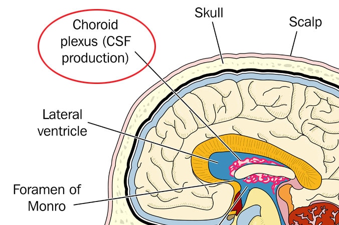
Vizcachas (Lagostomus maximus maximus, Chinchillidae) are nocturnal rodents living in burrows in many regions of Argentina, Bolivia, and Chile. We have studied the eye of the vizcacha using several light and electron microscopic procedures, with the purpose of understanding the role of vision in the behavior of this species. Our observations demonstrated an avascular, rod-rich retina, with a specialized region spanning through most of the equator of the eye. In this central band, all neural retinal layers exhibited a high cell density, whereas the photoreceptor layer was characterized by the presence of very long rods. In addition, the central region was associated with a distinct pigmentation pattern, including scarce granulation of the pigment epithelium, low pigmentation of the choroid, and the selective attachment of suprachoroidal cells to the inner scleral surface. These central modifications probably form the structural basis of a reflecting tapetum. The eye of the vizcacha received both long and short ciliary vessels, and a specialized cilio-sclero-choroidal vascular network appeared at the equatorial region. Our findings suggest that the equatorial region of the eye of the vizcacha could be a highly sensitive light detector related to foraging behaviors during crepuscular or nocturnal hours. (+info)The choroid plexus is a network of blood vessels and tissue located within each ventricle (fluid-filled space) of the brain. It plays a crucial role in the production of cerebrospinal fluid (CSF), which provides protection and nourishment to the brain and spinal cord.
The choroid plexus consists of modified ependymal cells, called plexus epithelial cells, that line the ventricular walls. These cells have finger-like projections called villi, which increase their surface area for efficient CSF production. The blood vessels within the choroid plexus transport nutrients, ions, and water to these epithelial cells, where they are actively secreted into the ventricles to form CSF.
In addition to its role in CSF production, the choroid plexus also acts as a barrier between the blood and the central nervous system (CNS), regulating the exchange of substances between them. This barrier function is primarily attributed to tight junctions present between the epithelial cells, which limit the paracellular movement of molecules.
Abnormalities in the choroid plexus can lead to various neurological conditions, such as hydrocephalus (excessive accumulation of CSF) or certain types of brain tumors.
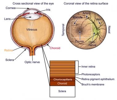
The choroid is a layer of the eye that contains blood vessels that supply oxygen and nutrients to the outer layers of the retina. It lies between the sclera (the white, protective coat of the eye) and the retina (the light-sensitive tissue at the back of the eye). The choroid is essential for maintaining the health and function of the retina, particularly the photoreceptor cells that detect light and transmit visual signals to the brain. Damage to the choroid can lead to vision loss or impairment.
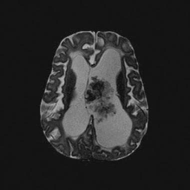
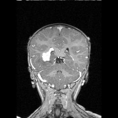
Choroid plexus neoplasms are rare types of brain tumors that arise from the choroid plexus, which are clusters of blood vessels in the ventricles (fluid-filled spaces) of the brain. These tumors can be benign (choroid plexus papilloma) or malignant (choroid plexus carcinoma). Choroid plexus neoplasms most commonly occur in children under the age of 2, but they can also affect adults. Symptoms may include increased head circumference, hydrocephalus (fluid buildup in the brain), vomiting, and developmental delays. Treatment typically involves surgical removal of the tumor, followed by radiation therapy or chemotherapy for malignant tumors.
A choroid plexus papilloma is a rare, benign (non-cancerous) tumor that develops in the choroid plexus, which are clusters of blood vessels and specialized cells in the ventricles of the brain. These tumors can occur at any age but are more common in children under the age of 10.
Choroid plexus papillomas arise from the ependymal cells that line the ventricular system and produce cerebrospinal fluid (CSF). The tumor grows slowly and tends to block the flow of CSF, leading to increased intracranial pressure and symptoms such as headaches, vomiting, irritability, and developmental delays in children.
The medical definition of choroid plexus papilloma is: "A benign, slow-growing tumor that arises from the ependymal cells of the choroid plexus in the ventricles of the brain. The tumor can obstruct the flow of cerebrospinal fluid and cause increased intracranial pressure."
It is important to note that while choroid plexus papillomas are generally benign, they can still cause significant symptoms due to their location in the brain and the obstruction of CSF flow. Treatment typically involves surgical removal of the tumor, followed by radiation therapy or chemotherapy if necessary.
Choroid neoplasms are abnormal growths that develop in the choroid, a layer of blood vessels that lies between the retina and the sclera (the white of the eye). These growths can be benign or malignant (cancerous). Benign choroid neoplasms include choroidal hemangiomas and choroidal osteomas. Malignant choroid neoplasms are typically choroidal melanomas, which are the most common primary eye tumors in adults. Other types of malignant choroid neoplasms include metastatic tumors that have spread to the eye from other parts of the body. Symptoms of choroid neoplasms can vary depending on the size and location of the growth, but may include blurred vision, floaters, or a dark spot in the visual field. Treatment options depend on the type, size, and location of the tumor, as well as the patient's overall health and personal preferences.
The choroid is a part of the eye located between the retina and the sclera, which contains a large number of blood vessels that supply oxygen and nutrients to the outer layers of the retina. Choroid diseases refer to various medical conditions that affect the health and function of the choroid. Here are some examples:
1. Choroidal neovascularization (CNV): This is a condition where new blood vessels grow from the choroid into the retina, leading to fluid accumulation, bleeding, and scarring. CNV can cause vision loss and is often associated with age-related macular degeneration, myopia, and inflammatory eye diseases.
2. Chorioretinitis: This is an infection or inflammation of the choroid and retina, which can be caused by various microorganisms such as bacteria, viruses, fungi, or parasites. Symptoms may include blurred vision, floaters, light sensitivity, and eye pain.
3. Choroidal hemorrhage: This is a rare but serious condition where there is bleeding into the choroid, often caused by trauma, high blood pressure, or blood clotting disorders. It can lead to sudden vision loss and requires urgent medical attention.
4. Choroideremia: This is a genetic disorder that affects the choroid, retina, and optic nerve, leading to progressive vision loss. It is caused by mutations in the CHM gene and primarily affects males.
5. Central serous retinopathy (CSR): This is a condition where fluid accumulates under the retina, often in the macula, causing distortion or blurring of vision. While the exact cause is unknown, CSR is thought to be related to stress, steroid use, and other factors that affect the choroid's ability to regulate fluid.
6. Polypoidal choroidal vasculopathy (PCV): This is a condition where abnormal blood vessels form in the choroid, leading to serous or hemorrhagic detachment of the retina. PCV is often associated with age-related macular degeneration and can cause vision loss if left untreated.
These are just a few examples of choroidal disorders that can affect vision. If you experience any sudden changes in your vision, it's important to seek medical attention promptly.
Cerebral ventricle neoplasms refer to tumors that develop within the cerebral ventricles, which are fluid-filled spaces in the brain. These tumors can arise from various types of cells within the ventricular system, including the ependymal cells that line the ventricles, choroid plexus cells that produce cerebrospinal fluid, or other surrounding tissues.
Cerebral ventricle neoplasms can cause a variety of symptoms depending on their size and location, such as headaches, nausea, vomiting, vision changes, imbalance, weakness, or difficulty with mental tasks. The treatment options for these tumors may include surgical resection, radiation therapy, and chemotherapy, depending on the type and extent of the tumor. Regular follow-up care is essential to monitor for recurrence and manage any long-term effects of treatment.
Cerebrospinal fluid (CSF) is a clear, colorless fluid that surrounds and protects the brain and spinal cord. It acts as a shock absorber for the central nervous system and provides nutrients to the brain while removing waste products. CSF is produced by specialized cells called ependymal cells in the choroid plexus of the ventricles (fluid-filled spaces) inside the brain. From there, it circulates through the ventricular system and around the outside of the brain and spinal cord before being absorbed back into the bloodstream. CSF analysis is an important diagnostic tool for various neurological conditions, including infections, inflammation, and cancer.
The Uvea, also known as the uveal tract or vascular tunic, is the middle layer of the eye between the sclera (the white, protective outer coat) and the retina (the light-sensitive inner layer). It consists of three main parts: the iris (the colored part of the eye), the ciliary body (structures that control the lens shape and produce aqueous humor), and the choroid (a layer of blood vessels that provides oxygen and nutrients to the retina). Inflammation of the uvea is called uveitis.
The ependyma is a type of epithelial tissue that lines the ventricular system of the brain and the central canal of the spinal cord. These cells are specialized glial cells that help to form the blood-brain barrier, regulate the cerebrospinal fluid (CSF) composition, and provide support and protection for the nervous tissue.
Ependymal cells have a cuboidal or columnar shape and possess numerous cilia on their apical surface, which helps to circulate CSF within the ventricles. They also have tight junctions that help to form the blood-brain barrier and prevent the passage of harmful substances from the blood into the CSF.
In addition to their role in maintaining the integrity of the CNS, ependymal cells can also differentiate into other types of cells, such as neurons and glial cells, under certain conditions. This property has made them a topic of interest in regenerative medicine and the study of neurodevelopmental disorders.
The pigment epithelium of the eye, also known as the retinal pigment epithelium (RPE), is a layer of cells located between the photoreceptor cells of the retina and the choroid, which is the vascular layer of the eye. The RPE plays a crucial role in maintaining the health and function of the photoreceptors by providing them with nutrients, removing waste products, and helping to regulate the light that enters the eye.
The RPE cells contain pigment granules that absorb excess light, preventing it from scattering within the eye and improving visual acuity. They also help to create a barrier between the retina and the choroid, which is important for maintaining the proper functioning of the photoreceptors. Additionally, the RPE plays a role in the regeneration of visual pigments in the photoreceptor cells, allowing us to see in different light conditions.
Damage to the RPE can lead to various eye diseases and conditions, including age-related macular degeneration (AMD), which is a leading cause of vision loss in older adults.
The sclera is the tough, white, fibrous outer coating of the eye in humans and other vertebrates, covering about five sixths of the eyeball's surface. It provides protection for the delicate inner structures of the eye and maintains its shape. The sclera is composed mainly of collagen and elastic fiber, making it strong and resilient. Its name comes from the Greek word "skleros," which means hard.
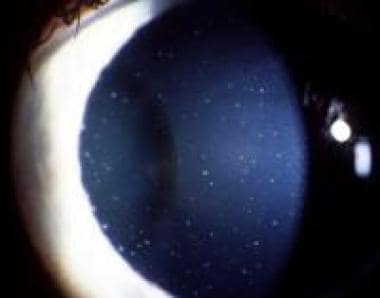
Gyrate atrophy is a rare inherited eye disorder that is characterized by progressive degeneration of the retina, which is the light-sensitive tissue at the back of the eye. It is caused by a deficiency in an enzyme called ornithine aminotransferase (OAT), which is necessary for the normal metabolism of an amino acid called ornithine.
The accumulation of ornithine in the retinal cells leads to their degeneration and the formation of well-demarcated, circular areas of atrophy (gyrates) in the retina. This can result in decreased vision, night blindness, and a progressive loss of visual field, which can ultimately lead to legal or complete blindness.
Gyrate atrophy is typically inherited as an autosomal recessive trait, meaning that an individual must inherit two copies of the mutated gene (one from each parent) in order to develop the condition. The disorder usually becomes apparent in childhood or adolescence and can progress slowly over several decades. There is currently no cure for gyrate atrophy, but dietary restrictions and supplements may help slow its progression.
Ornithine-oxo-acid transaminase (OAT), also known as ornithine aminotransferase, is a urea cycle enzyme that catalyzes the reversible transfer of an amino group from ornithine to α-ketoglutarate, producing glutamate semialdehyde and glutamate. This reaction is an essential part of the urea cycle, which is responsible for the detoxification of ammonia in the body. Deficiencies in OAT can lead to a genetic disorder called ornithine transcarbamylase deficiency (OTCD), which can cause hyperammonemia and neurological symptoms.
The eye is the organ of sight, primarily responsible for detecting and focusing on visual stimuli. It is a complex structure composed of various parts that work together to enable vision. Here are some of the main components of the eye:
1. Cornea: The clear front part of the eye that refracts light entering the eye and protects the eye from harmful particles and microorganisms.
2. Iris: The colored part of the eye that controls the amount of light reaching the retina by adjusting the size of the pupil.
3. Pupil: The opening in the center of the iris that allows light to enter the eye.
4. Lens: A biconvex structure located behind the iris that further refracts light and focuses it onto the retina.
5. Retina: A layer of light-sensitive cells (rods and cones) at the back of the eye that convert light into electrical signals, which are then transmitted to the brain via the optic nerve.
6. Optic Nerve: The nerve that carries visual information from the retina to the brain.
7. Vitreous: A clear, gel-like substance that fills the space between the lens and the retina, providing structural support to the eye.
8. Conjunctiva: A thin, transparent membrane that covers the front of the eye and the inner surface of the eyelids.
9. Extraocular Muscles: Six muscles that control the movement of the eye, allowing for proper alignment and focus.
The eye is a remarkable organ that allows us to perceive and interact with our surroundings. Various medical specialties, such as ophthalmology and optometry, are dedicated to the diagnosis, treatment, and management of various eye conditions and diseases.
The meninges are the protective membranes that cover the brain and spinal cord. They consist of three layers: the dura mater (the outermost, toughest layer), the arachnoid mater (middle layer), and the pia mater (the innermost, delicate layer). These membranes provide protection and support to the central nervous system, and contain blood vessels that supply nutrients and remove waste products. Inflammation or infection of the meninges is called meningitis, which can be a serious medical condition requiring prompt treatment.
The ciliary body is a part of the eye's internal structure that is located between the choroid and the iris. It is composed of muscle tissue and is responsible for adjusting the shape of the lens through a process called accommodation, which allows the eye to focus on objects at varying distances. Additionally, the ciliary body produces aqueous humor, the clear fluid that fills the anterior chamber of the eye and helps to nourish the eye's internal structures. The ciliary body is also responsible for maintaining the shape and position of the lens within the eye.
The retina is the innermost, light-sensitive layer of tissue in the eye of many vertebrates and some cephalopods. It receives light that has been focused by the cornea and lens, converts it into neural signals, and sends these to the brain via the optic nerve. The retina contains several types of photoreceptor cells including rods (which handle vision in low light) and cones (which are active in bright light and are capable of color vision).
In medical terms, any pathological changes or diseases affecting the retinal structure and function can lead to visual impairment or blindness. Examples include age-related macular degeneration, diabetic retinopathy, retinal detachment, and retinitis pigmentosa among others.
The Blood-Brain Barrier (BBB) is a highly specialized, selective interface between the central nervous system (CNS) and the circulating blood. It is formed by unique endothelial cells that line the brain's capillaries, along with tight junctions, astrocytic foot processes, and pericytes, which together restrict the passage of substances from the bloodstream into the CNS. This barrier serves to protect the brain from harmful agents and maintain a stable environment for proper neural function. However, it also poses a challenge in delivering therapeutics to the CNS, as most large and hydrophilic molecules cannot cross the BBB.
The retinal pigment epithelium (RPE) is a single layer of cells located between the photoreceptor cells of the retina and the choroid, which is a part of the eye containing blood vessels. The RPE plays a crucial role in maintaining the health and function of the photoreceptors by providing them with nutrients, removing waste products, and helping to regulate the light-sensitive visual pigments within the photoreceptors.
The RPE cells contain pigment granules that absorb excess light to prevent scattering within the eye and improve visual acuity. They also help to form the blood-retina barrier, which restricts the movement of certain molecules between the retina and the choroid, providing an important protective function for the retina.
Damage to the RPE can lead to a variety of eye conditions, including age-related macular degeneration (AMD), which is a leading cause of vision loss in older adults.
A cyst is a closed sac, having a distinct membrane and division between the sac and its surrounding tissue, that contains fluid, air, or semisolid material. Cysts can occur in various parts of the body, including the skin, internal organs, and bones. They can be caused by various factors, such as infection, genetic predisposition, or blockage of a duct or gland. Some cysts may cause symptoms, such as pain or discomfort, while others may not cause any symptoms at all. Treatment for cysts depends on the type and location of the cyst, as well as whether it is causing any problems. Some cysts may go away on their own, while others may need to be drained or removed through a surgical procedure.
Fluorescein angiography is a medical diagnostic procedure used in ophthalmology to examine the blood flow in the retina and choroid, which are the inner layers of the eye. This test involves injecting a fluorescent dye, Fluorescein, into a patient's arm vein. As the dye reaches the blood vessels in the eye, a specialized camera takes rapid sequences of photographs to capture the dye's circulation through the retina and choroid.
The images produced by fluorescein angiography can help doctors identify any damage to the blood vessels, leakage, or abnormal growth of new blood vessels. This information is crucial in diagnosing and managing various eye conditions such as age-related macular degeneration, diabetic retinopathy, retinal vein occlusions, and inflammatory eye diseases.
It's important to note that while fluorescein angiography is a valuable diagnostic tool, it does carry some risks, including temporary side effects like nausea, vomiting, or allergic reactions to the dye. In rare cases, severe adverse reactions can occur, so patients should discuss these potential risks with their healthcare provider before undergoing the procedure.
Prealbumin, also known as transthyretin, is a protein produced primarily in the liver and circulates in the blood. It plays a role in transporting thyroid hormones and vitamin A throughout the body. Prealbumin levels are often used as an indicator of nutritional status and liver function. Low prealbumin levels may suggest malnutrition or inflammation, while increased levels can be seen in certain conditions like hyperthyroidism. It is important to note that prealbumin levels should be interpreted in conjunction with other clinical findings and laboratory tests for a more accurate assessment of a patient's health status.
Cefadroxil is a type of antibiotic known as a cephalosporin. It works by interfering with the bacteria's ability to form a cell wall, which is necessary for its survival. Without a functional cell wall, the bacteria eventually die. Cefadroxil is used to treat a variety of infections caused by bacteria, including skin infections, ear infections, and urinary tract infections.
Cefadroxil is available as a prescription medication and is typically taken by mouth in the form of a tablet or liquid suspension. It is usually taken one to two times a day, depending on the severity of the infection and the individual patient's needs.
As with all antibiotics, it is important to take cefadroxil exactly as directed by your healthcare provider and to finish the entire course of treatment, even if you start to feel better. This will help ensure that the infection is fully treated and reduce the risk of the bacteria becoming resistant to the antibiotic.
Some common side effects of cefadroxil include nausea, vomiting, diarrhea, and stomach pain. In rare cases, more serious side effects may occur, such as an allergic reaction or severe skin reactions. If you experience any unusual symptoms while taking cefadroxil, it is important to contact your healthcare provider right away.
Choroidal neovascularization (CNV) is a medical term that refers to the growth of new, abnormal blood vessels in the choroid layer of the eye, which is located between the retina and the sclera. This condition typically occurs as a complication of age-related macular degeneration (AMD), although it can also be caused by other eye diseases or injuries.
In CNV, the new blood vessels that grow into the choroid layer are fragile and can leak fluid or blood, which can cause distortion or damage to the retina, leading to vision loss. Symptoms of CNV may include blurred or distorted vision, a blind spot in the center of the visual field, or changes in color perception.
Treatment for CNV typically involves medications that are designed to stop the growth of new blood vessels, such as anti-VEGF drugs, which target a protein called vascular endothelial growth factor (VEGF) that is involved in the development of new blood vessels. Laser surgery or photodynamic therapy may also be used in some cases to destroy the abnormal blood vessels and prevent further vision loss.
The cerebral ventricles are a system of interconnected fluid-filled cavities within the brain. They are located in the center of the brain and are filled with cerebrospinal fluid (CSF), which provides protection to the brain by cushioning it from impacts and helping to maintain its stability within the skull.
There are four ventricles in total: two lateral ventricles, one third ventricle, and one fourth ventricle. The lateral ventricles are located in each cerebral hemisphere, while the third ventricle is located between the thalami of the two hemispheres. The fourth ventricle is located at the base of the brain, above the spinal cord.
CSF flows from the lateral ventricles into the third ventricle through narrow passageways called the interventricular foramen. From there, it flows into the fourth ventricle through another narrow passageway called the cerebral aqueduct. CSF then leaves the fourth ventricle and enters the subarachnoid space surrounding the brain and spinal cord, where it can be absorbed into the bloodstream.
Abnormalities in the size or shape of the cerebral ventricles can indicate underlying neurological conditions, such as hydrocephalus (excessive accumulation of CSF) or atrophy (shrinkage) of brain tissue. Imaging techniques, such as computed tomography (CT) or magnetic resonance imaging (MRI), are often used to assess the size and shape of the cerebral ventricles in clinical settings.
Hydrocephalus is a medical condition characterized by an abnormal accumulation of cerebrospinal fluid (CSF) within the brain, leading to an increase in intracranial pressure and potentially causing damage to the brain tissues. This excessive buildup of CSF can result from either overproduction or impaired absorption of the fluid, which typically causes the ventricles (fluid-filled spaces) inside the brain to expand and put pressure on surrounding brain structures.
The condition can be congenital, present at birth due to genetic factors or abnormalities during fetal development, or acquired later in life as a result of injuries, infections, tumors, or other disorders affecting the brain's ability to regulate CSF flow and absorption. Symptoms may vary depending on age, severity, and duration but often include headaches, vomiting, balance problems, vision issues, cognitive impairment, and changes in behavior or personality.
Treatment for hydrocephalus typically involves surgically implanting a shunt system that diverts the excess CSF from the brain to another part of the body where it can be absorbed, such as the abdominal cavity. In some cases, endoscopic third ventriculostomy (ETV) might be an alternative treatment option, creating a new pathway for CSF flow within the brain. Regular follow-ups with neurosurgeons and other healthcare professionals are essential to monitor the condition and make any necessary adjustments to the treatment plan.
Ependymoma is a type of brain or spinal cord tumor that develops from the ependymal cells that line the ventricles (fluid-filled spaces) in the brain, or the central canal of the spinal cord. These tumors can be benign or malignant, and they can cause various symptoms depending on their location and size.
Ependymomas are relatively rare, accounting for about 2-3% of all primary brain and central nervous system tumors. They most commonly occur in children and young adults, but they can also affect older individuals. Treatment typically involves surgical removal of the tumor, followed by radiation therapy or chemotherapy, depending on the grade and location of the tumor. The prognosis for ependymomas varies widely, with some patients experiencing long-term survival and others having more aggressive tumors that are difficult to treat.
Brain diseases, also known as neurological disorders, refer to a wide range of conditions that affect the brain and nervous system. These diseases can be caused by various factors such as genetics, infections, injuries, degeneration, or structural abnormalities. They can affect different parts of the brain, leading to a variety of symptoms and complications.
Some examples of brain diseases include:
1. Alzheimer's disease - a progressive degenerative disorder that affects memory and cognitive function.
2. Parkinson's disease - a movement disorder characterized by tremors, stiffness, and difficulty with coordination and balance.
3. Multiple sclerosis - a chronic autoimmune disease that affects the nervous system and can cause a range of symptoms such as vision loss, muscle weakness, and cognitive impairment.
4. Epilepsy - a neurological disorder characterized by recurrent seizures.
5. Brain tumors - abnormal growths in the brain that can be benign or malignant.
6. Stroke - a sudden interruption of blood flow to the brain, which can cause paralysis, speech difficulties, and other neurological symptoms.
7. Meningitis - an infection of the membranes surrounding the brain and spinal cord.
8. Encephalitis - an inflammation of the brain that can be caused by viruses, bacteria, or autoimmune disorders.
9. Huntington's disease - a genetic disorder that affects muscle coordination, cognitive function, and mental health.
10. Migraine - a neurological condition characterized by severe headaches, often accompanied by nausea, vomiting, and sensitivity to light and sound.
Brain diseases can range from mild to severe and may be treatable or incurable. They can affect people of all ages and backgrounds, and early diagnosis and treatment are essential for improving outcomes and quality of life.
In medical terms, the iris refers to the colored portion of the eye that surrounds the pupil. It is a circular structure composed of thin, contractile muscle fibers (radial and circumferential) arranged in a regular pattern. These muscles are controlled by the autonomic nervous system and can adjust the size of the pupil in response to changes in light intensity or emotional arousal. By constricting or dilating the iris, the amount of light entering the eye can be regulated, which helps maintain optimal visual acuity under various lighting conditions.
The color of the iris is determined by the concentration and distribution of melanin pigments within the iris stroma. The iris also contains blood vessels, nerves, and connective tissue that support its structure and function. Anatomically, the iris is continuous with the ciliary body and the choroid, forming part of the uveal tract in the eye.
The brain is the central organ of the nervous system, responsible for receiving and processing sensory information, regulating vital functions, and controlling behavior, movement, and cognition. It is divided into several distinct regions, each with specific functions:
1. Cerebrum: The largest part of the brain, responsible for higher cognitive functions such as thinking, learning, memory, language, and perception. It is divided into two hemispheres, each controlling the opposite side of the body.
2. Cerebellum: Located at the back of the brain, it is responsible for coordinating muscle movements, maintaining balance, and fine-tuning motor skills.
3. Brainstem: Connects the cerebrum and cerebellum to the spinal cord, controlling vital functions such as breathing, heart rate, and blood pressure. It also serves as a relay center for sensory information and motor commands between the brain and the rest of the body.
4. Diencephalon: A region that includes the thalamus (a major sensory relay station) and hypothalamus (regulates hormones, temperature, hunger, thirst, and sleep).
5. Limbic system: A group of structures involved in emotional processing, memory formation, and motivation, including the hippocampus, amygdala, and cingulate gyrus.
The brain is composed of billions of interconnected neurons that communicate through electrical and chemical signals. It is protected by the skull and surrounded by three layers of membranes called meninges, as well as cerebrospinal fluid that provides cushioning and nutrients.
"Fundus Oculi" is a medical term that refers to the back part of the interior of the eye, including the optic disc, macula, fovea, retinal vasculature, and peripheral retina. It is the area where light is focused and then transmitted to the brain via the optic nerve, forming visual images. Examinations of the fundus oculi are crucial for detecting various eye conditions such as diabetic retinopathy, macular degeneration, glaucoma, and other retinal diseases. The examination is typically performed using an ophthalmoscope or a specialized camera called a retinal camera.
The vitreous body, also known simply as the vitreous, is the clear, gel-like substance that fills the space between the lens and the retina in the eye. It is composed mainly of water, but also contains collagen fibers, hyaluronic acid, and other proteins. The vitreous helps to maintain the shape of the eye and provides a transparent medium for light to pass through to reach the retina. With age, the vitreous can become more liquefied and may eventually separate from the retina, leading to symptoms such as floaters or flashes of light.
The fourth ventricle is a part of the cerebrospinal fluid-filled system in the brain, located in the posterior cranial fossa and continuous with the central canal of the medulla oblongata and the cerebral aqueduct. It is shaped like a cavity with a roof, floor, and lateral walls, and it communicates rostrally with the third ventricle through the cerebral aqueduct and caudally with the subarachnoid space through the median and lateral apertures (foramina of Luschka and Magendie). The fourth ventricle contains choroid plexus tissue, which produces cerebrospinal fluid. Its roof is formed by the cerebellar vermis and the superior medullary velum, while its floor is composed of the rhomboid fossa, which includes several important structures such as the vagal trigone, hypoglossal trigone, and striae medullares.
The lateral ventricles are a pair of fluid-filled cavities located within the brain. They are part of the ventricular system, which is a series of interconnected spaces filled with cerebrospinal fluid (CSF). The lateral ventricles are situated in the left and right hemispheres of the brain and are among the largest of the ventricles.
Each lateral ventricle has a complex structure and can be divided into several parts:
1. Anterior horn: This is the front part of the lateral ventricle, located in the frontal lobe of the brain.
2. Body: The central part of the lateral ventricle, which is continuous with the anterior horn and posterior horn.
3. Posterior horn: The back part of the lateral ventricle, located in the occipital lobe of the brain.
4. Temporal horn: An extension that projects into the temporal lobe of the brain.
The lateral ventricles are lined with ependymal cells, which produce cerebrospinal fluid. CSF circulates through the ventricular system, providing buoyancy and protection to the brain, and is eventually absorbed into the bloodstream. Abnormalities in the size or shape of the lateral ventricles can be associated with various neurological conditions, such as hydrocephalus, brain tumors, or neurodegenerative diseases.
Epithelium is the tissue that covers the outer surface of the body, lines the internal cavities and organs, and forms various glands. It is composed of one or more layers of tightly packed cells that have a uniform shape and size, and rest on a basement membrane. Epithelial tissues are avascular, meaning they do not contain blood vessels, and are supplied with nutrients by diffusion from the underlying connective tissue.
Epithelial cells perform a variety of functions, including protection, secretion, absorption, excretion, and sensation. They can be classified based on their shape and the number of cell layers they contain. The main types of epithelium are:
1. Squamous epithelium: composed of flat, scalelike cells that fit together like tiles on a roof. It forms the lining of blood vessels, air sacs in the lungs, and the outermost layer of the skin.
2. Cuboidal epithelium: composed of cube-shaped cells with equal height and width. It is found in glands, tubules, and ducts.
3. Columnar epithelium: composed of tall, rectangular cells that are taller than they are wide. It lines the respiratory, digestive, and reproductive tracts.
4. Pseudostratified epithelium: appears stratified or layered but is actually made up of a single layer of cells that vary in height. The nuclei of these cells appear at different levels, giving the tissue a stratified appearance. It lines the respiratory and reproductive tracts.
5. Transitional epithelium: composed of several layers of cells that can stretch and change shape to accommodate changes in volume. It is found in the urinary bladder and ureters.
Epithelial tissue provides a barrier between the internal and external environments, protecting the body from physical, chemical, and biological damage. It also plays a crucial role in maintaining homeostasis by regulating the exchange of substances between the body and its environment.
Retinal detachment is a serious eye condition that occurs when the retina, a thin layer of tissue at the back of the eye responsible for processing light and sending visual signals to the brain, pulls away from its normal position. This can lead to significant vision loss or even blindness if not promptly treated. Retinal detachment can be caused by various factors such as aging, trauma, eye disease, or an inflammatory condition. Symptoms of retinal detachment may include sudden flashes of light, floaters, a shadow in the peripheral vision, or a curtain-like covering over part of the visual field. Immediate medical attention is necessary to prevent further damage and preserve vision.
The arachnoid is one of the three membranes that cover the brain and the spinal cord, known as the meninges. It is located between the dura mater (the outermost layer) and the pia mater (the innermost layer). The arachnoid is a thin, delicate membrane that is filled with cerebrospinal fluid, which provides protection and nutrition to the central nervous system.
The arachnoid has a spider-web like appearance, hence its name, and it is composed of several layers of collagen fibers and elastic tissue. It is highly vascularized, meaning that it contains many blood vessels, and it plays an important role in regulating the flow of cerebrospinal fluid around the brain and spinal cord.
In some cases, the arachnoid can become inflamed or irritated, leading to a condition called arachnoiditis. This can cause a range of symptoms, including pain, muscle weakness, and sensory changes, and it may require medical treatment to manage.
Retinal vessels refer to the blood vessels that are located in the retina, which is the light-sensitive tissue that lines the inner surface of the eye. The retina contains two types of blood vessels: arteries and veins.
The central retinal artery supplies oxygenated blood to the inner layers of the retina, while the central retinal vein drains deoxygenated blood from the retina. These vessels can be visualized during a routine eye examination using an ophthalmoscope, which allows healthcare professionals to assess their health and any potential abnormalities.
Retinal vessels are essential for maintaining the health and function of the retina, and any damage or changes to these vessels can affect vision and lead to various eye conditions such as diabetic retinopathy, retinal vein occlusion, and hypertensive retinopathy.
Sturge-Weber syndrome is a rare neurocutaneous disorder characterized by the combination of a facial port-wine birthmark and neurological abnormalities. The facial birthmark, which is typically located on one side of the face, occurs due to the malformation of small blood vessels (capillaries) in the skin and eye.
Neurological features often include seizures that begin in infancy, muscle weakness or paralysis on one side of the body (hemiparesis), developmental delay, and intellectual disability. These neurological symptoms are caused by abnormal blood vessel formation in the brain (leptomeningeal angiomatosis) leading to increased pressure, reduced blood flow, and potential damage to the brain tissue.
Sturge-Weber syndrome can also affect the eyes, with glaucoma being a common occurrence due to increased pressure within the eye. Early diagnosis and appropriate management of this condition are crucial for improving the quality of life and reducing potential complications.
Macular degeneration, also known as age-related macular degeneration (AMD), is a medical condition that affects the central part of the retina, called the macula. The macula is responsible for sharp, detailed vision, which is necessary for activities such as reading, driving, and recognizing faces.
In AMD, there is a breakdown or deterioration of the macula, leading to gradual loss of central vision. There are two main types of AMD: dry (atrophic) and wet (exudative). Dry AMD is more common and progresses more slowly, while wet AMD is less common but can cause rapid and severe vision loss if left untreated.
The exact causes of AMD are not fully understood, but risk factors include age, smoking, family history, high blood pressure, obesity, and exposure to sunlight. While there is no cure for AMD, treatments such as vitamin supplements, laser therapy, and medication injections can help slow its progression and reduce the risk of vision loss.
Parasympathetic ganglia are collections of neurons located outside the central nervous system (CNS) that serve as relay stations for parasympathetic nerve impulses. The parasympathetic nervous system is one of the two subdivisions of the autonomic nervous system, which controls involuntary physiological responses.
The parasympathetic ganglia receive preganglionic fibers from the brainstem and sacral regions of the spinal cord. After synapsing in these ganglia, postganglionic fibers innervate target organs such as the heart, glands, and smooth muscles. The primary function of the parasympathetic nervous system is to promote rest, digestion, and energy conservation.
Parasympathetic ganglia are typically located close to or within the target organs they innervate. Examples include:
1. Ciliary ganglion: Innervates the ciliary muscle and iris sphincter in the eye, controlling accommodation and pupil constriction.
2. Pterygopalatine (sphenopalatine) ganglion: Supplies the lacrimal gland, mucous membranes of the nasal cavity, and palate, regulating tear production and nasal secretions.
3. Otic ganglion: Innervates the parotid gland, controlling salivary secretion.
4. Submandibular ganglion: Supplies the submandibular and sublingual salivary glands, regulating salivation.
5. Sacral parasympathetic ganglia: Located in the sacrum, they innervate the distal colon, rectum, and genitourinary organs, controlling defecation, urination, and sexual arousal.
These parasympathetic ganglia play crucial roles in maintaining homeostasis by regulating various bodily functions during rest and relaxation.
A coloboma is a congenital condition that results from incomplete closure of the optic fissure during fetal development. This results in a gap or hole in one or more structures of the eye, such as the iris, retina, choroid, or optic nerve. The size and location of the coloboma can vary widely, and it may affect one or both eyes.
Colobomas can cause a range of visual symptoms, depending on their size and location. Some people with colobomas may have no visual impairment, while others may experience reduced vision, double vision, or sensitivity to light. In severe cases, colobomas can lead to blindness.
Colobomas are usually diagnosed during routine eye exams and are typically not treatable, although some visual symptoms may be managed with glasses, contact lenses, or surgery in certain cases. Colobomas can occur as an isolated condition or as part of a genetic syndrome, so individuals with colobomas may benefit from genetic counseling to understand their risk of passing the condition on to their offspring.
Immunohistochemistry (IHC) is a technique used in pathology and laboratory medicine to identify specific proteins or antigens in tissue sections. It combines the principles of immunology and histology to detect the presence and location of these target molecules within cells and tissues. This technique utilizes antibodies that are specific to the protein or antigen of interest, which are then tagged with a detection system such as a chromogen or fluorophore. The stained tissue sections can be examined under a microscope, allowing for the visualization and analysis of the distribution and expression patterns of the target molecule in the context of the tissue architecture. Immunohistochemistry is widely used in diagnostic pathology to help identify various diseases, including cancer, infectious diseases, and immune-mediated disorders.
Fluorescein is not a medical condition or term, but rather a diagnostic dye used in various medical tests and procedures. Medically, it is referred to as Fluorescein Sodium, a fluorescent compound that absorbs light at one wavelength and emits light at another longer wavelength when excited.
In the field of ophthalmology (eye care), Fluorescein is commonly used in:
1. Fluorescein angiography: A diagnostic test to examine blood flow in the retina and choroid, often used to diagnose and manage conditions like diabetic retinopathy, age-related macular degeneration, and retinal vessel occlusions.
2. Tear film assessment: Fluorescein dye is used to evaluate the quality of tear film and diagnose dry eye syndrome by observing the staining pattern on the cornea.
3. Corneal abrasions/foreign body detection: Fluorescein dye can help identify corneal injuries, such as abrasions or foreign bodies, under a cobalt blue light.
In other medical fields, fluorescein is also used in procedures like:
1. Urinary tract imaging: To detect urinary tract abnormalities and evaluate kidney function.
2. Lymphangiography: A procedure to visualize the lymphatic system.
3. Surgical navigation: In some surgical procedures, fluorescein is used as a marker for better visualization of specific structures or areas.
Visna-maedi virus (VMV) is an retrovirus that belongs to the genus Lentivirus, which is part of the family Retroviridae. This virus is the causative agent of a slowly progressive, fatal disease in sheep known as maedi-visna. The term "visna" refers to a inflammatory disease of the central nervous system (CNS) and "maedi" refers to a progressive interstitial pneumonia.
The Visna-Maedi virus is closely related to the human immunodeficiency virus (HIV), which causes AIDS, as well as to other lentiviruses that affect animals such as caprine arthritis encephalitis virus (CAEV) and equine infectious anemia virus (EIAV).
Visna-maedi virus primarily targets the immune system cells, specifically monocytes/macrophages, leading to a weakened immune response in infected animals. This makes them more susceptible to other infections and diseases. The virus is transmitted through the respiratory route and infection can occur through inhalation of infectious aerosols or by ingestion of contaminated milk or colostrum from infected ewes.
There is no effective treatment or vaccine available for Visna-maedi virus infection, and control measures are focused on identifying and isolating infected animals to prevent the spread of the disease within sheep flocks.
Indocyanine green (ICG) is a sterile, water-soluble, tricarbocyanine dye that is used as a diagnostic agent in medical imaging. It is primarily used in ophthalmology for fluorescein angiography to examine blood flow in the retina and choroid, and in cardiac surgery to assess cardiac output and perfusion. When injected into the body, ICG binds to plasma proteins and fluoresces when exposed to near-infrared light, allowing for visualization of various tissues and structures. It is excreted primarily by the liver and has a half-life of approximately 3-4 minutes in the bloodstream.
Fetal diseases are medical conditions or abnormalities that affect a fetus during pregnancy. These diseases can be caused by genetic factors, environmental influences, or a combination of both. They can range from mild to severe and may impact various organ systems in the developing fetus. Examples of fetal diseases include congenital heart defects, neural tube defects, chromosomal abnormalities such as Down syndrome, and infectious diseases such as toxoplasmosis or rubella. Fetal diseases can be diagnosed through prenatal testing, including ultrasound, amniocentesis, and chorionic villus sampling. Treatment options may include medication, surgery, or delivery of the fetus, depending on the nature and severity of the disease.
Corrosion casting is a specialized technique used in anatomy and pathology to create detailed casts or molds of biological specimens, particularly vascular systems. This method is also known as "acid etching" or "corrosive casting." Here's the medical definition:
Corrosion casting is a process that involves injecting a special resin or plastic material into the vasculature or other hollow structures of a biological specimen, such as an organ or tissue. The injected material thoroughly fills the cavity and then hardens once it has set. After hardening, the surrounding tissues are corroded or dissolved using strong acids or bases, leaving behind only the cast or mold of the internal structures.
This technique results in a detailed three-dimensional representation of the complex internal networks, like blood vessels, which can be used for further study, research, and education. Corrosion casting is particularly useful in visualizing the intricate branching patterns and structural relationships within these systems.
Myopia, also known as nearsightedness, is a common refractive error of the eye. It occurs when the eye is either too long or the cornea (the clear front part of the eye) is too curved. As a result, light rays focus in front of the retina instead of directly on it, causing distant objects to appear blurry while close objects remain clear.
Myopia typically develops during childhood and can progress gradually or rapidly until early adulthood. It can be corrected with glasses, contact lenses, or refractive surgery such as LASIK. Regular eye examinations are essential for people with myopia to monitor any changes in their prescription and ensure proper correction.
While myopia is generally not a serious condition, high levels of nearsightedness can increase the risk of certain eye diseases, including cataracts, glaucoma, retinal detachment, and myopic degeneration. Therefore, it's crucial to manage myopia effectively and maintain regular follow-ups with an eye care professional.
An encyclopedia is a comprehensive reference work containing articles on various topics, usually arranged in alphabetical order. In the context of medicine, a medical encyclopedia is a collection of articles that provide information about a wide range of medical topics, including diseases and conditions, treatments, tests, procedures, and anatomy and physiology. Medical encyclopedias may be published in print or electronic formats and are often used as a starting point for researching medical topics. They can provide reliable and accurate information on medical subjects, making them useful resources for healthcare professionals, students, and patients alike. Some well-known examples of medical encyclopedias include the Merck Manual and the Stedman's Medical Dictionary.
MedlinePlus is not a medical term, but rather a consumer health website that provides high-quality, accurate, and reliable health information, written in easy-to-understand language. It is produced by the U.S. National Library of Medicine, the world's largest medical library, and is widely recognized as a trusted source of health information.
MedlinePlus offers information on various health topics, including conditions, diseases, tests, treatments, and wellness. It also provides access to drug information, medical dictionary, and encyclopedia, as well as links to clinical trials, medical news, and patient organizations. The website is available in both English and Spanish and can be accessed for free.
I'm sorry for any confusion, but "Gross Domestic Product" (GDP) is an economic term, not a medical one. GDP is the total monetary or market value of all the finished goods and services produced within a country's borders in a specific time period. It serves as a comprehensive measure of a nation’s overall economic activity.
If you have any medical questions or terms you would like defined, I would be happy to help!
 Choroid
Choroid plexus
Choroid glomus
Choroid veins
Choroid plexus tumor
Choroid plexus carcinoma
Choroid plexus papilloma
Choroid plexus cyst
Capillary lamina of choroid
Tela choroidea
Sattler's layer
Superior medullary velum
List of OMIM disorder codes
Aulus Cornelius Celsus
Glymphatic system
Macular scarring
Santosh G. Honavar
Ornithine aminotransferase deficiency
CII protein
Karl Wilhelm Ludwig Bruch
Ornithine aminotransferase
Ciliary muscle
Ronald Silverman
Suprachoroid lamina
Kearns-Sayre syndrome
Canine brain tumors
88Kasyo Junrei
Trisomy 18
Pia mater
Alpha-2-HS-glycoprotein
Ciliary body9
- Along with the ciliary body and iris, the choroid forms the uveal tract. (wikipedia.org)
- This parasympathetic ganglion contains 2 distinct types of neurons: choroid neurons, which project to vasculature in the eye's choroid layer and use somatostatin as a co-transmitter with ACh, and ciliary neurons, which innervate the ciliary body and iris and use ACh but no known peptide co-transmitter. (jneurosci.org)
- Intraocular melanoma is a rare cancer that forms from cells that make melanin in the iris, ciliary body, and choroid. (cancer.gov)
- The most common primary intraocular malignancy in adults is melanoma arising from the ciliary body and/or choroid. (aao.org)
- When this type of tumor grows to a significant size, it may extend beyond its site of origin (ie, from the choroid to the ciliary body and vice versa). (aao.org)
- If directly assigning SS2000, use the *Melanoma of the Cornea, Retina, Choroid, Ciliary Body, Eyeball, and Overlapping and Other Eye* chapter on page 262 of the [SS2000 on-line manual](http://seer.cancer.gov/tools/ssm/SSSM2000-122012.pdf#page=262). (cancer.gov)
- The sections show a large nodule of malignant melanoma apparently centred in the anterior choroid extending from the vicinity of the equator to the middle portion of the ciliary body, the latter being reflected axially by the tumour. (ox.ac.uk)
- They can be classified as anterior uveal melanomas when the tumor arises in the iris and as posterior uveal melanomas when it arises in either the choroid or the ciliary body. (medscape.com)
- The uvea is subdivided into the iris, ciliary body, and choroid. (medscape.com)
Sclera6
- choroid - The middle vascular tunic of the eye lying between the pigment epithelium and the sclera. (academic.ru)
- choroid - Middle layer of the vertebrate eye, between retina and sclera. (academic.ru)
- choroid - noun /ˈkəʊrɔɪd,ˈkoʊrɔɪd/ The vascular layer of the eye lying between the retina and the sclera. (academic.ru)
- The choroid is a layer at the back of the eye situated between the retina and sclera . (gene.vision)
- The choroid is the layer of the eyeball located between the retina and the sclera, it is a thin highly vascular dark brown membrane that absorbs excess light and prevents blurred vision. (freyjasforest.com)
- The eye is composed of the cornea (clear outer covering), conjunctiva (white part), iris (colored part), and the eye wall (choroid, retina, and sclera). (cdc.gov)
Carcinomas5
- The Surveillance and End Results (SEER) database was reviewed by Cannon et al for population-based outcomes of choroid plexus tumors (CPTs), including choroid plexus papillomas (CPPs), atypical CPPs (aCPPs), and choroid plexus carcinomas (CPCs). (medscape.com)
- Some choroid plexus carcinomas are linked to certain genetic changes passed down in families. (sparrow.org)
- 2 mitoses are present per 10 high-power field) and, to a lesser degree, histological features distinguish them from atypical choroid plexus papillomas (WHO grade 2) and choroid plexus carcinomas (WHO Grade 3) 7,10 . (radiopaedia.org)
- Choroid plexus carcinomas account for 10-20% of all choroid plexus tumors. (morganadamsfoundation.org)
- The Surveillance and End Results (SEER) database was reviewed for population-based outcomes of choroid plexus tumors (CPTs), including choroid plexus papillomas (CPP), atypical CPPs (aCPP), and choroid plexus carcinomas (CPC). (naqlafshk.com)
Cerebrospinal fluid9
- Choroid plexus papilloma (CPP) is a benign but rare central nervous system (CNS) neoplasm of the choroid plexus-a structure made from tufts of villi within the ventricular system that produces cerebrospinal fluid (CSF). (medscape.com)
- Take an in vitro approach to human neural biomarker discovery and central nervous system (CNS) permeability with human pluripotent stem cell (hPSC)-derived organoids patterned to the choroid plexus, the specialized brain epithelium that forms the blood-cerebrospinal fluid barrier. (stemcell.com)
- As NPC patients, who carry NPC1 mutations, have shown to share several pathological features with Alzheimer's disease (AD) and we and others have previously shown that AD is associated with a dysfunctionality of the blood-cerebrospinal fluid (CSF) barrier located at choroid plexus, we investigated the functionality of this latter barrier in NPC1 pathology. (ugent.be)
- We found a time-dependent uptake of IGF-1 in cerebrospinal fluid, decreasing with postnatal age, and a translocation of IGF-1 through the choroid plexus. (lu.se)
- The impact of systemic rhIGF-1/rhIGFBP-3 on IGF-1 receptor activation in the choroid plexus decreased with postnatal age, correlating with IGF-1 uptake in cerebrospinal fluid. (lu.se)
- Choroid plexus tumors are developed by brain tissue called "choroid plexus" by invading nearby tissue and spreading through the ventricles of the brain which are the interconnected cavities that contain cerebrospinal fluid. (morganadamsfoundation.org)
- Most choroid plexus tumors are noncancerous, though the cancerous form grows faster and is much more likely to spread through the cerebrospinal fluid and invade nearby tissue. (morganadamsfoundation.org)
- It grows out of brain tissue called the choroid plexus, which lines the ventricles of the brain and produces cerebrospinal fluid. (adventhealthneuroinstitute.com)
- Choroid plexus papillomas (CPPs) are benign neoplasms of the choroid plexus, a structure made from tufts of villi within the ventricular system that produces cerebrospinal fluid (CSF). (naqlafshk.com)
Called the choroid plexus2
- Choroid plexus carcinoma begins as a growth of cells in the part of the brain called the choroid plexus. (sparrow.org)
- OAK BROOK, Ill: Increased volume of an important structure in the brain called the choroid plexus is linked to greater cognitive impairment and Alzheimer's disease, according to a new study published in the journal Radiology. (medicaldialogues.in)
Tumors8
- Choroid plexus papillomas account for about 1% of all brain tumors, 2-4% of brain tumors in children younger than 15 years, 10-20% of brain tumors that occur in the first year of life, and 0.5% of adult brain tumors. (medscape.com)
- Symptoms seen with choroid plexus tumors generally result from blocking of the CSF pathways and debatably by secretion of CSF by tumor cells, both leading to increased fluid and, eventually, to hydrocephalus . (medscape.com)
- As choroid plexus tumors grow, resulting hydrocephalus and other complications usually lead to greater morbidity than that which occurs if tumors are removed when they are first discovered and are smaller. (medscape.com)
- If your child receives a diagnosis of choroid plexus carcinoma, ask your health care provider to refer you to a specialist who cares for children with brain tumors. (sparrow.org)
- Overall, choroid plexus tumors represent about 3% of brain tumors in children. (morganadamsfoundation.org)
- Choroid plexus tumors are most common in infants and represent 10-20% of brain tumors found in children younger than one year old. (morganadamsfoundation.org)
- LESSONS The known association between choroid plexus tumors and intracranial bleeding raised differential diagnosis issues. (uniroma1.it)
- Our goal is to develop at least three new, 'clinical trial-ready' treatments, specifically for children with choroid plexus carcinoma (CPC) and/or supratentorial C11orf95-RELA ependymoma (ST-EP-RELA) - two rare and aggressive childhood brain tumors. (uky.edu)
Choroidal5
- Choroidal metastases should be differentiated from uveal melanoma, where the latter is a primary tumour arising from the choroid itself. (wikipedia.org)
- CSF from the choroid plexus of the lateral ventricles is derived from the plasma filtration of blood capillaries which are from the anterior choroidal artery. (emedicodiary.com)
- The choroid plexuses-CSF system shapes the central nervous system response to inflammation at the adult stage, but little is known on the neuroimmune interactions that take place at the choroidal blood-CSF barrier during development. (biomedcentral.com)
- Combining light sheet microscopy imaging of choroid plexus, a differentiated model of the blood-CSF barrier, and multiplex cytokine assays, we showed that the choroidal epithelium responds to the bacterial insult by a specific pattern of cytokine secretion, leading to a selective accumulation of neutrophils in the choroid plexus and to their trafficking into CSF. (biomedcentral.com)
- N-acetylcysteine acted by blocking neutrophil migration across both the endothelium of choroidal stromal vessels and the epithelium forming the blood-CSF barrier, without interfering with neutrophil blood count, neutrophil tropism for choroid plexus, and choroidal chemokine-driven chemotaxis. (biomedcentral.com)
Epithelial4
- Our project goals are to define the factors involved in choroid plexus epithelial (CPe) cell development in mice, then apply this information to generate CPe cells from mouse and human embryonic stem cells (ESCs) for clinical applications. (ca.gov)
- Using NPC1(-/-) mice, we show that despite an increase in inflammatory gene expression in choroid plexus epithelial (CPE) cells, the blood-CSF barrier integrity is not dramatically affected. (ugent.be)
- Multiple Na,K-ATPase Subunits Colocalize in the Brush Border of Mouse Choroid Plexus Epithelial Cells. (bvsalud.org)
- The unusual accumulation of Na,K- ATPase complexes in the brush border membrane of choroid plexus epithelial cells have intrigued researchers for decades. (bvsalud.org)
Cells of the choroid1
- Although our systematic analysis yields no molecular traces of SARS-CoV-2 in the brain, we observe broad cellular perturbations indicating that barrier cells of the choroid plexus sense and relay peripheral inflammation into the brain and show that peripheral T cells infiltrate the parenchyma. (nature.com)
Epithelium1
- The choroid plexus epithelium (CPe) is primarily responsible for secreting CSF and regulating its composition by mechanisms currently not fully understood. (escholarship.org)
Retina and choroid1
- The main abnormality is an area of the choroid or the retina and choroid that fails to develop fully. (merckvetmanual.com)
Plexus cyst1
- It is also important to distinguish and not to confuse a cyst choroid plexus cyst with vascular origin, which occurs in the brain due to a stroke, aneurysm or infectious diseases. (vsebolezni.com)
Cornea1
- Slit lamp examination and dilated funduscopic examination can be helpful for determining whether anterior segment structures (conjunctiva, cornea, iris) or posterior segment structures (retina, nerve, choroid) are involved. (cdc.gov)
Cyst7
- How can convulsions and choroid fissure cyst be managed in my daughter? (ndtv.com)
- The CT scan showed choroid fissure cyst in her brain . (ndtv.com)
- The CT scan study shows incidental choroid fissure cyst. (ndtv.com)
- You should be assured that the choroid fissure cyst is not likely to trouble your child by 7 increasing in size or causing damage. (ndtv.com)
- A cyst of the choroid plexus in the fetus reveal is usually on the sixth to seventh month of pregnancy. (vsebolezni.com)
- A cyst of the choroid plexus during development of the fetus to discover in three percent of cases among all pregnant women. (vsebolezni.com)
- In General, a cyst of the choroid plexus does not affect the brain, does not lead to any deviations in development and does not require medical and surgical intervention. (vsebolezni.com)
Iris2
- Intraocular melanoma of the choroid is often larger and more likely to spread to other parts of the body than intraocular melanoma of the iris. (cancer.gov)
- It usually remains hidden behind the iris diaphragm, growing undetected for longer periods of time than melanoma in the iris or choroid. (medscape.com)
Malignant Melanoma1
- C693 Choroid **Note 1:** This schema is based on the UICC chapter *Malignant Melanoma of Uvea,* pages 284-290. (cancer.gov)
Eyeball1
- as, the choroid plexuses of the ventricles of the brain, and the choroid coat of the eyeball. (academic.ru)
Uveal1
- Most uveal nevi (>90%) develop in the choroid (see Chapter 17, Fig 17-4). (aao.org)
Vascular layer of the eye1
- The choroid, also known as the choroidea or choroid coat, is a part of the uvea, the vascular layer of the eye. (wikipedia.org)
Pathology2
- Moreover, we observed that EVs derived from the supernatant of NPC1(-/-) choroid plexus explants are able to induce typical brain pathology characteristics of NPC1(-/-), more specifically microgliosis and astrogliosis. (ugent.be)
- We found no relationship between choroid plexus volume and amyloid pathology but a clear relationship between the choroid plexus volume and cognitive impairment severity. (medicaldialogues.in)
Intraocular1
- Most intraocular melanomas begin in the choroid. (cancer.gov)
Parenchyma1
- Using a preterm rabbit pup model, we investigated the uptake of systemic recombinant human (rh) IGF-1 in complex with its main binding protein IGF-binding protein 3 (BP-3) to the brain parenchyma via the choroid plexus. (lu.se)
Synonyms1
- and Bruch's membrane (synonyms: Lamina basalis, Complexus basalis, Lamina vitra) - innermost layer of the choroid. (wikipedia.org)
Membrane2
- The ocular fundus is the back of the eye opposite the pupil and includes the retina, the membrane (the choroid) between the retina and the white of the eye, and the optic disk. (merckvetmanual.com)
- NKCC1 expression seems necessary for full brush border membrane accumulation of the Na,K- ATPase in the choroid plexus . (bvsalud.org)
Benign1
- Choroid plexus papillomas are an uncommon, benign (WHO grade 1) neuroepithelial intraventricular tumor, which can occur in both the pediatric (more common) and adult population. (radiopaedia.org)
Adults1
- The AdventHealth Neuroscience Institute is a state-of-the art facility for children and adults affected by choroid plexus carcinoma. (adventhealthneuroinstitute.com)
20211
- Involvement of the Choroid Plexus in the Pathogenesis of Niemann-Pick Disease Type C." FRONTIERS IN CELLULAR NEUROSCIENCE , vol. 15, 2021, doi:10.3389/fncel.2021.757482. (ugent.be)
Retinal1
- The investigative team from Turkey found chronic rhinosinusitis appears to cause thinning of the choroid and retinal nerve fiber layer (RNFL), especially in the superior and inferior quadrants and ganglion cell-inner plexiform layer (GCIPL). (reviewofoptometry.com)
Tapetum3
- In these animals, melanin is absent from a section of the choroid and within that section a layer of highly reflective tissue, the tapetum lucidum, helps to collect light by reflecting it in a controlled manner. (wikipedia.org)
- The uncontrolled reflection of light from dark choroid produces the photographic red-eye effect on photos, whereas the controlled reflection of light from the tapetum lucidum produces eyeshine (see Tapetum lucidum). (wikipedia.org)
- Well vascularized and also pigmented to throw light back onto the retina (the tapetum is an irridescent layer in the choroid of some eyes). (academic.ru)
Foramen1
- Choroid plexus papillomas of the foramen of Luschka: MR appearance. (ajnr.org)
Alzheimer's1
- Choroid plexus volume was greater in those with Alzheimer's dementia than in those without. (medicaldialogues.in)
Intraventricular1
- On imaging, choroid plexus papillomas are characterized by vividly enhancing masses, usually intraventricular. (radiopaedia.org)
Ventricles1
- The choroid plexus is a network of blood vessels, connective tissue and cells found in spaces of the brain called ventricles. (medicaldialogues.in)
Coat2
- The choroid coat of the eye. (academic.ru)
- See choroid coat. (academic.ru)
Connective tissue1
- The choroid is the layer of blood vessels and connective tissue between the white of the eye and retina (at the back of the eye). (medlineplus.gov)
Nerve1
- Nerve cells are not included in their composition, however, the choroid plexus have a direct effect on the formation of nerve tissue in the brain. (vsebolezni.com)
Humans2
- In humans and most other primates, melanin occurs throughout the choroid. (wikipedia.org)
- A collaboration was initiated to profile all of the genes expressed by the purified mouse ESC-derived CPECs, and to compare this profile to those expressed by the choroid plexus in developing mice and humans. (ca.gov)
Pigment1
- Melanin, a dark colored pigment, helps the choroid limit uncontrolled reflection within the eye that would potentially result in the perception of confusing images. (wikipedia.org)
Preterm1
- Here, we characterize the uptake of systemic rhIGF-1/rhIGFBP-3 to the preterm brain, and show that the interaction between systemic rhIGF-1/rhIGFBP-3 and choroid plexus varies over time. (lu.se)
Inflammation2
- Inflammation of the choroid is called choroiditis . (medlineplus.gov)
- They also point to the efficacy of N-acetylcysteine in reducing the deleterious effects of inflammation-associated perinatal injuries by a previously undescribed mechanism, i.e. the inhibition of innate immune cell migration across the choroid plexuses, without interfering with the systemic inflammatory response to infection. (biomedcentral.com)
Inflammatory response1
- The fundus displayed a pathologic phenomenon that was diagnosed as chorioretinitis, a form of posterior uveitis, which is an inflammatory response involving both the choroid layer, and the retina. (cdc.gov)
Findings2
- Findings consistent with choroid plexus papilloma. (radiopaedia.org)
- I think our findings on the choroid plexus can suggest it as a new potential MR imaging surrogate for an impaired clearance system and neuroinflammation," Dr. Moon said. (medicaldialogues.in)
Layer3
- We have found that the earliest developmental stage in which neurons with somatostatinlike immunoreactivity (SOM-IR) are consistently found in vivo is stage 30 (embryonic day 6.5), a time shortly after the extension of neurites to targets in the eye's choroid layer. (jneurosci.org)
- Thus, the expression of somatostatin in cultured CG neurons is regulated by a macromolecule produced by cells in the choroid layer, a target normally innervated in vivo by CG neurons expressing somatostatin. (jneurosci.org)
- The choroid is a layer of blood vessels that bring oxygen and nutrients to the eye. (cancer.gov)
Posterior1
- Similarly, the choroid plexus of the fourth ventricle is traversed by anterior inferior cerebellar artery & superior cerebellar arteries, branch of basilar artery & posterior inferior cerebellar artery, branch of the vertebral artery. (emedicodiary.com)
Chemotherapy2
- Treatment of a choroid plexus carcinoma is often surgery followed by chemotherapy, radiation therapy or both. (sparrow.org)
- Children with choroid plexus carcinoma often need chemotherapy and radiation therapy to kill any remaining cancer cells. (morganadamsfoundation.org)
Absent1
- The name, which implies a congenital absence of the choroid, is not very well chosen, as the choroid is not absent at birth. (eyehospital.nl)
Anatomy1
- Beginning with an introduction to the technique for obtaining clear images, and discussion on normal anatomy, the following sections offer step by step guidance on the interpretation of OCT images and data acquired by OCT, 'en face' OCT and OCT angiography. (ophthalmologyebooks.store)
Plexuses2
- Here we showed in neonatal rats exposed to P3C that the migration of neutrophils into the CSF, which occurred through the choroid plexuses, is abolished following administration of the antioxidant drug N-acetylcysteine. (biomedcentral.com)
- The CNS is protected against the invasion of immune cells by cellular barriers which comprise the cerebrovascular walls and the choroid plexuses (ChPs). (biomedcentral.com)
Gene expression2
- In addition, choroid plexus gene expression was observed to increase with postnatal age. (lu.se)
- Moreover, using choroid plexus in vitro cell cultures, gene expression and protein synthesis were further investigated upon rhIGF-1/rhIGFBP-3 stimulation as compared to rhIGF-1 alone, and found not to be differently altered. (lu.se)
Masses1
- Choroid plexus papillomas typically appear as cauliflower-like masses 4,10 . (radiopaedia.org)
Blood5
- We have developed software to measure blood flow in the choroid (Ch/d software) using mean fluorescence intensity of the disc as a reference value for fluorescent intensity of the choroid. (arvojournals.org)
- CSF is formed by an active transport mechanism followed by a passive diffusion process of blood plasma present in the blood capillaries of choroid plexus . (emedicodiary.com)
- Thus there is possible of passive diffusion of water from the blood capillaries of the choroid plexus into the ventricle. (emedicodiary.com)
- One potential transport mechanism of blood-borne IGF-1 to reach the central nervous system is via the choroid plexus (ChP). (lu.se)
- A pathological process consisting of the formation of new blood vessels in the CHOROID. (bvsalud.org)
Symptoms2
- What are the symptoms of a choroid plexus carcinoma? (morganadamsfoundation.org)
- The symptoms of a choroid plexus tumor depend on the location and of the mass. (morganadamsfoundation.org)
Accumulation1
- Thus, we assume that the abnormal status of choroid plexus is linked to the failure of clearance leading to waste and toxic protein accumulation in the brain and failure of immune surveillance leading to neuroinflammation. (medicaldialogues.in)
Molecular1
- A multitude of Na,K- ATPase subunits form molecular complexes in the choroid plexus brush border , which may bind to the cytoskeleton by various alternative actin binding proteins . (bvsalud.org)

![Recombinant Anti-p53 antibody [E26] KO Tested (ab32389) | Abcam](https://www.abcam.com/ps/products/32/ab32389/Images/ab32389-389819-antip53antibodyimmunocytochemistryimmunofluorescencehap1human.jpg)


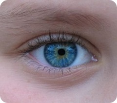


![10 Best Clinics for Esophageal Cancer Treatment in Thailand [2023 Prices]](https://www.mymeditravel.com/cdn-cgi/image/f=auto,fit=contain,quality=75/uploads/property/gallery/5af2758efa6b7e04401f8c27/5af51dadfa6b7e4212052361/preview.jpg)