Bile Ducts, Intrahepatic
Klatskin's Tumor
Bile Ducts, Extrahepatic
Opisthorchis
Hepatic Duct, Common
Cholangitis, Sclerosing
Lithiasis
Clonorchiasis
Fascioliasis
Bile Ducts
Fasciola hepatica
Clonorchis sinensis
Thailand
Cholangiopancreatography, Endoscopic Retrograde
Jaundice, Obstructive
Cholangitis
Biliary Tract Neoplasms
CA-19-9 Antigen
Prognosis
Carcinoma, Hepatocellular
Cholangiopancreatography, Magnetic Resonance
Thioacetamide
Retrospective Studies
Iridium Radioisotopes
Cholangiography
Food Parasitology
Survival Rate
Tumor Markers, Biological
Choledochal Cyst
Liver Transplantation
Lobar decrease in 99mTc-GSA accumulation in hilar cholangiocarcinoma. (1/1040)
Hilar cholangiocarcinoma can obstruct hepatic ducts and involve the portal veins. Both biliary stasis and decrease in portal venous flow are known to reduce 99mTc-diethylenetriamine pentaacetic acid-galactosyl human serum albumin (GSA) accumulation. The specific relationship between these pathological conditions due to hilar cholangiocarcinomas and 99mTc-GSA accumulation has never been clarified. METHODS: Sixteen patients with hilar cholangiocarcinomas who underwent 99mTc-GSA liver scintigraphy were reviewed. The relationship between significant decrease in 99mTc-GSA accumulation and lobar biliary stasis, or decrease in the portal venous flow, was evaluated. Average counts of region of interest placed in both right and left lobes were compared in the same transaxial SPECT section. Count ratios of right and left lobes were calculated. RESULTS: Significant lobar decrease in 99mTc-GSA accumulation was observed in 6 of the 16 patients. Ipsilateral portal venous stenosis or obstruction was seen in all these 6 patients, whereas ipsilateral portal venous stenosis or obstruction was seen in only 1 of the other 10 patients. Symmetric bile duct dilatation was seen in 13 patients, and asymmetric bile duct dilatation was seen in 3. Lobar decrease in 99mTc-GSA accumulation correlated well with decrease in ipsilateral portal venous flow (P < 0.0005). The count ratio was significantly reduced when unilateral portal venous flow decreased (P < 0.05). CONCLUSION: Using 99mTc-GSA liver scintigraphy, we can predict lobar decrease in ipsilateral portal venous flow and monitor hepatic functional lateralities in patients with hilar cholangiocarcinomas. (+info)Binding of tumour necrosis factor-alpha (TNF-alpha) to TNF-RI induces caspase(s)-dependent apoptosis in human cholangiocarcinoma cell lines. (2/1040)
Cholangiocarcinoma (CCA), a tumour of the bile duct epithelium, occurs with a higher incidence in South-east Asian countries than in Europe and North America. The prognosis is poor, due to the unavailability of early diagnosis and the tumours being relatively resistant to chemotherapy. In the present study one of the fatal routes of this tumour was studied. This death was stimulated by TNF-alpha. TNF-alpha at a concentration of 760 pg/ml and 100 pg/ml in the presence of 1 microgram/ml actinomycin D induced 50% cell death of the two established human cholangiocarcinoma cell lines HuCCA-1 and HuCCA-INu, respectively. Preincubation of both cell lines with MoAb to TNF-RI or TNF-RII before TNF-alpha treatment showed that only the MoAb specific to TNF-RI inhibited death. The death of these two cell lines was proved to be apoptosis. Western blot analysis of extracts from both cell lines demonstrated a cleavage of poly (ADP-ribose) polymerase (PARP) within 6-8 h following TNF-alpha treatment. The degradation of PARP was prevented by a MoAb to TNF-RI indicating that the TNF-RI but not TNF-RII was involved in TNF-induced apoptosis in these two human cholangiocarcinoma cell lines. Moreover, peptide inhibitor for caspase II subfamily, Ac-DEVD-CHO, reduced the cytolysis of TNF-alpha-treated cholangiocarcinoma cells. The inhibitor also prevented degradation of PARP. These results indicate that the interaction between TNF-alpha and TNF-RI alone generated a sufficient signal to activate a caspase II subfamily-dependent apoptosis in human cholangiocarcinoma cell lines. (+info)Lymph node metastasis in intrahepatic cholangiocarcinoma. (3/1040)
BACKGROUND: Lymph node metastasis is a significant prognostic factor in intrahepatic cholangiocarcinoma. This study was aimed at investigating lymph node metastasis in intrahepatic cholangiocarcinoma and to examine whether the extent of metastasis affects outcomes after surgery. METHODS: From 1980 through 1996, 70 patients with intrahepatic cholangiocarcinoma underwent hepatectomy, with a 50% curative resection rate. Lymph node dissection was performed in 51 patients, and the presence of lymph node metastasis was examined microscopically. The metastatic nodes were divided into groups N1, N2 or N3 using the classification proposed by the Liver Cancer Study Group of Japan. RESULTS: Twenty-three patients had lymph node metastasis. Metastasis was to N1 nodes in 10 patients, to N2 nodes in nine patients and to N3 nodes in four patients. Nineteen patients had metastatic nodes in the hepatoduodenal ligament, which was the most common metastatic site regardless of tumor location. The five-year survival rate in patients with lymph node metastasis (0%) was significantly lower (p < 0.0001) than that in patients without lymph node metastasis (51 %); however, five-year survival rates did not differ between patients with metastases to N1, N2 and N3 nodes. CONCLUSIONS: Lymph nodes in the hepatoduodenal ligament may be sentinel nodes for intrahepatic cholangiocarcinoma, and outcomes after surgery for patients with lymph node metastasis are poor regardless of the sites of nodal metastasis. (+info)Promoting effects of 3-chloro-4-(dichloromethyl)-5-hydroxy-2(5H)-furanone on rat glandular stomach carcinogenesis initiated with N-methyl-N'-nitro-N-nitrosoguanidine. (4/1040)
The modifying effects of 3-chloro-4-(dichloromethyl)-5-hydroxy-2(5H)-furanone (MX), a mutagenic by-product in chlorinated water, on the development of glandular stomach cancers were investigated in Wistar rats. A total of 120 males, 6 weeks of age, were divided into six groups. After initiation with 100 ppm N-methyl-N'-nitro-N-nitrosoguanidine (MNNG) solution and 5% NaCl diet for 8 weeks, 30 rats each in groups 1-3 were given MX in the drinking water at concentrations of 30, 10, or 0 ppm for the following 57 weeks. Ten animals each in groups 4-6 were administered the MX without prior carcinogen exposure. There were no statistical significant differences in final body weights between the groups. The incidences and multiplicities of adenocarcinomas in the glandular stomachs were significantly higher (P < 0.05) in the initiated 30 ppm MX group than those in the MNNG/NaCl group. The incidences of atypical hyperplasias in the glandular stomachs were also significantly increased (P < 0.05 or 0.01) by the MX treatments. With their multiplicity, the effects were clearly dose dependent. Interestingly, the 30 ppm MX alone itself induced atypical hyperplasias in the pylorus, although the incidences and severity were low. Moreover, MX showed a tendency to enhance the development of intrahepatic cholangiocellular tumors and thyroid follicular cell tumors in the MNNG-treated animals. The results of the present study thus indicate that MX exerts promoting effects when given during the postinitiation phase of two-stage glandular stomach carcinogenesis in rats. (+info)Expression of p73, a novel protein related to the p53 tumour suppressor p53, and apoptosis in cholangiocellular carcinoma of the liver. (5/1040)
p73, the first homologue of the tumour suppressor protein p53, was recently discovered on chromosome 1p36 and has been shown to induce apoptosis in a p53-like manner. The present study was performed with the aim of investigating the expression of p53, its new homologue p73 and the occurrence of apoptosis in cholangiocellular carcinoma. Protein levels of p73 were examined in 41 patients with curatively (R0-) resected cholangiocellular carcinomas with an antiserum, raised against a peptide in the N-terminal domain of p73. The incidence of mutations in the p53 gene was analysed by direct sequencing and also immunohistochemically. Apoptotic cell death was assessed using in-situ end-labelling (ISEL) technique in combination with morphological criteria. The results obtained were correlated with patient survival. Immunostaining of p73 protein was detected in 17/41 carcinomas examined (41%). The immunoreactivity was confined to the cell nucleus. In 15/41 patients (37%), mutations of the p53 gene were observed. Eleven out of these 15 patients stained also positive for p73. In contrast, out of 26 patients without any detectable p53 mutation, only six exhibited p73 immunostaining. We failed to observe a correlation between p73 expression or p53 and apoptosis within a given tumour. Survival analysis including the parameters stage and grade of disease, p73 and p53, and also apoptosis, showed that tumour stage and grade as well as p53 and p73 were significantly related to prognosis. In Cox regression survival analysis, however, only extent of primary tumour and lymph node status had an independent prognostic impact. Our results with a high prevalence of p73 within tumours harbouring mutated p53 gene suggest that p73 could compensate for p53 function. We failed to establish p73 or p53 as independent prognostic factors in cholangiocellular carcinoma of the liver. (+info)Intrahepatic peripheral cholangiocarcinoma: CT features in 18 pathologically proven cases. (6/1040)
OBJECTIVE: To determine the morphological features of 18 pathologically proven intrahepatic peripheral cholangiocarcinoma (IHPCC) cases in computerized tomography (CT) image. METHODS: All 18 patients had CT, using Picker I.Q.T/C and taking pre-contrast continuous 10-mm sections throughout the liver and post-contrast continuous 10-mm sections throughout the focuses. RESULTS: The disease was characterized in CT image by the following: all focuses were found in the periphery of the liver and shown as a lobulated or fused hypodense space-occupying mass; there were one or more divergent or confluent, circular or irregular cystic areas with much lower density, in the majority. All focuses could be enhanced slightly and most revealed a dim edge. Dilated bile ducts around the focus were found frequently; the dilated bile ducts especially seemed to encircle the focus (33.3%, 6/18). This phenomenon were referred to as "encysted sign of dilated bile ducts". CONCLUSIONS: CT scanning should be one of the most important investigative methods for IHPCC due to the disease characteristics identified in CT image, especially "encysted sign of dilated bile ducts" which possesses specificity in diagnosing the disease. (+info)Apoptosis and tumorigenesis in human cholangiocarcinoma cells. Involvement of Fas/APO-1 (CD95) and calmodulin. (7/1040)
We have previously demonstrated that tamoxifen inhibits the growth of human cholangiocarcinoma cells in culture and inhibits tumor growth when cells are injected into nude mice. However, the mechanism of action of tamoxifen remains unknown. Here we demonstrate that tamoxifen and trifluoperazine, both potent calmodulin antagonists, induce apoptosis in vitro, probably acting via the Fas system, in human cholangiocarcinoma cells. Human cholangiocarcinoma cell lines heterogeneously express Fas antigen on their surface. Fas-negative and Fas-positive surface-expressing cells were isolated, cloned, and cultured. Fas antibody, tamoxifen, and trifluoperazine induced dose-dependent apoptosis only in Fas-positive cells; Fas-negative cells were unaffected. Furthermore, apoptosis induced by tamoxifen in Fas-positive cells was blocked by an inhibitory Fas antibody. Tamoxifen was not acting through an anti-estrogenic mechanism, because neither Fas-negative nor Fas-positive cells expressed estrogen receptors and the pure anti-estrogen compound, ICI 182780, did not induce apoptosis in either cell line. Fas-negative cells, but not Fas-positive cells, were able to produce tumors when subcutaneously injected into nude mice. These findings suggest Fas may be a candidate oncogene involved in the pathogenesis of cholangiocarcinoma. Furthermore, the similarity between the pro-apoptotic effects of tamoxifen and trifluoperazine support an underlying molecular mechanism for Fas-mediated apoptosis that involves calmodulin. (+info)Recurrent thrombo-embolic episodes: the association of cholangiocarcinoma with antiphospholipid syndrome. (8/1040)
Antiphospholipid syndrome is a disorder of recurrent vascular thrombosis, pregnancy loss and thrombocytopenia associated with persistently elevated levels of antiphospholipid antibodies. It was first described in a group of patients with systemic lupus erythematosus but has since been associated with a wide range of conditions, including other autoimmune disorders and malignancy. It can also occur in isolation, the so-called primary antiphospholipid syndrome. We describe an elderly woman with the antiphospholipid syndrome thought to be associated with a cholangiocarcinoma. (+info)Cholangiocarcinoma is a type of cancer that arises from the cells that line the bile ducts, which are small tubes that carry digestive enzymes from the liver to the small intestine. It can occur in different parts of the bile duct system, including the bile ducts inside the liver (intrahepatic), the bile ducts outside the liver (extrahepatic), and the area where the bile ducts join the pancreas and small intestine (ampulla of Vater).
Cholangiocarcinoma is a relatively rare cancer, but its incidence has been increasing in recent years. It can be difficult to diagnose because its symptoms are often nonspecific and similar to those of other conditions, such as gallstones or pancreatitis. Treatment options depend on the location and stage of the cancer, and may include surgery, radiation therapy, chemotherapy, or a combination of these approaches.
Bile duct neoplasms, also known as cholangiocarcinomas, refer to a group of malignancies that arise from the bile ducts. These are the tubes that carry bile from the liver to the gallbladder and small intestine. Bile duct neoplasms can be further classified based on their location as intrahepatic (within the liver), perihilar (at the junction of the left and right hepatic ducts), or distal (in the common bile duct).
These tumors are relatively rare, but their incidence has been increasing in recent years. They can cause a variety of symptoms, including jaundice, abdominal pain, weight loss, and fever. The diagnosis of bile duct neoplasms typically involves imaging studies such as CT or MRI scans, as well as blood tests to assess liver function. In some cases, a biopsy may be necessary to confirm the diagnosis.
Treatment options for bile duct neoplasms depend on several factors, including the location and stage of the tumor, as well as the patient's overall health. Surgical resection is the preferred treatment for early-stage tumors, while chemotherapy and radiation therapy may be used in more advanced cases. For patients who are not candidates for surgery, palliative treatments such as stenting or bypass procedures may be recommended to relieve symptoms and improve quality of life.
Intrahepatic bile ducts are the small tubular structures inside the liver that collect bile from the liver cells (hepatocytes). Bile is a digestive fluid produced by the liver that helps in the absorption of fats and fat-soluble vitamins from food. The intrahepatic bile ducts merge to form larger ducts, which eventually exit the liver and join with the cystic duct from the gallbladder to form the common bile duct. The common bile duct then empties into the duodenum, the first part of the small intestine, where bile aids in digestion. Intrahepatic bile ducts can become obstructed or damaged due to various conditions such as gallstones, tumors, or inflammation, leading to complications like jaundice, liver damage, and infection.
Adenoma of the bile duct is a benign (noncancerous) tumor that develops in the bile ducts, which are tiny tubes that carry bile from the liver to the gallbladder and small intestine. Bile is a digestive fluid produced by the liver.
Bile duct adenomas are rare and usually do not cause any symptoms. However, if they grow large enough, they may obstruct the flow of bile and cause jaundice (yellowing of the skin and whites of the eyes), abdominal pain, or itching. In some cases, bile duct adenomas may become cancerous and develop into bile duct carcinomas.
The exact cause of bile duct adenomas is not known, but they are more common in people with certain genetic disorders, such as Gardner's syndrome and von Hippel-Lindau disease. Treatment for bile duct adenomas typically involves surgical removal of the tumor.
A Klatskin's tumor, also known as a perihilar cholangiocarcinoma, is a rare and aggressive form of cancer that occurs at the junction where the right and left hepatic ducts come together to form the common hepatic duct, which then becomes the common bile duct. This type of tumor can obstruct the flow of bile from the liver into the small intestine, leading to jaundice, itching, abdominal pain, and other symptoms. Klatskin's tumors are often difficult to diagnose and treat due to their location and tendency to spread quickly. Surgical resection is the preferred treatment option when possible, although chemotherapy and radiation therapy may also be used in some cases.
Extrahepatic bile ducts refer to the portion of the biliary system that lies outside the liver. The biliary system is responsible for producing, storing, and transporting bile, a digestive fluid produced by the liver.
The extrahepatic bile ducts include:
1. The common hepatic duct: This duct is formed by the union of the right and left hepatic ducts, which drain bile from the corresponding lobes of the liver.
2. The cystic duct: This short duct connects the gallbladder to the common hepatic duct, allowing bile to flow into the gallbladder for storage and concentration.
3. The common bile duct: This is the result of the fusion of the common hepatic duct and the cystic duct. It transports bile from the liver and gallbladder to the duodenum, the first part of the small intestine, where it aids in fat digestion.
4. The ampulla of Vater (or hepatopancreatic ampulla): This is a dilated area where the common bile duct and the pancreatic duct join and empty their contents into the duodenum through a shared opening called the major duodenal papilla.
Extrahepatic bile ducts can be affected by various conditions, such as gallstones, inflammation (cholangitis), strictures, or tumors, which may require medical or surgical intervention.
Opisthorchis is a genus of trematode flatworms that are commonly known as liver flukes. These parasites primarily infect the bile ducts and liver of various mammals, including humans. The most common species that infect humans are Opisthorchis viverrini and Opisthorchis felineus.
Humans become infected with these parasites by consuming raw or undercooked fish that contain the larval stage of the fluke (metacercariae). Once ingested, the metacercariae excyst in the small intestine and migrate to the bile ducts, where they mature into adults. Adult Opisthorchis worms are thin and elongated, with a length of 7-15 mm and a width of 1-3 mm. They have a characteristic brownish color due to their diet, which consists mainly of blood and bile.
Infection with Opisthorchis can lead to chronic inflammation of the bile ducts and liver, which may result in symptoms such as abdominal pain, diarrhea, weight loss, and fatigue. Long-term infection has been linked to an increased risk of cholangiocarcinoma, a rare but aggressive form of liver cancer.
Prevention of Opisthorchis infection involves avoiding the consumption of raw or undercooked fish, particularly in areas where the parasite is endemic. Infection can also be treated with anti-parasitic drugs such as praziquantel.
Opisthorchiasis is a parasitic infection caused by the trematode flatworms of the genus Opisthorchiidae, specifically Opisthorchis viverrini and Opisthorchis felineus. These flatworms are transmitted to humans through the consumption of raw or undercooked fish that contain the infective larval stage (metacercariae) of the parasite.
Once ingested, the metacercariae excyst in the small intestine and migrate to the bile ducts of the liver, where they mature into adult worms and reside. The adults can live for several years in the host's body, producing eggs that are released into the bile and then passed through the stool.
The infection can cause a range of symptoms, including abdominal pain, diarrhea, liver enlargement, and bile duct inflammation. Chronic opisthorchiasis can lead to more severe complications such as cholangitis, cholecystitis, gallstones, and liver cirrhosis. In some cases, it may also increase the risk of developing cholangiocarcinoma, a rare but aggressive form of bile duct cancer.
Preventive measures include avoiding the consumption of raw or undercooked fish, particularly in areas where the infection is endemic, and practicing good personal hygiene to prevent fecal-oral transmission. Treatment typically involves the use of anti-parasitic drugs such as praziquantel or albendazole to kill the adult worms and prevent further complications.
Biliary tract surgical procedures refer to a range of operations that involve the biliary system, which includes the liver, gallbladder, and bile ducts. These procedures can be performed for various reasons, including the treatment of gallstones, bile duct injuries, tumors, or other conditions affecting the biliary tract. Here are some examples of biliary tract surgical procedures:
1. Cholecystectomy: This is the surgical removal of the gallbladder, which is often performed to treat symptomatic gallstones or chronic cholecystitis (inflammation of the gallbladder). It can be done as an open procedure or laparoscopically.
2. Bile duct exploration: This procedure involves opening the common bile duct to remove stones, strictures, or tumors. It is often performed during a cholecystectomy if there is suspicion of common bile duct involvement.
3. Hepaticojejunostomy: This operation connects the liver's bile ducts directly to a portion of the small intestine called the jejunum, bypassing a damaged or obstructed segment of the biliary tract. It is often performed for benign or malignant conditions affecting the bile ducts.
4. Roux-en-Y hepaticojejunostomy: This procedure involves creating a Y-shaped limb of jejunum and connecting it to the liver's bile ducts, bypassing the common bile duct and duodenum. It is often performed for complex biliary tract injuries or malignancies.
5. Whipple procedure (pancreaticoduodenectomy): This extensive operation involves removing the head of the pancreas, the duodenum, a portion of the jejunum, the gallbladder, and the common bile duct. It is performed for malignancies involving the pancreas, bile duct, or duodenum.
6. Liver resection: This procedure involves removing a portion of the liver to treat primary liver tumors (hepatocellular carcinoma or cholangiocarcinoma) or metastatic cancer from other organs.
7. Biliary stenting or bypass: These minimally invasive procedures involve placing a stent or creating a bypass to relieve bile duct obstructions caused by tumors, strictures, or stones. They can be performed endoscopically (ERCP) or percutaneously (PTC).
8. Cholecystectomy: This procedure involves removing the gallbladder, often for symptomatic cholelithiasis (gallstones) or cholecystitis (inflammation of the gallbladder). It can be performed laparoscopically or open.
9. Biliary drainage: This procedure involves placing a catheter to drain bile from the liver or bile ducts, often for acute or chronic obstructions caused by tumors, strictures, or stones. It can be performed endoscopically (ERCP) or percutaneously (PTC).
10. Bilioenteric anastomosis: This procedure involves connecting the biliary tract to a portion of the small intestine, often for benign or malignant conditions affecting the bile ducts or pancreas. It can be performed open or laparoscopically.
The common hepatic duct is a medical term that refers to the duct in the liver responsible for carrying bile from the liver. More specifically, it is the duct that results from the convergence of the right and left hepatic ducts, which themselves carry bile from the right and left lobes of the liver, respectively. The common hepatic duct then joins with the cystic duct from the gallbladder to form the common bile duct, which ultimately drains into the duodenum, a part of the small intestine.
The primary function of the common hepatic duct is to transport bile, a digestive juice produced by the liver, to the small intestine. Bile helps break down fats during the digestion process, making it possible for the body to absorb them properly. Any issues or abnormalities in the common hepatic duct can lead to problems with bile flow and potentially cause health complications such as jaundice, gallstones, or liver damage.
Sclerosing cholangitis is a chronic progressive disease characterized by inflammation and scarring (fibrosis) of the bile ducts, leading to their narrowing or obstruction. This results in impaired bile flow from the liver to the small intestine, which can cause damage to the liver cells and eventually result in cirrhosis and liver failure.
The condition often affects both the intrahepatic (within the liver) and extrahepatic (outside the liver) bile ducts. The exact cause of sclerosing cholangitis is not known, but it is believed to involve an autoimmune response, genetic predisposition, and environmental factors.
Symptoms of sclerosing cholangitis may include jaundice (yellowing of the skin and eyes), itching, abdominal pain, fatigue, weight loss, dark urine, and light-colored stools. The diagnosis is typically made through imaging tests such as magnetic resonance cholangiopancreatography (MRCP) or endoscopic retrograde cholangiopancreatography (ERCP), which can visualize the bile ducts and detect any abnormalities.
Treatment for sclerosing cholangitis is aimed at managing symptoms, preventing complications, and slowing down the progression of the disease. This may include medications to relieve itching, antibiotics to treat infections, and drugs to reduce inflammation and improve bile flow. In severe cases, a liver transplant may be necessary.
Liver neoplasms refer to abnormal growths in the liver that can be benign or malignant. Benign liver neoplasms are non-cancerous tumors that do not spread to other parts of the body, while malignant liver neoplasms are cancerous tumors that can invade and destroy surrounding tissue and spread to other organs.
Liver neoplasms can be primary, meaning they originate in the liver, or secondary, meaning they have metastasized (spread) to the liver from another part of the body. Primary liver neoplasms can be further classified into different types based on their cell of origin and behavior, including hepatocellular carcinoma, cholangiocarcinoma, and hepatic hemangioma.
The diagnosis of liver neoplasms typically involves a combination of imaging studies, such as ultrasound, CT scan, or MRI, and biopsy to confirm the type and stage of the tumor. Treatment options depend on the type and extent of the neoplasm and may include surgery, radiation therapy, chemotherapy, or liver transplantation.
Hepatectomy is a surgical procedure that involves the removal of part or all of the liver. This procedure can be performed for various reasons, such as removing cancerous or non-cancerous tumors, treating liver trauma, or donating a portion of the liver to another person in need of a transplant (live donor hepatectomy). The extent of the hepatectomy depends on the medical condition and overall health of the patient. It is a complex procedure that requires significant expertise and experience from the surgical team due to the liver's unique anatomy, blood supply, and regenerative capabilities.
Lithiasis is a medical term that refers to the formation of stones or calculi in various organs of the body. These stones can develop in the kidneys (nephrolithiasis), gallbladder (cholelithiasis), urinary bladder (cystolithiasis), or salivary glands (sialolithiasis). The stones are usually composed of minerals and organic substances, and their formation can be influenced by various factors such as diet, dehydration, genetic predisposition, and chronic inflammation. Lithiasis can cause a range of symptoms depending on the location and size of the stone, including pain, obstruction, infection, and damage to surrounding tissues. Treatment may involve medication, shock wave lithotripsy, or surgical removal of the stones.
Clonorchiasis is a parasitic infection caused by the trematode worm Clonorchis sinensis, also known as the Chinese liver fluke. This flatworm infects the bile ducts and liver of humans and other animals, leading to inflammation, obstruction, and potential complications such as cholangitis, cirrhosis, and cholangiocarcinoma (bile duct cancer).
Humans become infected with Clonorchis sinensis by consuming raw or undercooked freshwater fish that contain metacercariae, the infective larval stage of the parasite. The larvae excyst in the small intestine and migrate to the bile ducts, where they mature into adult worms and reproduce. Eggs are released into the stool and can contaminate water sources if proper sanitation is not maintained.
Symptoms of clonorchiasis may include abdominal pain, diarrhea, nausea, vomiting, and liver enlargement. In severe cases, patients may experience jaundice, ascites (fluid accumulation in the abdomen), and weight loss. Diagnosis is typically made by detecting eggs in stool samples or identifying the parasite in biopsied tissue. Treatment involves administering anthelmintic drugs such as praziquantel to eliminate the infection. Preventive measures include avoiding consumption of raw or undercooked fish and maintaining good hygiene practices.
Fascioliasis is a parasitic infection caused by two species of flatworms (trematodes) called Fasciola hepatica and Fasciola gigantica. These worms are commonly known as liver flukes. The infection occurs when people consume raw or undercooked watercress, watercress salad, or other contaminated vegetables.
The life cycle of these parasites involves a complex series of stages involving snails and aquatic vegetation. When humans ingest the larval stage of the parasite, it migrates through the intestinal wall, enters the abdominal cavity, and eventually reaches the liver. Here, it causes damage to the bile ducts and liver parenchyma, leading to symptoms such as fever, abdominal pain, diarrhea, and jaundice.
Fascioliasis is more common in areas where livestock farming is prevalent, particularly in parts of South America, Africa, and Asia. However, it can also occur in travelers who have consumed contaminated food or water while visiting endemic areas. Treatment typically involves the use of anti-parasitic medications such as triclabendazole or praziquantel.
Bile ducts are tubular structures that carry bile from the liver to the gallbladder for storage or directly to the small intestine to aid in digestion. There are two types of bile ducts: intrahepatic and extrahepatic. Intrahepatic bile ducts are located within the liver and drain bile from liver cells, while extrahepatic bile ducts are outside the liver and include the common hepatic duct, cystic duct, and common bile duct. These ducts can become obstructed or inflamed, leading to various medical conditions such as cholestasis, cholecystitis, and gallstones.
'Fasciola hepatica' is a medical term that refers to a type of flatworm, specifically a liver fluke, which is a parasitic flatworm that infects the livers of various animals, including sheep, cattle, and humans. The parasite has a complex life cycle involving aquatic snails as an intermediate host and can cause significant damage to the liver and bile ducts in its definitive host. Infection with Fasciola hepatica is known as fascioliasis, which can lead to symptoms such as abdominal pain, fever, and jaundice.
Clonorchis sinensis is a tiny parasitic flatworm, also known as the Chinese liver fluke. It belongs to the class Trematoda and the family Opisthorchiidae. This parasite infects the bile ducts of humans and other animals, causing a disease called clonorchiasis.
Humans become infected with C. sinensis by consuming raw or undercooked freshwater fish that carry the parasite's larvae. Once inside the human body, the larvae migrate to the bile ducts and mature into adult flukes, which can live for several years. The presence of these flukes in the bile ducts can cause inflammation, obstruction, and scarring, leading to symptoms such as abdominal pain, diarrhea, jaundice, and liver damage.
Preventing clonorchiasis involves avoiding the consumption of raw or undercooked freshwater fish, particularly in areas where the parasite is endemic. Proper cooking and freezing of fish can kill the larvae and prevent infection. In addition, improving sanitation and hygiene practices can help reduce the spread of the parasite from infected individuals to others.
I'm sorry for any confusion, but "Thailand" is not a medical term. It is a country located in Southeast Asia. If you have any questions about medical terms or concepts, I would be happy to help answer those for you!
Endoscopic retrograde cholangiopancreatography (ERCP) is a medical procedure that combines upper gastrointestinal (GI) endoscopy and fluoroscopy to diagnose and treat certain problems of the bile ducts and pancreas.
During ERCP, a flexible endoscope (a long, thin, lighted tube with a camera on the end) is passed through the patient's mouth and throat, then through the stomach and into the first part of the small intestine (duodenum). A narrow plastic tube (catheter) is then inserted through the endoscope and into the bile ducts and/or pancreatic duct. Contrast dye is injected through the catheter, and X-rays are taken to visualize the ducts.
ERCP can be used to diagnose a variety of conditions affecting the bile ducts and pancreas, including gallstones, tumors, strictures (narrowing of the ducts), and chronic pancreatitis. It can also be used to treat certain conditions, such as removing gallstones from the bile duct or placing stents to keep the ducts open in cases of stricture.
ERCP is an invasive procedure that carries a risk of complications, including pancreatitis, infection, bleeding, and perforation (a tear in the lining of the GI tract). It should only be performed by experienced medical professionals in a hospital setting.
Gallbladder neoplasms refer to abnormal growths in the tissue of the gallbladder, which can be benign or malignant. Benign neoplasms are non-cancerous and typically do not spread to other parts of the body. Malignant neoplasms, also known as gallbladder cancer, can invade nearby tissues and organs and may metastasize (spread) to distant parts of the body. Gallbladder neoplasms can cause symptoms such as abdominal pain, jaundice, and nausea, but they are often asymptomatic until they have advanced to an advanced stage. The exact causes of gallbladder neoplasms are not fully understood, but risk factors include gallstones, chronic inflammation of the gallbladder, and certain inherited genetic conditions.
Obstructive Jaundice is a medical condition characterized by the yellowing of the skin, sclera (whites of the eyes), and mucous membranes due to the accumulation of bilirubin in the bloodstream. This occurs when there is an obstruction or blockage in the bile ducts that transport bile from the liver to the small intestine.
Bile, which contains bilirubin, aids in digestion and is usually released from the liver into the small intestine. When the flow of bile is obstructed, bilirubin builds up in the blood, causing jaundice. The obstruction can be caused by various factors, such as gallstones, tumors, or strictures in the bile ducts.
Obstructive jaundice may present with additional symptoms like dark urine, light-colored stools, itching, abdominal pain, and weight loss, depending on the cause and severity of the obstruction. It is essential to seek medical attention if jaundice is observed, as timely diagnosis and management can prevent potential complications, such as liver damage or infection.
Cholangitis is a medical condition characterized by inflammation of the bile ducts, which are the tubes that carry bile from the liver to the small intestine. Bile is a digestive juice produced by the liver that helps break down fats in food.
There are two types of cholangitis: acute and chronic. Acute cholangitis is a sudden and severe infection that can cause symptoms such as abdominal pain, fever, jaundice (yellowing of the skin and eyes), and dark urine. It is usually caused by a bacterial infection that enters the bile ducts through a blockage or obstruction.
Chronic cholangitis, on the other hand, is a long-term inflammation of the bile ducts that can lead to scarring and narrowing of the ducts. This can cause symptoms such as abdominal pain, itching, and jaundice. Chronic cholangitis can be caused by various factors, including primary sclerosing cholangitis (an autoimmune disease), bile duct stones, or tumors in the bile ducts.
Treatment for cholangitis depends on the underlying cause of the condition. Antibiotics may be used to treat bacterial infections, and surgery may be necessary to remove blockages or obstructions in the bile ducts. In some cases, medications may be prescribed to manage symptoms and prevent further complications.
Biliary tract neoplasms refer to abnormal growths or tumors that develop in the biliary system, which includes the gallbladder, bile ducts inside and outside the liver, and the ducts that connect the liver to the small intestine. These neoplasms can be benign (non-cancerous) or malignant (cancerous).
Malignant biliary tract neoplasms are often referred to as cholangiocarcinoma if they originate in the bile ducts, or gallbladder cancer if they arise in the gallbladder. These cancers are relatively rare but can be aggressive and difficult to treat. They can cause symptoms such as jaundice (yellowing of the skin and eyes), abdominal pain, weight loss, and dark urine.
Risk factors for biliary tract neoplasms include chronic inflammation of the biliary system, primary sclerosing cholangitis, liver cirrhosis, hepatitis B or C infection, parasitic infections, and certain genetic conditions. Early detection and treatment can improve outcomes for patients with these neoplasms.
A cell line that is derived from tumor cells and has been adapted to grow in culture. These cell lines are often used in research to study the characteristics of cancer cells, including their growth patterns, genetic changes, and responses to various treatments. They can be established from many different types of tumors, such as carcinomas, sarcomas, and leukemias. Once established, these cell lines can be grown and maintained indefinitely in the laboratory, allowing researchers to conduct experiments and studies that would not be feasible using primary tumor cells. It is important to note that tumor cell lines may not always accurately represent the behavior of the original tumor, as they can undergo genetic changes during their time in culture.
CA 19-9 antigen, also known as carbohydrate antigen 19-9, is a tumor marker that is commonly found in the blood. It is a type of sialylated Lewis blood group antigen, which is a complex carbohydrate molecule found on the surface of many cells in the body.
CA 19-9 antigen is often elevated in people with certain types of cancer, particularly pancreatic cancer, bile duct cancer, and colon cancer. However, it can also be elevated in noncancerous conditions such as pancreatitis, liver cirrhosis, and cholestasis. Therefore, CA 19-9 antigen is not a specific or sensitive marker for cancer, and its use as a screening test for cancer is not recommended.
Instead, CA 19-9 antigen is often used as a tumor marker to monitor the response to treatment in people with known cancers, particularly pancreatic cancer. A decrease in CA 19-9 antigen levels may indicate that the cancer is responding to treatment, while an increase may suggest that the cancer is growing or has recurred. However, it is important to note that CA 19-9 antigen levels can also be affected by other factors, such as the size and location of the tumor, the presence of obstructive jaundice, and the patient's overall health status. Therefore, CA 19-9 antigen should always be interpreted in conjunction with other clinical and diagnostic findings.
Drainage, in medical terms, refers to the removal of excess fluid or accumulated collections of fluids from various body parts or spaces. This is typically accomplished through the use of medical devices such as catheters, tubes, or drains. The purpose of drainage can be to prevent the buildup of fluids that may cause discomfort, infection, or other complications, or to treat existing collections of fluid such as abscesses, hematomas, or pleural effusions. Drainage may also be used as a diagnostic tool to analyze the type and composition of the fluid being removed.
Prognosis is a medical term that refers to the prediction of the likely outcome or course of a disease, including the chances of recovery or recurrence, based on the patient's symptoms, medical history, physical examination, and diagnostic tests. It is an important aspect of clinical decision-making and patient communication, as it helps doctors and patients make informed decisions about treatment options, set realistic expectations, and plan for future care.
Prognosis can be expressed in various ways, such as percentages, categories (e.g., good, fair, poor), or survival rates, depending on the nature of the disease and the available evidence. However, it is important to note that prognosis is not an exact science and may vary depending on individual factors, such as age, overall health status, and response to treatment. Therefore, it should be used as a guide rather than a definitive forecast.
Hepatocellular carcinoma (HCC) is the most common type of primary liver cancer in adults. It originates from the hepatocytes, which are the main functional cells of the liver. This type of cancer is often associated with chronic liver diseases such as cirrhosis caused by hepatitis B or C virus infection, alcohol abuse, non-alcoholic fatty liver disease (NAFLD), and aflatoxin exposure.
The symptoms of HCC can vary but may include unexplained weight loss, lack of appetite, abdominal pain or swelling, jaundice, and fatigue. The diagnosis of HCC typically involves imaging tests such as ultrasound, CT scan, or MRI, as well as blood tests to measure alpha-fetoprotein (AFP) levels. Treatment options for Hepatocellular carcinoma depend on the stage and extent of the cancer, as well as the patient's overall health and liver function. Treatment options may include surgery, radiation therapy, chemotherapy, targeted therapy, or liver transplantation.
Magnetic resonance cholangiopancreatography (MRCP) is a non-invasive medical imaging technique that uses magnetic resonance imaging (MRI) to visualize the bile ducts and pancreatic duct. This diagnostic test does not use radiation like other imaging techniques such as computed tomography (CT) scans or endoscopic retrograde cholangiopancreatography (ERCP).
During an MRCP, the patient lies on a table that slides into the MRI machine. Contrast agents may be used to enhance the visibility of the ducts. The MRI machine uses a strong magnetic field and radio waves to produce detailed images of the internal structures, allowing radiologists to assess any abnormalities or blockages in the bile and pancreatic ducts.
MRCP is often used to diagnose conditions such as gallstones, tumors, inflammation, or strictures in the bile or pancreatic ducts. It can also be used to monitor the effectiveness of treatments for these conditions. However, it does not allow for therapeutic interventions like ERCP, which can remove stones or place stents.
Thioacetamide is not a medical term, but a chemical compound with the formula TAA or CH3CSNH2. It's used in research and industry, and can be harmful or fatal if swallowed, inhaled, or absorbed through the skin. It can cause damage to the eyes, skin, respiratory system, and digestive tract, and may be harmful to the liver and kidneys with long-term exposure.
However, in a medical context, thioacetamide is sometimes used as a laboratory animal model of hepatotoxicity (liver toxicity) because it can cause centrilobular necrosis (death of cells in the center of liver lobules) and other liver damage when given repeatedly in small doses.
Retrospective studies, also known as retrospective research or looking back studies, are a type of observational study that examines data from the past to draw conclusions about possible causal relationships between risk factors and outcomes. In these studies, researchers analyze existing records, medical charts, or previously collected data to test a hypothesis or answer a specific research question.
Retrospective studies can be useful for generating hypotheses and identifying trends, but they have limitations compared to prospective studies, which follow participants forward in time from exposure to outcome. Retrospective studies are subject to biases such as recall bias, selection bias, and information bias, which can affect the validity of the results. Therefore, retrospective studies should be interpreted with caution and used primarily to generate hypotheses for further testing in prospective studies.
Iridium radioisotopes are unstable isotopes or variants of the element iridium that emit radiation as they decay into more stable forms. These isotopes can be used in various medical applications, such as brachytherapy, a type of cancer treatment where a small amount of radioactive material is placed inside the body near the tumor site to deliver targeted radiation therapy.
Iridium-192 is one commonly used iridium radioisotope for this purpose. It has a half-life of 74.2 days and emits gamma rays, making it useful for treating various types of cancer, including breast, gynecological, prostate, and head and neck cancers.
It's important to note that handling and using radioisotopes requires specialized training and equipment due to the potential radiation hazards associated with them.
Cholangiography is a medical procedure that involves taking X-ray images of the bile ducts (the tubes that carry bile from the liver to the small intestine). This is typically done by injecting a contrast dye into the bile ducts through an endoscope or a catheter that has been inserted into the body.
There are several types of cholangiography, including:
* Endoscopic retrograde cholangiopancreatography (ERCP): This procedure involves inserting an endoscope through the mouth and down the throat into the small intestine. A dye is then injected into the bile ducts through a small tube that is passed through the endoscope.
* Percutaneous transhepatic cholangiography (PTC): This procedure involves inserting a needle through the skin and into the liver to inject the contrast dye directly into the bile ducts.
* Operative cholangiography: This procedure is performed during surgery to examine the bile ducts for any abnormalities or blockages.
Cholangiography can help diagnose a variety of conditions that affect the bile ducts, such as gallstones, tumors, or inflammation. It can also be used to guide treatment decisions, such as whether surgery is necessary to remove a blockage.
Food parasitology is not a commonly used term in medical or scientific communities. However, it generally refers to the study of parasites that are transmitted through food, including parasitic protozoa, helminths (worms), and arthropods (e.g., tapeworms, roundworms, Giardia, Cryptosporidium, etc.). Food parasitology involves understanding the life cycles, epidemiology, diagnosis, treatment, and prevention of these foodborne parasites. It is an important field within medical and veterinary parasitology, as well as food safety and public health.
Medical survival rate is a statistical measure used to determine the percentage of patients who are still alive for a specific period of time after their diagnosis or treatment for a certain condition or disease. It is often expressed as a five-year survival rate, which refers to the proportion of people who are alive five years after their diagnosis. Survival rates can be affected by many factors, including the stage of the disease at diagnosis, the patient's age and overall health, the effectiveness of treatment, and other health conditions that the patient may have. It is important to note that survival rates are statistical estimates and do not necessarily predict an individual patient's prognosis.
Tumor markers are substances that can be found in the body and their presence can indicate the presence of certain types of cancer or other conditions. Biological tumor markers refer to those substances that are produced by cancer cells or by other cells in response to cancer or certain benign (non-cancerous) conditions. These markers can be found in various bodily fluids such as blood, urine, or tissue samples.
Examples of biological tumor markers include:
1. Proteins: Some tumor markers are proteins that are produced by cancer cells or by other cells in response to the presence of cancer. For example, prostate-specific antigen (PSA) is a protein produced by normal prostate cells and in higher amounts by prostate cancer cells.
2. Genetic material: Tumor markers can also include genetic material such as DNA, RNA, or microRNA that are shed by cancer cells into bodily fluids. For example, circulating tumor DNA (ctDNA) is genetic material from cancer cells that can be found in the bloodstream.
3. Metabolites: Tumor markers can also include metabolic products produced by cancer cells or by other cells in response to cancer. For example, lactate dehydrogenase (LDH) is an enzyme that is released into the bloodstream when cancer cells break down glucose for energy.
It's important to note that tumor markers are not specific to cancer and can be elevated in non-cancerous conditions as well. Therefore, they should not be used alone to diagnose cancer but rather as a tool in conjunction with other diagnostic tests and clinical evaluations.
A Choledochal cyst is a congenital dilatation or abnormal enlargement of the bile ducts, which are the tubes that carry bile from the liver to the small intestine. Bile is a digestive juice produced by the liver that helps in the digestion of fats.
Choledochal cysts can be classified into several types based on their location and the anatomy of the biliary tree. The most common type, called Type I, involves dilatation of the common bile duct. Other types include dilatation of the intrahepatic bile ducts (Type II), dilatation of both the intrahepatic and extrahepatic bile ducts (Type III), and multiple cystic dilatations of the bile ducts (Type IV).
Choledochal cysts are more common in females than males, and they can present at any age. Symptoms may include abdominal pain, jaundice, vomiting, and fever. Complications of choledochal cysts can include bile duct stones, infection, and cancer. Treatment typically involves surgical removal of the cyst, followed by reconstruction of the biliary tree.
The portal vein is the large venous trunk that carries blood from the gastrointestinal tract, spleen, pancreas, and gallbladder to the liver. It is formed by the union of the superior mesenteric vein (draining the small intestine and a portion of the large intestine) and the splenic vein (draining the spleen and pancreas). The portal vein then divides into right and left branches within the liver, where the blood flows through the sinusoids and gets enriched with oxygen and nutrients before being drained by the hepatic veins into the inferior vena cava. This unique arrangement allows the liver to process and detoxify the absorbed nutrients, remove waste products, and regulate metabolic homeostasis.
Liver transplantation is a surgical procedure in which a diseased or failing liver is replaced with a healthy one from a deceased donor or, less commonly, a portion of a liver from a living donor. The goal of the procedure is to restore normal liver function and improve the patient's overall health and quality of life.
Liver transplantation may be recommended for individuals with end-stage liver disease, acute liver failure, certain genetic liver disorders, or liver cancers that cannot be treated effectively with other therapies. The procedure involves complex surgery to remove the diseased liver and implant the new one, followed by a period of recovery and close medical monitoring to ensure proper function and minimize the risk of complications.
The success of liver transplantation has improved significantly in recent years due to advances in surgical techniques, immunosuppressive medications, and post-transplant care. However, it remains a major operation with significant risks and challenges, including the need for lifelong immunosuppression to prevent rejection of the new liver, as well as potential complications such as infection, bleeding, and organ failure.
Neoplasm invasiveness is a term used in pathology and oncology to describe the aggressive behavior of cancer cells as they invade surrounding tissues and organs. This process involves the loss of cell-to-cell adhesion, increased motility and migration, and the ability of cancer cells to degrade the extracellular matrix (ECM) through the production of enzymes such as matrix metalloproteinases (MMPs).
Invasive neoplasms are cancers that have spread beyond the original site where they first developed and have infiltrated adjacent tissues or structures. This is in contrast to non-invasive or in situ neoplasms, which are confined to the epithelial layer where they originated and have not yet invaded the underlying basement membrane.
The invasiveness of a neoplasm is an important prognostic factor in cancer diagnosis and treatment, as it can indicate the likelihood of metastasis and the potential effectiveness of various therapies. In general, more invasive cancers are associated with worse outcomes and require more aggressive treatment approaches.
Cell proliferation is the process by which cells increase in number, typically through the process of cell division. In the context of biology and medicine, it refers to the reproduction of cells that makes up living tissue, allowing growth, maintenance, and repair. It involves several stages including the transition from a phase of quiescence (G0 phase) to an active phase (G1 phase), DNA replication in the S phase, and mitosis or M phase, where the cell divides into two daughter cells.
Abnormal or uncontrolled cell proliferation is a characteristic feature of many diseases, including cancer, where deregulated cell cycle control leads to excessive and unregulated growth of cells, forming tumors that can invade surrounding tissues and metastasize to distant sites in the body.
Treatment outcome is a term used to describe the result or effect of medical treatment on a patient's health status. It can be measured in various ways, such as through symptoms improvement, disease remission, reduced disability, improved quality of life, or survival rates. The treatment outcome helps healthcare providers evaluate the effectiveness of a particular treatment plan and make informed decisions about future care. It is also used in clinical research to compare the efficacy of different treatments and improve patient care.
 Cholangiocarcinoma
Klatskin tumor
Joni Craighead
Opisthorchiasis
Opisthorchis viverrini
Carcinogen
Koi (dish)
Liver cancer
Gemcitabine
Alcohol and cancer
Carcinogenic parasite
Clonorchis sinensis
Liver tumor
Bile duct
Deaths in October 2000
Photodynamic therapy
Deaths in September 2023
Deaths in November 2013
Integrated Opisthorchiasis Control Program
Deaths in October 2009
Gastrointestinal cancer
Pemigatinib
Pancreaticoduodenectomy
Retroperitoneal fibrosis
Hepatectomy
Upendra Devkota
Ivosidenib
Deaths in December 1991
Deaths in November 1999
Futibatinib
Cholangiocarcinoma - Wikipedia
 Cholangiocarcinoma: MedlinePlus Medical Encyclopedia
 Cholangiocarcinoma: Practice Essentials, Pathophysiology, Epidemiology
 Cholangiocarcinoma Treatment Information | Moffitt
 Bile Duct Cancer (Cholangiocarcinoma) | Johns Hopkins Medicine
 Questions About Bile Duct Cancer | Cholangiocarcinoma Questions | American Cancer Society
 JCM | Free Full-Text | Management of Intrahepatic Cholangiocarcinoma
 Practical considerations in screening for genetic alterations in cholangiocarcinoma
Cholangiocarcinoma Treatment & Management: Approach Considerations, Stent Placement, Photodynamic Therapy
 ABSTRACTS: PHOTODYNAMIC THERAPY TREATMENT IN CHOLANGIOCARCINOMA | Article | NursingCenter
 FDA Approves Lytgobi for Advanced Cholangiocarcinoma
JCI - WNT signaling drives cholangiocarcinoma growth and can be pharmacologically inhibited
 Cholangiocarcinoma
Vesalius Clinical Folios: Cholangiocarcinoma (Klatskin Tumor)
 Arsenic-contaminated drinking water and cholangiocarcinoma : European Journal of Cancer Prevention
 Frontiers | Somatic BRCA Mutation in a Cholangiocarcinoma Patient for HBOC Syndrome Detection
 Pathological classification of intrahepatic cholangiocarcinoma based on a new concept
 Laparoscopic Cholecystectomy in situs Inversus Totalis patient Complicated by Cholangiocarcinoma - SAGES Abstract Archives
 Cholangiocarcinoma | Bile Duct Cancer, Asbestos Link
Prognostic impact of preoperative skeletal muscle change from diagnosis to surgery in patients with perihilar cholangiocarcinoma
 Consumer Health Tech Company Ciitizen and Cholangiocarcinoma Foundation Announce Partnership Enabling Early Access for...
 FDA Approves Taiho's LYTGOBI® (futibatinib) Tablets for Previously Treated, Unresectable, Locally Advanced or Metastatic...
 Bile Duct Cancer (Cholangiocarcinoma) Global Clinical Trials Review, H1, 2020
 Reciprocal Interaction of Cancer Stem Cells of Cholangiocarcinoma with Macrophage | Stem Cell Reviews and Reports
Cholangiocarcinoma: Practice Essentials, Pathophysiology, Epidemiology
Secular trends of intrahepatic cholangiocarcinoma in a high endemic area: A population-based study
 2024 Cholangiocarcinoma Annual Conference Wednesday Parking - Apr 17 | BestParking
 Current Status of Endoscopic Treatment of Advanced Hilar Cholangiocarcinoma | OMICS International | Abstract
 Novel CK2 Inhibitor Silmitasertib Shows Promise in Cholangiocarcinoma - The ASCO Post
Guidelines for the diagnosis and treatment of cholangiocarcinoma: an update | Gut
Hilar10
- Cancer in bile ducts outside but closer to the liver is called hilar cholangiocarcinoma. (mesothelioma.net)
- Hilar cholangiocarcinoma is a specific type of extrahepatic cholangiocarcinoma that involves the hepatic hilum and has a worse prognosis. (omicsonline.org)
- Endoscopic biliary drainage via endoscopic retrograde cholangiopancreatography, the modality of choice, for the advanced hilar type is more difficult and complex than those in distal cholangiocarcinoma. (omicsonline.org)
- Hilar cholangiocarcinoma. (medscape.com)
- EUS-guided fine-needle aspiration of suspected hilar cholangiocarcinoma in potentially operable patients with negative brush cytology. (medscape.com)
- A Novel Endoscopic Approach to Brachytherapy in the Management of Hilar Cholangiocarcinoma. (medscape.com)
- Prognostic Significance of Tumor Necrosis in Hilar Cholangiocarcinoma. (qxmd.com)
- However, little is known regarding their influence on tumor progression and prognosis in hilar cholangiocarcinoma (HC). (qxmd.com)
- A Klatskin tumor (also known as hilar cholangiocarcinoma) is a special type of extrahepatic cholangiocarcinoma that arises at the junction of the right and left hepatic bile ducts. (mypathologyreport.ca)
- Systematic review and meta-analysis of the role of vascular resection in the treatment of hilar cholangiocarcinoma. (cancercentrum.se)
20231
- ZUG, Switzerland I July 4, 2023 I Taiho Oncology Europe GmbH and Taiho Pharmaceutical Co., Ltd., announced today that the European Commission has granted conditional marketing authorization for LYTGOBI ® (futibatinib) monotherapy for the treatment of adult patients with locally advanced or metastatic cholangiocarcinoma (CCA) with a fibroblast growth factor receptor 2 (FGFR2) fusion or rearrangement that have progressed after at least one prior line of systemic therapy. (pipelinereview.com)
Hepatocellular carcinoma8
- Hepatocellular carcinoma (HCC) and cholangiocarcinoma (CCA) are the two most common primary liver cancers, yet there have been no significant advances in effective therapeutics. (jcancer.org)
- Submit your clinical case on the topic of Transplant Oncology - Liver Transplantation for Hepatocellular Carcinoma and Intrahepatic Cholangiocarcinoma for your chance to present at the Case Discussion Session at ILTS-ILCA 2024 Consensus Conference. (ilts.org)
- This tumor should be distinguished from separate hepatocellular carcinoma and cholangiocarcinoma arising in the same liver. (mycancergenome.org)
- Combined hepatocellular carcinoma and cholangiocarcinomas most frequently harbor alterations in TP53, PBRM1, CDKN2B, CDKN2A, and BAP1 [ 2 ]. (mycancergenome.org)
- TP53 Mutation, TP53 c.217-c.1178 Missense, TP53 Missense, CDKN2B Loss, and CDKN2A Loss are the most common alterations in combined hepatocellular carcinoma and cholangiocarcinoma [ 2 ]. (mycancergenome.org)
- There is 1 clinical trial for combined hepatocellular carcinoma and cholangiocarcinoma, of which 0 are open and 1 is completed or closed. (mycancergenome.org)
- Of the trial that contains combined hepatocellular carcinoma and cholangiocarcinoma as an inclusion criterion, 1 is phase 2 (0 open). (mycancergenome.org)
- Carboplatin, paclitaxel, and pevonedistat are the most common interventions in combined hepatocellular carcinoma and cholangiocarcinoma clinical trials. (mycancergenome.org)
Locally advanced or metastatic cholangiocarcinoma5
- futibatinib) for the treatment of patients with previously treated locally advanced or metastatic cholangiocarcinoma - a type of cancer that originates in the bile ducts - that has FGFR2 gene rearrangements, including gene fusions. (curetoday.com)
- Taiho Oncology Inc. and Taiho Pharmaceutical Co. Ltd. announced that the U.S. Food and Drug Administration accepted for priority review the New Drug Application (NDA) for futibatinib in the treatment of patients with previously treated locally advanced or metastatic cholangiocarcinoma (CCA) harboring FGFR2 gene rearrangements, including gene fusions. (pharmalive.com)
- The U.S. Food and Drug Administration approved Truseltiq (infigratinib) under the accelerated approval program for the treatment of patients with previously treated locally advanced or metastatic cholangiocarcinoma (CCA) harboring an FGFR2 fusion or rearrangement. (pharmalive.com)
- A novel inhibitor of casein kinase 2 (CK2) has shown preliminary evidence of efficacy in patients with locally advanced or metastatic cholangiocarcinoma, according to a phase I/IIb study presented at the 2021 Gastrointestinal Cancers Symposium. (ascopost.com)
- 1. Borad MJ, Bai L-Y, Chen M-H, et al: Silmitasertib (CX-4945) in combination with gemcitabine and cisplatin as first-line treatment for patients with locally advanced or metastatic cholangiocarcinoma: A phase Ib/II study. (ascopost.com)
Tumor8
- The most common physical indications of cholangiocarcinoma are abnormal liver function tests, jaundice (yellowing of the eyes and skin occurring when bile ducts are blocked by tumor), abdominal pain (30-50%), generalized itching (66%), weight loss (30-50%), fever (up to 20%), and changes in the color of stool or urine. (wikipedia.org)
- To some extent, the symptoms depend upon the location of the tumor: people with cholangiocarcinoma in the extrahepatic bile ducts (outside the liver) are more likely to have jaundice, while those with tumors of the bile ducts within the liver more often have pain without jaundice. (wikipedia.org)
- After working together to develop an individualized cholangiocarcinoma treatment plan for a patient, these experts meet weekly as a tumor board to review the patient's progress and refine the plan as necessary. (moffitt.org)
- Cholangiocarcinoma, or bile duct cancer, occurs when a malignant (cancerous) tumor grows in one of the ducts that transport bile from the liver to the small intestine. (hopkinsmedicine.org)
- Cholangiocarcinoma is a tumor that arises as a result of differentiation of the cholangiocytes and can develop from anywhere in the biliary tree. (mdpi.com)
- Cholangiocarcinoma (CCA) is an aggressive tumor occurring in bile ducts and associated with dismal outcomes. (lww.com)
- A rare tumor containing unequivocal elements of both hepatocellular and cholangiocarcinoma that are intimately admixed. (mycancergenome.org)
- Foods for Intrahepatic Cholangiocarcinoma should be personalized for each individual and also must adapt when cancer treatment or tumor genetic change. (addon.life)
Perihilar cholangiocarcinoma2
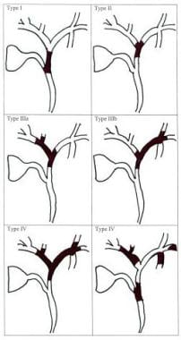
- Bismuth classification for perihilar cholangiocarcinoma. (medscape.com)
- The treatment for perihilar cholangiocarcinoma (PHC) is a highly invasive surgery. (nih.gov)
Extrahepatic cholangiocarcinoma6
- It can be classified according to anatomical location as intrahepatic cholangiocarcinoma (ICC) or extrahepatic cholangiocarcinoma (ECC). (lww.com)
- Extrahepatic cholangiocarcinoma occurs in ducts outside of the liver, closest to the small intestine. (mesothelioma.net)
- What is the difference between intrahepatic cholangiocarcinoma and extrahepatic cholangiocarcinoma? (mypathologyreport.ca)
- Intrahepatic and extrahepatic cholangiocarcinoma are very similar types of cancer. (mypathologyreport.ca)
- The main difference is that intrahepatic cholangiocarcinoma starts from one of the small bile ducts found inside the liver while extrahepatic cholangiocarcinoma starts from one of the larger bile ducts found outside the liver. (mypathologyreport.ca)
- Extrahepatic cholangiocarcinoma frequently causes jaundice (yellowing of the eyes and skin). (mypathologyreport.ca)
Treated advanced cholangiocarcinoma1
- The Food and Drug Administration approved Lytgobi for patients with previously treated advanced cholangiocarcinoma with FGFR2 gene rearrangements. (curetoday.com)
Diagnosis9
- Cholangiocarcinoma is typically incurable at diagnosis which is why early detection is ideal. (wikipedia.org)
- Cholangiocarcinoma (CCA) encompasses diverse epithelial tumors historically associated with poor outcomes due to an aggressive disease course, late diagnosis, and limited benefit of standard chemotherapy for advanced disease. (nih.gov)
- As most patients present with non resectable disease at the time of diagnosis, palliative modalities have a crucial role in the treatment of cholangiocarcinoma. (nursingcenter.com)
- Approximately 8,000 people per year receive a diagnosis of cholangiocarcinoma, compared to 3,000 mesothelioma diagnoses in the U.S. Bile duct cancer is more common in southeast Asia due to a parasite. (mesothelioma.net)
- The diagnosis of cholangiocarcinoma was not histologically confirmed in all the cases. (bmj.com)
- The diagnosis of cholangiocarcinoma can be made after tissue is examined under the microscope by a pathologist. (mypathologyreport.ca)
- And if what I eat matters - then how does one identify foods which are recommended for Intrahepatic Cholangiocarcinoma and is it the same answer for everyone with the same diagnosis or genetic risk? (addon.life)
- Among patients treated with pancreatoduodenectomy, diagnosis was pancreatic ductal adenocarcinoma in 1380 (50.0 per cent), distal cholangiocarcinoma in 284 (10.3 per cent), ampullary cancer in 376 (13.6 per cent), duodenal cancer in 160 (5.8 per cent) and other diagnoses in 560 (20.3 per cent) patients. (lu.se)
- The preoperative diagnosis corresponded to the postoperative in 1177 (67.5 per cent) patients for pancreatic ductal adenocarcinoma, 162 (37.4 per cent) patients for distal cholangiocarcinoma, 220 (61.3 per cent) patients for ampullary cancer and 120 (53.6 per cent) patients for duodenal cancer. (lu.se)
Cancer of th2
- Cholangiocarcinoma is a cancer of the bile ducts. (mesothelioma.net)
- As a whole, cholangiocarcinoma is an aggressive cancer of the bile ducts and is diagnosed in approximately 8,000 individuals each year in the U.S. 1 This includes both intrahepatic (inside the liver) and extrahepatic (outside the liver) forms of the disease. (biospace.com)
Cure for cholangiocarcinoma2
- Complete surgical resection is the only therapy to afford a chance of cure for cholangiocarcinoma. (medscape.com)
- We are excited to work with CCF and support their efforts in finding a cure for cholangiocarcinoma," said Premal Shah , President and Co-Founder of Ciitizen. (prnewswire.com)
Tumors4
- These tumors are also known as intrahepatic cholangiocarcinomas. (tri-kobe.org)
- Tumors of this region are also known as perihilar cholangiocarcinomas or Klatskin tumors. (tri-kobe.org)
- Tumors of this region are also known as extrahepatic cholangiocarcinomas (Figure 2 ). (tri-kobe.org)
- Cholangiocarcinomas (CCAs) are rare and aggressive tumors that display features of biliary differentiation. (uni-luebeck.de)
Cancers4
- At the 4th Annual Cholangiocarcinoma Summit, held on October 13-15, 2022, in Aurora, CO, experts in the field of gastroenterology, interventional radiology, nursing, and surgery discussed the importance of using a multidisciplinary approach to supportive care and the management of complications in patients with biliary tract cancers (BTCs). (valuebasedcancer.com)
- A very common nutrition question asked by cancer patients and individuals at-genetic risk of cancer is - for cancers like Intrahepatic Cholangiocarcinoma does it matter what foods I eat and which I do not? (addon.life)
- All cancers like Intrahepatic Cholangiocarcinoma can be characterized by a unique set of biochemical pathways - the signature pathways of Intrahepatic Cholangiocarcinoma. (addon.life)
- Background: The prevalence of different periampullary cancers (pancreatic ductal adenocarcinoma, distal cholangiocarcinoma, ampullary cancer and duodenal cancer) is heterogeneous in the literature. (lu.se)
Patients24
- For inflammatory bowel disease patients with altered DNA repair functions, the progression from PSC to cholangiocarcinoma may be a consequence of DNA damage resulting from biliary inflammation and bile acids. (wikipedia.org)
- Additionally, our compassionate oncology nurses, social workers and dietitians offer a full range of supportive care designed specifically for patients who are being treated for cholangiocarcinoma. (moffitt.org)
- Through our robust clinical trials, we offer our patients opportunities to be among the first to benefit from promising new treatments for cholangiocarcinoma, such as anti-angiogenesis therapies, epidermal growth factor receptor (EGFR) inhibitors, photodynamic therapy (PDT), molecular therapy, immunotherapy, hyperthermia therapy and radiosensitizers. (moffitt.org)
- PDT is effective in restoring biliary drainage and improving quality of life in patients with nonresectable disseminated cholangiocarcinomas. (medscape.com)
- A study of 66 patients with unresectable intrahepatic cholangiocarcinoma treated with hypofractionated radiation therapy reported 2-year outcomes of 84% local control and 58% overall survival. (medscape.com)
- In a study of 1636 patients with unresectable localized intrahepatic cholangiocarcinoma, the addition of radiation to chemotherapy was associated with an improvement in overall survival. (medscape.com)
- The approval is based on findings from the TAS-120-101 clinical trial, which showed 42% of patients with previously treated, unresectable, locally advanced, or metastatic intrahepatic cholangiocarcinoma harboring a FGFR2 gene fusion or other rearrangement have their disease shrink as a result of treatment (a statistic called overall response rate). (curetoday.com)
- However, one expert explained that the majority of patients with cholangiocarcinoma will still receive upfront standard of care , namely Gemzar (gemcitabine) and cisplatin. (curetoday.com)
- PALO ALTO, Calif. , Aug. 12, 2019 /PRNewswire/ -- Ciitizen , a consumer health tech company building a platform that helps patients collect, organize, and share their medical records digitally, and the Cholangiocarcinoma Foundation (CCF), whose mission is to find a cure and improve the quality of life for those affected by cholangiocarcinoma, have formed a partnership. (prnewswire.com)
- PRINCETON, N.J., Sept. 30, 2022 /PRNewswire/ -- Taiho Oncology, Inc. and Taiho Pharmaceutical Co., Ltd. announced today that the U.S. Food and Drug Administration (FDA) has approved LYTGOBI ® tablets for the treatment of adult patients with previously treated, unresectable, locally advanced or metastatic intrahepatic cholangiocarcinoma (iCCA) harboring fibroblast growth factor receptor 2 (FGFR2) gene fusions or other rearrangements. (biospace.com)
- LYTGOBI is indicated for the treatment of adult patients with previously treated, unresectable, locally advanced or metastatic intrahepatic cholangiocarcinoma harboring fibroblast growth factor receptor 2 (FGFR2) gene fusions or other rearrangements. (biospace.com)
- This triplet was evaluated in the open-label multicenter phase Ib/II S4-13-001 study of 88 patients with previously untreated locally advanced unresectable or metastatic cholangiocarcinoma. (ascopost.com)
- Silmitasertib, a novel inhibitor of casein kinase 2 (CK2), was combined with gemcitabine and cisplatin (the standard of care) in patients with previously untreated advanced cholangiocarcinoma. (ascopost.com)
- In a cohort study reported in JAMA Surgery , Franssen et al found little difference in median overall survival with hepatic arterial infusion pump (HAIP) floxuridine chemotherapy vs resection in patients with multifocal intrahepatic cholangiocarcinoma. (ascopost.com)
- This cohort study found that patients with multifocal intrahepatic cholangiocarcinoma had similar overall survival after HAIP floxuridine chemotherapy vs resection. (ascopost.com)
- I believe that LYTGOBI may be part of a new era in the treatment of CCA, one in which the power of personalised medicine may touch the lives of patients in ways we haven't seen before with traditional chemotherapy," said Helen Morement, CEO of the AAMF - The Cholangiocarcinoma Charity and the UK's only charity dedicated to this cause. (pipelinereview.com)
- Cholangiocarcinoma in patients with primary sclerosing cholangitis: a multicenter case-control study. (medscape.com)
- Approximately 90% of patients diagnosed with cholangiocarcinoma in Western countries do not have a recognized risk factor. (cdc.gov)
- First-line standard of care for patients with advanced cholangiocarcinoma has evolved from a chemotherapy-only regimen with gemcitabine/cisplatin to include the immune checkpoint inhibitor durvalumab based on results from TOPAZ-1 showing that the addition of durvalumab improved progression-free survival and overall survival compared with GemCis alone. (valuebasedcancer.com)
- Personalized medicine has expanded the treatment options for patients with cholangiocarcinoma (CCA). (valuebasedcancer.com)
- At the 2021 Annual Meeting of the Cholangiocarcinoma Foundation (CCF), Ghassan K. Abou-Alfa, MD, MBA, Professor of Medicine, Memorial Sloan Kettering Cancer Center, New York, NY, discussed recent developments in personalized therapies, highlighting genomic alterations that are informing the new therapies for patients with CCA. (valuebasedcancer.com)
- This chapter details all aspects of the general principles of palliative care for advanced stage cholangiocarcinoma patients . (bvsalud.org)
- AJCC 7th edition of TNM staging accurately discriminates outcomes of patients with resectable intrahepatic cholangiocarcinoma: By the AFC-IHCC-2009 study group. (cancercentrum.se)
- The value of F-18 FDG PET in patients with primary sclerosing cholangitis and cholangiocarcinoma using visual and semiquantitative analysis. (cancercentrum.se)
Treat cholangiocarcinoma2
- PDT is a relatively new local, minimally invasive procedure that can be used to treat cholangiocarcinoma. (nursingcenter.com)
- Today's approval will add to the list of targeted drugs available to treat cholangiocarcinoma. (curetoday.com)
Causes cholangiocarcinoma2
- What Causes Cholangiocarcinoma? (mesothelioma.net)
- Experts do not fully understand what causes cholangiocarcinoma. (mesothelioma.net)
Symptoms3
- Call your provider if you have jaundice or other symptoms of cholangiocarcinoma. (medlineplus.gov)
- What are the symptoms of cholangiocarcinoma? (mypathologyreport.ca)
- The most common symptoms of cholangiocarcinoma are abdominal pain, weight loss, and weakness. (mypathologyreport.ca)
Hepatic2
- Cholangiocarcinoma is a high-mortality primary hepatic malignancy. (omicsonline.org)
- Singal AK, Vauthey JN, Grady JJ, Stroehlein JR. Intra-hepatic cholangiocarcinoma--frequency and demographic patterns: thirty-year data from the M.D. Anderson Cancer Center. (medscape.com)
Treatment6
- Bile duct cancer (cholangiocarcinoma) treatment (PDQ) -- health professional version. (medlineplus.gov)
- Moffitt Cancer Center takes a multispecialty approach to the treatment of cholangiocarcinoma. (moffitt.org)
- To learn more about treatment options for cholangiocarcinoma, call 1-888-663-3488 or request an appointment by completing a new patient registration form online. (moffitt.org)
- PDT as a treatment for cholangiocarcinoma has been shown to increase survival. (nursingcenter.com)
- Cholangiocarcinoma is an aggressive type of liver cancer with few effective treatment options. (springer.com)
- Implications of CA19-9 elevation for survival, staging, and treatment sequencing in intrahepatic cholangiocarcinoma: A national cohort analysis. (medscape.com)
Aggressive2
- Cholangiocarcinoma (CCA) is an aggressive type of liver cancer, which arises from the epithelium of the biliary tract. (springer.com)
- Cholangiocarcinoma (CCA), a rare and aggressive form of cancer, has seen a steady increase in incidence in the United States, with a predominant increase in intrahepatic CCA, from 2001 to 2017, according to a recent epidemiological study. (valuebasedcancer.com)
Chronic3
- People with chronic liver disease, whether in the form of viral hepatitis (e.g. hepatitis B or hepatitis C), alcoholic liver disease, or cirrhosis of the liver due to other causes, are at significantly increased risk of cholangiocarcinoma. (wikipedia.org)
- Intrahepatic cholangiocarcinoma may be associated with chronic ulcerative colitis and chronic cholecystitis . (medscape.com)
- Small duct cholangiocarcinoma is caused by chronic viral infections of the liver (e.g., hepatitis B and hepatitis C) and excessive alcohol consumption, both of which are associated with cirrhosis. (mypathologyreport.ca)
Bile duct14
- Cholangiocarcinoma, also known as bile duct cancer, is a type of cancer that forms in the bile ducts. (wikipedia.org)
- Cholangiocarcinoma is a type of cancer that develops within the bile duct system, which is a series of small tubes that transport bile fluid from the liver (where it is produced) to the gallbladder (where it is stored). (moffitt.org)
- Based on where the cancer initially develops within the bile duct system, cholangiocarcinoma is classified into three general categories. (moffitt.org)
- Distal cholangiocarcinomas are found further down the bile duct, closer to the small intestine. (moffitt.org)
- The majority of cholangiocarcinomas are adenocarcinomas that develop within the mucous gland cells that line the inside of the bile duct. (moffitt.org)
- Cholangiocarcinoma is bile duct cancer, a serious and potentially life-threatening type of cancer. (mesothelioma.net)
- One study that examined thousands of Europeans diagnosed with bile duct cancer discovered that those exposed to asbestos in the workplace were more likely to develop cholangiocarcinoma, specifically intrahepatic bile duct cancer. (mesothelioma.net)
- Founded in 2006, the Cholangiocarcinoma Foundation is a global 501(c) (3) non-profit organization whose mission is to find a cure and improve the quality of life for those affected by bile duct cancer. (prnewswire.com)
- This clinical trial report, "Bile Duct Cancer (Cholangiocarcinoma) Global Clinical Trials Review, H1, 2020" provides an overview of Bile Duct Cancer (Cholangiocarcinoma) Clinical trials scenario. (researchandmarkets.com)
- This report provides top line data relating to the clinical trials on Bile Duct Cancer (Cholangiocarcinoma). (researchandmarkets.com)
- Cholangiocarcinoma (CCA) is a fundamental adenocarcinoma and malignancy arising from the epithelium of the bile duct. (scholarsresearchlibrary.com)
- Cancer of the bile duct (also called cholangiocarcinoma) is extremely rare. (tri-kobe.org)
- Cholangiocarcinoma is a type of bile duct cancer. (mypathologyreport.ca)
- Metastatic cholangiocarcinoma means that tumour cells have spread from the bile duct to another part of the body such as a lymph node or the lungs. (mypathologyreport.ca)
Malignancies2
- Cholangiocarcinomas (CCCs) are malignancies of the biliary duct system that may originate in the liver and extrahepatic bile ducts, which terminate at the ampulla of Vater. (medscape.com)
- BRCA-associated hereditary breast and ovarian cancer syndrome (HBOC) is characterized by an increased risk of developing other malignancies including cholangiocarcinoma (CCA). (frontiersin.org)
Chemotherapy1
- Cholangiocarcinoma (CC) is typically diagnosed at an advanced stage and is refractory to surgical intervention and chemotherapy. (jci.org)
Subtypes1
- Subtypes of cholangiocarcinoma are differentiated based on their location in the biliary tree. (mdpi.com)
Liver fluke infection2
- In the absence of detection of liver flukes, there is no test available that can determine if liver fluke infection is the underlying cause of cholangiocarcinoma or other hepatobiliary conditions. (cdc.gov)
- Large duct cholangiocarcinoma is caused by primary sclerosing cholangitis, hepatolithiasis (gallstones in the large bile ducts), and liver fluke infection. (mypathologyreport.ca)
Hepatobiliary cancer1
- Cholangiocarcinoma (CCA) is a type of hepatobiliary cancer characterized by uncontrolled cell proliferation, with a poor prognosis and high mortality. (ijbs.com)
Incidence2
- Control programs (Integrated Opisthorchiasis Control Program) aimed at discouraging the consumption of raw and undercooked food have been successful at reducing the incidence of cholangiocarcinoma in some countries. (wikipedia.org)
- A higher incidence of cholangiocarcinoma was reported in Asia, especially Southeast Asia, than in Western countries. (omicsonline.org)
Prognosis1
- More than one tumour increases the tumour stage (pT) for intrahepatic cholangiocarcinoma and is associated with a worse prognosis . (mypathologyreport.ca)
Malignant1
- Cholangiocarcinoma (CCA) is a rare cancerous (malignant) growth in one of the ducts that carries bile from the liver to the small intestine. (medlineplus.gov)
Malignancy1
- Objective Cholangiocarcinoma (CCA) is the most common malignancy in a Northeast Thai population. (bmj.com)
Primary4
- Risk factors for cholangiocarcinoma include primary sclerosing cholangitis (an inflammatory disease of the bile ducts), ulcerative colitis, cirrhosis, hepatitis C, hepatitis B, infection with certain liver flukes, and some congenital liver malformations. (wikipedia.org)
- Intrahepatic cholangiocarcinoma (iCCA) is a primary liver cancer epidemiologically linked with liver injury, which has poorly understood incipient stages and lacks early diagnostics and effective therapies. (aacrjournals.org)
- Detection of cholangiocarcinoma in primary sclerosing cholangitis by positron emission tomography. (medscape.com)
- Cholangiocarcinoma in primary sclerosing cholangitis. (cancercentrum.se)
Risk factors2
- Although most people present without any known risk factors evident, a number of risk factors for the development of cholangiocarcinoma have been described. (wikipedia.org)
- There are some known risk factors for cholangiocarcinoma. (mesothelioma.net)
Biliary duct system1
- Cholangiocarcinoma (CCA) is a rare cancer of the biliary duct system, which is comprised of the gallbladder and bile ducts. (cdc.gov)
Gallbladder1
- Each year, approximately 2500 cases of cholangiocarcinoma occur, compared with 5000 cases of gallbladder cancer and 15,000 cases of hepatocellular cancer. (medscape.com)
Consensus Conference1
- The recent European HepatoPancreatoBiliary Association Consensus Conference on Cholangiocarcinoma guidelines have also been used as a source. (bmj.com)
Glands3
- Well differentiated cholangiocarcinoma - More than 95% of the tumour is made up of glands. (mypathologyreport.ca)
- Moderately differentiated cholangiocarcinoma - 50 to 95% of the tumour is made up of glands. (mypathologyreport.ca)
- Poorly differentiated cholangiocarcinoma - Less than 50% of the tumour is made up of glands. (mypathologyreport.ca)
Epidemiology1
- Epidemiology of cholangiocarcinoma (CCA) is high in Thailand and Southeast Asia. (scholarsresearchlibrary.com)
Proliferation1
- Sun, X. Nobiletin suppresses cholangiocarcinoma proliferation via inhibiting GSK3β. (ijbs.com)
Typically2
- Small duct cholangiocarcinoma is typically found near the periphery (outside) of the liver. (mypathologyreport.ca)
- In contrast, large duct cholangiocarcinoma typically involves one of the large bile ducts near the hilum (middle) of the liver. (mypathologyreport.ca)
Presented at the 20211
- Key topics related to intrahepatic cholangiocarcinoma (CCA) were presented at the 2021 Society of Interventional Oncology (SIO) and the Society of Interventional Radiology (SIR) meetings and were discussed during the CCA Summit. (valuebasedcancer.com)
Type1
- The causes associated with cholangiocarcinoma vary by type. (mypathologyreport.ca)