Cell Movement
Movement
Gastrulation
Gastrula
Morphogenesis
Zebrafish
Dictyostelium
Chemotaxis
Zebrafish Proteins
Pseudopodia
Head Movements
Cell Polarity
Primitive Streak
Embryo, Nonmammalian
Movement Disorders
Body Patterning
Actins
Time-Lapse Imaging
Models, Biological
Microscopy, Video
Signal Transduction
Plant Viral Movement Proteins
Cytoskeleton
Cell Surface Extensions
Gene Expression Regulation, Developmental
Fetal Movement
Mesoderm
Psychomotor Performance
Video Recording
Cell Aggregation
Motion Pictures as Topic
Wnt Proteins
Xenopus Proteins
Chick Embryo
Microfilament Proteins
Saccades
Myxococcus xanthus
Somites
Electromyography
Biomechanical Phenomena
Cadherins
rac GTP-Binding Proteins
Mutation
Notochord
Cell Communication
In Situ Hybridization
Hand
rhoA GTP-Binding Protein
Green Fluorescent Proteins
Adherens Junctions
Molecular Sequence Data
Fixation, Ocular
Xenopus laevis
Cells, Cultured
Microscopy, Fluorescence
Wiskott-Aldrich Syndrome Protein Family
Actin Cytoskeleton
Microtubules
Oligoribonucleotides, Antisense
Cell Membrane
Microscopy, Phase-Contrast
Motor Cortex
Xenopus
rho GTP-Binding Proteins
Pursuit, Smooth
Neural Crest
Focal Adhesions
Animals, Genetically Modified
Luminescent Proteins
Receptors, Eph Family
NIH 3T3 Cells
Chemotactic Factors
Blastoderm
Drosophila
Drosophila Proteins
Metencephalon
Microscopy, Confocal
Organizers, Embryonic
Microscopy, Interference
Amino Acid Sequence
Myosin Type II
Photic Stimulation
Periodicity
Embryonic Induction
Membrane Proteins
Cell Lineage
Neurons
Extracellular Matrix
Blastula
Cytoskeletal Proteins
Embryo, Mammalian
Rotation
Fibroblasts
cdc42 GTP-Binding Protein
Cell Adhesion Molecules
Nerve Tissue Proteins
Gene Knockdown Techniques
Fibroblast Growth Factors
Guanine Nucleotide Exchange Factors
Phenotype
Myxococcales
Adaptor Proteins, Signal Transducing
Computer Simulation
Microscopy, Electron, Scanning
Seminiferous Epithelium
Proprioception
Intercellular Junctions
Rhombencephalon
Myosins
Ephrins
Organogenesis
Macaca mulatta
Functional Laterality
Receptor, EphA4
Antigens, CD29
Cell Division
Epithelial Cells
Actomyosin
Tail
Fibronectins
Epithelium
Cell Tracking
Electrooculography
Protein Transport
Recombinant Fusion Proteins
Chemotaxis, Leukocyte
Sleep, REM
Phosphoproteins
Actin-Related Protein 2-3 Complex
Feedback, Sensory
rac1 GTP-Binding Protein
Integrins
Blood-Testis Barrier
Carrier Proteins
Protein Binding
rho-Associated Kinases
Image Processing, Computer-Assisted
Blastomeres
Cytoplasm
Protein Structure, Tertiary
Kinesthesis
Cortactin
Locomotion
Cell Differentiation
Focal Adhesion Protein-Tyrosine Kinases
Proto-Oncogene Proteins c-fyn
Microinjections
Vertebrates
Germ Layers
Cyclic AMP
LIM Domain Proteins
Nervous System
Actin Depolymerizing Factors
Reflex, Vestibulo-Ocular
Transcription Factors
Caenorhabditis elegans
Visual Perception
Phosphorylation
Homeodomain Proteins
Dyskinesias
Muscle, Skeletal
Immunohistochemistry
Base Sequence
Microscopy, Electron
Monomeric GTP-Binding Proteins
Motion
Intracellular Signaling Peptides and Proteins
Laminin
Fluorescent Antibody Technique
Head
Glycoproteins
Immunoprecipitation
Nonbehavioral selection for pawns, mutants of Paramecium aurelia with decreased excitability. (1/32044)
The reversal response in Paramecium aurelia is mediated by calcium which carries the inward current during excitation. Electrophysiological studies indicate that strontium and barium can also carry the inward current. Exposure to high concentrations of barium rapidly paralyzes and later kills wild-type paramecia. Following mutagenesis with nitrosoguanidine, seven mutants which continued to swim in the ;high-barium' solution were selected. All of the mutants show decreased reversal behavior, with phenotypes ranging from extremely non-reversing (;extreme' pawns) to nearly wild-type reversal behavior (;partial' pawns). The mutations fall into three complementation groups, identical to the pwA, pwB, and pwC genes of Kunget al. (1975). All of the pwA and pwB mutants withstand longer exposure to barium, the pwB mutants surviving longer than the pwA mutants. Among mutants of each gene, survival is correlated with loss of reversal behavior. Double mutants (A-B, A-C, B-C), identified in the exautogamous progeny of crosses between ;partial' mutants, exhibited a more extreme non-reversing phenotype than either of their single-mutant (;partial' pawn) parents.---Inability to reverse could be expected from an alteration in the calcium-activated reversal mechanism or in excitation. A normal calcium-activated structure was demonstrated in all pawns by chlorpromazine treatment. In a separate report (Schein, Bennett and Katz 1976) the results of electrophysiological investigations directly demonstrate decreased excitability in all of the mutants, a decrease due to an altered calcium activation. The studies of the genetics, the survival in barium and the electro-physiology of the pawns demonstrate that the pwA and pwB genes have different effects on calcium activation. (+info)Polarized distribution of Bcr-Abl in migrating myeloid cells and co-localization of Bcr-Abl and its target proteins. (2/32044)
Bcr-Abl plays a critical role in the pathogenesis of Philadelphia chromosome-positive leukemia. Although a large number of substrates and interacting proteins of Bcr-Abl have been identified, it remains unclear whether Bcr-Abl assembles multi-protein complexes and if it does where these complexes are within cells. We have investigated the localization of Bcr-Abl in 32D myeloid cells attached to the extracellular matrix. We have found that Bcr-Abl displays a polarized distribution, colocalizing with a subset of filamentous actin at trailing portions of migrating 32D cells, and localizes on the cortical F-actin and on vesicle-like structures in resting 32D cells. Deletion of the actin binding domain of Bcr-Abl (Bcr-AbI-AD) dramatically enhances the localization of Bcr-Abl on the vesicle-like structures. These distinct localization patterns of Bcr-Abl and Bcr-Abl-AD enabled us to examine the localization of Bcr-Abl substrate and interacting proteins in relation to Bcr-Abl. We found that a subset of biochemically defined target proteins of Bcr-Abl redistributed and co-localized with Bcr-Abl on F-actin and on vesicle-like structures. The co-localization of signaling proteins with Bcr-Abl at its sites of localization supports the idea that Bcr-Abl forms a multi-protein signaling complex, while the polarized distribution and vesicle-like localization of Bcr-Abl may play a role in leukemogenesis. (+info)The LIM-only protein PINCH directly interacts with integrin-linked kinase and is recruited to integrin-rich sites in spreading cells. (3/32044)
PINCH is a widely expressed and evolutionarily conserved protein comprising primarily five LIM domains, which are cysteine-rich consensus sequences implicated in mediating protein-protein interactions. We report here that PINCH is a binding protein for integrin-linked kinase (ILK), an intracellular serine/threonine protein kinase that plays important roles in the cell adhesion, growth factor, and Wnt signaling pathways. The interaction between ILK and PINCH has been consistently observed under a variety of experimental conditions. They have interacted in yeast two-hybrid assays, in solution, and in solid-phase-based binding assays. Furthermore, ILK, but not vinculin or focal adhesion kinase, has been coisolated with PINCH from mammalian cells by immunoaffinity chromatography, indicating that PINCH and ILK associate with each other in vivo. The PINCH-ILK interaction is mediated by the N-terminal-most LIM domain (LIM1, residues 1 to 70) of PINCH and multiple ankyrin (ANK) repeats located within the N-terminal domain (residues 1 to 163) of ILK. Additionally, biochemical studies indicate that ILK, through the interaction with PINCH, is capable of forming a ternary complex with Nck-2, an SH2/SH3-containing adapter protein implicated in growth factor receptor kinase and small GTPase signaling pathways. Finally, we have found that PINCH is concentrated in peripheral ruffles of cells spreading on fibronectin and have detected clusters of PINCH that are colocalized with the alpha5beta1 integrins. These results demonstrate a specific protein recognition mechanism utilizing a specific LIM domain and multiple ANK repeats and suggest that PINCH functions as an adapter protein connecting ILK and the integrins with components of growth factor receptor kinase and small GTPase signaling pathways. (+info)Transduction of glioma cells using a high-titer retroviral vector system and their subsequent migration in brain tumors. (4/32044)
The intracranial migration of transduced glioma cells was investigated in order to improve the treatment of malignant glioma by gene therapy using retroviral vectors. In this study, about half the volume of the tumor mass could be transduced in 14 days after only a single implantation of 3 x 10(5) retrovirus-producing cells into a tumor mass with a diameter of 5 mm. Moreover, we were able to follow the migration of glioma cells transduced by the lacZ-harboring retroviruses originating from the high-titer retrovirus-producing cells. Besides the importance of using a high-titer retroviral vector system, our results also indicate that the implantation site of the virus-producing cells and the interval between the implantation of the virus-producing cells and the subsequent administration of ganciclovir are important factors for the efficient killing of glioma cells. (+info)Prolonged eosinophil accumulation in allergic lung interstitium of ICAM-2 deficient mice results in extended hyperresponsiveness. (5/32044)
ICAM-2-deficient mice exhibit prolonged accumulation of eosinophils in lung interstitium concomitant with a delayed increase in eosinophil numbers in the airway lumen during the development of allergic lung inflammation. The ICAM-2-dependent increased and prolonged accumulation of eosinophils in lung interstitium results in prolonged, heightened airway hyperresponsiveness. These findings reveal an essential role for ICAM-2 in the development of the inflammatory and respiratory components of allergic lung disease. This phenotype is caused by the lack of ICAM-2 expression on non-hematopoietic cells. ICAM-2 deficiency on endothelial cells causes reduced eosinophil transmigration in vitro. ICAM-2 is not essential for lymphocyte homing or the development of leukocytes, with the exception of megakaryocyte progenitors, which are significantly reduced. (+info)Anti-monocyte chemoattractant protein-1/monocyte chemotactic and activating factor antibody inhibits neointimal hyperplasia in injured rat carotid arteries. (6/32044)
Monocyte chemoattractant protein-1 (MCP-1)/monocyte chemotactic and activating factor (MCAF) has been suggested to promote atherogenesis. The effects of in vivo neutralization of MCP-1 in a rat model were examined in an effort to clarify the role of MCP-1 in the development of neointimal hyperplasia. Competitive polymerase chain reaction analysis revealed maximum MCP-1 mRNA expression at 4 hours after carotid arterial injury. Increased immunoreactivities of MCP-1 were also detected at 2 and 8 hours after injury. Either anti-MCP-1 antibody or nonimmunized goat IgG (10 mg/kg) was then administered every 12 hours to rats that had undergone carotid arterial injury. Treatment with 3 consecutive doses of anti-MCP-1 antibody within 24 hours (experiment 1) and every 12 hours for 5 days (experiment 2) significantly inhibited neointimal hyperplasia at day 14, resulting in a 27.8% reduction of the mean intima/media ratio (P<0.05) in experiment 1 and a 43.6% reduction (P<0.01) in experiment 2. This effect was still apparent at day 56 (55.6% inhibition; P<0.05). The number of vascular smooth muscle cells in the neointima at day 4 was significantly reduced by anti-MCP-1 treatment, demonstrating the important role of MCP-1 in early neointimal lesion formation. However, recombinant MCP-1 did not stimulate chemotaxis of vascular smooth muscle cells in an in vitro migration assay. These results suggest that MCP-1 promotes neointimal hyperplasia in early neointimal lesion formation and that neutralization of MCP-1 before, and immediately after, arterial injury may be effective in preventing restenosis after angioplasty. Further studies are needed to clarify the mechanism underlying the promotion of neointimal hyperplasia by MCP-1. (+info)Non-serum-dependent chemotactic factors produced by Candida albicans stimulate chemotaxis by binding to the formyl peptide receptor on neutrophils and to an unknown receptor on macrophages. (7/32044)
Serum-free culture filtrates of six Candida species and Saccharomyces cerevisiae were found to contain chemoattractants for human polymorphonuclear leukocytes (PMNs) and a mouse macrophage-like cell line, J774. The chemotactic factors differed for the PMN and J774 cells, however, in terms of heat stability, kinetics of liberation by the yeast cells, and divalent cation requirements for production. The chemoattractant in Candida albicans culture filtrates appeared to act through the formyl peptide receptor (FPR) of PMNs, since it was found to induce chemotaxis of Chinese hamster ovary (CHO) cells that were expressing the human FPR but did not induce chemotaxis of wild-type CHO cells. The C. albicans culture filtrates also induced migration of PMNs across confluent monolayers of a human gastrointestinal epithelial cell line, T84; migration occurred in the basolateral-to-apical direction but not the reverse direction, unless the epithelial tight junctions were disrupted. J774 cells did not migrate toward the formylated peptide (fMet-Leu-Phe; fMLF), and chemotaxis toward the C. albicans culture filtrate was not inhibited by an FPR antagonist (t-butoxycarbonyl-Met-Leu-Phe), suggesting that a different receptor mediated J774 cell chemotaxis. In conclusion, we have identified a receptor by which a non-serum-dependent chemotactic factor (NSCF) produced by C. albicans induced chemotaxis of PMNs. Additionally, we have shown that NSCF was active across epithelial monolayers. These findings suggest that NSCFs produced by C. albicans and other yeast species may influence host-pathogen interactions at the gastrointestinal tract mucosal surface by inducing phagocytic-cell infiltration. (+info)Role of the extracellular signal-regulated protein kinase cascade in human neutrophil killing of Staphylococcus aureus and Candida albicans and in migration. (8/32044)
Killing of Staphylococcus aureus and Candida albicans by neutrophils involves adherence of the microorganisms, phagocytosis, and a collaborative action of oxygen reactive species and components of the granules. While a number of intracellular signalling pathways have been proposed to regulate neutrophil responses, the extent to which each pathway contributes to the killing of S. aureus and C. albicans has not been clearly defined. We have therefore examined the effect of blocking one such pathway, the extracellular signal-regulated protein kinase (ERK) cascade, using the specific inhibitor of the mitogen-activated protein kinase/ERK kinase, PD98059, on the ability of human neutrophils to kill S. aureus and C. albicans. Our data demonstrate the presence of ERK2 and a 43-kDa form of ERK but not ERK1 in human neutrophils. Upon stimulation with formyl methionyl leucyl phenylalanine (fMLP), the activities of both ERK2 and the 43-kDa form were stimulated. Despite abrogating the activity of both ERK forms, PD98059 only slightly reduced the ability of neutrophils to kill S. aureus or C. albicans. This is consistent with our finding that PD98059 had no effect on neutrophil adherence or degranulation, although pretreatment of neutrophils with PD98059 inhibited fMLP-stimulated superoxide production by 50%, suggesting that a change in superoxide production per se is not strictly correlated with microbicidal activity. However, fMLP-stimulated chemokinesis was markedly inhibited, while random migration and fMLP-stimulated chemotaxis were partially inhibited, by PD98059. These data demonstrate, for the first time, that the ERK cascade plays only a minor role in the microbicidal activity of neutrophils and that the ERK cascade is involved primarily in regulating neutrophil migration in response to fMLP. (+info)Cell movement, also known as cell motility, refers to the ability of cells to move independently and change their location within tissue or inside the body. This process is essential for various biological functions, including embryonic development, wound healing, immune responses, and cancer metastasis.
There are several types of cell movement, including:
1. **Crawling or mesenchymal migration:** Cells move by extending and retracting protrusions called pseudopodia or filopodia, which contain actin filaments. This type of movement is common in fibroblasts, immune cells, and cancer cells during tissue invasion and metastasis.
2. **Amoeboid migration:** Cells move by changing their shape and squeezing through tight spaces without forming protrusions. This type of movement is often observed in white blood cells (leukocytes) as they migrate through the body to fight infections.
3. **Pseudopodial extension:** Cells extend pseudopodia, which are temporary cytoplasmic projections containing actin filaments. These protrusions help the cell explore its environment and move forward.
4. **Bacterial flagellar motion:** Bacteria use a whip-like structure called a flagellum to propel themselves through their environment. The rotation of the flagellum is driven by a molecular motor in the bacterial cell membrane.
5. **Ciliary and ependymal movement:** Ciliated cells, such as those lining the respiratory tract and fallopian tubes, have hair-like structures called cilia that beat in coordinated waves to move fluids or mucus across the cell surface.
Cell movement is regulated by a complex interplay of signaling pathways, cytoskeletal rearrangements, and adhesion molecules, which enable cells to respond to environmental cues and navigate through tissues.
In the context of medicine and healthcare, "movement" refers to the act or process of changing physical location or position. It involves the contraction and relaxation of muscles, which allows for the joints to move and the body to be in motion. Movement can also refer to the ability of a patient to move a specific body part or limb, which is assessed during physical examinations. Additionally, "movement" can describe the progression or spread of a disease within the body.
Gastrulation is a fundamental process in embryonic development, characterized by the transformation of a initially flat layer of cells called the blastula into a three-layered structure known as the gastrula. This complex series of cellular movements and rearrangements establishes the foundation for the formation of the three primary germ layers: the ectoderm, mesoderm, and endoderm. These germ layers further differentiate to give rise to all the diverse cell types and tissues in the developing organism, including the nervous system, muscles, bones, and internal organs.
The precise mechanisms of gastrulation vary among different animal groups; however, common features include:
1. Formation of a blastopore: A small indentation or opening that forms on the surface of the blastula, which eventually develops into the primitive gut or anus in the gastrula.
2. Invagination: The process by which cells at the blastopore fold inward and migrate towards the interior of the embryo, forming the endodermal layer.
3. Epiboly: A coordinated movement of cells that spreads over and encloses the yolk within the embryo, contributing to the formation of the ectodermal layer.
4. Delamination: The separation and migration of cells from the epiblast (the outer layer of the blastula) to form the mesodermal layer in between the ectoderm and endoderm.
Gastrulation is a critical period in embryonic development, as errors during this process can lead to severe congenital abnormalities or even embryonic lethality. A thorough understanding of gastrulation has important implications for regenerative medicine, stem cell research, and the study of evolutionary developmental biology (Evo-Devo).
Eye movements, also known as ocular motility, refer to the voluntary or involuntary motion of the eyes that allows for visual exploration of our environment. There are several types of eye movements, including:
1. Saccades: rapid, ballistic movements that quickly shift the gaze from one point to another.
2. Pursuits: smooth, slow movements that allow the eyes to follow a moving object.
3. Vergences: coordinated movements of both eyes in opposite directions, usually in response to a three-dimensional stimulus.
4. Vestibulo-ocular reflex (VOR): automatic eye movements that help stabilize the gaze during head movement.
5. Optokinetic nystagmus (OKN): rhythmic eye movements that occur in response to large moving visual patterns, such as when looking out of a moving vehicle.
Abnormalities in eye movements can indicate neurological or ophthalmological disorders and are often assessed during clinical examinations.
A gastrula is a stage in the early development of many animals, including humans, that occurs following fertilization and cleavage of the zygote. During this stage, the embryo undergoes a process called gastrulation, which involves a series of cell movements that reorganize the embryo into three distinct layers: the ectoderm, mesoderm, and endoderm. These germ layers give rise to all the different tissues and organs in the developing organism.
The gastrula is characterized by the presence of a central cavity called the archenteron, which will eventually become the gut or gastrointestinal tract. The opening of the archenteron is called the blastopore, which will give rise to either the mouth or anus, depending on the animal group.
In summary, a gastrula is a developmental stage in which an embryo undergoes gastrulation to form three germ layers and a central cavity, which will eventually develop into various organs and tissues of the body.
Morphogenesis is a term used in developmental biology and refers to the process by which cells give rise to tissues and organs with specific shapes, structures, and patterns during embryonic development. This process involves complex interactions between genes, cells, and the extracellular environment that result in the coordinated movement and differentiation of cells into specialized functional units.
Morphogenesis is a dynamic and highly regulated process that involves several mechanisms, including cell proliferation, death, migration, adhesion, and differentiation. These processes are controlled by genetic programs and signaling pathways that respond to environmental cues and regulate the behavior of individual cells within a developing tissue or organ.
The study of morphogenesis is important for understanding how complex biological structures form during development and how these processes can go awry in disease states such as cancer, birth defects, and degenerative disorders.
A zebrafish is a freshwater fish species belonging to the family Cyprinidae and the genus Danio. Its name is derived from its distinctive striped pattern that resembles a zebra's. Zebrafish are often used as model organisms in scientific research, particularly in developmental biology, genetics, and toxicology studies. They have a high fecundity rate, transparent embryos, and a rapid development process, making them an ideal choice for researchers. However, it is important to note that providing a medical definition for zebrafish may not be entirely accurate or relevant since they are primarily used in biological research rather than clinical medicine.
'Dictyostelium' is a genus of social amoebae that are commonly found in soil and decaying organic matter. These microscopic organisms have a unique life cycle, starting as individual cells that feed on bacteria. When food becomes scarce, the cells undergo a developmental process where they aggregate together to form a multicellular slug-like structure called a pseudoplasmodium or grex. This grex then moves and differentiates into a fruiting body that can release spores for further reproduction.
Dictyostelium discoideum is the most well-studied species in this genus, serving as a valuable model organism for research in various fields such as cell biology, developmental biology, and evolutionary biology. The study of Dictyostelium has contributed significantly to our understanding of fundamental biological processes like chemotaxis, signal transduction, and cell differentiation.
Chemotaxis is a term used in biology and medicine to describe the movement of an organism or cell towards or away from a chemical stimulus. This process plays a crucial role in various biological phenomena, including immune responses, wound healing, and the development and progression of diseases such as cancer.
In chemotaxis, cells can detect and respond to changes in the concentration of specific chemicals, known as chemoattractants or chemorepellents, in their environment. These chemicals bind to receptors on the cell surface, triggering a series of intracellular signaling events that ultimately lead to changes in the cytoskeleton and directed movement of the cell towards or away from the chemical gradient.
For example, during an immune response, white blood cells called neutrophils use chemotaxis to migrate towards sites of infection or inflammation, where they can attack and destroy invading pathogens. Similarly, cancer cells can use chemotaxis to migrate towards blood vessels and metastasize to other parts of the body.
Understanding chemotaxis is important for developing new therapies and treatments for a variety of diseases, including cancer, infectious diseases, and inflammatory disorders.
Zebrafish proteins refer to the diverse range of protein molecules that are produced by the organism Danio rerio, commonly known as the zebrafish. These proteins play crucial roles in various biological processes such as growth, development, reproduction, and response to environmental stimuli. They are involved in cellular functions like enzymatic reactions, signal transduction, structural support, and regulation of gene expression.
Zebrafish is a popular model organism in biomedical research due to its genetic similarity with humans, rapid development, and transparent embryos that allow for easy observation of biological processes. As a result, the study of zebrafish proteins has contributed significantly to our understanding of protein function, structure, and interaction in both zebrafish and human systems.
Some examples of zebrafish proteins include:
* Transcription factors that regulate gene expression during development
* Enzymes involved in metabolic pathways
* Structural proteins that provide support to cells and tissues
* Receptors and signaling molecules that mediate communication between cells
* Heat shock proteins that assist in protein folding and protect against stress
The analysis of zebrafish proteins can be performed using various techniques, including biochemical assays, mass spectrometry, protein crystallography, and computational modeling. These methods help researchers to identify, characterize, and understand the functions of individual proteins and their interactions within complex networks.
Pseudopodia are temporary projections or extensions of the cytoplasm in certain types of cells, such as white blood cells (leukocytes) and some amoebas. They are used for locomotion and engulfing particles or other cells through a process called phagocytosis.
In simpler terms, pseudopodia are like "false feet" that some cells use to move around and interact with their environment. The term comes from the Greek words "pseudes," meaning false, and "podos," meaning foot.
Head movements refer to the voluntary or involuntary motion of the head in various directions. These movements can occur in different planes, including flexion (moving the head forward), extension (moving the head backward), rotation (turning the head to the side), and lateral bending (leaning the head to one side).
Head movements can be a result of normal physiological processes, such as when nodding in agreement or shaking the head to indicate disagreement. They can also be caused by neurological conditions, such as abnormal head movements in patients with Parkinson's disease or cerebellar disorders. Additionally, head movements may occur in response to sensory stimuli, such as turning the head toward a sound.
In a medical context, an examination of head movements can provide important clues about a person's neurological function and help diagnose various conditions affecting the brain and nervous system.
Cell polarity refers to the asymmetric distribution of membrane components, cytoskeleton, and organelles in a cell. This asymmetry is crucial for various cellular functions such as directed transport, cell division, and signal transduction. The plasma membrane of polarized cells exhibits distinct domains with unique protein and lipid compositions that define apical, basal, and lateral surfaces of the cell.
In epithelial cells, for example, the apical surface faces the lumen or external environment, while the basolateral surface interacts with other cells or the extracellular matrix. The establishment and maintenance of cell polarity are regulated by various factors including protein complexes, lipids, and small GTPases. Loss of cell polarity has been implicated in several diseases, including cancer and neurological disorders.
The Primitive Streak is a transient structure that forms in the epiblast (the outermost layer of cells) of a developing embryo during gastrulation, which is a critical phase of embryonic development in many animals, including humans. In human embryos, this process starts around 14-16 days after fertilization.
The Primitive Streak is the site of important events that establish the three primary germ layers of the developing embryo: the ectoderm, mesoderm, and endoderm. These germ layers give rise to all the different cell types and tissues in the body. The Primitive Streak itself is formed by a narrow band of cells that migrate inward from the epiblast, creating a linear groove on the surface of the embryo.
As gastrulation proceeds, cells continue to move through the Primitive Streak, undergoing an epithelial-to-mesenchymal transition (EMT) and differentiating into various cell types that will form the mesoderm and endoderm. The ectoderm remains on the exterior of the embryo and eventually forms the skin and nervous system.
The Primitive Streak is a crucial structure in early human development, as its formation and subsequent events set the stage for proper body plan establishment and organogenesis. Any abnormalities during this process can lead to severe birth defects or developmental disorders.
A nonmammalian embryo refers to the developing organism in animals other than mammals, from the fertilized egg (zygote) stage until hatching or birth. In nonmammalian species, the developmental stages and terminology differ from those used in mammals. The term "embryo" is generally applied to the developing organism up until a specific stage of development that is characterized by the formation of major organs and structures. After this point, the developing organism is referred to as a "larva," "juvenile," or other species-specific terminology.
The study of nonmammalian embryos has played an important role in our understanding of developmental biology and evolutionary developmental biology (evo-devo). By comparing the developmental processes across different animal groups, researchers can gain insights into the evolutionary origins and diversification of body plans and structures. Additionally, nonmammalian embryos are often used as model systems for studying basic biological processes, such as cell division, gene regulation, and pattern formation.
Movement disorders are a group of neurological conditions that affect the control and coordination of voluntary movements. These disorders can result from damage to or dysfunction of the cerebellum, basal ganglia, or other parts of the brain that regulate movement. Symptoms may include tremors, rigidity, bradykinesia (slowness of movement), akathisia (restlessness and inability to remain still), dystonia (sustained muscle contractions leading to abnormal postures), chorea (rapid, unpredictable movements), tics, and gait disturbances. Examples of movement disorders include Parkinson's disease, Huntington's disease, Tourette syndrome, and dystonic disorders.
"Body patterning" is a general term that refers to the process of forming and organizing various tissues and structures into specific patterns during embryonic development. This complex process involves a variety of molecular mechanisms, including gene expression, cell signaling, and cell-cell interactions. It results in the creation of distinct body regions, such as the head, trunk, and limbs, as well as the organization of internal organs and systems.
In medical terminology, "body patterning" may refer to specific developmental processes or abnormalities related to embryonic development. For example, in genetic disorders such as Poland syndrome or Holt-Oram syndrome, mutations in certain genes can lead to abnormal body patterning, resulting in the absence or underdevelopment of certain muscles, bones, or other structures.
It's important to note that "body patterning" is not a formal medical term with a specific definition, but rather a general concept used in developmental biology and genetics.
Actin is a type of protein that forms part of the contractile apparatus in muscle cells, and is also found in various other cell types. It is a globular protein that polymerizes to form long filaments, which are important for many cellular processes such as cell division, cell motility, and the maintenance of cell shape. In muscle cells, actin filaments interact with another type of protein called myosin to enable muscle contraction. Actins can be further divided into different subtypes, including alpha-actin, beta-actin, and gamma-actin, which have distinct functions and expression patterns in the body.
Time-lapse imaging is a medical imaging technique where images are captured at regular intervals over a period of time and then played back at a faster rate to show the progression or changes that occur during that time frame. This technique is often used in various fields of medicine, including microbiology, pathology, and reproductive medicine. In microbiology, for example, time-lapse imaging can be used to observe bacterial growth or the movement of individual cells. In pathology, it might help track the development of a lesion or the response of a tumor to treatment. In reproductive medicine, time-lapse imaging is commonly employed in embryo culture during in vitro fertilization (IVF) procedures to assess the development and quality of embryos before implantation.
Biological models, also known as physiological models or organismal models, are simplified representations of biological systems, processes, or mechanisms that are used to understand and explain the underlying principles and relationships. These models can be theoretical (conceptual or mathematical) or physical (such as anatomical models, cell cultures, or animal models). They are widely used in biomedical research to study various phenomena, including disease pathophysiology, drug action, and therapeutic interventions.
Examples of biological models include:
1. Mathematical models: These use mathematical equations and formulas to describe complex biological systems or processes, such as population dynamics, metabolic pathways, or gene regulation networks. They can help predict the behavior of these systems under different conditions and test hypotheses about their underlying mechanisms.
2. Cell cultures: These are collections of cells grown in a controlled environment, typically in a laboratory dish or flask. They can be used to study cellular processes, such as signal transduction, gene expression, or metabolism, and to test the effects of drugs or other treatments on these processes.
3. Animal models: These are living organisms, usually vertebrates like mice, rats, or non-human primates, that are used to study various aspects of human biology and disease. They can provide valuable insights into the pathophysiology of diseases, the mechanisms of drug action, and the safety and efficacy of new therapies.
4. Anatomical models: These are physical representations of biological structures or systems, such as plastic models of organs or tissues, that can be used for educational purposes or to plan surgical procedures. They can also serve as a basis for developing more sophisticated models, such as computer simulations or 3D-printed replicas.
Overall, biological models play a crucial role in advancing our understanding of biology and medicine, helping to identify new targets for therapeutic intervention, develop novel drugs and treatments, and improve human health.
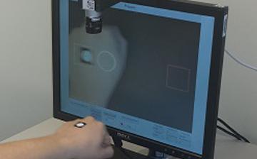
Video microscopy is a medical technique that involves the use of a microscope equipped with a video camera to capture and display real-time images of specimens on a monitor. This allows for the observation and documentation of dynamic processes, such as cell movement or chemical reactions, at a level of detail that would be difficult or impossible to achieve with the naked eye. Video microscopy can also be used in conjunction with image analysis software to measure various parameters, such as size, shape, and motion, of individual cells or structures within the specimen.
There are several types of video microscopy, including brightfield, darkfield, phase contrast, fluorescence, and differential interference contrast (DIC) microscopy. Each type uses different optical techniques to enhance contrast and reveal specific features of the specimen. For example, fluorescence microscopy uses fluorescent dyes or proteins to label specific structures within the specimen, allowing them to be visualized against a dark background.
Video microscopy is used in various fields of medicine, including pathology, microbiology, and neuroscience. It can help researchers and clinicians diagnose diseases, study disease mechanisms, develop new therapies, and understand fundamental biological processes at the cellular and molecular level.
Signal transduction is the process by which a cell converts an extracellular signal, such as a hormone or neurotransmitter, into an intracellular response. This involves a series of molecular events that transmit the signal from the cell surface to the interior of the cell, ultimately resulting in changes in gene expression, protein activity, or metabolism.
The process typically begins with the binding of the extracellular signal to a receptor located on the cell membrane. This binding event activates the receptor, which then triggers a cascade of intracellular signaling molecules, such as second messengers, protein kinases, and ion channels. These molecules amplify and propagate the signal, ultimately leading to the activation or inhibition of specific cellular responses.
Signal transduction pathways are highly regulated and can be modulated by various factors, including other signaling molecules, post-translational modifications, and feedback mechanisms. Dysregulation of these pathways has been implicated in a variety of diseases, including cancer, diabetes, and neurological disorders.
Cell adhesion refers to the binding of cells to extracellular matrices or to other cells, a process that is fundamental to the development, function, and maintenance of multicellular organisms. Cell adhesion is mediated by various cell surface receptors, such as integrins, cadherins, and immunoglobulin-like cell adhesion molecules (Ig-CAMs), which interact with specific ligands in the extracellular environment. These interactions lead to the formation of specialized junctions, such as tight junctions, adherens junctions, and desmosomes, that help to maintain tissue architecture and regulate various cellular processes, including proliferation, differentiation, migration, and survival. Disruptions in cell adhesion can contribute to a variety of diseases, including cancer, inflammation, and degenerative disorders.
Plant viral movement proteins (MPs) are specialized proteins encoded by plant viruses that play a crucial role in the infection process. These proteins are responsible for the cell-to-cell movement of the virus, allowing it to spread throughout the infected plant. MPs facilitate the transport of viral genetic material from infected cells to neighboring uninfected cells, often through plasmodesmata, which are specialized channels that connect the cytoplasm of adjacent plant cells.
Movement proteins can increase the size exclusion limit (SEL) of plasmodesmata, creating a larger pore through which viral RNA or DNA can pass. They also form complexes with viral nucleic acids and other MPs to create movement protein-viral RNA/DNA complexes that are transported between cells. The precise mechanisms by which MPs function vary among different virus families, but their role in facilitating the spread of plant viruses is well established.
It's important to note that understanding the structure and function of plant viral movement proteins can provide valuable insights into plant-virus interactions and contribute to the development of novel strategies for controlling plant virus diseases.
The cytoskeleton is a complex network of various protein filaments that provides structural support, shape, and stability to the cell. It plays a crucial role in maintaining cellular integrity, intracellular organization, and enabling cell movement. The cytoskeleton is composed of three major types of protein fibers: microfilaments (actin filaments), intermediate filaments, and microtubules. These filaments work together to provide mechanical support, participate in cell division, intracellular transport, and help maintain the cell's architecture. The dynamic nature of the cytoskeleton allows cells to adapt to changing environmental conditions and respond to various stimuli.
Cell surface extensions, also known as cellular processes or protrusions, are specialized structures that extend from the plasma membrane of a eukaryotic cell. These extensions include various types of projections such as cilia, flagella, and filopodia, as well as larger and more complex structures like lamellipodia and pseudopodia.
Cilia and flagella are hair-like structures that are involved in cell movement and the sensation of external stimuli. They are composed of a core of microtubules surrounded by the plasma membrane.
Filopodia are thin, finger-like protrusions that contain bundles of actin filaments and are involved in cell motility, sensing the environment, and establishing cell-cell contacts.
Lamellipodia are sheet-like extensions composed of a branched network of actin filaments and are involved in cell migration.
Pseudopodia are large, irregularly shaped protrusions that contain a mixture of actin filaments and other cytoskeletal elements, and are involved in phagocytosis and cell motility.
These cell surface extensions play important roles in various biological processes, including cell motility, sensing the environment, establishing cell-cell contacts, and the uptake of extracellular material.
Developmental gene expression regulation refers to the processes that control the activation or repression of specific genes during embryonic and fetal development. These regulatory mechanisms ensure that genes are expressed at the right time, in the right cells, and at appropriate levels to guide proper growth, differentiation, and morphogenesis of an organism.
Developmental gene expression regulation is a complex and dynamic process involving various molecular players, such as transcription factors, chromatin modifiers, non-coding RNAs, and signaling molecules. These regulators can interact with cis-regulatory elements, like enhancers and promoters, to fine-tune the spatiotemporal patterns of gene expression during development.
Dysregulation of developmental gene expression can lead to various congenital disorders and developmental abnormalities. Therefore, understanding the principles and mechanisms governing developmental gene expression regulation is crucial for uncovering the etiology of developmental diseases and devising potential therapeutic strategies.
Fetal movement, also known as quickening, refers to the first perceived movements of the fetus in the uterus during pregnancy. These movements are often described as a fluttering sensation in the lower abdomen and are usually felt by pregnant individuals between 18 and 25 weeks of gestation, although they may occur earlier or later depending on various factors such as the position of the placenta and whether it is a first-time pregnancy.
Fetal movements are an important sign of fetal well-being, and pregnant individuals are typically advised to monitor them regularly starting from around 28 weeks of gestation. A decrease in fetal movement or the absence of fetal movement for an extended period may indicate a problem and should be reported to a healthcare provider immediately.
Fetal movements can be described as kicks, rolls, jabs, or turns, and they become stronger and more frequent as the pregnancy progresses. By 32 weeks of gestation, most fetuses move around 10 times per hour, and by 37 weeks, they typically move around 30 times per day. However, it is important to note that every fetus has its own pattern of movements, and what is normal for one may not be normal for another.
In medical and embryological terms, the mesoderm is one of the three primary germ layers in the very early stages of embryonic development. It forms between the ectoderm and endoderm during gastrulation, and it gives rise to a wide variety of cell types, tissues, and organs in the developing embryo.
The mesoderm contributes to the formation of structures such as:
1. The connective tissues (including tendons, ligaments, and most of the bones)
2. Muscular system (skeletal, smooth, and cardiac muscles)
3. Circulatory system (heart, blood vessels, and blood cells)
4. Excretory system (kidneys and associated structures)
5. Reproductive system (gonads, including ovaries and testes)
6. Dermis of the skin
7. Parts of the eye and inner ear
8. Several organs in the urogenital system
Dysfunctions or abnormalities in mesoderm development can lead to various congenital disorders and birth defects, highlighting its importance during embryogenesis.
Psychomotor performance refers to the integration and coordination of mental processes (cognitive functions) with physical movements. It involves the ability to perform complex tasks that require both cognitive skills, such as thinking, remembering, and perceiving, and motor skills, such as gross and fine motor movements. Examples of psychomotor performances include driving a car, playing a musical instrument, or performing surgical procedures.
In a medical context, psychomotor performance is often used to assess an individual's ability to perform activities of daily living (ADLs) and instrumental activities of daily living (IADLs), such as bathing, dressing, cooking, cleaning, and managing medications. Deficits in psychomotor performance can be a sign of neurological or psychiatric disorders, such as dementia, Parkinson's disease, or depression.
Assessment of psychomotor performance may involve tests that measure reaction time, coordination, speed, precision, and accuracy of movements, as well as cognitive functions such as attention, memory, and problem-solving skills. These assessments can help healthcare professionals develop appropriate treatment plans and monitor the progression of diseases or the effectiveness of interventions.
I apologize, but "video recording" is not a term that has a specific medical definition. It generally refers to the process of capturing and storing moving visual images on electronic media, which is used in various medical fields for different purposes such as surgical training, telemedicine consultations, or monitoring patient conditions. However, there is no unique medical meaning associated with this term.
Cell aggregation is the process by which individual cells come together and adhere to each other to form a group or cluster. This phenomenon can occur naturally during embryonic development, tissue repair, and wound healing, as well as in the formation of multicellular organisms such as slime molds. In some cases, cell aggregation may also be induced in the laboratory setting through the use of various techniques, including the use of cell culture surfaces that promote cell-to-cell adhesion or the addition of factors that stimulate the expression of adhesion molecules on the cell surface.
Cell aggregation can be influenced by a variety of factors, including the type and properties of the cells involved, as well as environmental conditions such as pH, temperature, and nutrient availability. The ability of cells to aggregate is often mediated by the presence of adhesion molecules on the cell surface, such as cadherins, integrins, and immunoglobulin-like cell adhesion molecules (Ig-CAMs). These molecules interact with each other and with extracellular matrix components to promote cell-to-cell adhesion and maintain the stability of the aggregate.
In some contexts, abnormal or excessive cell aggregation can contribute to the development of diseases such as cancer, fibrosis, and inflammatory disorders. For example, the aggregation of cancer cells can facilitate their invasion and metastasis, while the accumulation of fibrotic cells in tissues can lead to organ dysfunction and failure. Understanding the mechanisms that regulate cell aggregation is therefore an important area of research with potential implications for the development of new therapies and treatments for a variety of diseases.
I'm sorry for any confusion, but "Motion Pictures as Topic" is not a medical term or concept. It is actually a subject heading used in library and information sciences to categorize materials related to the study or analysis of motion pictures as a medium or art form. This could include books, articles, and other resources about film theory, film history, film criticism, and so on.
If you have any questions about medical terminology or concepts, I would be happy to help!
Wnt proteins are a family of secreted signaling molecules that play crucial roles in the regulation of fundamental biological processes, including cell proliferation, differentiation, migration, and survival. They were first discovered in 1982 through genetic studies in Drosophila melanogaster (fruit flies) and have since been found to be highly conserved across various species, from invertebrates to humans.
Wnt proteins exert their effects by binding to specific receptors on the target cell surface, leading to the activation of several intracellular signaling pathways:
1. Canonical Wnt/β-catenin pathway: In the absence of Wnt ligands, β-catenin is continuously degraded by a destruction complex consisting of Axin, APC (Adenomatous polyposis coli), and GSK3β (Glycogen synthase kinase 3 beta). When Wnt proteins bind to their receptors Frizzled and LRP5/6, the formation of a "signalosome" complex leads to the inhibition of the destruction complex, allowing β-catenin to accumulate in the cytoplasm and translocate into the nucleus. Here, it interacts with TCF/LEF (T-cell factor/lymphoid enhancer-binding factor) transcription factors to regulate the expression of target genes involved in cell proliferation, differentiation, and survival.
2. Non-canonical Wnt pathways: These include the Wnt/Ca^2+^ pathway and the planar cell polarity (PCP) pathway. In the Wnt/Ca^2+^ pathway, Wnt ligands bind to Frizzled receptors and activate heterotrimeric G proteins, leading to an increase in intracellular Ca^2+^ levels and activation of downstream targets such as protein kinase C (PKC) and calcium/calmodulin-dependent protein kinase II (CAMKII). These signaling events ultimately regulate cell movement, adhesion, and gene expression. In the PCP pathway, Wnt ligands bind to Frizzled receptors and coreceptor complexes containing Ror2 or Ryk, leading to activation of small GTPases such as RhoA and Rac1, which control cytoskeletal organization and cell polarity.
Dysregulation of Wnt signaling has been implicated in various human diseases, including cancer, developmental disorders, and degenerative conditions. In cancer, aberrant activation of the canonical Wnt/β-catenin pathway contributes to tumor initiation, progression, and metastasis by promoting cell proliferation, survival, and epithelial-mesenchymal transition (EMT). Inhibitors targeting different components of the Wnt signaling pathway are currently being developed as potential therapeutic strategies for cancer treatment.
"Xenopus proteins" refer to the proteins that are expressed or isolated from the Xenopus species, which are primarily used as model organisms in biological and biomedical research. The most commonly used Xenopus species for research are the African clawed frogs, Xenopus laevis and Xenopus tropicalis. These proteins play crucial roles in various cellular processes and functions, and they serve as valuable tools to study different aspects of molecular biology, developmental biology, genetics, and biochemistry.
Some examples of Xenopus proteins that are widely studied include:
1. Xenopus Histones: These are the proteins that package DNA into nucleosomes, which are the fundamental units of chromatin in eukaryotic cells. They play a significant role in gene regulation and epigenetic modifications.
2. Xenopus Cyclins and Cyclin-dependent kinases (CDKs): These proteins regulate the cell cycle and control cell division, differentiation, and apoptosis.
3. Xenopus Transcription factors: These proteins bind to specific DNA sequences and regulate gene expression during development and in response to various stimuli.
4. Xenopus Signaling molecules: These proteins are involved in intracellular signaling pathways that control various cellular processes, such as cell growth, differentiation, migration, and survival.
5. Xenopus Cytoskeletal proteins: These proteins provide structural support to the cells and regulate their shape, motility, and organization.
6. Xenopus Enzymes: These proteins catalyze various biochemical reactions in the cell, such as metabolic pathways, DNA replication, transcription, and translation.
Overall, Xenopus proteins are essential tools for understanding fundamental biological processes and have contributed significantly to our current knowledge of molecular biology, genetics, and developmental biology.
A chick embryo refers to the developing organism that arises from a fertilized chicken egg. It is often used as a model system in biological research, particularly during the stages of development when many of its organs and systems are forming and can be easily observed and manipulated. The study of chick embryos has contributed significantly to our understanding of various aspects of developmental biology, including gastrulation, neurulation, organogenesis, and pattern formation. Researchers may use various techniques to observe and manipulate the chick embryo, such as surgical alterations, cell labeling, and exposure to drugs or other agents.
Microfilament proteins are a type of structural protein that form part of the cytoskeleton in eukaryotic cells. They are made up of actin monomers, which polymerize to form long, thin filaments. These filaments are involved in various cellular processes such as muscle contraction, cell division, and cell motility. Microfilament proteins also interact with other cytoskeletal components like intermediate filaments and microtubules to maintain the overall shape and integrity of the cell. Additionally, they play a crucial role in the formation of cell-cell junctions and cell-matrix adhesions, which are essential for tissue structure and function.
In the field of medicine, "time factors" refer to the duration of symptoms or time elapsed since the onset of a medical condition, which can have significant implications for diagnosis and treatment. Understanding time factors is crucial in determining the progression of a disease, evaluating the effectiveness of treatments, and making critical decisions regarding patient care.
For example, in stroke management, "time is brain," meaning that rapid intervention within a specific time frame (usually within 4.5 hours) is essential to administering tissue plasminogen activator (tPA), a clot-busting drug that can minimize brain damage and improve patient outcomes. Similarly, in trauma care, the "golden hour" concept emphasizes the importance of providing definitive care within the first 60 minutes after injury to increase survival rates and reduce morbidity.
Time factors also play a role in monitoring the progression of chronic conditions like diabetes or heart disease, where regular follow-ups and assessments help determine appropriate treatment adjustments and prevent complications. In infectious diseases, time factors are crucial for initiating antibiotic therapy and identifying potential outbreaks to control their spread.
Overall, "time factors" encompass the significance of recognizing and acting promptly in various medical scenarios to optimize patient outcomes and provide effective care.
A saccade is a quick, rapid, and ballistic conjugate eye movement that shifts the point of fixation from one target to another. It helps in rapidly repositioning the fovea (the central part of the retina with the highest visual acuity) to focus on different targets of interest in the visual scene. Saccades are essential for efficient scanning and exploration of our environment, allowing us to direct our high-resolution vision towards various points of interest. They typically take only about 20-200 milliseconds to complete and can reach peak velocities of up to 500 degrees per second or more, depending on the amplitude of the movement. Saccades are a critical component of normal visual function and are often studied in fields such as ophthalmology, neurology, and neuroscience.
Cell shape refers to the physical form or configuration of a cell, which is determined by the cytoskeleton (the internal framework of the cell) and the extracellular matrix (the external environment surrounding the cell). The shape of a cell can vary widely depending on its type and function. For example, some cells are spherical, such as red blood cells, while others are elongated or irregularly shaped. Changes in cell shape can be indicative of various physiological or pathological processes, including development, differentiation, migration, and disease.
I apologize for any confusion, but "Myxococcus xanthus" is not a medical term or concept. It is actually the scientific name of a type of soil-dwelling bacteria that belongs to the family Myxococcaceae. These bacteria are known for their social behavior and complex life cycle, which includes the formation of multicellular structures under certain conditions. They have been studied extensively in the field of microbiology due to their unique biological characteristics.
In medical terms, the arm refers to the upper limb of the human body, extending from the shoulder to the wrist. It is composed of three major bones: the humerus in the upper arm, and the radius and ulna in the lower arm. The arm contains several joints, including the shoulder joint, elbow joint, and wrist joint, which allow for a wide range of motion. The arm also contains muscles, blood vessels, nerves, and other soft tissues that are essential for normal function.
Somites are transient, segmentally repeated embryonic structures that form along the anterior-posterior body axis during vertebrate development. They are derived from the paraxial mesoderm and give rise to various tissues, including the sclerotome (which forms the vertebrae and ribs), myotome (which forms the skeletal muscles of the back and limbs), and dermatome (which forms the dermis of the skin).
Each somite is a block-like structure that is arranged in a repeating pattern along the notochord, which is a flexible rod-like structure that provides mechanical support to the developing embryo. The formation of somites is a critical step in the development of the vertebrate body plan, as they help to establish the segmental organization of the musculoskeletal system and contribute to the formation of other important structures such as the dermis and the circulatory system.
The process of somitogenesis, or the formation of somites, is a highly regulated and coordinated event that involves the interaction of various signaling molecules and genetic pathways. Defects in somite formation can lead to a range of developmental abnormalities, including spinal deformities, muscle weakness, and skin defects.
Electromyography (EMG) is a medical diagnostic procedure that measures the electrical activity of skeletal muscles during contraction and at rest. It involves inserting a thin needle electrode into the muscle to record the electrical signals generated by the muscle fibers. These signals are then displayed on an oscilloscope and may be heard through a speaker.
EMG can help diagnose various neuromuscular disorders, such as muscle weakness, numbness, or pain, and can distinguish between muscle and nerve disorders. It is often used in conjunction with other diagnostic tests, such as nerve conduction studies, to provide a comprehensive evaluation of the nervous system.
EMG is typically performed by a neurologist or a physiatrist, and the procedure may cause some discomfort or pain, although this is usually minimal. The results of an EMG can help guide treatment decisions and monitor the progression of neuromuscular conditions over time.
Biomechanics is the application of mechanical laws to living structures and systems, particularly in the field of medicine and healthcare. A biomechanical phenomenon refers to a observable event or occurrence that involves the interaction of biological tissues or systems with mechanical forces. These phenomena can be studied at various levels, from the molecular and cellular level to the tissue, organ, and whole-body level.
Examples of biomechanical phenomena include:
1. The way that bones and muscles work together to produce movement (known as joint kinematics).
2. The mechanical behavior of biological tissues such as bone, cartilage, tendons, and ligaments under various loads and stresses.
3. The response of cells and tissues to mechanical stimuli, such as the way that bone tissue adapts to changes in loading conditions (known as Wolff's law).
4. The biomechanics of injury and disease processes, such as the mechanisms of joint injury or the development of osteoarthritis.
5. The use of mechanical devices and interventions to treat medical conditions, such as orthopedic implants or assistive devices for mobility impairments.
Understanding biomechanical phenomena is essential for developing effective treatments and prevention strategies for a wide range of medical conditions, from musculoskeletal injuries to neurological disorders.
Cadherins are a type of cell adhesion molecule that play a crucial role in the development and maintenance of intercellular junctions. They are transmembrane proteins that mediate calcium-dependent homophilic binding between adjacent cells, meaning that they bind to identical cadherin molecules on neighboring cells.
There are several types of cadherins, including classical cadherins, desmosomal cadherins, and protocadherins, each with distinct functions and localization in tissues. Classical cadherins, also known as type I cadherins, are the most well-studied and are essential for the formation of adherens junctions, which help to maintain cell-to-cell contact and tissue architecture.
Desmosomal cadherins, on the other hand, are critical for the formation and maintenance of desmosomes, which are specialized intercellular junctions that provide mechanical strength and stability to tissues. Protocadherins are a diverse family of cadherin-related proteins that have been implicated in various developmental processes, including neuronal connectivity and tissue patterning.
Mutations in cadherin genes have been associated with several human diseases, including cancer, neurological disorders, and heart defects. Therefore, understanding the structure, function, and regulation of cadherins is essential for elucidating their roles in health and disease.
Rac (Ras-related C3 botulinum toxin substrate) GTP-binding proteins are a subfamily of the Rho family of small GTPases, which function as molecular switches that regulate various cellular processes, including actin cytoskeleton organization, cell adhesion, and gene transcription.
Rac GTP-binding proteins cycle between an inactive GDP-bound state and an active GTP-bound state. When Rac is in its active state, it interacts with downstream effectors to regulate various signaling pathways that control cell behavior. Activation of Rac promotes the formation of lamellipodia and membrane ruffles, which are important for cell migration and invasion.
Rac GTP-binding proteins have been implicated in a variety of physiological and pathological processes, including embryonic development, immune function, and cancer. Dysregulation of Rac signaling has been associated with various diseases, such as inflammatory disorders, neurological disorders, and cancer. Therefore, understanding the regulation and function of Rac GTP-binding proteins is crucial for developing therapeutic strategies to target these diseases.
A mutation is a permanent change in the DNA sequence of an organism's genome. Mutations can occur spontaneously or be caused by environmental factors such as exposure to radiation, chemicals, or viruses. They may have various effects on the organism, ranging from benign to harmful, depending on where they occur and whether they alter the function of essential proteins. In some cases, mutations can increase an individual's susceptibility to certain diseases or disorders, while in others, they may confer a survival advantage. Mutations are the driving force behind evolution, as they introduce new genetic variability into populations, which can then be acted upon by natural selection.
The notochord is a flexible, rod-shaped structure that is present in the embryos of chordates, including humans. It is composed of cells called chordocytes and is surrounded by a sheath. The notochord runs along the length of the body, providing support and flexibility. In human embryos, the notochord eventually becomes part of the discs between the vertebrae in the spine. An abnormal or absent notochord can lead to developmental problems with the spine and nervous system.
Cell communication, also known as cell signaling, is the process by which cells exchange and transmit signals between each other and their environment. This complex system allows cells to coordinate their functions and maintain tissue homeostasis. Cell communication can occur through various mechanisms including:
1. Autocrine signaling: When a cell releases a signal that binds to receptors on the same cell, leading to changes in its behavior or function.
2. Paracrine signaling: When a cell releases a signal that binds to receptors on nearby cells, influencing their behavior or function.
3. Endocrine signaling: When a cell releases a hormone into the bloodstream, which then travels to distant target cells and binds to specific receptors, triggering a response.
4. Synaptic signaling: In neurons, communication occurs through the release of neurotransmitters that cross the synapse and bind to receptors on the postsynaptic cell, transmitting electrical or chemical signals.
5. Contact-dependent signaling: When cells physically interact with each other, allowing for the direct exchange of signals and information.
Cell communication is essential for various physiological processes such as growth, development, differentiation, metabolism, immune response, and tissue repair. Dysregulation in cell communication can contribute to diseases, including cancer, diabetes, and neurological disorders.
Ectoderm is the outermost of the three primary germ layers in a developing embryo, along with the endoderm and mesoderm. The ectoderm gives rise to the outer covering of the body, including the skin, hair, nails, glands, and the nervous system, which includes the brain, spinal cord, and peripheral nerves. It also forms the lining of the mouth, anus, nose, and ears. Essentially, the ectoderm is responsible for producing all the epidermal structures and the neural crest cells that contribute to various derivatives such as melanocytes, adrenal medulla, smooth muscle, and peripheral nervous system components.
In situ hybridization (ISH) is a molecular biology technique used to detect and localize specific nucleic acid sequences, such as DNA or RNA, within cells or tissues. This technique involves the use of a labeled probe that is complementary to the target nucleic acid sequence. The probe can be labeled with various types of markers, including radioisotopes, fluorescent dyes, or enzymes.
During the ISH procedure, the labeled probe is hybridized to the target nucleic acid sequence in situ, meaning that the hybridization occurs within the intact cells or tissues. After washing away unbound probe, the location of the labeled probe can be visualized using various methods depending on the type of label used.
In situ hybridization has a wide range of applications in both research and diagnostic settings, including the detection of gene expression patterns, identification of viral infections, and diagnosis of genetic disorders.
In medical terms, a hand is the part of the human body that is attached to the forearm and consists of the carpus (wrist), metacarpus, and phalanges. It is made up of 27 bones, along with muscles, tendons, ligaments, and other soft tissues. The hand is a highly specialized organ that is capable of performing a wide range of complex movements and functions, including grasping, holding, manipulating objects, and communicating through gestures. It is also richly innervated with sensory receptors that provide information about touch, temperature, pain, and proprioception (the sense of the position and movement of body parts).
RhoA (Ras Homolog Family Member A) is a small GTPase protein that acts as a molecular switch, cycling between an inactive GDP-bound state and an active GTP-bound state. It plays a crucial role in regulating various cellular processes such as actin cytoskeleton organization, gene expression, cell cycle progression, and cell migration.
RhoA GTP-binding protein becomes activated when it binds to GTP, and this activation leads to the recruitment of downstream effectors that mediate its functions. The activity of RhoA is tightly regulated by several proteins, including guanine nucleotide exchange factors (GEFs) that promote the exchange of GDP for GTP, GTPase-activating proteins (GAPs) that stimulate the intrinsic GTPase activity of RhoA to hydrolyze GTP to GDP and return it to an inactive state, and guanine nucleotide dissociation inhibitors (GDIs) that sequester RhoA in the cytoplasm and prevent its association with the membrane.
Mutations or dysregulation of RhoA GTP-binding protein have been implicated in various human diseases, including cancer, neurological disorders, and cardiovascular diseases.
Green Fluorescent Protein (GFP) is not a medical term per se, but a scientific term used in the field of molecular biology. GFP is a protein that exhibits bright green fluorescence when exposed to light, particularly blue or ultraviolet light. It was originally discovered in the jellyfish Aequorea victoria.
In medical and biological research, scientists often use recombinant DNA technology to introduce the gene for GFP into other organisms, including bacteria, plants, and animals, including humans. This allows them to track the expression and localization of specific genes or proteins of interest in living cells, tissues, or even whole organisms.
The ability to visualize specific cellular structures or processes in real-time has proven invaluable for a wide range of research areas, from studying the development and function of organs and organ systems to understanding the mechanisms of diseases and the effects of therapeutic interventions.
Adherens junctions are specialized types of cell-cell contacts that play a crucial role in maintaining the integrity and stability of tissues. They are composed of transmembrane cadherin proteins, which connect to the actin cytoskeleton inside the cell through intracellular adaptor proteins such as catenins.
The cadherins on opposing cells interact with each other to form adhesive bonds that help to anchor the cells together and regulate various cellular processes, including cell growth, differentiation, and migration. Adherens junctions are essential for many physiological processes, such as embryonic development, wound healing, and tissue homeostasis, and their dysfunction has been implicated in a variety of diseases, including cancer and degenerative disorders.
Molecular sequence data refers to the specific arrangement of molecules, most commonly nucleotides in DNA or RNA, or amino acids in proteins, that make up a biological macromolecule. This data is generated through laboratory techniques such as sequencing, and provides information about the exact order of the constituent molecules. This data is crucial in various fields of biology, including genetics, evolution, and molecular biology, allowing for comparisons between different organisms, identification of genetic variations, and studies of gene function and regulation.
Endoderm is the innermost of the three primary germ layers in a developing embryo, along with the ectoderm and mesoderm. The endoderm gives rise to several internal tissues and organs, most notably those found in the digestive system and respiratory system. Specifically, it forms the lining of the gut tube, which eventually becomes the epithelial lining of the gastrointestinal tract, liver, pancreas, lungs, and other associated structures.
During embryonic development, the endoderm arises from the inner cell mass of the blastocyst, following a series of cell divisions and migrations that help to establish the basic body plan of the organism. As the embryo grows and develops, the endoderm continues to differentiate into more specialized tissues and structures, playing a critical role in the formation of many essential bodily functions.
Ocular fixation is a term used in ophthalmology and optometry to refer to the ability of the eyes to maintain steady gaze or visual focus on an object. It involves the coordinated movement of the extraocular muscles that control eye movements, allowing for clear and stable vision.
In medical terminology, fixation specifically refers to the state in which the eyes are aligned and focused on a single point in space. This is important for maintaining visual perception and preventing blurring or double vision. Ocular fixation can be affected by various factors such as muscle weakness, nerve damage, or visual processing disorders.
Assessment of ocular fixation is often used in eye examinations to evaluate visual acuity, eye alignment, and muscle function. Abnormalities in fixation may indicate the presence of underlying eye conditions or developmental delays that require further investigation and treatment.
"Xenopus laevis" is not a medical term itself, but it refers to a specific species of African clawed frog that is often used in scientific research, including biomedical and developmental studies. Therefore, its relevance to medicine comes from its role as a model organism in laboratories.
In a broader sense, Xenopus laevis has contributed significantly to various medical discoveries, such as the understanding of embryonic development, cell cycle regulation, and genetic research. For instance, the Nobel Prize in Physiology or Medicine was awarded in 1963 to John R. B. Gurdon and Sir Michael J. Bishop for their discoveries concerning the genetic mechanisms of organism development using Xenopus laevis as a model system.
"Cells, cultured" is a medical term that refers to cells that have been removed from an organism and grown in controlled laboratory conditions outside of the body. This process is called cell culture and it allows scientists to study cells in a more controlled and accessible environment than they would have inside the body. Cultured cells can be derived from a variety of sources, including tissues, organs, or fluids from humans, animals, or cell lines that have been previously established in the laboratory.
Cell culture involves several steps, including isolation of the cells from the tissue, purification and characterization of the cells, and maintenance of the cells in appropriate growth conditions. The cells are typically grown in specialized media that contain nutrients, growth factors, and other components necessary for their survival and proliferation. Cultured cells can be used for a variety of purposes, including basic research, drug development and testing, and production of biological products such as vaccines and gene therapies.
It is important to note that cultured cells may behave differently than they do in the body, and results obtained from cell culture studies may not always translate directly to human physiology or disease. Therefore, it is essential to validate findings from cell culture experiments using additional models and ultimately in clinical trials involving human subjects.
Fluorescence microscopy is a type of microscopy that uses fluorescent dyes or proteins to highlight and visualize specific components within a sample. In this technique, the sample is illuminated with high-energy light, typically ultraviolet (UV) or blue light, which excites the fluorescent molecules causing them to emit lower-energy, longer-wavelength light, usually visible light in the form of various colors. This emitted light is then collected by the microscope and detected to produce an image.
Fluorescence microscopy has several advantages over traditional brightfield microscopy, including the ability to visualize specific structures or molecules within a complex sample, increased sensitivity, and the potential for quantitative analysis. It is widely used in various fields of biology and medicine, such as cell biology, neuroscience, and pathology, to study the structure, function, and interactions of cells and proteins.
There are several types of fluorescence microscopy techniques, including widefield fluorescence microscopy, confocal microscopy, two-photon microscopy, and total internal reflection fluorescence (TIRF) microscopy, each with its own strengths and limitations. These techniques can provide valuable insights into the behavior of cells and proteins in health and disease.
The Wiskott-Aldrich Syndrome Protein (WASP) family is a group of proteins that play crucial roles in actin cytoskeleton regulation, which is essential for various cellular processes such as cell motility, membrane trafficking, and immune synapse formation. The family includes WASP, N-WASP (Neural WASP), WAVE1 (WASP-family verprolin homologous protein 1), WAVE2, WAVE3, and WHAMM (WASP Homology Associated with Actin, Membranes and Microtubules). These proteins share a common structural feature called the WASP homology domain 2 (WH2) that binds to actin monomers, and a C-terminal verprolin homology domain (VHD) that interacts with various regulatory factors. Mutations in the gene encoding WASP can lead to Wiskott-Aldrich syndrome, an X-linked recessive disorder characterized by microthrombocytopenia, eczema, and recurrent infections.
The actin cytoskeleton is a complex, dynamic network of filamentous (threadlike) proteins that provides structural support and shape to cells, allows for cell movement and division, and plays a role in intracellular transport. Actin filaments are composed of actin monomers that polymerize to form long, thin fibers. These filaments can be organized into different structures, such as stress fibers, which provide tension and support, or lamellipodia and filopodia, which are involved in cell motility. The actin cytoskeleton is constantly remodeling in response to various intracellular and extracellular signals, allowing for changes in cell shape and behavior.
Microtubules are hollow, cylindrical structures composed of tubulin proteins in the cytoskeleton of eukaryotic cells. They play crucial roles in various cellular processes such as maintaining cell shape, intracellular transport, and cell division (mitosis and meiosis). Microtubules are dynamic, undergoing continuous assembly and disassembly, which allows them to rapidly reorganize in response to cellular needs. They also form part of important cellular structures like centrioles, basal bodies, and cilia/flagella.
Oligoribonucleotides are short, single-stranded RNA molecules that consist of fewer than 200 nucleotides. Antisense oligoribonucleotides (ORNs) are a type of oligoribonucleotide that are designed to be complementary to a specific target RNA molecule. They work by binding to the target RNA through base-pairing, which can prevent the target RNA from being translated into protein or can trigger its degradation by cellular enzymes. Antisense ORNs have potential therapeutic applications in the treatment of various diseases, including viral infections and genetic disorders.
A cell membrane, also known as the plasma membrane, is a thin semi-permeable phospholipid bilayer that surrounds all cells in animals, plants, and microorganisms. It functions as a barrier to control the movement of substances in and out of the cell, allowing necessary molecules such as nutrients, oxygen, and signaling molecules to enter while keeping out harmful substances and waste products. The cell membrane is composed mainly of phospholipids, which have hydrophilic (water-loving) heads and hydrophobic (water-fearing) tails. This unique structure allows the membrane to be flexible and fluid, yet selectively permeable. Additionally, various proteins are embedded in the membrane that serve as channels, pumps, receptors, and enzymes, contributing to the cell's overall functionality and communication with its environment.
Phase-contrast microscopy is a type of optical microscopy that allows visualization of transparent or translucent specimens, such as living cells and their organelles, by increasing the contrast between areas with different refractive indices within the sample. This technique works by converting phase shifts in light passing through the sample into changes in amplitude, which can then be observed as differences in brightness and contrast.
In a phase-contrast microscope, a special condenser and objective are used to create an optical path difference between the direct and diffracted light rays coming from the specimen. The condenser introduces a phase shift for the diffracted light, while the objective contains a phase ring that compensates for this shift in the direct light. This results in the direct light appearing brighter than the diffracted light, creating contrast between areas with different refractive indices within the sample.
Phase-contrast microscopy is particularly useful for observing unstained living cells and their dynamic processes, such as cell division, motility, and secretion, without the need for stains or dyes that might affect their viability or behavior.
Eye movement measurements, also known as oculometry, refer to the measurement and analysis of eye movements. This can include assessing the direction, speed, range, and patterns of eye movement. These measurements are often used in research and clinical settings to understand various aspects of vision, perception, and cognition. They can be used to diagnose and monitor conditions that affect eye movement, such as strabismus (crossed eyes), amblyopia (lazy eye), or neurological disorders. Additionally, eye movement measurements are also used in areas such as human-computer interaction, marketing research, and virtual reality to understand how individuals interact with their environment.
The motor cortex is a region in the frontal lobe of the brain that is responsible for controlling voluntary movements. It is involved in planning, initiating, and executing movements of the limbs, body, and face. The motor cortex contains neurons called Betz cells, which have large cell bodies and are responsible for transmitting signals to the spinal cord to activate muscles. Damage to the motor cortex can result in various movement disorders such as hemiplegia or paralysis on one side of the body.
"Xenopus" is not a medical term, but it is a genus of highly invasive aquatic frogs native to sub-Saharan Africa. They are often used in scientific research, particularly in developmental biology and genetics. The most commonly studied species is Xenopus laevis, also known as the African clawed frog.
In a medical context, Xenopus might be mentioned when discussing their use in research or as a model organism to study various biological processes or diseases.
Rho GTP-binding proteins are a subfamily of the Ras superfamily of small GTPases, which function as molecular switches in various cellular signaling pathways. These proteins play crucial roles in regulating diverse cellular processes such as actin cytoskeleton dynamics, gene expression, cell cycle progression, and cell migration.
Rho GTP-binding proteins cycle between an active GTP-bound state and an inactive GDP-bound state. In the active state, they interact with various downstream effectors to regulate their respective cellular functions. Guanine nucleotide exchange factors (GEFs) activate Rho GTP-binding proteins by promoting the exchange of GDP for GTP, while GTPase-activating proteins (GAPs) inactivate them by enhancing their intrinsic GTP hydrolysis activity.
There are several members of the Rho GTP-binding protein family, including RhoA, RhoB, RhoC, Rac1, Rac2, Rac3, Cdc42, and Rnd proteins, each with distinct functions and downstream effectors. Dysregulation of Rho GTP-binding proteins has been implicated in various human diseases, including cancer, cardiovascular disease, neurological disorders, and inflammatory diseases.
In the context of medical terminology, I believe you may be referring to "pursuit" as it relates to neurological tests. A smooth pursuit is a type of eye movement in which the eyes smoothly and slowly follow a moving object. It requires coordination between the extraocular muscles, vestibular system, and visual system. If there are issues with any of these systems, smooth pursuit can be affected, leading to abnormalities such as jerky or saccadic movements.
Therefore, "smooth pursuit" is a medical term used to describe the normal, coordinated movement of the eyes that allows for the tracking of moving objects in a smooth and continuous manner.
The neural crest is a transient, multipotent embryonic cell population that originates from the ectoderm (outermost layer) of the developing neural tube (precursor to the central nervous system). These cells undergo an epithelial-to-mesenchymal transition and migrate throughout the embryo, giving rise to a diverse array of cell types and structures.
Neural crest cells differentiate into various tissues, including:
1. Peripheral nervous system (PNS) components: sensory neurons, sympathetic and parasympathetic ganglia, and glial cells (e.g., Schwann cells).
2. Facial bones and cartilage, as well as connective tissue of the skull.
3. Melanocytes, which are pigment-producing cells in the skin.
4. Smooth muscle cells in major blood vessels, heart, gastrointestinal tract, and other organs.
5. Secretory cells in endocrine glands (e.g., chromaffin cells of the adrenal medulla).
6. Parts of the eye, such as the cornea and iris stroma.
7. Dental tissues, including dentin, cementum, and dental pulp.
Due to their wide-ranging contributions to various tissues and organs, neural crest cells play a crucial role in embryonic development and organogenesis. Abnormalities in neural crest cell migration or differentiation can lead to several congenital disorders, such as neurocristopathies.
Focal adhesions are specialized structures found in cells that act as points of attachment between the intracellular cytoskeleton and the extracellular matrix (ECM). They are composed of a complex network of proteins, including integrins, talin, vinculin, paxillin, and various others.
Focal adhesions play a crucial role in cellular processes such as adhesion, migration, differentiation, and signal transduction. They form when integrin receptors in the cell membrane bind to specific ligands within the ECM, leading to the clustering of these receptors and the recruitment of various adaptor and structural proteins. This results in the formation of a stable linkage between the cytoskeleton and the ECM, which helps maintain cell shape, provide mechanical stability, and facilitate communication between the intracellular and extracellular environments.
Focal adhesions are highly dynamic structures that can undergo rapid assembly and disassembly in response to various stimuli, allowing cells to adapt and respond to changes in their microenvironment. Dysregulation of focal adhesion dynamics has been implicated in several pathological conditions, including cancer metastasis, fibrosis, and impaired wound healing.
Genetically modified animals (GMAs) are those whose genetic makeup has been altered using biotechnological techniques. This is typically done by introducing one or more genes from another species into the animal's genome, resulting in a new trait or characteristic that does not naturally occur in that species. The introduced gene is often referred to as a transgene.
The process of creating GMAs involves several steps:
1. Isolation: The desired gene is isolated from the DNA of another organism.
2. Transfer: The isolated gene is transferred into the target animal's cells, usually using a vector such as a virus or bacterium.
3. Integration: The transgene integrates into the animal's chromosome, becoming a permanent part of its genetic makeup.
4. Selection: The modified cells are allowed to multiply, and those that contain the transgene are selected for further growth and development.
5. Breeding: The genetically modified individuals are bred to produce offspring that carry the desired trait.
GMAs have various applications in research, agriculture, and medicine. In research, they can serve as models for studying human diseases or testing new therapies. In agriculture, GMAs can be developed to exhibit enhanced growth rates, improved disease resistance, or increased nutritional value. In medicine, GMAs may be used to produce pharmaceuticals or other therapeutic agents within their bodies.
Examples of genetically modified animals include mice with added genes for specific proteins that make them useful models for studying human diseases, goats that produce a human protein in their milk to treat hemophilia, and pigs with enhanced resistance to certain viruses that could potentially be used as organ donors for humans.
It is important to note that the use of genetically modified animals raises ethical concerns related to animal welfare, environmental impact, and potential risks to human health. These issues must be carefully considered and addressed when developing and implementing GMA technologies.
Luminescent proteins are a type of protein that emit light through a chemical reaction, rather than by absorbing and re-emitting light like fluorescent proteins. This process is called bioluminescence. The light emitted by luminescent proteins is often used in scientific research as a way to visualize and track biological processes within cells and organisms.
One of the most well-known luminescent proteins is Green Fluorescent Protein (GFP), which was originally isolated from jellyfish. However, GFP is actually a fluorescent protein, not a luminescent one. A true example of a luminescent protein is the enzyme luciferase, which is found in fireflies and other bioluminescent organisms. When luciferase reacts with its substrate, luciferin, it produces light through a process called oxidation.
Luminescent proteins have many applications in research, including as reporters for gene expression, as markers for protein-protein interactions, and as tools for studying the dynamics of cellular processes. They are also used in medical imaging and diagnostics, as well as in the development of new therapies.
Eph family receptors are a group of tyrosine kinase receptors that play crucial roles in the development and function of the nervous system, as well as in other tissues. They are named after the first discovered member of this family, EPH (Erythropoietin-Producing Human Hepatocellular carcinoma) receptor.
These receptors are divided into two subfamilies: EphA and EphB, based on their binding preferences for ephrin ligands. Ephrins are membrane-bound proteins that can be either GPI-anchored (ephrin-A) or transmembrane (ephrin-B), and they interact with Eph receptors in a bidirectional manner, activating both forward signaling in the receptor-expressing cell and reverse signaling in the ephrin-expressing cell.
Eph receptors and ephrins are essential for axon guidance, topographic mapping, and synaptic plasticity during neural development. They also participate in various processes in adult tissues, such as angiogenesis, tumorigenesis, and immune responses. Dysregulation of Eph family receptors has been implicated in several diseases, including cancer, neurological disorders, and vascular diseases.
NIH 3T3 cells are a type of mouse fibroblast cell line that was developed by the National Institutes of Health (NIH). The "3T3" designation refers to the fact that these cells were derived from embryonic Swiss mouse tissue and were able to be passaged (i.e., subcultured) more than three times in tissue culture.
NIH 3T3 cells are widely used in scientific research, particularly in studies involving cell growth and differentiation, signal transduction, and gene expression. They have also been used as a model system for studying the effects of various chemicals and drugs on cell behavior. NIH 3T3 cells are known to be relatively easy to culture and maintain, and they have a stable, flat morphology that makes them well-suited for use in microscopy studies.
It is important to note that, as with any cell line, it is essential to verify the identity and authenticity of NIH 3T3 cells before using them in research, as contamination or misidentification can lead to erroneous results.
Chemotactic factors are substances that attract or repel cells, particularly immune cells, by stimulating directional movement in response to a chemical gradient. These factors play a crucial role in the body's immune response and inflammation process. They include:
1. Chemokines: A family of small signaling proteins that direct the migration of immune cells to sites of infection or tissue damage.
2. Cytokines: A broad category of signaling molecules that mediate and regulate immunity, inflammation, and hematopoiesis. Some cytokines can also act as chemotactic factors.
3. Complement components: Cleavage products of the complement system can attract immune cells to the site of infection or tissue injury.
4. Growth factors: Certain growth factors, like colony-stimulating factors (CSFs), can stimulate the migration and proliferation of specific cell types.
5. Lipid mediators: Products derived from arachidonic acid metabolism, such as leukotrienes and prostaglandins, can also act as chemotactic factors.
6. Formyl peptides: Bacterial-derived formylated peptides can attract and activate neutrophils during an infection.
7. Extracellular matrix (ECM) components: Fragments of ECM proteins, like collagen and fibronectin, can serve as chemotactic factors for immune cells.
These factors help orchestrate the immune response by guiding the movement of immune cells to specific locations in the body where they are needed.
Reaction time, in the context of medicine and physiology, refers to the time period between the presentation of a stimulus and the subsequent initiation of a response. This complex process involves the central nervous system, particularly the brain, which perceives the stimulus, processes it, and then sends signals to the appropriate muscles or glands to react.
There are different types of reaction times, including simple reaction time (responding to a single, expected stimulus) and choice reaction time (choosing an appropriate response from multiple possibilities). These measures can be used in clinical settings to assess various aspects of neurological function, such as cognitive processing speed, motor control, and alertness.
However, it is important to note that reaction times can be influenced by several factors, including age, fatigue, attention, and the use of certain medications or substances.
The blastoderm is the layer of cells that forms on the surface of a developing embryo, during the blastula stage of embryonic development. In mammals, this layer of cells is also known as the epiblast. The blastoderm is responsible for giving rise to all of the tissues and organs of the developing organism. It is formed by the cleavage of the fertilized egg, or zygote, and is typically a single layer of cells that surrounds a fluid-filled cavity called the blastocoel. The blastoderm plays a critical role in the early stages of embryonic development, and any disruptions to its formation or function can lead to developmental abnormalities or death of the embryo.
In medical terms, fingers are not specifically defined as they are common anatomical structures. However, I can provide you with a general anatomy definition:
Fingers are the terminal parts of the upper limb in primates, including humans, consisting of four digits (thumb, index, middle, and ring fingers) and one opposable thumb. They contain bones called phalanges, connected by joints that allow for movement and flexibility. Each finger has a nail, nerve endings for sensation, and blood vessels to supply nutrients and oxygen. Fingers are crucial for various activities such as grasping, manipulating objects, and tactile exploration of the environment.
"Drosophila" is a genus of small flies, also known as fruit flies. The most common species used in scientific research is "Drosophila melanogaster," which has been a valuable model organism for many areas of biological and medical research, including genetics, developmental biology, neurobiology, and aging.
The use of Drosophila as a model organism has led to numerous important discoveries in genetics and molecular biology, such as the identification of genes that are associated with human diseases like cancer, Parkinson's disease, and obesity. The short reproductive cycle, large number of offspring, and ease of genetic manipulation make Drosophila a powerful tool for studying complex biological processes.
'Drosophila proteins' refer to the proteins that are expressed in the fruit fly, Drosophila melanogaster. This organism is a widely used model system in genetics, developmental biology, and molecular biology research. The study of Drosophila proteins has contributed significantly to our understanding of various biological processes, including gene regulation, cell signaling, development, and aging.
Some examples of well-studied Drosophila proteins include:
1. HSP70 (Heat Shock Protein 70): A chaperone protein involved in protein folding and protection from stress conditions.
2. TUBULIN: A structural protein that forms microtubules, important for cell division and intracellular transport.
3. ACTIN: A cytoskeletal protein involved in muscle contraction, cell motility, and maintenance of cell shape.
4. BETA-GALACTOSIDASE (LACZ): A reporter protein often used to monitor gene expression patterns in transgenic flies.
5. ENDOGLIN: A protein involved in the development of blood vessels during embryogenesis.
6. P53: A tumor suppressor protein that plays a crucial role in preventing cancer by regulating cell growth and division.
7. JUN-KINASE (JNK): A signaling protein involved in stress response, apoptosis, and developmental processes.
8. DECAPENTAPLEGIC (DPP): A member of the TGF-β (Transforming Growth Factor Beta) superfamily, playing essential roles in embryonic development and tissue homeostasis.
These proteins are often studied using various techniques such as biochemistry, genetics, molecular biology, and structural biology to understand their functions, interactions, and regulation within the cell.
The metencephalon is a term used in the field of neuroanatomy, which refers to the portion of the brain that develops from the anterior rhombencephalic vesicle during embryonic development. It gives rise to two major structures in the adult brain: the pons and the cerebellum.
The pons is a region located in the brainstem that plays important roles in relaying sensory information, regulating respiration, and controlling facial movements. The cerebellum, on the other hand, is a structure located at the back of the brain that is responsible for coordinating muscle movements, maintaining balance, and contributing to cognitive functions such as attention and language processing.
Overall, the metencephalon is an essential part of the brain that plays critical roles in sensory perception, motor control, and various other physiological processes.
Confocal microscopy is a powerful imaging technique used in medical and biological research to obtain high-resolution, contrast-rich images of thick samples. This super-resolution technology provides detailed visualization of cellular structures and processes at various depths within a specimen.
In confocal microscopy, a laser beam focused through a pinhole illuminates a small spot within the sample. The emitted fluorescence or reflected light from this spot is then collected by a detector, passing through a second pinhole that ensures only light from the focal plane reaches the detector. This process eliminates out-of-focus light, resulting in sharp images with improved contrast compared to conventional widefield microscopy.
By scanning the laser beam across the sample in a raster pattern and collecting fluorescence at each point, confocal microscopy generates optical sections of the specimen. These sections can be combined to create three-dimensional reconstructions, allowing researchers to study cellular architecture and interactions within complex tissues.
Confocal microscopy has numerous applications in medical research, including studying protein localization, tracking intracellular dynamics, analyzing cell morphology, and investigating disease mechanisms at the cellular level. Additionally, it is widely used in clinical settings for diagnostic purposes, such as analyzing skin lesions or detecting pathogens in patient samples.
Embryonic organizers are specialized cells or tissues in developing embryos that provide critical signals to guide the organization and development of surrounding cells and tissues. They play a crucial role in establishing the body plan and patterning of the organism during embryogenesis. A well-known example is the Spemann-Mangold organizer, first described in amphibians, which induces the formation of the neural tissue and organizes the surrounding tissues to form the body axis. Embryonic organizers have been identified in various animal models, including mammals, birds, and fish, and they are essential for normal embryonic development.
Embryonic development is the series of growth and developmental stages that occur during the formation and early growth of the embryo. In humans, this stage begins at fertilization (when the sperm and egg cell combine) and continues until the end of the 8th week of pregnancy. During this time, the fertilized egg (now called a zygote) divides and forms a blastocyst, which then implants into the uterus. The cells in the blastocyst begin to differentiate and form the three germ layers: the ectoderm, mesoderm, and endoderm. These germ layers will eventually give rise to all of the different tissues and organs in the body.
Embryonic development is a complex and highly regulated process that involves the coordinated interaction of genetic and environmental factors. It is characterized by rapid cell division, migration, and differentiation, as well as programmed cell death (apoptosis) and tissue remodeling. Abnormalities in embryonic development can lead to birth defects or other developmental disorders.
It's important to note that the term "embryo" is used to describe the developing organism from fertilization until the end of the 8th week of pregnancy in humans, after which it is called a fetus.
I could not find a medical definition for "animal fins" as a single concept. However, in the field of comparative anatomy and evolutionary biology, fins are specialized limbs that some aquatic animals use for movement, stability, or sensory purposes. Fins can be found in various forms among different animal groups, including fish, amphibians, reptiles, and even mammals like whales and dolphins.
Fins consist of either bony or cartilaginous structures that support webs of skin or connective tissue. They may contain muscles, blood vessels, nerves, and sensory organs, which help animals navigate their underwater environment efficiently. The specific structure and function of fins can vary greatly depending on the animal's taxonomic group and lifestyle adaptations.
In a medical context, studying animal fins could provide insights into the evolution of limbs in vertebrates or contribute to the development of biomimetic technologies inspired by nature. However, there is no standalone medical definition for 'animal fins.'
Interference microscopy is a type of microscopy that uses the interference of light waves to enhance contrast and visualize details in a specimen. It is often used to measure thin transparent samples, such as cells or tissues, with very high precision. One common method of interference microscopy is phase contrast microscopy, which converts differences in the optical path length of light passing through the sample into changes in amplitude and/or phase of the transmitted light. This results in enhanced contrast and visibility of details that may be difficult to see using other forms of microscopy. Other types of interference microscopy include differential interference contrast (DIC) microscopy, which uses polarized light to enhance contrast, and holographic microscopy, which records and reconstructs the wavefront of light passing through the sample to create a 3D image.
An amino acid sequence is the specific order of amino acids in a protein or peptide molecule, formed by the linking of the amino group (-NH2) of one amino acid to the carboxyl group (-COOH) of another amino acid through a peptide bond. The sequence is determined by the genetic code and is unique to each type of protein or peptide. It plays a crucial role in determining the three-dimensional structure and function of proteins.
Myosin Type II, also known as myosin II or heavy meromyosin, is a type of motor protein involved in muscle contraction and other cellular movements. It is a hexameric protein composed of two heavy chains and four light chains. The heavy chains have a head domain that binds to actin filaments and an tail domain that forms a coiled-coil structure, allowing the formation of filaments. Myosin II uses the energy from ATP hydrolysis to move along actin filaments, generating force and causing muscle contraction or other cell movements. It plays a crucial role in various cellular processes such as cytokinesis, cell motility, and maintenance of cell shape.
Photic stimulation is a medical term that refers to the exposure of the eyes to light, specifically repetitive pulses of light, which is used as a method in various research and clinical settings. In neuroscience, it's often used in studies related to vision, circadian rhythms, and brain function.
In a clinical context, photic stimulation is sometimes used in the diagnosis of certain medical conditions such as seizure disorders (like epilepsy). By observing the response of the brain to this light stimulus, doctors can gain valuable insights into the functioning of the brain and the presence of any neurological disorders.
However, it's important to note that photic stimulation should be conducted under the supervision of a trained healthcare professional, as improper use can potentially trigger seizures in individuals who are susceptible to them.
In the context of medicine, "periodicity" refers to the occurrence of events or phenomena at regular intervals or cycles. This term is often used in reference to recurring symptoms or diseases that have a pattern of appearing and disappearing over time. For example, some medical conditions like menstrual cycles, sleep-wake disorders, and certain infectious diseases exhibit periodicity. It's important to note that the duration and frequency of these cycles can vary depending on the specific condition or individual.
Motor skills are defined as the abilities required to plan, control and execute physical movements. They involve a complex interplay between the brain, nerves, muscles, and the environment. Motor skills can be broadly categorized into two types: fine motor skills, which involve small, precise movements (such as writing or picking up small objects), and gross motor skills, which involve larger movements using the arms, legs, and torso (such as crawling, walking, or running).
Motor skills development is an essential aspect of child growth and development, and it continues to evolve throughout adulthood. Difficulties with motor skills can impact a person's ability to perform daily activities and can be associated with various neurological and musculoskeletal conditions.
Embryonic induction is a process that occurs during the development of a multicellular organism, where one group of cells in the embryo signals and influences the developmental fate of another group of cells. This interaction leads to the formation of specific structures or organs in the developing embryo. The signaling cells that initiate the process are called organizers, and they release signaling molecules known as morphogens that bind to receptors on the target cells and trigger a cascade of intracellular signals that ultimately lead to changes in gene expression and cell fate. Embryonic induction is a crucial step in the development of complex organisms and plays a key role in establishing the body plan and organizing the different tissues and organs in the developing embryo.
Membrane proteins are a type of protein that are embedded in the lipid bilayer of biological membranes, such as the plasma membrane of cells or the inner membrane of mitochondria. These proteins play crucial roles in various cellular processes, including:
1. Cell-cell recognition and signaling
2. Transport of molecules across the membrane (selective permeability)
3. Enzymatic reactions at the membrane surface
4. Energy transduction and conversion
5. Mechanosensation and signal transduction
Membrane proteins can be classified into two main categories: integral membrane proteins, which are permanently associated with the lipid bilayer, and peripheral membrane proteins, which are temporarily or loosely attached to the membrane surface. Integral membrane proteins can further be divided into three subcategories based on their topology:
1. Transmembrane proteins, which span the entire width of the lipid bilayer with one or more alpha-helices or beta-barrels.
2. Lipid-anchored proteins, which are covalently attached to lipids in the membrane via a glycosylphosphatidylinositol (GPI) anchor or other lipid modifications.
3. Monotopic proteins, which are partially embedded in the membrane and have one or more domains exposed to either side of the bilayer.
Membrane proteins are essential for maintaining cellular homeostasis and are targets for various therapeutic interventions, including drug development and gene therapy. However, their structural complexity and hydrophobicity make them challenging to study using traditional biochemical methods, requiring specialized techniques such as X-ray crystallography, nuclear magnetic resonance (NMR) spectroscopy, and single-particle cryo-electron microscopy (cryo-EM).
'Cell lineage' is a term used in biology and medicine to describe the developmental history or relationship of a cell or group of cells to other cells, tracing back to the original progenitor or stem cell. It refers to the series of cell divisions and differentiation events that give rise to specific types of cells in an organism over time.
In simpler terms, cell lineage is like a family tree for cells, showing how they are related to each other through a chain of cell division and specialization events. This concept is important in understanding the development, growth, and maintenance of tissues and organs in living beings.
Neurons, also known as nerve cells or neurocytes, are specialized cells that constitute the basic unit of the nervous system. They are responsible for receiving, processing, and transmitting information and signals within the body. Neurons have three main parts: the dendrites, the cell body (soma), and the axon. The dendrites receive signals from other neurons or sensory receptors, while the axon transmits these signals to other neurons, muscles, or glands. The junction between two neurons is called a synapse, where neurotransmitters are released to transmit the signal across the gap (synaptic cleft) to the next neuron. Neurons vary in size, shape, and structure depending on their function and location within the nervous system.
The extracellular matrix (ECM) is a complex network of biomolecules that provides structural and biochemical support to cells in tissues and organs. It is composed of various proteins, glycoproteins, and polysaccharides, such as collagens, elastin, fibronectin, laminin, and proteoglycans. The ECM plays crucial roles in maintaining tissue architecture, regulating cell behavior, and facilitating communication between cells. It provides a scaffold for cell attachment, migration, and differentiation, and helps to maintain the structural integrity of tissues by resisting mechanical stresses. Additionally, the ECM contains various growth factors, cytokines, and chemokines that can influence cellular processes such as proliferation, survival, and differentiation. Overall, the extracellular matrix is essential for the normal functioning of tissues and organs, and its dysregulation can contribute to various pathological conditions, including fibrosis, cancer, and degenerative diseases.
A blastula is a stage in the early development of many animals, including mammals. It is a hollow ball of cells that forms as a result of cleavage, which is the process of cell division during embryonic development. The blastula is typically characterized by the presence of a fluid-filled cavity called the blastocoel, which is surrounded by a single layer of cells known as the blastoderm.
In mammals, the blastula stage follows the morula stage, which is a solid mass of cells that results from cleavage of the fertilized egg. During further cell division and rearrangement, the cells in the morula become organized into an inner cell mass and an outer layer of cells, called the trophoblast. The inner cell mass will eventually give rise to the embryo proper, while the trophoblast will contribute to the formation of the placenta.
As the morula continues to divide and expand, it forms a cavity within the inner cell mass, which becomes the blastocoel. The single layer of cells surrounding the blastocoel is called the blastoderm. At this stage, the blastula is capable of further development through a process called gastrulation, during which the three germ layers of the embryo (ectoderm, mesoderm, and endoderm) are formed.
It's important to note that not all animals go through a blastula stage in their development. Some animals, such as insects and nematodes, have different patterns of early development that do not include a blastula stage.
Cytoskeletal proteins are a type of structural proteins that form the cytoskeleton, which is the internal framework of cells. The cytoskeleton provides shape, support, and structure to the cell, and plays important roles in cell division, intracellular transport, and maintenance of cell shape and integrity.
There are three main types of cytoskeletal proteins: actin filaments, intermediate filaments, and microtubules. Actin filaments are thin, rod-like structures that are involved in muscle contraction, cell motility, and cell division. Intermediate filaments are thicker than actin filaments and provide structural support to the cell. Microtubules are hollow tubes that are involved in intracellular transport, cell division, and maintenance of cell shape.
Cytoskeletal proteins are composed of different subunits that polymerize to form filamentous structures. These proteins can be dynamically assembled and disassembled, allowing cells to change their shape and move. Mutations in cytoskeletal proteins have been linked to various human diseases, including cancer, neurological disorders, and muscular dystrophies.
A mammalian embryo is the developing offspring of a mammal, from the time of implantation of the fertilized egg (blastocyst) in the uterus until the end of the eighth week of gestation. During this period, the embryo undergoes rapid cell division and organ differentiation to form a complex structure with all the major organs and systems in place. This stage is followed by fetal development, which continues until birth. The study of mammalian embryos is important for understanding human development, evolution, and reproductive biology.
In the context of medicine, particularly in anatomy and physiology, "rotation" refers to the movement of a body part around its own axis or the long axis of another structure. This type of motion is three-dimensional and can occur in various planes. A common example of rotation is the movement of the forearm bones (radius and ulna) around each other during pronation and supination, which allows the hand to be turned palm up or down. Another example is the rotation of the head during mastication (chewing), where the mandible moves in a circular motion around the temporomandibular joint.
Fibroblasts are specialized cells that play a critical role in the body's immune response and wound healing process. They are responsible for producing and maintaining the extracellular matrix (ECM), which is the non-cellular component present within all tissues and organs, providing structural support and biochemical signals for surrounding cells.
Fibroblasts produce various ECM proteins such as collagens, elastin, fibronectin, and laminins, forming a complex network of fibers that give tissues their strength and flexibility. They also help in the regulation of tissue homeostasis by controlling the turnover of ECM components through the process of remodeling.
In response to injury or infection, fibroblasts become activated and start to proliferate rapidly, migrating towards the site of damage. Here, they participate in the inflammatory response, releasing cytokines and chemokines that attract immune cells to the area. Additionally, they deposit new ECM components to help repair the damaged tissue and restore its functionality.
Dysregulation of fibroblast activity has been implicated in several pathological conditions, including fibrosis (excessive scarring), cancer (where they can contribute to tumor growth and progression), and autoimmune diseases (such as rheumatoid arthritis).
CDC42 is a small GTP-binding protein that belongs to the Rho family of GTPases. It acts as a molecular switch, cycling between an inactive GDP-bound state and an active GTP-bound state, and plays a critical role in regulating various cellular processes, including actin cytoskeleton organization, cell polarity, and membrane trafficking.
When CDC42 is activated by Guanine nucleotide exchange factors (GEFs), it interacts with downstream effectors to modulate the assembly of actin filaments and the formation of membrane protrusions, such as lamellipodia and filopodia. These cellular structures are essential for cell migration, adhesion, and morphogenesis.
CDC42 also plays a role in intracellular signaling pathways that regulate gene expression, cell cycle progression, and apoptosis. Dysregulation of CDC42 has been implicated in various human diseases, including cancer, neurodegenerative disorders, and immune disorders.
In summary, CDC42 is a crucial GTP-binding protein involved in regulating multiple cellular processes, and its dysfunction can contribute to the development of several pathological conditions.
Cell adhesion molecules (CAMs) are a type of protein found on the surface of cells that mediate the attachment or adhesion of cells to either other cells or to the extracellular matrix (ECM), which is the network of proteins and carbohydrates that provides structural and biochemical support to surrounding cells.
CAMs play crucial roles in various biological processes, including tissue development, differentiation, repair, and maintenance of tissue architecture and function. They are also involved in cell signaling, migration, and regulation of the immune response.
There are several types of CAMs, classified based on their structure and function, such as immunoglobulin-like CAMs (IgCAMs), cadherins, integrins, and selectins. Dysregulation of CAMs has been implicated in various diseases, including cancer, inflammation, and neurological disorders.
Nerve tissue proteins are specialized proteins found in the nervous system that provide structural and functional support to nerve cells, also known as neurons. These proteins include:
1. Neurofilaments: These are type IV intermediate filaments that provide structural support to neurons and help maintain their shape and size. They are composed of three subunits - NFL (light), NFM (medium), and NFH (heavy).
2. Neuronal Cytoskeletal Proteins: These include tubulins, actins, and spectrins that provide structural support to the neuronal cytoskeleton and help maintain its integrity.
3. Neurotransmitter Receptors: These are specialized proteins located on the postsynaptic membrane of neurons that bind neurotransmitters released by presynaptic neurons, triggering a response in the target cell.
4. Ion Channels: These are transmembrane proteins that regulate the flow of ions across the neuronal membrane and play a crucial role in generating and transmitting electrical signals in neurons.
5. Signaling Proteins: These include enzymes, receptors, and adaptor proteins that mediate intracellular signaling pathways involved in neuronal development, differentiation, survival, and death.
6. Adhesion Proteins: These are cell surface proteins that mediate cell-cell and cell-matrix interactions, playing a crucial role in the formation and maintenance of neural circuits.
7. Extracellular Matrix Proteins: These include proteoglycans, laminins, and collagens that provide structural support to nerve tissue and regulate neuronal migration, differentiation, and survival.
Contact inhibition is a biological phenomenon primarily observed in cell culture systems, where cells come into contact with each other and stop growing or dividing. This process helps to regulate cell growth and prevent overcrowding, allowing the cells to form a monolayer that covers the surface of the culture dish evenly.
In more detail, when normal animal cells come into contact with neighboring cells during migration or proliferation, they stop growing and dividing, and may even retract their processes or move away from each other. This behavior is thought to be mediated by a variety of mechanisms, including the activation of specific signaling pathways that inhibit cell cycle progression and promote cytoskeletal changes leading to retraction of cellular protrusions.
Contact inhibition plays an important role in maintaining tissue homeostasis and preventing uncontrolled cell growth, which can lead to tumor formation. In some cases, cancer cells may lose contact inhibition, allowing them to continue growing and dividing even when they come into contact with other cells, leading to the formation of tumors and invasive growth patterns.
Gene knockdown techniques are methods used to reduce the expression or function of specific genes in order to study their role in biological processes. These techniques typically involve the use of small RNA molecules, such as siRNAs (small interfering RNAs) or shRNAs (short hairpin RNAs), which bind to and promote the degradation of complementary mRNA transcripts. This results in a decrease in the production of the protein encoded by the targeted gene.
Gene knockdown techniques are often used as an alternative to traditional gene knockout methods, which involve completely removing or disrupting the function of a gene. Knockdown techniques allow for more subtle and reversible manipulation of gene expression, making them useful for studying genes that are essential for cell survival or have redundant functions.
These techniques are widely used in molecular biology research to investigate gene function, genetic interactions, and disease mechanisms. However, it is important to note that gene knockdown can have off-target effects and may not completely eliminate the expression of the targeted gene, so results should be interpreted with caution.
Fibroblast Growth Factors (FGFs) are a family of growth factors that play crucial roles in various biological processes, including cell survival, proliferation, migration, and differentiation. They bind to specific tyrosine kinase receptors (FGFRs) on the cell surface, leading to intracellular signaling cascades that regulate gene expression and downstream cellular responses. FGFs are involved in embryonic development, tissue repair, and angiogenesis (the formation of new blood vessels). There are at least 22 distinct FGFs identified in humans, each with unique functions and patterns of expression. Some FGFs, like FGF1 and FGF2, have mitogenic effects on fibroblasts and other cell types, while others, such as FGF7 and FGF10, are essential for epithelial-mesenchymal interactions during organ development. Dysregulation of FGF signaling has been implicated in various pathological conditions, including cancer, fibrosis, and developmental disorders.
Guanine Nucleotide Exchange Factors (GEFs) are a group of regulatory proteins that play a crucial role in the activation of GTPases, which are enzymes that regulate various cellular processes such as signal transduction, cytoskeleton reorganization, and vesicle trafficking.
GEFs function by promoting the exchange of guanosine diphosphate (GDP) for guanosine triphosphate (GTP) on GTPases. GTP is the active form of the GTPase, and its binding to the GTPase leads to a conformational change that activates the enzyme's function.
In the absence of GEFs, GTPases remain in their inactive GDP-bound state, and cellular signaling pathways are not activated. Therefore, GEFs play a critical role in regulating the activity of GTPases and ensuring proper signal transduction in cells.
There are many different GEFs that are specific to various GTPase families, including Ras, Rho, and Arf families. Dysregulation of GEFs has been implicated in various diseases, including cancer and neurological disorders.
Wound healing is a complex and dynamic process that occurs after tissue injury, aiming to restore the integrity and functionality of the damaged tissue. It involves a series of overlapping phases: hemostasis, inflammation, proliferation, and remodeling.
1. Hemostasis: This initial phase begins immediately after injury and involves the activation of the coagulation cascade to form a clot, which stabilizes the wound and prevents excessive blood loss.
2. Inflammation: Activated inflammatory cells, such as neutrophils and monocytes/macrophages, infiltrate the wound site to eliminate pathogens, remove debris, and release growth factors that promote healing. This phase typically lasts for 2-5 days post-injury.
3. Proliferation: In this phase, various cell types, including fibroblasts, endothelial cells, and keratinocytes, proliferate and migrate to the wound site to synthesize extracellular matrix (ECM) components, form new blood vessels (angiogenesis), and re-epithelialize the wounded area. This phase can last up to several weeks depending on the size and severity of the wound.
4. Remodeling: The final phase of wound healing involves the maturation and realignment of collagen fibers, leading to the restoration of tensile strength in the healed tissue. This process can continue for months to years after injury, although the tissue may never fully regain its original structure and function.
It is important to note that wound healing can be compromised by several factors, including age, nutrition, comorbidities (e.g., diabetes, vascular disease), and infection, which can result in delayed healing or non-healing chronic wounds.
A phenotype is the physical or biochemical expression of an organism's genes, or the observable traits and characteristics resulting from the interaction of its genetic constitution (genotype) with environmental factors. These characteristics can include appearance, development, behavior, and resistance to disease, among others. Phenotypes can vary widely, even among individuals with identical genotypes, due to differences in environmental influences, gene expression, and genetic interactions.
Myxococcales is an order of delta proteobacteria that are known for their complex social behavior and unique life cycle. They are gram-negative bacteria that commonly exist in soil and aquatic environments. Myxococcales species can form multicellular structures called fruiting bodies, which consist of many individual cells that differentiate into dormant spores to survive unfavorable conditions. They move using gliding motility and feed on other bacteria by forming cooperative groups that collectively produce antibiotic-like compounds to kill and digest prey. Myxococcus xanthus is one of the most well-studied species within this order, serving as a model organism for studying cellular differentiation, social behavior, and biofilm formation in bacteria.
Adaptor proteins are a type of protein that play a crucial role in intracellular signaling pathways by serving as a link between different components of the signaling complex. Specifically, "signal transducing adaptor proteins" refer to those adaptor proteins that are involved in signal transduction processes, where they help to transmit signals from the cell surface receptors to various intracellular effectors. These proteins typically contain modular domains that allow them to interact with multiple partners, thereby facilitating the formation of large signaling complexes and enabling the integration of signals from different pathways.
Signal transducing adaptor proteins can be classified into several families based on their structural features, including the Src homology 2 (SH2) domain, the Src homology 3 (SH3) domain, and the phosphotyrosine-binding (PTB) domain. These domains enable the adaptor proteins to recognize and bind to specific motifs on other signaling molecules, such as receptor tyrosine kinases, G protein-coupled receptors, and cytokine receptors.
One well-known example of a signal transducing adaptor protein is the growth factor receptor-bound protein 2 (Grb2), which contains an SH2 domain that binds to phosphotyrosine residues on activated receptor tyrosine kinases. Grb2 also contains an SH3 domain that interacts with proline-rich motifs on other signaling proteins, such as the guanine nucleotide exchange factor SOS. This interaction facilitates the activation of the Ras small GTPase and downstream signaling pathways involved in cell growth, differentiation, and survival.
Overall, signal transducing adaptor proteins play a critical role in regulating various cellular processes by modulating intracellular signaling pathways in response to extracellular stimuli. Dysregulation of these proteins has been implicated in various diseases, including cancer and inflammatory disorders.
A computer simulation is a process that involves creating a model of a real-world system or phenomenon on a computer and then using that model to run experiments and make predictions about how the system will behave under different conditions. In the medical field, computer simulations are used for a variety of purposes, including:
1. Training and education: Computer simulations can be used to create realistic virtual environments where medical students and professionals can practice their skills and learn new procedures without risk to actual patients. For example, surgeons may use simulation software to practice complex surgical techniques before performing them on real patients.
2. Research and development: Computer simulations can help medical researchers study the behavior of biological systems at a level of detail that would be difficult or impossible to achieve through experimental methods alone. By creating detailed models of cells, tissues, organs, or even entire organisms, researchers can use simulation software to explore how these systems function and how they respond to different stimuli.
3. Drug discovery and development: Computer simulations are an essential tool in modern drug discovery and development. By modeling the behavior of drugs at a molecular level, researchers can predict how they will interact with their targets in the body and identify potential side effects or toxicities. This information can help guide the design of new drugs and reduce the need for expensive and time-consuming clinical trials.
4. Personalized medicine: Computer simulations can be used to create personalized models of individual patients based on their unique genetic, physiological, and environmental characteristics. These models can then be used to predict how a patient will respond to different treatments and identify the most effective therapy for their specific condition.
Overall, computer simulations are a powerful tool in modern medicine, enabling researchers and clinicians to study complex systems and make predictions about how they will behave under a wide range of conditions. By providing insights into the behavior of biological systems at a level of detail that would be difficult or impossible to achieve through experimental methods alone, computer simulations are helping to advance our understanding of human health and disease.
Scanning electron microscopy (SEM) is a type of electron microscopy that uses a focused beam of electrons to scan the surface of a sample and produce a high-resolution image. In SEM, a beam of electrons is scanned across the surface of a specimen, and secondary electrons are emitted from the sample due to interactions between the electrons and the atoms in the sample. These secondary electrons are then detected by a detector and used to create an image of the sample's surface topography. SEM can provide detailed images of the surface of a wide range of materials, including metals, polymers, ceramics, and biological samples. It is commonly used in materials science, biology, and electronics for the examination and analysis of surfaces at the micro- and nanoscale.
A cell line is a culture of cells that are grown in a laboratory for use in research. These cells are usually taken from a single cell or group of cells, and they are able to divide and grow continuously in the lab. Cell lines can come from many different sources, including animals, plants, and humans. They are often used in scientific research to study cellular processes, disease mechanisms, and to test new drugs or treatments. Some common types of human cell lines include HeLa cells (which come from a cancer patient named Henrietta Lacks), HEK293 cells (which come from embryonic kidney cells), and HUVEC cells (which come from umbilical vein endothelial cells). It is important to note that cell lines are not the same as primary cells, which are cells that are taken directly from a living organism and have not been grown in the lab.
Posture is the position or alignment of body parts supported by the muscles, especially the spine and head in relation to the vertebral column. It can be described as static (related to a stationary position) or dynamic (related to movement). Good posture involves training your body to stand, walk, sit, and lie in positions where the least strain is placed on supporting muscles and ligaments during movement or weight-bearing activities. Poor posture can lead to various health issues such as back pain, neck pain, headaches, and respiratory problems.
The seminiferous epithelium is a specialized type of epithelial tissue that lines the seminiferous tubules within the testes. It is composed of various cell types, including germ cells in different stages of development (spermatogonia, primary and secondary spermatocytes, spermatids) and supportive cells called Sertoli cells.
The primary function of the seminiferous epithelium is to support sperm production (spermatogenesis). The Sertoli cells provide structural support and nourishment to the developing germ cells, helping them to differentiate into mature spermatozoa (sperm). This process involves a series of complex cellular events, including mitosis, meiosis, and spermiogenesis.
In addition to its role in sperm production, the seminiferous epithelium also plays a crucial part in maintaining the blood-testis barrier, which separates the testicular environment from the systemic circulation. This barrier helps protect developing germ cells from potential immune attacks and maintains an optimal microenvironment for spermatogenesis.
Proprioception is the unconscious perception of movement and spatial orientation arising from stimuli within the body itself. It is sometimes described as the "sixth sense" and it's all about knowing where your body parts are, how they are moving, and the effort being used to move them. This information is crucial for motor control, balance, and coordination.
The proprioceptive system includes sensory receptors called proprioreceptors located in muscles, tendons, and joints that send messages to the brain through nerves regarding body position and movement. These messages are then integrated with information from other senses, such as vision and vestibular sense (related to balance), to create a complete understanding of the body's position and motion in space.
Deficits in proprioception can lead to problems with coordination, balance, and fine motor skills.
Cell size refers to the volume or spatial dimensions of a cell, which can vary widely depending on the type and function of the cell. In general, eukaryotic cells (cells with a true nucleus) tend to be larger than prokaryotic cells (cells without a true nucleus). The size of a cell is determined by various factors such as genetic makeup, the cell's role in the organism, and its environment.
The study of cell size and its relationship to cell function is an active area of research in biology, with implications for our understanding of cellular processes, evolution, and disease. For example, changes in cell size have been linked to various pathological conditions, including cancer and neurodegenerative disorders. Therefore, measuring and analyzing cell size can provide valuable insights into the health and function of cells and tissues.
Intercellular junctions are specialized areas of contact between two or more adjacent cells in multicellular organisms. They play crucial roles in maintaining tissue structure and function by regulating the movement of ions, molecules, and even larger cellular structures from one cell to another. There are several types of intercellular junctions, including:
1. Tight Junctions (Zonulae Occludentes): These are the most apical structures in epithelial and endothelial cells, forming a virtually impermeable barrier to prevent the paracellular passage of solutes and water between the cells. They create a tight seal by connecting the transmembrane proteins of adjacent cells, such as occludin and claudins.
2. Adherens Junctions: These are located just below the tight junctions and help maintain cell-to-cell adhesion and tissue integrity. Adherens junctions consist of cadherin proteins that form homophilic interactions with cadherins on adjacent cells, as well as intracellular adaptor proteins like catenins, which connect to the actin cytoskeleton.
3. Desmosomes: These are another type of cell-to-cell adhesion structure, primarily found in tissues that experience mechanical stress, such as the skin and heart. Desmosomes consist of cadherin proteins (desmocadherins) that interact with each other and connect to intermediate filaments (keratin in epithelial cells) via plakoglobin and desmoplakin.
4. Gap Junctions: These are specialized channels that directly connect the cytoplasm of adjacent cells, allowing for the exchange of small molecules, ions, and second messengers. Gap junctions consist of connexin proteins that form hexameric structures called connexons in the plasma membrane of each cell. When two connexons align, they create a continuous pore or channel between the cells.
In summary, intercellular junctions are essential for maintaining tissue structure and function by regulating paracellular transport, cell-to-cell adhesion, and intercellular communication.
The rhombencephalon is a term used in the field of neuroanatomy, which refers to the most posterior region of the developing brain during embryonic development. It is also known as the hindbrain and it gives rise to several important structures in the adult brain.
More specifically, the rhombencephalon can be further divided into two main parts: the metencephalon and the myelencephalon. The metencephalon eventually develops into the pons and cerebellum, while the myelencephalon becomes the medulla oblongata.
The rhombencephalon plays a crucial role in several critical functions of the nervous system, including regulating heart rate and respiration, maintaining balance and posture, and coordinating motor movements. Defects or abnormalities in the development of the rhombencephalon can lead to various neurological disorders, such as cerebellar hypoplasia, Chiari malformation, and certain forms of brainstem tumors.
Myosins are a large family of motor proteins that play a crucial role in various cellular processes, including muscle contraction and intracellular transport. They consist of heavy chains, which contain the motor domain responsible for generating force and motion, and light chains, which regulate the activity of the myosin. Based on their structural and functional differences, myosins are classified into over 35 classes, with classes II, V, and VI being the most well-studied.
Class II myosins, also known as conventional myosins, are responsible for muscle contraction in skeletal, cardiac, and smooth muscles. They form filaments called thick filaments, which interact with actin filaments to generate force and movement during muscle contraction.
Class V myosins, also known as unconventional myosins, are involved in intracellular transport and organelle positioning. They have a long tail that can bind to various cargoes, such as vesicles, mitochondria, and nuclei, and a motor domain that moves along actin filaments to transport the cargoes to their destinations.
Class VI myosins are also unconventional myosins involved in intracellular transport and organelle positioning. They have two heads connected by a coiled-coil tail, which can bind to various cargoes. Class VI myosins move along actin filaments in a unique hand-over-hand motion, allowing them to transport their cargoes efficiently.
Overall, myosins are essential for many cellular functions and have been implicated in various diseases, including cardiovascular diseases, neurological disorders, and cancer.
Ephrins are a family of membrane-bound proteins that play crucial roles in various biological processes, including cell migration, axon guidance, and tissue boundary formation during embryonic development. They interact with Eph receptors, which are tyrosine kinase receptors found on the surface of neighboring cells. This interaction results in bidirectional signaling between the two cells, affecting their behaviors and influencing the organization of tissues and organs.
There are two main types of ephrins: Ephrin-A (also known as GPI-anchored ephrins) and Ephrin-B (transmembrane ephrins). Ephrin-A proteins are attached to the cell membrane through a glycosylphosphatidylinositol (GPI) anchor, while Ephrin-B proteins have a transmembrane domain and a cytoplasmic tail. Both types of ephrins interact with Eph receptors, leading to the initiation of intracellular signaling cascades that regulate various cellular responses.
Dysregulation of ephrin/Eph receptor interactions has been implicated in several human diseases, including cancer, where they can contribute to tumor growth, progression, and metastasis. Therefore, understanding the functions and regulation of ephrins and their receptors is essential for developing novel therapeutic strategies to treat various diseases.
Organogenesis is the process of formation and development of organs during embryonic growth. It involves the complex interactions of cells, tissues, and signaling molecules that lead to the creation of specialized structures in the body. This process begins in the early stages of embryonic development, around week 4-8, and continues until birth. During organogenesis, the three primary germ layers (ectoderm, mesoderm, and endoderm) differentiate into various cell types and organize themselves into specific structures that will eventually form the functional organs of the body. Abnormalities in organogenesis can result in congenital disorders or birth defects.
Motion perception is the ability to interpret and understand the movement of objects in our environment. It is a complex process that involves multiple areas of the brain and the visual system. In medical terms, motion perception refers to the specific function of the visual system to detect and analyze the movement of visual stimuli. This allows us to perceive and respond to moving objects in our environment, which is crucial for activities such as driving, sports, and even maintaining balance. Disorders in motion perception can lead to conditions like motion sickness or difficulty with depth perception.
Fetal proteins are a type of proteins that are produced by the fetus during pregnancy and can be detected in various biological samples, such as amniotic fluid or maternal blood. These proteins can provide valuable information about the health and development of the fetus. One commonly studied fetal protein is human chorionic gonadotropin (hCG), which is produced by the placenta and can be used as a marker for pregnancy and to detect potential complications, such as Down syndrome or spinal cord defects. Other examples of fetal proteins include alpha-fetoprotein (AFP) and human placental lactogen (hPL).
"Macaca mulatta" is the scientific name for the Rhesus macaque, a species of monkey that is native to South, Central, and Southeast Asia. They are often used in biomedical research due to their genetic similarity to humans.
Functional laterality, in a medical context, refers to the preferential use or performance of one side of the body over the other for specific functions. This is often demonstrated in hand dominance, where an individual may be right-handed or left-handed, meaning they primarily use their right or left hand for tasks such as writing, eating, or throwing.
However, functional laterality can also apply to other bodily functions and structures, including the eyes (ocular dominance), ears (auditory dominance), or legs. It's important to note that functional laterality is not a strict binary concept; some individuals may exhibit mixed dominance or no strong preference for one side over the other.
In clinical settings, assessing functional laterality can be useful in diagnosing and treating various neurological conditions, such as stroke or traumatic brain injury, where understanding any resulting lateralized impairments can inform rehabilitation strategies.
EphA4 is a type of receptor tyrosine kinase that belongs to the Eph (Erythropoietin-producing hepatocellular) family of receptors. It is a transmembrane protein found on the surface of various types of cells, including neurons and glial cells in the nervous system.
EphA4 receptors play critical roles in several biological processes, such as cell migration, axon guidance, and synaptic plasticity during development and throughout adulthood. They interact with ephrin proteins, which are ligands (molecules that bind to receptors) found on adjacent cells. The interaction between EphA4 and ephrins triggers a cascade of intracellular signaling events that ultimately influence cell behavior.
In summary, EphA4 is a type of receptor involved in cell-cell communication, particularly during the development and functioning of the nervous system. Its dysfunction has been implicated in several neurological disorders, such as spinal cord injuries, Alzheimer's disease, and various forms of cancer.
"Motor activity" is a general term used in the field of medicine and neuroscience to refer to any kind of physical movement or action that is generated by the body's motor system. The motor system includes the brain, spinal cord, nerves, and muscles that work together to produce movements such as walking, talking, reaching for an object, or even subtle actions like moving your eyes.
Motor activity can be voluntary, meaning it is initiated intentionally by the individual, or involuntary, meaning it is triggered automatically by the nervous system without conscious control. Examples of voluntary motor activity include deliberately lifting your arm or kicking a ball, while examples of involuntary motor activity include heartbeat, digestion, and reflex actions like jerking your hand away from a hot stove.
Abnormalities in motor activity can be a sign of neurological or muscular disorders, such as Parkinson's disease, cerebral palsy, or multiple sclerosis. Assessment of motor activity is often used in the diagnosis and treatment of these conditions.
CD29, also known as integrin β1, is a type of cell surface protein called an integrin that forms heterodimers with various α subunits to form different integrin receptors. These integrin receptors play important roles in various biological processes such as cell adhesion, migration, and signaling.
CD29/integrin β1 is widely expressed on many types of cells including leukocytes, endothelial cells, epithelial cells, and fibroblasts. It can bind to several extracellular matrix proteins such as collagen, laminin, and fibronectin, and mediate cell-matrix interactions. CD29/integrin β1 also participates in intracellular signaling pathways that regulate cell survival, proliferation, differentiation, and migration.
CD29/integrin β1 can function as an antigen, which is a molecule capable of inducing an immune response. Antibodies against CD29/integrin β1 have been found in some autoimmune diseases such as rheumatoid arthritis and systemic lupus erythematosus (SLE). These antibodies can contribute to the pathogenesis of these diseases by activating complement, inducing inflammation, and damaging tissues.
Therefore, CD29/integrin β1 is an important molecule in both physiological and pathological processes, and its functions as an antigen have been implicated in some autoimmune disorders.
Cell division is the process by which a single eukaryotic cell (a cell with a true nucleus) divides into two identical daughter cells. This complex process involves several stages, including replication of DNA, separation of chromosomes, and division of the cytoplasm. There are two main types of cell division: mitosis and meiosis.
Mitosis is the type of cell division that results in two genetically identical daughter cells. It is a fundamental process for growth, development, and tissue repair in multicellular organisms. The stages of mitosis include prophase, prometaphase, metaphase, anaphase, and telophase, followed by cytokinesis, which divides the cytoplasm.
Meiosis, on the other hand, is a type of cell division that occurs in the gonads (ovaries and testes) during the production of gametes (sex cells). Meiosis results in four genetically unique daughter cells, each with half the number of chromosomes as the parent cell. This process is essential for sexual reproduction and genetic diversity. The stages of meiosis include meiosis I and meiosis II, which are further divided into prophase, prometaphase, metaphase, anaphase, and telophase.
In summary, cell division is the process by which a single cell divides into two daughter cells, either through mitosis or meiosis. This process is critical for growth, development, tissue repair, and sexual reproduction in multicellular organisms.
Epithelial cells are types of cells that cover the outer surfaces of the body, line the inner surfaces of organs and glands, and form the lining of blood vessels and body cavities. They provide a protective barrier against the external environment, regulate the movement of materials between the internal and external environments, and are involved in the sense of touch, temperature, and pain. Epithelial cells can be squamous (flat and thin), cuboidal (square-shaped and of equal height), or columnar (tall and narrow) in shape and are classified based on their location and function.
Actomyosin is a contractile protein complex that consists of actin and myosin filaments. It plays an essential role in muscle contraction, cell motility, and cytokinesis (the process of cell division where the cytoplasm is divided into two daughter cells). The interaction between actin and myosin generates force and movement through a mechanism called sliding filament theory. In this process, myosin heads bind to actin filaments and then undergo a power stroke, which results in the sliding of one filament relative to the other and ultimately leads to muscle contraction or cellular movements. Actomyosin complexes are also involved in various non-muscle cellular processes such as cytoplasmic streaming, intracellular transport, and maintenance of cell shape.
In the context of human anatomy, the term "tail" is not used to describe any part of the body. Humans are considered tailless primates, and there is no structure or feature that corresponds directly to the tails found in many other animals.
However, there are some medical terms related to the lower end of the spine that might be confused with a tail:
1. Coccyx (Tailbone): The coccyx is a small triangular bone at the very bottom of the spinal column, formed by the fusion of several rudimentary vertebrae. It's also known as the tailbone because it resembles the end of an animal's tail in its location and appearance.
2. Cauda Equina (Horse's Tail): The cauda equina is a bundle of nerve roots at the lower end of the spinal cord, just above the coccyx. It got its name because it looks like a horse's tail due to the numerous rootlets radiating from the conus medullaris (the tapering end of the spinal cord).
These two structures are not tails in the traditional sense but rather medical terms related to the lower end of the human spine.
Fibronectin is a high molecular weight glycoprotein that is found in many tissues and body fluids, including plasma, connective tissue, and the extracellular matrix. It is composed of two similar subunits that are held together by disulfide bonds. Fibronectin plays an important role in cell adhesion, migration, and differentiation by binding to various cell surface receptors, such as integrins, and other extracellular matrix components, such as collagen and heparan sulfate proteoglycans.
Fibronectin has several isoforms that are produced by alternative splicing of a single gene transcript. These isoforms differ in their biological activities and can be found in different tissues and developmental stages. Fibronectin is involved in various physiological processes, such as wound healing, tissue repair, and embryonic development, and has been implicated in several pathological conditions, including fibrosis, tumor metastasis, and thrombosis.
Epithelium is the tissue that covers the outer surface of the body, lines the internal cavities and organs, and forms various glands. It is composed of one or more layers of tightly packed cells that have a uniform shape and size, and rest on a basement membrane. Epithelial tissues are avascular, meaning they do not contain blood vessels, and are supplied with nutrients by diffusion from the underlying connective tissue.
Epithelial cells perform a variety of functions, including protection, secretion, absorption, excretion, and sensation. They can be classified based on their shape and the number of cell layers they contain. The main types of epithelium are:
1. Squamous epithelium: composed of flat, scalelike cells that fit together like tiles on a roof. It forms the lining of blood vessels, air sacs in the lungs, and the outermost layer of the skin.
2. Cuboidal epithelium: composed of cube-shaped cells with equal height and width. It is found in glands, tubules, and ducts.
3. Columnar epithelium: composed of tall, rectangular cells that are taller than they are wide. It lines the respiratory, digestive, and reproductive tracts.
4. Pseudostratified epithelium: appears stratified or layered but is actually made up of a single layer of cells that vary in height. The nuclei of these cells appear at different levels, giving the tissue a stratified appearance. It lines the respiratory and reproductive tracts.
5. Transitional epithelium: composed of several layers of cells that can stretch and change shape to accommodate changes in volume. It is found in the urinary bladder and ureters.
Epithelial tissue provides a barrier between the internal and external environments, protecting the body from physical, chemical, and biological damage. It also plays a crucial role in maintaining homeostasis by regulating the exchange of substances between the body and its environment.
Cell tracking is a technique used in medical research and clinical applications to monitor the movement, behavior, and fate of cells over time. This process typically involves labeling cells with a marker such as a dye, fluorescent protein, or magnetic nanoparticle, which allows researchers to observe and analyze the cells using various imaging techniques.
The labeled cells can be tracked individually or in groups, enabling the study of cell-cell interactions, migration patterns, proliferation rates, and other biological processes. Cell tracking has numerous applications in fields such as regenerative medicine, cancer research, developmental biology, and drug discovery.
There are different methods for cell tracking, including:
1. Intravital microscopy: This technique involves surgically implanting a microscope into a living organism to directly observe cells in their native environment over time.
2. Two-photon microscopy: Using laser pulses to excite fluorescent markers, this method allows for deep tissue imaging with minimal photodamage.
3. Magnetic resonance imaging (MRI): By labeling cells with magnetic nanoparticles, researchers can use MRI to non-invasively track cell movement and distribution within an organism.
4. Positron emission tomography (PET) and computed tomography (CT) scans: Radioactive tracers can be used to label cells for tracking via PET or CT imaging techniques.
5. Image analysis software: Specialized software can be used to analyze images captured through various imaging techniques, enabling researchers to track cell movement and behavior over time.
Overall, cell tracking is an essential tool in medical research, providing valuable insights into the dynamics of cellular processes and contributing to advancements in diagnostic and therapeutic strategies.
Electrooculography (EOG) is a technique for measuring the resting potential of the eye and the changes in this potential that occur with eye movements. It involves placing electrodes near the eyes to detect the small electric fields generated by the movement of the eyeball within the surrounding socket. This technique is used in research and clinical settings to study eye movements and their control, as well as in certain diagnostic applications such as assessing the function of the oculomotor system in patients with neurological disorders.
Protein transport, in the context of cellular biology, refers to the process by which proteins are actively moved from one location to another within or between cells. This is a crucial mechanism for maintaining proper cell function and regulation.
Intracellular protein transport involves the movement of proteins within a single cell. Proteins can be transported across membranes (such as the nuclear envelope, endoplasmic reticulum, Golgi apparatus, or plasma membrane) via specialized transport systems like vesicles and transport channels.
Intercellular protein transport refers to the movement of proteins from one cell to another, often facilitated by exocytosis (release of proteins in vesicles) and endocytosis (uptake of extracellular substances via membrane-bound vesicles). This is essential for communication between cells, immune response, and other physiological processes.
It's important to note that any disruption in protein transport can lead to various diseases, including neurological disorders, cancer, and metabolic conditions.
Recombinant fusion proteins are artificially created biomolecules that combine the functional domains or properties of two or more different proteins into a single protein entity. They are generated through recombinant DNA technology, where the genes encoding the desired protein domains are linked together and expressed as a single, chimeric gene in a host organism, such as bacteria, yeast, or mammalian cells.
The resulting fusion protein retains the functional properties of its individual constituent proteins, allowing for novel applications in research, diagnostics, and therapeutics. For instance, recombinant fusion proteins can be designed to enhance protein stability, solubility, or immunogenicity, making them valuable tools for studying protein-protein interactions, developing targeted therapies, or generating vaccines against infectious diseases or cancer.
Examples of recombinant fusion proteins include:
1. Etaglunatide (ABT-523): A soluble Fc fusion protein that combines the heavy chain fragment crystallizable region (Fc) of an immunoglobulin with the extracellular domain of the human interleukin-6 receptor (IL-6R). This fusion protein functions as a decoy receptor, neutralizing IL-6 and its downstream signaling pathways in rheumatoid arthritis.
2. Etanercept (Enbrel): A soluble TNF receptor p75 Fc fusion protein that binds to tumor necrosis factor-alpha (TNF-α) and inhibits its proinflammatory activity, making it a valuable therapeutic option for treating autoimmune diseases like rheumatoid arthritis, ankylosing spondylitis, and psoriasis.
3. Abatacept (Orencia): A fusion protein consisting of the extracellular domain of cytotoxic T-lymphocyte antigen 4 (CTLA-4) linked to the Fc region of an immunoglobulin, which downregulates T-cell activation and proliferation in autoimmune diseases like rheumatoid arthritis.
4. Belimumab (Benlysta): A monoclonal antibody that targets B-lymphocyte stimulator (BLyS) protein, preventing its interaction with the B-cell surface receptor and inhibiting B-cell activation in systemic lupus erythematosus (SLE).
5. Romiplostim (Nplate): A fusion protein consisting of a thrombopoietin receptor agonist peptide linked to an immunoglobulin Fc region, which stimulates platelet production in patients with chronic immune thrombocytopenia (ITP).
6. Darbepoetin alfa (Aranesp): A hyperglycosylated erythropoiesis-stimulating protein that functions as a longer-acting form of recombinant human erythropoietin, used to treat anemia in patients with chronic kidney disease or cancer.
7. Palivizumab (Synagis): A monoclonal antibody directed against the F protein of respiratory syncytial virus (RSV), which prevents RSV infection and is administered prophylactically to high-risk infants during the RSV season.
8. Ranibizumab (Lucentis): A recombinant humanized monoclonal antibody fragment that binds and inhibits vascular endothelial growth factor A (VEGF-A), used in the treatment of age-related macular degeneration, diabetic retinopathy, and other ocular disorders.
9. Cetuximab (Erbitux): A chimeric monoclonal antibody that binds to epidermal growth factor receptor (EGFR), used in the treatment of colorectal cancer and head and neck squamous cell carcinoma.
10. Adalimumab (Humira): A fully humanized monoclonal antibody that targets tumor necrosis factor-alpha (TNF-α), used in the treatment of various inflammatory diseases, including rheumatoid arthritis, psoriasis, and Crohn's disease.
11. Bevacizumab (Avastin): A recombinant humanized monoclonal antibody that binds to VEGF-A, used in the treatment of various cancers, including colorectal, lung, breast, and kidney cancer.
12. Trastuzumab (Herceptin): A humanized monoclonal antibody that targets HER2/neu receptor, used in the treatment of breast cancer.
13. Rituximab (Rituxan): A chimeric monoclonal antibody that binds to CD20 antigen on B cells, used in the treatment of non-Hodgkin's lymphoma and rheumatoid arthritis.
14. Palivizumab (Synagis): A humanized monoclonal antibody that binds to the F protein of respiratory syncytial virus, used in the prevention of respiratory syncytial virus infection in high-risk infants.
15. Infliximab (Remicade): A chimeric monoclonal antibody that targets TNF-α, used in the treatment of various inflammatory diseases, including Crohn's disease, ulcerative colitis, rheumatoid arthritis, and ankylosing spondylitis.
16. Natalizumab (Tysabri): A humanized monoclonal antibody that binds to α4β1 integrin, used in the treatment of multiple sclerosis and Crohn's disease.
17. Adalimumab (Humira): A fully human monoclonal antibody that targets TNF-α, used in the treatment of various inflammatory diseases, including rheumatoid arthritis, psoriatic arthritis, ankylosing spondylitis, Crohn's disease, and ulcerative colitis.
18. Golimumab (Simponi): A fully human monoclonal antibody that targets TNF-α, used in the treatment of rheumatoid arthritis, psoriatic arthritis, ankylosing spondylitis, and ulcerative colitis.
19. Certolizumab pegol (Cimzia): A PEGylated Fab' fragment of a humanized monoclonal antibody that targets TNF-α, used in the treatment of rheumatoid arthritis, psoriatic arthritis, ankylosing spondylitis, and Crohn's disease.
20. Ustekinumab (Stelara): A fully human monoclonal antibody that targets IL-12 and IL-23, used in the treatment of psoriasis, psoriatic arthritis, and Crohn's disease.
21. Secukinumab (Cosentyx): A fully human monoclonal antibody that targets IL-17A, used in the treatment of psoriasis, psoriatic arthritis, and ankylosing spondylitis.
22. Ixekizumab (Taltz): A fully human monoclonal antibody that targets IL-17A, used in the treatment of psoriasis and psoriatic arthritis.
23. Brodalumab (Siliq): A fully human monoclonal antibody that targets IL-17 receptor A, used in the treatment of psoriasis.
24. Sarilumab (Kevzara): A fully human monoclonal antibody that targets the IL-6 receptor, used in the treatment of rheumatoid arthritis.
25. Tocilizumab (Actemra): A humanized monoclonal antibody that targets the IL-6 receptor, used in the treatment of rheumatoid arthritis, systemic juvenile idiopathic arthritis, polyarticular juvenile idiopathic arthritis, giant cell arteritis, and chimeric antigen receptor T-cell-induced cytokine release syndrome.
26. Siltuximab (Sylvant): A chimeric monoclonal antibody that targets IL-6, used in the treatment of multicentric Castleman disease.
27. Satralizumab (Enspryng): A humanized monoclonal antibody that targets IL-6 receptor alpha, used in the treatment of neuromyelitis optica spectrum disorder.
28. Sirukumab (Plivensia): A human monoclonal antibody that targets IL-6, used in the treatment
Chemotaxis, Leukocyte is the movement of leukocytes (white blood cells) towards a higher concentration of a particular chemical substance, known as a chemotactic factor. This process plays a crucial role in the immune system's response to infection and injury.
When there is an infection or tissue damage, certain cells release chemotactic factors, which are small molecules or proteins that can attract leukocytes to the site of inflammation. Leukocytes have receptors on their surface that can detect these chemotactic factors and move towards them through a process called chemotaxis.
Once they reach the site of inflammation, leukocytes can help eliminate pathogens or damaged cells by phagocytosis (engulfing and destroying) or releasing toxic substances that kill the invading microorganisms. Chemotaxis is an essential part of the immune system's defense mechanisms and helps to maintain tissue homeostasis and prevent the spread of infection.
REM sleep, or Rapid Eye Movement sleep, is a stage of sleep characterized by rapid eye movements, low muscle tone, and active brain activity. It is one of the two main types of sleep along with non-REM sleep and is marked by vivid dreaming, increased brain metabolism, and altered brain wave patterns. REM sleep is often referred to as "paradoxical sleep" because of the seemingly contradictory nature of its characteristics - an active brain in a state of relaxation. It is thought to play a role in memory consolidation, learning, and mood regulation. A typical night's sleep cycle includes several episodes of REM sleep, with each episode becoming longer as the night progresses.
Phosphoproteins are proteins that have been post-translationally modified by the addition of a phosphate group (-PO3H2) onto specific amino acid residues, most commonly serine, threonine, or tyrosine. This process is known as phosphorylation and is mediated by enzymes called kinases. Phosphoproteins play crucial roles in various cellular processes such as signal transduction, cell cycle regulation, metabolism, and gene expression. The addition or removal of a phosphate group can activate or inhibit the function of a protein, thereby serving as a switch to control its activity. Phosphoproteins can be detected and quantified using techniques such as Western blotting, mass spectrometry, and immunofluorescence.
The Actin-Related Protein 2-3 (Arp2/3) complex is a group of seven proteins that play a crucial role in the regulation of actin dynamics within cells. The complex is composed of two actin-related proteins, Arp2 and Arp3, as well as five other subunits (ARPC1-5).
The primary function of the Arp2/3 complex is to initiate the formation of new actin filaments by nucleating and branching off from existing ones. This process helps in various cellular processes such as cell motility, cytokinesis, and vesicle trafficking. The activation of the Arp2/3 complex is tightly regulated by various proteins, including nucleation-promoting factors (NPFs), which bind to and stimulate the complex to induce actin polymerization.
Dysregulation of the Arp2/3 complex has been implicated in several human diseases, such as cancer and neurological disorders, highlighting its importance in maintaining proper cellular functions.
Sensory feedback refers to the information that our senses (such as sight, sound, touch, taste, and smell) provide to our nervous system about our body's interaction with its environment. This information is used by our brain and muscles to make adjustments in movement, posture, and other functions to maintain balance, coordination, and stability.
For example, when we walk, our sensory receptors in the skin, muscles, and joints provide feedback to our brain about the position and movement of our limbs. This information is used to adjust our muscle contractions and make small corrections in our gait to maintain balance and avoid falling. Similarly, when we touch a hot object, sensory receptors in our skin send signals to our brain that activate the withdrawal reflex, causing us to quickly pull away our hand.
In summary, sensory feedback is an essential component of our nervous system's ability to monitor and control our body's movements and responses to the environment.
Rac1 (Ras-related C3 botulinum toxin substrate 1) is a GTP-binding protein, which belongs to the Rho family of small GTPases. These proteins function as molecular switches that regulate various cellular processes such as actin cytoskeleton organization, gene expression, cell proliferation, and differentiation.
Rac1 cycles between an inactive GDP-bound state and an active GTP-bound state. When Rac1 is in its active form (GTP-bound), it interacts with various downstream effectors to modulate the actin cytoskeleton dynamics, cell adhesion, and motility. Activation of Rac1 has been implicated in several cellular responses, including cell migration, membrane ruffling, and filopodia formation.
Rac1 GTP-binding protein plays a crucial role in many physiological processes, such as embryonic development, angiogenesis, and wound healing. However, dysregulation of Rac1 activity has been associated with various pathological conditions, including cancer, inflammation, and neurological disorders.
Integrins are a type of cell-adhesion molecule that play a crucial role in cell-cell and cell-extracellular matrix (ECM) interactions. They are heterodimeric transmembrane receptors composed of non-covalently associated α and β subunits, which form more than 24 distinct integrin heterodimers in humans.
Integrins bind to specific ligands, such as ECM proteins (e.g., collagen, fibronectin, laminin), cell surface molecules, and soluble factors, through their extracellular domains. The intracellular domains of integrins interact with the cytoskeleton and various signaling proteins, allowing them to transduce signals from the ECM into the cell (outside-in signaling) and vice versa (inside-out signaling).
These molecular interactions are essential for numerous biological processes, including cell adhesion, migration, proliferation, differentiation, survival, and angiogenesis. Dysregulation of integrin function has been implicated in various pathological conditions, such as cancer, fibrosis, inflammation, and autoimmune diseases.
The Blood-Testis Barrier (BTB) is a unique structural and functional feature of the seminiferous epithelium in the testes, which forms a tight junction between adjacent Sertoli cells in the semi-niferous tubules. This barrier selectively restricts the passage of molecules, including potentially harmful substances and immune cells, from the systemic circulation into the adluminal compartment of the seminiferous epithelium where spermatogenesis occurs. This helps to maintain a immunologically privileged microenvironment that is essential for the survival and maturation of developing sperm cells, preventing an immune response against them. The BTB also regulates the movement of molecules required for spermatogenesis, such as nutrients, hormones, and signaling molecules, from the basal compartment to the adluminal compartment.
Carrier proteins, also known as transport proteins, are a type of protein that facilitates the movement of molecules across cell membranes. They are responsible for the selective and active transport of ions, sugars, amino acids, and other molecules from one side of the membrane to the other, against their concentration gradient. This process requires energy, usually in the form of ATP (adenosine triphosphate).
Carrier proteins have a specific binding site for the molecule they transport, and undergo conformational changes upon binding, which allows them to move the molecule across the membrane. Once the molecule has been transported, the carrier protein returns to its original conformation, ready to bind and transport another molecule.
Carrier proteins play a crucial role in maintaining the balance of ions and other molecules inside and outside of cells, and are essential for many physiological processes, including nerve impulse transmission, muscle contraction, and nutrient uptake.
Protein binding, in the context of medical and biological sciences, refers to the interaction between a protein and another molecule (known as the ligand) that results in a stable complex. This process is often reversible and can be influenced by various factors such as pH, temperature, and concentration of the involved molecules.
In clinical chemistry, protein binding is particularly important when it comes to drugs, as many of them bind to proteins (especially albumin) in the bloodstream. The degree of protein binding can affect a drug's distribution, metabolism, and excretion, which in turn influence its therapeutic effectiveness and potential side effects.
Protein-bound drugs may be less available for interaction with their target tissues, as only the unbound or "free" fraction of the drug is active. Therefore, understanding protein binding can help optimize dosing regimens and minimize adverse reactions.
Rho-associated kinases (ROCKs) are serine/threonine kinases that are involved in the regulation of various cellular processes, including actin cytoskeleton organization, cell migration, and gene expression. They are named after their association with the small GTPase RhoA, which activates them upon binding.
ROCKs exist as two isoforms, ROCK1 and ROCK2, which share a high degree of sequence homology and have similar functions. They contain several functional domains, including a kinase domain, a coiled-coil region that mediates protein-protein interactions, and a Rho-binding domain (RBD) that binds to active RhoA.
Once activated by RhoA, ROCKs phosphorylate a variety of downstream targets, including myosin light chain (MLC), LIM kinase (LIMK), and moesin, leading to the regulation of actomyosin contractility, stress fiber formation, and focal adhesion turnover. Dysregulation of ROCK signaling has been implicated in various pathological conditions, such as cancer, cardiovascular diseases, neurological disorders, and fibrosis. Therefore, ROCKs have emerged as promising therapeutic targets for the treatment of these diseases.
Computer-assisted image processing is a medical term that refers to the use of computer systems and specialized software to improve, analyze, and interpret medical images obtained through various imaging techniques such as X-ray, CT (computed tomography), MRI (magnetic resonance imaging), ultrasound, and others.
The process typically involves several steps, including image acquisition, enhancement, segmentation, restoration, and analysis. Image processing algorithms can be used to enhance the quality of medical images by adjusting contrast, brightness, and sharpness, as well as removing noise and artifacts that may interfere with accurate diagnosis. Segmentation techniques can be used to isolate specific regions or structures of interest within an image, allowing for more detailed analysis.
Computer-assisted image processing has numerous applications in medical imaging, including detection and characterization of lesions, tumors, and other abnormalities; assessment of organ function and morphology; and guidance of interventional procedures such as biopsies and surgeries. By automating and standardizing image analysis tasks, computer-assisted image processing can help to improve diagnostic accuracy, efficiency, and consistency, while reducing the potential for human error.
Blastomeres are early stage embryonic cells that result from the initial rounds of cell division in a fertilized egg, also known as a zygote. These cells are typically smaller and have a more simple organization compared to more mature cells. They are important for the normal development of the embryo and contribute to the formation of the blastocyst, which is an early stage embryonic structure that will eventually give rise to the fetus. The process of cell division that produces blastomeres is called cleavage.
Cytochalasin B is a fungal metabolite that inhibits actin polymerization in cells, which can disrupt the cytoskeleton and affect various cellular processes such as cell division and motility. It is often used in research to study actin dynamics and cell shape.
Cytoplasm is the material within a eukaryotic cell (a cell with a true nucleus) that lies between the nuclear membrane and the cell membrane. It is composed of an aqueous solution called cytosol, in which various organelles such as mitochondria, ribosomes, endoplasmic reticulum, Golgi apparatus, lysosomes, and vacuoles are suspended. Cytoplasm also contains a variety of dissolved nutrients, metabolites, ions, and enzymes that are involved in various cellular processes such as metabolism, signaling, and transport. It is where most of the cell's metabolic activities take place, and it plays a crucial role in maintaining the structure and function of the cell.
In medical terms, "volition" refers to the conscious and deliberate process of making decisions and initiating actions based on personal choice. It is the ability to choose or decide on a course of action and then carry it out willfully. Volition involves the integration of cognitive, emotional, and motor functions to achieve a specific goal-oriented behavior.
Volitional processes are often impaired in certain neurological and psychiatric conditions, such as dementia, Parkinson's disease, schizophrenia, and depression, among others. Assessing volition is important for evaluating an individual's capacity to make informed decisions and take responsibility for their actions.
Tertiary protein structure refers to the three-dimensional arrangement of all the elements (polypeptide chains) of a single protein molecule. It is the highest level of structural organization and results from interactions between various side chains (R groups) of the amino acids that make up the protein. These interactions, which include hydrogen bonds, ionic bonds, van der Waals forces, and disulfide bridges, give the protein its unique shape and stability, which in turn determines its function. The tertiary structure of a protein can be stabilized by various factors such as temperature, pH, and the presence of certain ions. Any changes in these factors can lead to denaturation, where the protein loses its tertiary structure and thus its function.
Kinesthesia, also known as proprioception, refers to the perception or awareness of the position and movement of the body parts in space. It is a type of sensory information that comes from receptors located in muscles, tendons, ligaments, and joints, which detect changes in tension, length, and pressure of these tissues during movement. This information is then sent to the brain, where it is integrated with visual and vestibular (inner ear) inputs to create a sense of body position and movement.
Kinesthesia allows us to perform complex movements and maintain balance without having to consciously think about each movement. It helps us to coordinate our movements, adjust our posture, and navigate through our environment with ease. Deficits in kinesthetic perception can lead to difficulties with motor coordination, balance, and mobility.
Cortactin is a protein that is involved in the regulation of the actin cytoskeleton, which is a network of fibers made up of actin proteins that provides structure and shape to cells. Cortactin plays a role in various cellular processes such as cell motility, adhesion, and membrane dynamics. It does this by interacting with other proteins and enzymes that are involved in the regulation of the actin cytoskeleton.
Cortactin is composed of several functional domains, including an N-terminal acidic region, a central repeating unit, and a C-terminal SH3 domain. The central repeating unit contains binding sites for actin filaments, while the SH3 domain interacts with other proteins that regulate actin dynamics. Cortactin also has a binding site for Arp2/3 complex, which is a protein complex that nucleates new actin filaments and promotes their branching.
Mutations in the gene encoding cortactin have been associated with certain types of cancer, such as breast cancer and leukemia, suggesting that cortactin may play a role in tumorigenesis. Additionally, cortactin has been implicated in various other diseases, including neurological disorders and infectious diseases.
Locomotion, in a medical context, refers to the ability to move independently and change location. It involves the coordinated movement of the muscles, bones, and nervous system that enables an individual to move from one place to another. This can include walking, running, jumping, or using assistive devices such as wheelchairs or crutches. Locomotion is a fundamental aspect of human mobility and is often assessed in medical evaluations to determine overall health and functioning.
Cell differentiation is the process by which a less specialized cell, or stem cell, becomes a more specialized cell type with specific functions and structures. This process involves changes in gene expression, which are regulated by various intracellular signaling pathways and transcription factors. Differentiation results in the development of distinct cell types that make up tissues and organs in multicellular organisms. It is a crucial aspect of embryonic development, tissue repair, and maintenance of homeostasis in the body.
Focal adhesion protein-tyrosine kinases (FAKs) are a group of non-receptor tyrosine kinases that play crucial roles in the regulation of various cellular processes, including cell adhesion, migration, proliferation, and survival. They are primarily localized at focal adhesions, which are specialized structures formed at the sites of integrin-mediated attachment of cells to the extracellular matrix (ECM).
FAKs consist of two major domains: an N-terminal FERM (4.1 protein, ezrin, radixin, moesin) domain and a C-terminal kinase domain. The FERM domain is responsible for the interaction with various proteins, including integrins, growth factor receptors, and cytoskeletal components, while the kinase domain possesses enzymatic activity that phosphorylates tyrosine residues on target proteins.
FAKs are activated in response to various extracellular signals, such as ECM stiffness, growth factors, and integrin engagement. Once activated, FAKs initiate a cascade of intracellular signaling events that ultimately regulate cell behavior. Dysregulation of FAK signaling has been implicated in several pathological conditions, including cancer, fibrosis, and cardiovascular diseases.
In summary, focal adhesion protein-tyrosine kinases are essential regulators of cellular processes that localize to focal adhesions and modulate intracellular signaling pathways in response to extracellular cues.
Proto-oncogene proteins, such as c-Fyn, are normal cellular proteins that play crucial roles in various cellular processes, including signal transduction, cell growth, differentiation, and survival. They are involved in the regulation of the cell cycle and apoptosis (programmed cell death). Proto-oncogenes can become oncogenes when they undergo mutations or aberrant regulations, leading to uncontrolled cell growth and tumor formation.
The c-Fyn protein is a member of the Src family of non-receptor tyrosine kinases. It is encoded by the FYN gene, which is a proto-oncogene. The c-Fyn protein is involved in various signaling pathways that regulate cellular functions, such as:
1. Cell adhesion and motility: c-Fyn helps to regulate the formation of focal adhesions, structures that allow cells to interact with the extracellular matrix and move.
2. Immune response: c-Fyn is essential for T-cell activation and signaling, contributing to the immune response.
3. Neuronal development and function: c-Fyn plays a role in neurite outgrowth, synaptic plasticity, and learning and memory processes.
4. Cell proliferation and survival: c-Fyn can contribute to the regulation of cell cycle progression and apoptosis, depending on the context and specific signaling pathways it is involved in.
Dysregulation or mutations in the FYN gene or its protein product, c-Fyn, have been implicated in several diseases, including cancer, neurodegenerative disorders, and immune system dysfunctions.
Microinjection is a medical technique that involves the use of a fine, precise needle to inject small amounts of liquid or chemicals into microscopic structures, cells, or tissues. This procedure is often used in research settings to introduce specific substances into individual cells for study purposes, such as introducing DNA or RNA into cell nuclei to manipulate gene expression.
In clinical settings, microinjections may be used in various medical and cosmetic procedures, including:
1. Intracytoplasmic Sperm Injection (ICSI): A type of assisted reproductive technology where a single sperm is injected directly into an egg to increase the chances of fertilization during in vitro fertilization (IVF) treatments.
2. Botulinum Toxin Injections: Microinjections of botulinum toxin (Botox, Dysport, or Xeomin) are used for cosmetic purposes to reduce wrinkles and fine lines by temporarily paralyzing the muscles responsible for their formation. They can also be used medically to treat various neuromuscular disorders, such as migraines, muscle spasticity, and excessive sweating (hyperhidrosis).
3. Drug Delivery: Microinjections may be used to deliver drugs directly into specific tissues or organs, bypassing the systemic circulation and potentially reducing side effects. This technique can be particularly useful in treating localized pain, delivering growth factors for tissue regeneration, or administering chemotherapy agents directly into tumors.
4. Gene Therapy: Microinjections of genetic material (DNA or RNA) can be used to introduce therapeutic genes into cells to treat various genetic disorders or diseases, such as cystic fibrosis, hemophilia, or cancer.
Overall, microinjection is a highly specialized and precise technique that allows for the targeted delivery of substances into small structures, cells, or tissues, with potential applications in research, medical diagnostics, and therapeutic interventions.
A group of chordate animals (Phylum Chordata) that have a vertebral column, or backbone, made up of individual vertebrae. This group includes mammals, birds, reptiles, amphibians, and fish. Vertebrates are characterized by the presence of a notochord, which is a flexible, rod-like structure that runs along the length of the body during development; a dorsal hollow nerve cord; and pharyngeal gill slits at some stage in their development. The vertebral column provides support and protection for the spinal cord and allows for the development of complex movements and behaviors.
Germ layers refer to the primary layers of cells that form during embryonic development and give rise to the various tissues and organs in the body. In humans, there are three germ layers: the ectoderm, mesoderm, and endoderm. Each germ layer differentiates into distinct cell types and structures during the process of gastrulation. The ectoderm gives rise to the nervous system, sensory organs, and skin; the mesoderm forms muscles, bones, blood vessels, and the circulatory system; and the endoderm develops into the respiratory and digestive systems, including the lungs, liver, and pancreas.
Cyclic adenosine monophosphate (cAMP) is a key secondary messenger in many biological processes, including the regulation of metabolism, gene expression, and cellular excitability. It is synthesized from adenosine triphosphate (ATP) by the enzyme adenylyl cyclase and is degraded by the enzyme phosphodiesterase.
In the body, cAMP plays a crucial role in mediating the effects of hormones and neurotransmitters on target cells. For example, when a hormone binds to its receptor on the surface of a cell, it can activate a G protein, which in turn activates adenylyl cyclase to produce cAMP. The increased levels of cAMP then activate various effector proteins, such as protein kinases, which go on to regulate various cellular processes.
Overall, the regulation of cAMP levels is critical for maintaining proper cellular function and homeostasis, and abnormalities in cAMP signaling have been implicated in a variety of diseases, including cancer, diabetes, and neurological disorders.
LIM domain proteins are a group of transcription factors that contain LIM domains, which are cysteine-rich zinc-binding motifs. These proteins play crucial roles in various cellular processes such as gene regulation, cell proliferation, differentiation, and migration. They are involved in the development and functioning of several organ systems including the nervous system, cardiovascular system, and musculoskeletal system. LIM domain proteins can interact with other proteins and DNA to regulate gene expression and have been implicated in various diseases such as cancer and neurological disorders.
I'm sorry for any confusion, but "Protozoan Proteins" is not a specific medical or scientific term. Protozoa are single-celled eukaryotic organisms, and proteins are large biological molecules consisting of one or more chains of amino acid residues. Therefore, "Protozoan Proteins" generally refers to the various types of proteins found in protozoa.
However, if you're looking for information about proteins specific to certain protozoan parasites with medical relevance (such as Plasmodium falciparum, which causes malaria), I would be happy to help! Please provide more context or specify the particular protozoan of interest.
The nervous system is a complex, highly organized network of specialized cells called neurons and glial cells that communicate with each other via electrical and chemical signals to coordinate various functions and activities in the body. It consists of two main parts: the central nervous system (CNS), including the brain and spinal cord, and the peripheral nervous system (PNS), which includes all the nerves and ganglia outside the CNS.
The primary function of the nervous system is to receive, process, and integrate information from both internal and external environments and then respond by generating appropriate motor outputs or behaviors. This involves sensing various stimuli through specialized receptors, transmitting this information through afferent neurons to the CNS for processing, integrating this information with other inputs and memories, making decisions based on this processed information, and finally executing responses through efferent neurons that control effector organs such as muscles and glands.
The nervous system can be further divided into subsystems based on their functions, including the somatic nervous system, which controls voluntary movements and reflexes; the autonomic nervous system, which regulates involuntary physiological processes like heart rate, digestion, and respiration; and the enteric nervous system, which is a specialized subset of the autonomic nervous system that controls gut functions. Overall, the nervous system plays a critical role in maintaining homeostasis, regulating behavior, and enabling cognition and consciousness.
Actin depolymerizing factors (ADFs) are a group of proteins that play a crucial role in the regulation of actin dynamics within cells. Actin is a major component of the cytoskeleton, which provides structural support and enables cell movement, division, and other processes. ADFs function by promoting the disassembly of actin filaments, also known as depolymerization, thereby allowing for the rapid turnover and reorganization of actin networks.
There are several isoforms of ADFs found in various organisms, with the most well-studied being cofilin in mammals. These proteins contain a conserved actin-depolymerizing factor (ADF) homology domain that enables them to bind and sever actin filaments. The activity of ADFs is tightly regulated through post-translational modifications, such as phosphorylation and binding to various regulatory partners, ensuring proper control of actin dynamics during cellular functions.
Dysregulation of ADF function has been implicated in several human diseases, including cancer, neurodegenerative disorders, and cardiovascular diseases, highlighting the importance of understanding their roles in maintaining cellular homeostasis.
A medical definition of the wrist is the complex joint that connects the forearm to the hand, composed of eight carpal bones arranged in two rows. The wrist allows for movement and flexibility in the hand, enabling us to perform various activities such as grasping, writing, and typing. It also provides stability and support for the hand during these movements. Additionally, numerous ligaments, tendons, and nerves pass through or near the wrist, making it susceptible to injuries and conditions like carpal tunnel syndrome.
A chimera, in the context of medicine and biology, is a single organism that is composed of cells with different genetics. This can occur naturally in some situations, such as when fraternal twins do not fully separate in utero and end up sharing some organs or tissues. The term "chimera" can also refer to an organism that contains cells from two different species, which can happen in certain types of genetic research or medical treatments. For example, a patient's cells might be genetically modified in a lab and then introduced into their body to treat a disease; if some of these modified cells mix with the patient's original cells, the result could be a chimera.
It's worth noting that the term "chimera" comes from Greek mythology, where it referred to a fire-breathing monster that was part lion, part goat, and part snake. In modern scientific usage, the term has a specific technical meaning related to genetics and organisms, but it may still evoke images of fantastical creatures for some people.
A vestibulo-ocular reflex (VOR) is a automatic motion of the eyes that helps to stabilize images on the retina during head movement. It is mediated by the vestibular system, which includes the semicircular canals and otolith organs in the inner ear.
When the head moves, the movement is detected by the vestibular system, which sends signals to the oculomotor nuclei in the brainstem. These nuclei then generate an eye movement that is equal and opposite to the head movement, allowing the eyes to remain fixed on a target while the head is moving. This reflex helps to maintain visual stability during head movements and is essential for activities such as reading, walking, and driving.
The VOR can be tested clinically by having the patient follow a target with their eyes while their head is moved passively. If the VOR is functioning properly, the eyes should remain fixed on the target despite the head movement. Abnormalities in the VOR can indicate problems with the vestibular system or the brainstem.
Transcription factors are proteins that play a crucial role in regulating gene expression by controlling the transcription of DNA to messenger RNA (mRNA). They function by binding to specific DNA sequences, known as response elements, located in the promoter region or enhancer regions of target genes. This binding can either activate or repress the initiation of transcription, depending on the properties and interactions of the particular transcription factor. Transcription factors often act as part of a complex network of regulatory proteins that determine the precise spatiotemporal patterns of gene expression during development, differentiation, and homeostasis in an organism.
'Caenorhabditis elegans' is a species of free-living, transparent nematode (roundworm) that is widely used as a model organism in scientific research, particularly in the fields of biology and genetics. It has a simple anatomy, short lifespan, and fully sequenced genome, making it an ideal subject for studying various biological processes and diseases.
Some notable features of C. elegans include:
* Small size: Adult hermaphrodites are about 1 mm in length.
* Short lifespan: The average lifespan of C. elegans is around 2-3 weeks, although some strains can live up to 4 weeks under laboratory conditions.
* Development: C. elegans has a well-characterized developmental process, with adults developing from eggs in just 3 days at 20°C.
* Transparency: The transparent body of C. elegans allows researchers to observe its internal structures and processes easily.
* Genetics: C. elegans has a fully sequenced genome, which contains approximately 20,000 genes. Many of these genes have human homologs, making it an excellent model for studying human diseases.
* Neurobiology: C. elegans has a simple nervous system, with only 302 neurons in the hermaphrodite and 383 in the male. This simplicity makes it an ideal organism for studying neural development, function, and behavior.
Research using C. elegans has contributed significantly to our understanding of various biological processes, including cell division, apoptosis, aging, learning, and memory. Additionally, studies on C. elegans have led to the discovery of many genes associated with human diseases such as cancer, neurodegenerative disorders, and metabolic conditions.
Visual perception refers to the ability to interpret and organize information that comes from our eyes to recognize and understand what we are seeing. It involves several cognitive processes such as pattern recognition, size estimation, movement detection, and depth perception. Visual perception allows us to identify objects, navigate through space, and interact with our environment. Deficits in visual perception can lead to learning difficulties and disabilities.
Phosphorylation is the process of adding a phosphate group (a molecule consisting of one phosphorus atom and four oxygen atoms) to a protein or other organic molecule, which is usually done by enzymes called kinases. This post-translational modification can change the function, localization, or activity of the target molecule, playing a crucial role in various cellular processes such as signal transduction, metabolism, and regulation of gene expression. Phosphorylation is reversible, and the removal of the phosphate group is facilitated by enzymes called phosphatases.
Homeodomain proteins are a group of transcription factors that play crucial roles in the development and differentiation of cells in animals and plants. They are characterized by the presence of a highly conserved DNA-binding domain called the homeodomain, which is typically about 60 amino acids long. The homeodomain consists of three helices, with the third helix responsible for recognizing and binding to specific DNA sequences.
Homeodomain proteins are involved in regulating gene expression during embryonic development, tissue maintenance, and organismal growth. They can act as activators or repressors of transcription, depending on the context and the presence of cofactors. Mutations in homeodomain proteins have been associated with various human diseases, including cancer, congenital abnormalities, and neurological disorders.
Some examples of homeodomain proteins include PAX6, which is essential for eye development, HOX genes, which are involved in body patterning, and NANOG, which plays a role in maintaining pluripotency in stem cells.
Dyskinesias are a type of movement disorder characterized by involuntary, erratic, and often repetitive muscle movements. These movements can affect any part of the body and can include twisting, writhing, or jerking motions, as well as slow, writhing contortions. Dyskinesias can be caused by a variety of factors, including certain medications (such as those used to treat Parkinson's disease), brain injury, stroke, infection, or exposure to toxins. They can also be a side effect of some medical treatments, such as radiation therapy or chemotherapy.
Dyskinesias can have a significant impact on a person's daily life, making it difficult for them to perform routine tasks and affecting their overall quality of life. Treatment for dyskinesias depends on the underlying cause and may include medication adjustments, surgery, or physical therapy. In some cases, dyskinesias may be managed with the use of assistive devices or by modifying the person's environment to make it easier for them to move around.
In a medical context, "orientation" typically refers to an individual's awareness and understanding of their personal identity, place, time, and situation. It is a critical component of cognitive functioning and mental status. Healthcare professionals often assess a person's orientation during clinical evaluations, using tests that inquire about their name, location, the current date, and the circumstances of their hospitalization or visit.
There are different levels of orientation:
1. Person (or self): The individual knows their own identity, including their name, age, and other personal details.
2. Place: The individual is aware of where they are, such as the name of the city, hospital, or healthcare facility.
3. Time: The individual can accurately state the current date, day of the week, month, and year.
4. Situation or event: The individual understands why they are in the healthcare setting, what happened leading to their hospitalization or visit, and the nature of any treatments or procedures they are undergoing.
Impairments in orientation can be indicative of various neurological or psychiatric conditions, such as delirium, dementia, or substance intoxication or withdrawal. It is essential for healthcare providers to monitor and address orientation issues to ensure appropriate diagnosis, treatment, and patient safety.
Skeletal muscle, also known as striated or voluntary muscle, is a type of muscle that is attached to bones by tendons or aponeuroses and functions to produce movements and support the posture of the body. It is composed of long, multinucleated fibers that are arranged in parallel bundles and are characterized by alternating light and dark bands, giving them a striped appearance under a microscope. Skeletal muscle is under voluntary control, meaning that it is consciously activated through signals from the nervous system. It is responsible for activities such as walking, running, jumping, and lifting objects.
Immunohistochemistry (IHC) is a technique used in pathology and laboratory medicine to identify specific proteins or antigens in tissue sections. It combines the principles of immunology and histology to detect the presence and location of these target molecules within cells and tissues. This technique utilizes antibodies that are specific to the protein or antigen of interest, which are then tagged with a detection system such as a chromogen or fluorophore. The stained tissue sections can be examined under a microscope, allowing for the visualization and analysis of the distribution and expression patterns of the target molecule in the context of the tissue architecture. Immunohistochemistry is widely used in diagnostic pathology to help identify various diseases, including cancer, infectious diseases, and immune-mediated disorders.
A base sequence in the context of molecular biology refers to the specific order of nucleotides in a DNA or RNA molecule. In DNA, these nucleotides are adenine (A), guanine (G), cytosine (C), and thymine (T). In RNA, uracil (U) takes the place of thymine. The base sequence contains genetic information that is transcribed into RNA and ultimately translated into proteins. It is the exact order of these bases that determines the genetic code and thus the function of the DNA or RNA molecule.
Electron microscopy (EM) is a type of microscopy that uses a beam of electrons to create an image of the sample being examined, resulting in much higher magnification and resolution than light microscopy. There are several types of electron microscopy, including transmission electron microscopy (TEM), scanning electron microscopy (SEM), and reflection electron microscopy (REM).
In TEM, a beam of electrons is transmitted through a thin slice of the sample, and the electrons that pass through the sample are focused to form an image. This technique can provide detailed information about the internal structure of cells, viruses, and other biological specimens, as well as the composition and structure of materials at the atomic level.
In SEM, a beam of electrons is scanned across the surface of the sample, and the electrons that are scattered back from the surface are detected to create an image. This technique can provide information about the topography and composition of surfaces, as well as the structure of materials at the microscopic level.
REM is a variation of SEM in which the beam of electrons is reflected off the surface of the sample, rather than scattered back from it. This technique can provide information about the surface chemistry and composition of materials.
Electron microscopy has a wide range of applications in biology, medicine, and materials science, including the study of cellular structure and function, disease diagnosis, and the development of new materials and technologies.
Monomeric GTP-binding proteins, also known as small GTPases, are a family of proteins that bind and hydrolyze guanosine triphosphate (GTP) to guanosine diphosphate (GDP). These proteins function as molecular switches, cycling between an inactive GDP-bound state and an active GTP-bound state. They play crucial roles in regulating various cellular processes such as signal transduction, vesicle trafficking, cytoskeleton organization, and cell cycle progression. Examples of monomeric GTP-binding proteins include Ras, Rho, Rab, and Ran families.
Neoplasm invasiveness is a term used in pathology and oncology to describe the aggressive behavior of cancer cells as they invade surrounding tissues and organs. This process involves the loss of cell-to-cell adhesion, increased motility and migration, and the ability of cancer cells to degrade the extracellular matrix (ECM) through the production of enzymes such as matrix metalloproteinases (MMPs).
Invasive neoplasms are cancers that have spread beyond the original site where they first developed and have infiltrated adjacent tissues or structures. This is in contrast to non-invasive or in situ neoplasms, which are confined to the epithelial layer where they originated and have not yet invaded the underlying basement membrane.
The invasiveness of a neoplasm is an important prognostic factor in cancer diagnosis and treatment, as it can indicate the likelihood of metastasis and the potential effectiveness of various therapies. In general, more invasive cancers are associated with worse outcomes and require more aggressive treatment approaches.
A cell line that is derived from tumor cells and has been adapted to grow in culture. These cell lines are often used in research to study the characteristics of cancer cells, including their growth patterns, genetic changes, and responses to various treatments. They can be established from many different types of tumors, such as carcinomas, sarcomas, and leukemias. Once established, these cell lines can be grown and maintained indefinitely in the laboratory, allowing researchers to conduct experiments and studies that would not be feasible using primary tumor cells. It is important to note that tumor cell lines may not always accurately represent the behavior of the original tumor, as they can undergo genetic changes during their time in culture.
In the context of medical terminology, "motion" generally refers to the act or process of moving or changing position. It can also refer to the range of movement of a body part or joint. However, there is no single specific medical definition for the term "motion." The meaning may vary depending on the context in which it is used.
Intracellular signaling peptides and proteins are molecules that play a crucial role in transmitting signals within cells, which ultimately lead to changes in cell behavior or function. These signals can originate from outside the cell (extracellular) or within the cell itself. Intracellular signaling molecules include various types of peptides and proteins, such as:
1. G-protein coupled receptors (GPCRs): These are seven-transmembrane domain receptors that bind to extracellular signaling molecules like hormones, neurotransmitters, or chemokines. Upon activation, they initiate a cascade of intracellular signals through G proteins and secondary messengers.
2. Receptor tyrosine kinases (RTKs): These are transmembrane receptors that bind to growth factors, cytokines, or hormones. Activation of RTKs leads to autophosphorylation of specific tyrosine residues, creating binding sites for intracellular signaling proteins such as adapter proteins, phosphatases, and enzymes like Ras, PI3K, and Src family kinases.
3. Second messenger systems: Intracellular second messengers are small molecules that amplify and propagate signals within the cell. Examples include cyclic adenosine monophosphate (cAMP), cyclic guanosine monophosphate (cGMP), diacylglycerol (DAG), inositol triphosphate (IP3), calcium ions (Ca2+), and nitric oxide (NO). These second messengers activate or inhibit various downstream effectors, leading to changes in cellular responses.
4. Signal transduction cascades: Intracellular signaling proteins often form complex networks of interacting molecules that relay signals from the plasma membrane to the nucleus. These cascades involve kinases (protein kinases A, B, C, etc.), phosphatases, and adapter proteins, which ultimately regulate gene expression, cell cycle progression, metabolism, and other cellular processes.
5. Ubiquitination and proteasome degradation: Intracellular signaling pathways can also control protein stability by modulating ubiquitin-proteasome degradation. E3 ubiquitin ligases recognize specific substrates and conjugate them with ubiquitin molecules, targeting them for proteasomal degradation. This process regulates the abundance of key signaling proteins and contributes to signal termination or amplification.
In summary, intracellular signaling pathways involve a complex network of interacting proteins that relay signals from the plasma membrane to various cellular compartments, ultimately regulating gene expression, metabolism, and other cellular processes. Dysregulation of these pathways can contribute to disease development and progression, making them attractive targets for therapeutic intervention.
Laminin is a family of proteins that are an essential component of the basement membrane, which is a specialized type of extracellular matrix. Laminins are large trimeric molecules composed of three different chains: α, β, and γ. There are five different α chains, three different β chains, and three different γ chains that can combine to form at least 15 different laminin isoforms.
Laminins play a crucial role in maintaining the structure and integrity of basement membranes by interacting with other components of the extracellular matrix, such as collagen IV, and cell surface receptors, such as integrins. They are involved in various biological processes, including cell adhesion, differentiation, migration, and survival.
Laminin dysfunction has been implicated in several human diseases, including cancer, diabetic nephropathy, and muscular dystrophy.
The Fluorescent Antibody Technique (FAT) is a type of immunofluorescence assay used in laboratory medicine and pathology for the detection and localization of specific antigens or antibodies in tissues, cells, or microorganisms. In this technique, a fluorescein-labeled antibody is used to selectively bind to the target antigen or antibody, forming an immune complex. When excited by light of a specific wavelength, the fluorescein label emits light at a longer wavelength, typically visualized as green fluorescence under a fluorescence microscope.
The FAT is widely used in diagnostic microbiology for the identification and characterization of various bacteria, viruses, fungi, and parasites. It has also been applied in the diagnosis of autoimmune diseases and certain cancers by detecting specific antibodies or antigens in patient samples. The main advantage of FAT is its high sensitivity and specificity, allowing for accurate detection and differentiation of various pathogens and disease markers. However, it requires specialized equipment and trained personnel to perform and interpret the results.
In medical terms, the "head" is the uppermost part of the human body that contains the brain, skull, face, eyes, nose, mouth, and ears. It is connected to the rest of the body by the neck and is responsible for many vital functions such as sight, hearing, smell, taste, touch, and thought processing. The head also plays a crucial role in maintaining balance, speech, and eating.
Glycoproteins are complex proteins that contain oligosaccharide chains (glycans) covalently attached to their polypeptide backbone. These glycans are linked to the protein through asparagine residues (N-linked) or serine/threonine residues (O-linked). Glycoproteins play crucial roles in various biological processes, including cell recognition, cell-cell interactions, cell adhesion, and signal transduction. They are widely distributed in nature and can be found on the outer surface of cell membranes, in extracellular fluids, and as components of the extracellular matrix. The structure and composition of glycoproteins can vary significantly depending on their function and location within an organism.
Immunoprecipitation (IP) is a research technique used in molecular biology and immunology to isolate specific antigens or antibodies from a mixture. It involves the use of an antibody that recognizes and binds to a specific antigen, which is then precipitated out of solution using various methods, such as centrifugation or chemical cross-linking.
In this technique, an antibody is first incubated with a sample containing the antigen of interest. The antibody specifically binds to the antigen, forming an immune complex. This complex can then be captured by adding protein A or G agarose beads, which bind to the constant region of the antibody. The beads are then washed to remove any unbound proteins, leaving behind the precipitated antigen-antibody complex.
Immunoprecipitation is a powerful tool for studying protein-protein interactions, post-translational modifications, and signal transduction pathways. It can also be used to detect and quantify specific proteins in biological samples, such as cells or tissues, and to identify potential biomarkers of disease.
'Drosophila melanogaster' is the scientific name for a species of fruit fly that is commonly used as a model organism in various fields of biological research, including genetics, developmental biology, and evolutionary biology. Its small size, short generation time, large number of offspring, and ease of cultivation make it an ideal subject for laboratory studies. The fruit fly's genome has been fully sequenced, and many of its genes have counterparts in the human genome, which facilitates the understanding of genetic mechanisms and their role in human health and disease.
Here is a brief medical definition:
Drosophila melanogaster (droh-suh-fih-luh meh-lon-guh-ster): A species of fruit fly used extensively as a model organism in genetic, developmental, and evolutionary research. Its genome has been sequenced, revealing many genes with human counterparts, making it valuable for understanding genetic mechanisms and their role in human health and disease.
Analysis of Variance (ANOVA) is a statistical technique used to compare the means of two or more groups and determine whether there are any significant differences between them. It is a way to analyze the variance in a dataset to determine whether the variability between groups is greater than the variability within groups, which can indicate that the groups are significantly different from one another.
ANOVA is based on the concept of partitioning the total variance in a dataset into two components: variance due to differences between group means (also known as "between-group variance") and variance due to differences within each group (also known as "within-group variance"). By comparing these two sources of variance, ANOVA can help researchers determine whether any observed differences between groups are statistically significant, or whether they could have occurred by chance.
ANOVA is a widely used technique in many areas of research, including biology, psychology, engineering, and business. It is often used to compare the means of two or more experimental groups, such as a treatment group and a control group, to determine whether the treatment had a significant effect. ANOVA can also be used to compare the means of different populations or subgroups within a population, to identify any differences that may exist between them.
GTPase-activating proteins (GAPs) are a group of regulatory proteins that play a crucial role in the regulation of intracellular signaling pathways, particularly those involving GTP-binding proteins. GTPases are enzymes that can bind and hydrolyze guanosine triphosphate (GTP) to guanosine diphosphate (GDP). This biochemical reaction is essential for the regulation of various cellular processes, such as signal transduction, vesicle trafficking, and cytoskeleton organization.
GAPs function as negative regulators of GTPases by accelerating the rate of GTP hydrolysis, thereby promoting the inactive GDP-bound state of the GTPase. By doing so, GAPs help terminate GTPase-mediated signaling events and ensure proper control of downstream cellular responses.
There are various families of GAPs, each with specificity towards particular GTPases. Some well-known GAP families include:
1. p50/RhoGAP: Regulates Rho GTPases involved in cytoskeleton organization and cell migration.
2. GIT (G protein-coupled receptor kinase interactor 1) family: Regulates Arf GTPases involved in vesicle trafficking and actin remodeling.
3. IQGAPs (IQ motif-containing GTPase-activating proteins): Regulate Rac and Cdc42 GTPases, which are involved in cell adhesion, migration, and cytoskeleton organization.
In summary, GTPase-activating proteins (GAPs) are regulatory proteins that accelerate the GTP hydrolysis of GTPases, thereby acting as negative regulators of various intracellular signaling pathways and ensuring proper control of downstream cellular responses.
Antisense oligonucleotides (ASOs) are short synthetic single stranded DNA-like molecules that are designed to complementarily bind to a specific RNA sequence through base-pairing, with the goal of preventing the translation of the target RNA into protein or promoting its degradation.
The antisense oligonucleotides work by hybridizing to the targeted messenger RNA (mRNA) molecule and inducing RNase H-mediated degradation, sterically blocking ribosomal translation, or modulating alternative splicing of the pre-mRNA.
ASOs have shown promise as therapeutic agents for various genetic diseases, viral infections, and cancers by specifically targeting disease-causing genes. However, their clinical application is still facing challenges such as off-target effects, stability, delivery, and potential immunogenicity.
Nocturnal Myoclonus Syndrome, also known as Periodic Limb Movement Disorder (PLMD), is a condition characterized by recurring involuntary jerking movements of the limbs during sleep, particularly the legs. These movements typically occur every 20-40 seconds and can last for an hour or more throughout the night. They often disrupt normal sleep patterns, causing insomnia or excessive daytime sleepiness.
The movements are usually jerky, rapid, and rhythmic, involving extension of the big toe and flexion of the ankle, knee, or hip. In some cases, these movements can be so forceful that they cause the person to wake up, although often individuals with this condition may not be aware of their nighttime leg movements.
Nocturnal Myoclonus Syndrome is different from another common sleep disorder called Restless Legs Syndrome (RLS), as RLS primarily causes discomfort or an irresistible urge to move the legs while awake and still, whereas Nocturnal Myoclonus Syndrome involves involuntary movements during sleep. However, up to 80% of people with RLS also have PLMD.
The exact cause of Nocturnal Myoclonus Syndrome is not fully understood, but it may be associated with abnormalities in the brain's regulation of muscle activity during sleep. Certain medications, neurological conditions, and iron deficiency anemia have been linked to an increased risk of developing this disorder. Treatment options include medication, lifestyle changes, and addressing any underlying medical conditions that may contribute to the development or worsening of symptoms.
The corneal epithelium is the outermost layer of the cornea, which is the clear, dome-shaped surface at the front of the eye. It is a stratified squamous epithelium, consisting of several layers of flat, scale-like cells that are tightly packed together. The corneal epithelium serves as a barrier to protect the eye from microorganisms, dust, and other foreign particles. It also provides a smooth surface for the refraction of light, contributes to the maintenance of corneal transparency, and plays a role in the eye's sensitivity to touch and pain. The corneal epithelium is constantly being renewed through the process of cell division and shedding, with new cells produced by stem cells located at the limbus, the border between the cornea and the conjunctiva.
In the context of medicine and physiology, acceleration refers to the process of increasing or quickening a function or process. For example, heart rate acceleration is an increase in the speed at which the heart beats. It can also refer to the rate at which something increases, such as the acceleration of muscle strength during rehabilitation. In physics terms, acceleration refers to the rate at which an object changes its velocity, but this definition is not typically used in a medical context.
An algorithm is not a medical term, but rather a concept from computer science and mathematics. In the context of medicine, algorithms are often used to describe step-by-step procedures for diagnosing or managing medical conditions. These procedures typically involve a series of rules or decision points that help healthcare professionals make informed decisions about patient care.
For example, an algorithm for diagnosing a particular type of heart disease might involve taking a patient's medical history, performing a physical exam, ordering certain diagnostic tests, and interpreting the results in a specific way. By following this algorithm, healthcare professionals can ensure that they are using a consistent and evidence-based approach to making a diagnosis.
Algorithms can also be used to guide treatment decisions. For instance, an algorithm for managing diabetes might involve setting target blood sugar levels, recommending certain medications or lifestyle changes based on the patient's individual needs, and monitoring the patient's response to treatment over time.
Overall, algorithms are valuable tools in medicine because they help standardize clinical decision-making and ensure that patients receive high-quality care based on the latest scientific evidence.
'Task Performance and Analysis' is not a commonly used medical term, but it can be found in the field of rehabilitation medicine and ergonomics. It refers to the process of evaluating and understanding how a specific task is performed, in order to identify any physical or cognitive demands placed on an individual during the performance of that task. This information can then be used to inform the design of interventions, such as workplace modifications or rehabilitation programs, aimed at improving task performance or reducing the risk of injury.
In a medical context, task performance and analysis may be used in the assessment and treatment of individuals with disabilities or injuries, to help them return to work or other activities of daily living. The analysis involves breaking down the task into its component parts, observing and measuring the physical and cognitive demands of each part, and evaluating the individual's ability to perform those demands. Based on this analysis, recommendations may be made for modifications to the task or the environment, training or education, or assistive devices that can help the individual perform the task more safely and efficiently.
Overall, task performance and analysis is a valuable tool in promoting safe and effective task performance, reducing the risk of injury, and improving functional outcomes for individuals with disabilities or injuries.
The elbow joint, also known as the cubitus joint, is a hinge joint that connects the humerus bone of the upper arm to the radius and ulna bones of the forearm. It allows for flexion and extension movements of the forearm, as well as some degree of rotation. The main articulation occurs between the trochlea of the humerus and the trochlear notch of the ulna, while the radial head of the radius also contributes to the joint's stability and motion. Ligaments, muscles, and tendons surround and support the elbow joint, providing strength and protection during movement.
Muscle contraction is the physiological process in which muscle fibers shorten and generate force, leading to movement or stability of a body part. This process involves the sliding filament theory where thick and thin filaments within the sarcomeres (the functional units of muscles) slide past each other, facilitated by the interaction between myosin heads and actin filaments. The energy required for this action is provided by the hydrolysis of adenosine triphosphate (ATP). Muscle contractions can be voluntary or involuntary, and they play a crucial role in various bodily functions such as locomotion, circulation, respiration, and posture maintenance.
In the context of medicine and pharmacology, "kinetics" refers to the study of how a drug moves throughout the body, including its absorption, distribution, metabolism, and excretion (often abbreviated as ADME). This field is called "pharmacokinetics."
1. Absorption: This is the process of a drug moving from its site of administration into the bloodstream. Factors such as the route of administration (e.g., oral, intravenous, etc.), formulation, and individual physiological differences can affect absorption.
2. Distribution: Once a drug is in the bloodstream, it gets distributed throughout the body to various tissues and organs. This process is influenced by factors like blood flow, protein binding, and lipid solubility of the drug.
3. Metabolism: Drugs are often chemically modified in the body, typically in the liver, through processes known as metabolism. These changes can lead to the formation of active or inactive metabolites, which may then be further distributed, excreted, or undergo additional metabolic transformations.
4. Excretion: This is the process by which drugs and their metabolites are eliminated from the body, primarily through the kidneys (urine) and the liver (bile).
Understanding the kinetics of a drug is crucial for determining its optimal dosing regimen, potential interactions with other medications or foods, and any necessary adjustments for special populations like pediatric or geriatric patients, or those with impaired renal or hepatic function.
Robotics, in the medical context, refers to the branch of technology that deals with the design, construction, operation, and application of robots in medical fields. These machines are capable of performing a variety of tasks that can aid or replicate human actions, often with high precision and accuracy. They can be used for various medical applications such as surgery, rehabilitation, prosthetics, patient care, and diagnostics. Surgical robotics, for example, allows surgeons to perform complex procedures with increased dexterity, control, and reduced fatigue, while minimizing invasiveness and improving patient outcomes.
The eye is the organ of sight, primarily responsible for detecting and focusing on visual stimuli. It is a complex structure composed of various parts that work together to enable vision. Here are some of the main components of the eye:
1. Cornea: The clear front part of the eye that refracts light entering the eye and protects the eye from harmful particles and microorganisms.
2. Iris: The colored part of the eye that controls the amount of light reaching the retina by adjusting the size of the pupil.
3. Pupil: The opening in the center of the iris that allows light to enter the eye.
4. Lens: A biconvex structure located behind the iris that further refracts light and focuses it onto the retina.
5. Retina: A layer of light-sensitive cells (rods and cones) at the back of the eye that convert light into electrical signals, which are then transmitted to the brain via the optic nerve.
6. Optic Nerve: The nerve that carries visual information from the retina to the brain.
7. Vitreous: A clear, gel-like substance that fills the space between the lens and the retina, providing structural support to the eye.
8. Conjunctiva: A thin, transparent membrane that covers the front of the eye and the inner surface of the eyelids.
9. Extraocular Muscles: Six muscles that control the movement of the eye, allowing for proper alignment and focus.
The eye is a remarkable organ that allows us to perceive and interact with our surroundings. Various medical specialties, such as ophthalmology and optometry, are dedicated to the diagnosis, treatment, and management of various eye conditions and diseases.
Electroencephalography (EEG) is a medical procedure that records electrical activity in the brain. It uses small, metal discs called electrodes, which are attached to the scalp with paste or a specialized cap. These electrodes detect tiny electrical charges that result from the activity of brain cells, and the EEG machine then amplifies and records these signals.
EEG is used to diagnose various conditions related to the brain, such as seizures, sleep disorders, head injuries, infections, and degenerative diseases like Alzheimer's or Parkinson's. It can also be used during surgery to monitor brain activity and ensure that surgical procedures do not interfere with vital functions.
EEG is a safe and non-invasive procedure that typically takes about 30 minutes to an hour to complete, although longer recordings may be necessary in some cases. Patients are usually asked to relax and remain still during the test, as movement can affect the quality of the recording.
"Air movements" is not a medical term or concept. It generally refers to the movement or circulation of air, which can occur naturally (such as through wind) or mechanically (such as through fans or ventilation systems). In some contexts, it may refer specifically to the movement of air in operating rooms or other controlled environments for medical purposes. However, without more specific context, it is difficult to provide a precise definition or medical interpretation of "air movements."
Chemokine (C-X-C motif) ligand 12 (CXCL12), also known as stromal cell-derived factor 1 (SDF-1), is a small signaling protein belonging to the chemokine family. Chemokines are a group of cytokines, or signaling molecules, that play important roles in immune responses and inflammation by recruiting and activating various immune cells.
CXCL12 is produced by several types of cells, including stromal cells, endothelial cells, and certain immune cells. It exerts its effects by binding to a specific receptor called C-X-C chemokine receptor type 4 (CXCR4), which is found on the surface of various cell types, including immune cells, stem cells, and some cancer cells.
The CXCL12-CXCR4 axis plays crucial roles in various physiological processes, such as embryonic development, tissue homeostasis, hematopoiesis (the formation of blood cells), and neurogenesis (the formation of neurons). Additionally, this signaling pathway has been implicated in several pathological conditions, including cancer metastasis, inflammatory diseases, and HIV infection.
In summary, Chemokine CXCL12 is a small signaling protein that binds to the CXCR4 receptor and plays essential roles in various physiological processes and pathological conditions.
'Animal behavior' refers to the actions or responses of animals to various stimuli, including their interactions with the environment and other individuals. It is the study of the actions of animals, whether they are instinctual, learned, or a combination of both. Animal behavior includes communication, mating, foraging, predator avoidance, and social organization, among other things. The scientific study of animal behavior is called ethology. This field seeks to understand the evolutionary basis for behaviors as well as their physiological and psychological mechanisms.
Sleep stages are distinct patterns of brain activity that occur during sleep, as measured by an electroencephalogram (EEG). They are part of the sleep cycle and are used to describe the different types of sleep that humans go through during a normal night's rest. The sleep cycle includes several repeating stages:
1. Stage 1 (N1): This is the lightest stage of sleep, where you transition from wakefulness to sleep. During this stage, muscle activity and brain waves begin to slow down.
2. Stage 2 (N2): In this stage, your heart rate slows, body temperature decreases, and eye movements stop. Brain wave activity becomes slower, with occasional bursts of electrical activity called sleep spindles.
3. Stage 3 (N3): Also known as deep non-REM sleep, this stage is characterized by slow delta waves. It is during this stage that the body undergoes restorative processes such as tissue repair, growth, and immune function enhancement.
4. REM (Rapid Eye Movement) sleep: This is the stage where dreaming typically occurs. Your eyes move rapidly beneath closed eyelids, heart rate and respiration become irregular, and brain wave activity increases to levels similar to wakefulness. REM sleep is important for memory consolidation and learning.
The sleep cycle progresses through these stages multiple times during the night, with REM sleep periods becoming longer towards morning. Understanding sleep stages is crucial in diagnosing and treating various sleep disorders.
Articular Range of Motion (AROM) is a term used in physiotherapy and orthopedics to describe the amount of movement available in a joint, measured in degrees of a circle. It refers to the range through which synovial joints can actively move without causing pain or injury. AROM is assessed by measuring the degree of motion achieved by active muscle contraction, as opposed to passive range of motion (PROM), where the movement is generated by an external force.
Assessment of AROM is important in evaluating a patient's functional ability and progress, planning treatment interventions, and determining return to normal activities or sports participation. It is also used to identify any restrictions in joint mobility that may be due to injury, disease, or surgery, and to monitor the effectiveness of rehabilitation programs.
Electric stimulation, also known as electrical nerve stimulation or neuromuscular electrical stimulation, is a therapeutic treatment that uses low-voltage electrical currents to stimulate nerves and muscles. It is often used to help manage pain, promote healing, and improve muscle strength and mobility. The electrical impulses can be delivered through electrodes placed on the skin or directly implanted into the body.
In a medical context, electric stimulation may be used for various purposes such as:
1. Pain management: Electric stimulation can help to block pain signals from reaching the brain and promote the release of endorphins, which are natural painkillers produced by the body.
2. Muscle rehabilitation: Electric stimulation can help to strengthen muscles that have become weak due to injury, illness, or surgery. It can also help to prevent muscle atrophy and improve range of motion.
3. Wound healing: Electric stimulation can promote tissue growth and help to speed up the healing process in wounds, ulcers, and other types of injuries.
4. Urinary incontinence: Electric stimulation can be used to strengthen the muscles that control urination and reduce symptoms of urinary incontinence.
5. Migraine prevention: Electric stimulation can be used as a preventive treatment for migraines by applying electrical impulses to specific nerves in the head and neck.
It is important to note that electric stimulation should only be administered under the guidance of a qualified healthcare professional, as improper use can cause harm or discomfort.
In the context of physiology and medicine, "kinesis" refers to a type of movement or motion that is spontaneous and not under the direct control of willful thought. It is a broad term that can encompass various forms of involuntary movements in the body, including muscle contractions, heartbeats, and peristalsis (the wave-like muscular contractions that move food through the digestive system).
It's worth noting that "kinesis" is also a term used in the field of psychology to refer to an individual's range of motion or mobility, but this usage is less common in medical contexts.
Brain mapping is a broad term that refers to the techniques used to understand the structure and function of the brain. It involves creating maps of the various cognitive, emotional, and behavioral processes in the brain by correlating these processes with physical locations or activities within the nervous system. Brain mapping can be accomplished through a variety of methods, including functional magnetic resonance imaging (fMRI), positron emission tomography (PET) scans, electroencephalography (EEG), and others. These techniques allow researchers to observe which areas of the brain are active during different tasks or thoughts, helping to shed light on how the brain processes information and contributes to our experiences and behaviors. Brain mapping is an important area of research in neuroscience, with potential applications in the diagnosis and treatment of neurological and psychiatric disorders.
Space perception, in the context of neuroscience and psychology, refers to the ability to perceive and understand the spatial arrangement of objects and their relationship to oneself. It involves integrating various sensory inputs such as visual, auditory, tactile, and proprioceptive information to create a coherent three-dimensional representation of our environment.
This cognitive process enables us to judge distances, sizes, shapes, and movements of objects around us. It also helps us navigate through space, reach for objects, avoid obstacles, and maintain balance. Disorders in space perception can lead to difficulties in performing everyday activities and may be associated with neurological conditions such as stroke, brain injury, or neurodevelopmental disorders like autism.
Calcium is an essential mineral that is vital for various physiological processes in the human body. The medical definition of calcium is as follows:
Calcium (Ca2+) is a crucial cation and the most abundant mineral in the human body, with approximately 99% of it found in bones and teeth. It plays a vital role in maintaining structural integrity, nerve impulse transmission, muscle contraction, hormonal secretion, blood coagulation, and enzyme activation.
Calcium homeostasis is tightly regulated through the interplay of several hormones, including parathyroid hormone (PTH), calcitonin, and vitamin D. Dietary calcium intake, absorption, and excretion are also critical factors in maintaining optimal calcium levels in the body.
Hypocalcemia refers to low serum calcium levels, while hypercalcemia indicates high serum calcium levels. Both conditions can have detrimental effects on various organ systems and require medical intervention to correct.
The oculomotor muscles are a group of extraocular muscles that control the movements of the eye. They include:
1. Superior rectus: This muscle is responsible for elevating the eye and helping with inward rotation (intorsion) when looking downwards.
2. Inferior rectus: It depresses the eye and helps with outward rotation (extorsion) when looking upwards.
3. Medial rectus: This muscle adducts, or moves, the eye towards the midline of the face.
4. Inferior oblique: The inferior oblique muscle intorts and elevates the eye.
5. Superior oblique: It extorts and depresses the eye.
These muscles work together to allow for smooth and precise movements of the eyes, enabling tasks such as tracking moving objects, reading, and maintaining visual fixation on a single point in space.
The term "extremities" in a medical context refers to the most distant parts of the body, including the hands and feet (both fingers and toes), as well as the arms and legs. These are the farthest parts from the torso and head. Medical professionals may examine a patient's extremities for various reasons, such as checking circulation, assessing nerve function, or looking for injuries or abnormalities.
Messenger RNA (mRNA) is a type of RNA (ribonucleic acid) that carries genetic information copied from DNA in the form of a series of three-base code "words," each of which specifies a particular amino acid. This information is used by the cell's machinery to construct proteins, a process known as translation. After being transcribed from DNA, mRNA travels out of the nucleus to the ribosomes in the cytoplasm where protein synthesis occurs. Once the protein has been synthesized, the mRNA may be degraded and recycled. Post-transcriptional modifications can also occur to mRNA, such as alternative splicing and addition of a 5' cap and a poly(A) tail, which can affect its stability, localization, and translation efficiency.
Neurological models are simplified representations or simulations of various aspects of the nervous system, including its structure, function, and processes. These models can be theoretical, computational, or physical and are used to understand, explain, and predict neurological phenomena. They may focus on specific neurological diseases, disorders, or functions, such as memory, learning, or movement. The goal of these models is to provide insights into the complex workings of the nervous system that cannot be easily observed or understood through direct examination alone.
Phosphatidylinositol 3-Kinases (PI3Ks) are a family of enzymes that play a crucial role in intracellular signal transduction. They phosphorylate the 3-hydroxyl group of the inositol ring in phosphatidylinositol and its derivatives, which results in the production of second messengers that regulate various cellular processes such as cell growth, proliferation, differentiation, motility, and survival.
PI3Ks are divided into three classes based on their structure and substrate specificity. Class I PI3Ks are further subdivided into two categories: class IA and class IB. Class IA PI3Ks are heterodimers consisting of a catalytic subunit (p110α, p110β, or p110δ) and a regulatory subunit (p85α, p85β, p55γ, or p50γ). They are primarily activated by receptor tyrosine kinases and G protein-coupled receptors. Class IB PI3Ks consist of a catalytic subunit (p110γ) and a regulatory subunit (p101 or p84/87). They are mainly activated by G protein-coupled receptors.
Dysregulation of PI3K signaling has been implicated in various human diseases, including cancer, diabetes, and autoimmune disorders. Therefore, PI3Ks have emerged as important targets for drug development in these areas.
GTP (Guanosine Triphosphate) Phosphohydrolases are a group of enzymes that catalyze the hydrolysis of GTP to GDP (Guanosine Diphosphate) and inorganic phosphate. This reaction plays a crucial role in regulating various cellular processes, including signal transduction pathways, protein synthesis, and vesicle trafficking.
The human genome encodes several different types of GTP Phosphohydrolases, such as GTPase-activating proteins (GAPs), GTPase effectors, and G protein-coupled receptors (GPCRs). These enzymes share a common mechanism of action, in which they utilize the energy released from GTP hydrolysis to drive conformational changes that enable them to interact with downstream effector molecules and modulate their activity.
Dysregulation of GTP Phosphohydrolases has been implicated in various human diseases, including cancer, neurodegenerative disorders, and infectious diseases. Therefore, understanding the structure, function, and regulation of these enzymes is essential for developing novel therapeutic strategies to target these conditions.
Ocular motility disorders refer to a group of conditions that affect the movement of the eyes. These disorders can result from nerve damage, muscle dysfunction, or brain injuries. They can cause abnormal eye alignment, limited range of motion, and difficulty coordinating eye movements. Common symptoms include double vision, blurry vision, strabismus (crossed eyes), nystagmus (involuntary eye movement), and difficulty tracking moving objects. Ocular motility disorders can be congenital or acquired and may require medical intervention to correct or manage the condition.
Sequence homology, amino acid, refers to the similarity in the order of amino acids in a protein or a portion of a protein between two or more species. This similarity can be used to infer evolutionary relationships and functional similarities between proteins. The higher the degree of sequence homology, the more likely it is that the proteins are related and have similar functions. Sequence homology can be determined through various methods such as pairwise alignment or multiple sequence alignment, which compare the sequences and calculate a score based on the number and type of matching amino acids.
The retina is the innermost, light-sensitive layer of tissue in the eye of many vertebrates and some cephalopods. It receives light that has been focused by the cornea and lens, converts it into neural signals, and sends these to the brain via the optic nerve. The retina contains several types of photoreceptor cells including rods (which handle vision in low light) and cones (which are active in bright light and are capable of color vision).
In medical terms, any pathological changes or diseases affecting the retinal structure and function can lead to visual impairment or blindness. Examples include age-related macular degeneration, diabetic retinopathy, retinal detachment, and retinitis pigmentosa among others.
According to the National Institutes of Health (NIH), stem cells are "initial cells" or "precursor cells" that have the ability to differentiate into many different cell types in the body. They can also divide without limit to replenish other cells for as long as the person or animal is still alive.
There are two main types of stem cells: embryonic stem cells, which come from human embryos, and adult stem cells, which are found in various tissues throughout the body. Embryonic stem cells have the ability to differentiate into all cell types in the body, while adult stem cells have more limited differentiation potential.
Stem cells play an essential role in the development and repair of various tissues and organs in the body. They are currently being studied for their potential use in the treatment of a wide range of diseases and conditions, including cancer, diabetes, heart disease, and neurological disorders. However, more research is needed to fully understand the properties and capabilities of these cells before they can be used safely and effectively in clinical settings.
RNA interference (RNAi) is a biological process in which RNA molecules inhibit the expression of specific genes. This process is mediated by small RNA molecules, including microRNAs (miRNAs) and small interfering RNAs (siRNAs), that bind to complementary sequences on messenger RNA (mRNA) molecules, leading to their degradation or translation inhibition.
RNAi plays a crucial role in regulating gene expression and defending against foreign genetic elements, such as viruses and transposons. It has also emerged as an important tool for studying gene function and developing therapeutic strategies for various diseases, including cancer and viral infections.
Cell proliferation is the process by which cells increase in number, typically through the process of cell division. In the context of biology and medicine, it refers to the reproduction of cells that makes up living tissue, allowing growth, maintenance, and repair. It involves several stages including the transition from a phase of quiescence (G0 phase) to an active phase (G1 phase), DNA replication in the S phase, and mitosis or M phase, where the cell divides into two daughter cells.
Abnormal or uncontrolled cell proliferation is a characteristic feature of many diseases, including cancer, where deregulated cell cycle control leads to excessive and unregulated growth of cells, forming tumors that can invade surrounding tissues and metastasize to distant sites in the body.
'Caenorhabditis elegans' (C. elegans) is a type of free-living, transparent nematode (roundworm) that is often used as a model organism in scientific research. C. elegans proteins refer to the various types of protein molecules that are produced by the organism's genes and play crucial roles in maintaining its biological functions.
Proteins are complex molecules made up of long chains of amino acids, and they are involved in virtually every cellular process, including metabolism, DNA replication, signal transduction, and transportation of molecules within the cell. In C. elegans, proteins are encoded by genes, which are transcribed into messenger RNA (mRNA) molecules that are then translated into protein sequences by ribosomes.
Studying C. elegans proteins is important for understanding the basic biology of this organism and can provide insights into more complex biological systems, including humans. Because C. elegans has a relatively simple nervous system and a short lifespan, it is often used to study neurobiology, aging, and development. Additionally, because many of the genes and proteins in C. elegans have counterparts in other organisms, including humans, studying them can provide insights into human disease processes and potential therapeutic targets.
Western blotting is a laboratory technique used in molecular biology to detect and quantify specific proteins in a mixture of many different proteins. This technique is commonly used to confirm the expression of a protein of interest, determine its size, and investigate its post-translational modifications. The name "Western" blotting distinguishes this technique from Southern blotting (for DNA) and Northern blotting (for RNA).
The Western blotting procedure involves several steps:
1. Protein extraction: The sample containing the proteins of interest is first extracted, often by breaking open cells or tissues and using a buffer to extract the proteins.
2. Separation of proteins by electrophoresis: The extracted proteins are then separated based on their size by loading them onto a polyacrylamide gel and running an electric current through the gel (a process called sodium dodecyl sulfate-polyacrylamide gel electrophoresis or SDS-PAGE). This separates the proteins according to their molecular weight, with smaller proteins migrating faster than larger ones.
3. Transfer of proteins to a membrane: After separation, the proteins are transferred from the gel onto a nitrocellulose or polyvinylidene fluoride (PVDF) membrane using an electric current in a process called blotting. This creates a replica of the protein pattern on the gel but now immobilized on the membrane for further analysis.
4. Blocking: The membrane is then blocked with a blocking agent, such as non-fat dry milk or bovine serum albumin (BSA), to prevent non-specific binding of antibodies in subsequent steps.
5. Primary antibody incubation: A primary antibody that specifically recognizes the protein of interest is added and allowed to bind to its target protein on the membrane. This step may be performed at room temperature or 4°C overnight, depending on the antibody's properties.
6. Washing: The membrane is washed with a buffer to remove unbound primary antibodies.
7. Secondary antibody incubation: A secondary antibody that recognizes the primary antibody (often coupled to an enzyme or fluorophore) is added and allowed to bind to the primary antibody. This step may involve using a horseradish peroxidase (HRP)-conjugated or alkaline phosphatase (AP)-conjugated secondary antibody, depending on the detection method used later.
8. Washing: The membrane is washed again to remove unbound secondary antibodies.
9. Detection: A detection reagent is added to visualize the protein of interest by detecting the signal generated from the enzyme-conjugated or fluorophore-conjugated secondary antibody. This can be done using chemiluminescent, colorimetric, or fluorescent methods.
10. Analysis: The resulting image is analyzed to determine the presence and quantity of the protein of interest in the sample.
Western blotting is a powerful technique for identifying and quantifying specific proteins within complex mixtures. It can be used to study protein expression, post-translational modifications, protein-protein interactions, and more. However, it requires careful optimization and validation to ensure accurate and reproducible results.
A forelimb is a term used in animal anatomy to refer to the upper limbs located in the front of the body, primarily involved in movement and manipulation of the environment. In humans, this would be equivalent to the arms, while in quadrupedal animals (those that move on four legs), it includes the structures that are comparable to both the arms and legs of humans, such as the front legs of dogs or the forepaws of cats. The bones that make up a typical forelimb include the humerus, radius, ulna, carpals, metacarpals, and phalanges.
DNA-binding proteins are a type of protein that have the ability to bind to DNA (deoxyribonucleic acid), the genetic material of organisms. These proteins play crucial roles in various biological processes, such as regulation of gene expression, DNA replication, repair and recombination.
The binding of DNA-binding proteins to specific DNA sequences is mediated by non-covalent interactions, including electrostatic, hydrogen bonding, and van der Waals forces. The specificity of binding is determined by the recognition of particular nucleotide sequences or structural features of the DNA molecule.
DNA-binding proteins can be classified into several categories based on their structure and function, such as transcription factors, histones, and restriction enzymes. Transcription factors are a major class of DNA-binding proteins that regulate gene expression by binding to specific DNA sequences in the promoter region of genes and recruiting other proteins to modulate transcription. Histones are DNA-binding proteins that package DNA into nucleosomes, the basic unit of chromatin structure. Restriction enzymes are DNA-binding proteins that recognize and cleave specific DNA sequences, and are widely used in molecular biology research and biotechnology applications.
Tobacco is not a medical term, but it refers to the leaves of the plant Nicotiana tabacum that are dried and fermented before being used in a variety of ways. Medically speaking, tobacco is often referred to in the context of its health effects. According to the World Health Organization (WHO), "tobacco" can also refer to any product prepared from the leaf of the tobacco plant for smoking, sucking, chewing or snuffing.
Tobacco use is a major risk factor for a number of diseases, including cancer, heart disease, stroke, lung disease, and various other medical conditions. The smoke produced by burning tobacco contains thousands of chemicals, many of which are toxic and can cause serious health problems. Nicotine, one of the primary active constituents in tobacco, is highly addictive and can lead to dependence.
In medical terms, the jaw is referred to as the mandible (in humans and some other animals), which is the lower part of the face that holds the lower teeth in place. It's a large, horseshoe-shaped bone that forms the lower jaw and serves as a attachment point for several muscles that are involved in chewing and moving the lower jaw.
In addition to the mandible, the upper jaw is composed of two bones known as the maxillae, which fuse together at the midline of the face to form the upper jaw. The upper jaw holds the upper teeth in place and forms the roof of the mouth, as well as a portion of the eye sockets and nasal cavity.
Together, the mandible and maxillae allow for various functions such as speaking, eating, and breathing.
Parkinson's disease is a progressive neurodegenerative disorder that affects movement. It is characterized by the death of dopamine-producing cells in the brain, specifically in an area called the substantia nigra. The loss of these cells leads to a decrease in dopamine levels, which results in the motor symptoms associated with Parkinson's disease. These symptoms can include tremors at rest, stiffness or rigidity of the limbs and trunk, bradykinesia (slowness of movement), and postural instability (impaired balance and coordination). In addition to these motor symptoms, non-motor symptoms such as cognitive impairment, depression, anxiety, and sleep disturbances are also common in people with Parkinson's disease. The exact cause of Parkinson's disease is unknown, but it is thought to be a combination of genetic and environmental factors. There is currently no cure for Parkinson's disease, but medications and therapies can help manage the symptoms and improve quality of life.
N-Formylmethionine Leucyl-Phenylalanine (fMLP) is not a medical condition, but rather a synthetic peptide that is often used in laboratory settings for research purposes. It is a formylated methionine residue linked to a leucine and phenylalanine tripeptide.
fMLP is a potent chemoattractant for certain types of white blood cells, including neutrophils and monocytes. When these cells encounter fMLP, they are stimulated to migrate towards the source of the peptide and release various inflammatory mediators. As such, fMLP is often used in studies of inflammation, immune cell function, and signal transduction pathways.
It's important to note that while fMLP has important research applications, it is not a substance that would be encountered or used in clinical medicine.
Physiological adaptation refers to the changes or modifications that occur in an organism's biological functions or structures as a result of environmental pressures or changes. These adaptations enable the organism to survive and reproduce more successfully in its environment. They can be short-term, such as the constriction of blood vessels in response to cold temperatures, or long-term, such as the evolution of longer limbs in animals that live in open environments.
In the context of human physiology, examples of physiological adaptation include:
1. Acclimatization: The process by which the body adjusts to changes in environmental conditions, such as altitude or temperature. For example, when a person moves to a high-altitude location, their body may produce more red blood cells to compensate for the lower oxygen levels, leading to improved oxygen delivery to tissues.
2. Exercise adaptation: Regular physical activity can lead to various physiological adaptations, such as increased muscle strength and endurance, enhanced cardiovascular function, and improved insulin sensitivity.
3. Hormonal adaptation: The body can adjust hormone levels in response to changes in the environment or internal conditions. For instance, during prolonged fasting, the body releases stress hormones like cortisol and adrenaline to help maintain energy levels and prevent muscle wasting.
4. Sensory adaptation: Our senses can adapt to different stimuli over time. For example, when we enter a dark room after being in bright sunlight, it takes some time for our eyes to adjust to the new light level. This process is known as dark adaptation.
5. Aging-related adaptations: As we age, various physiological changes occur that help us adapt to the changing environment and maintain homeostasis. These include changes in body composition, immune function, and cognitive abilities.
Small interfering RNA (siRNA) is a type of short, double-stranded RNA molecule that plays a role in the RNA interference (RNAi) pathway. The RNAi pathway is a natural cellular process that regulates gene expression by targeting and destroying specific messenger RNA (mRNA) molecules, thereby preventing the translation of those mRNAs into proteins.
SiRNAs are typically 20-25 base pairs in length and are generated from longer double-stranded RNA precursors called hairpin RNAs or dsRNAs by an enzyme called Dicer. Once generated, siRNAs associate with a protein complex called the RNA-induced silencing complex (RISC), which uses one strand of the siRNA (the guide strand) to recognize and bind to complementary sequences in the target mRNA. The RISC then cleaves the target mRNA, leading to its degradation and the inhibition of protein synthesis.
SiRNAs have emerged as a powerful tool for studying gene function and have shown promise as therapeutic agents for a variety of diseases, including viral infections, cancer, and genetic disorders. However, their use as therapeutics is still in the early stages of development, and there are challenges associated with delivering siRNAs to specific cells and tissues in the body.
Hand strength refers to the measure of force or power that an individual can generate using the muscles of the hand and forearm. It is often assessed through various tests, such as grip strength dynamometry, which measures the maximum force exerted by the hand when squeezing a device called a handgrip dynanometer. Hand strength is important for performing daily activities, maintaining independence, and can be indicative of overall health and well-being. Reduced hand strength may be associated with conditions such as neuromuscular disorders, arthritis, or injuries.
In the context of medicine, "cues" generally refer to specific pieces of information or signals that can help healthcare professionals recognize and respond to a particular situation or condition. These cues can come in various forms, such as:
1. Physical examination findings: For example, a patient's abnormal heart rate or blood pressure reading during a physical exam may serve as a cue for the healthcare professional to investigate further.
2. Patient symptoms: A patient reporting chest pain, shortness of breath, or other concerning symptoms can act as a cue for a healthcare provider to consider potential diagnoses and develop an appropriate treatment plan.
3. Laboratory test results: Abnormal findings on laboratory tests, such as elevated blood glucose levels or abnormal liver function tests, may serve as cues for further evaluation and diagnosis.
4. Medical history information: A patient's medical history can provide valuable cues for healthcare professionals when assessing their current health status. For example, a history of smoking may increase the suspicion for chronic obstructive pulmonary disease (COPD) in a patient presenting with respiratory symptoms.
5. Behavioral or environmental cues: In some cases, behavioral or environmental factors can serve as cues for healthcare professionals to consider potential health risks. For instance, exposure to secondhand smoke or living in an area with high air pollution levels may increase the risk of developing respiratory conditions.
Overall, "cues" in a medical context are essential pieces of information that help healthcare professionals make informed decisions about patient care and treatment.
Biological transport refers to the movement of molecules, ions, or solutes across biological membranes or through cells in living organisms. This process is essential for maintaining homeostasis, regulating cellular functions, and enabling communication between cells. There are two main types of biological transport: passive transport and active transport.
Passive transport does not require the input of energy and includes:
1. Diffusion: The random movement of molecules from an area of high concentration to an area of low concentration until equilibrium is reached.
2. Osmosis: The diffusion of solvent molecules (usually water) across a semi-permeable membrane from an area of lower solute concentration to an area of higher solute concentration.
3. Facilitated diffusion: The assisted passage of polar or charged substances through protein channels or carriers in the cell membrane, which increases the rate of diffusion without consuming energy.
Active transport requires the input of energy (in the form of ATP) and includes:
1. Primary active transport: The direct use of ATP to move molecules against their concentration gradient, often driven by specific transport proteins called pumps.
2. Secondary active transport: The coupling of the movement of one substance down its electrochemical gradient with the uphill transport of another substance, mediated by a shared transport protein. This process is also known as co-transport or counter-transport.
Dystonia is a neurological movement disorder characterized by involuntary muscle contractions, leading to repetitive or twisting movements. These movements can be painful and may affect one part of the body (focal dystonia) or multiple parts (generalized dystonia). The exact cause of dystonia varies, with some cases being inherited and others resulting from damage to the brain. Treatment options include medications, botulinum toxin injections, and deep brain stimulation surgery.
Gene expression is the process by which the information encoded in a gene is used to synthesize a functional gene product, such as a protein or RNA molecule. This process involves several steps: transcription, RNA processing, and translation. During transcription, the genetic information in DNA is copied into a complementary RNA molecule, known as messenger RNA (mRNA). The mRNA then undergoes RNA processing, which includes adding a cap and tail to the mRNA and splicing out non-coding regions called introns. The resulting mature mRNA is then translated into a protein on ribosomes in the cytoplasm through the process of translation.
The regulation of gene expression is a complex and highly controlled process that allows cells to respond to changes in their environment, such as growth factors, hormones, and stress signals. This regulation can occur at various stages of gene expression, including transcriptional activation or repression, RNA processing, mRNA stability, and translation. Dysregulation of gene expression has been implicated in many diseases, including cancer, genetic disorders, and neurological conditions.
SRC-family kinases (SFKs) are a group of non-receptor tyrosine kinases that play important roles in various cellular processes, including cell proliferation, differentiation, survival, and migration. They are named after the founding member, SRC, which was first identified as an oncogene in Rous sarcoma virus.
SFKs share a common structure, consisting of an N-terminal unique domain, a SH3 domain, a SH2 domain, a catalytic kinase domain, and a C-terminal regulatory tail with a negative regulatory tyrosine residue (Y527 in human SRC). In their inactive state, SFKs are maintained in a closed conformation through intramolecular interactions between the SH3 domain, SH2 domain, and the phosphorylated C-terminal tyrosine.
Upon activation by various signals, such as growth factors, cytokines, or integrin engagement, SFKs are activated through a series of events that involve dephosphorylation of the regulatory tyrosine residue, recruitment to membrane receptors via their SH2 and SH3 domains, and trans-autophosphorylation of the activation loop in the kinase domain.
Once activated, SFKs can phosphorylate a wide range of downstream substrates, including other protein kinases, adaptor proteins, and cytoskeletal components, thereby regulating various signaling pathways that control cell behavior. Dysregulation of SFK activity has been implicated in various diseases, including cancer, inflammation, and neurological disorders.
The Central Nervous System (CNS) is the part of the nervous system that consists of the brain and spinal cord. It is called the "central" system because it receives information from, and sends information to, the rest of the body through peripheral nerves, which make up the Peripheral Nervous System (PNS).
The CNS is responsible for processing sensory information, controlling motor functions, and regulating various autonomic processes like heart rate, respiration, and digestion. The brain, as the command center of the CNS, interprets sensory stimuli, formulates thoughts, and initiates actions. The spinal cord serves as a conduit for nerve impulses traveling to and from the brain and the rest of the body.
The CNS is protected by several structures, including the skull (which houses the brain) and the vertebral column (which surrounds and protects the spinal cord). Despite these protective measures, the CNS remains vulnerable to injury and disease, which can have severe consequences due to its crucial role in controlling essential bodily functions.
Animal migration is a seasonal movement of animals from one place to another, typically over long distances, to find food, reproduce, or escape harsh conditions. This phenomenon is observed in various species, including birds, mammals, fish, and insects. The routes and destinations of these migrations are often genetically programmed and can be quite complex. Animal migration has important ecological consequences and is influenced by factors such as climate change, habitat loss, and human activities.
Motor neurons are specialized nerve cells in the brain and spinal cord that play a crucial role in controlling voluntary muscle movements. They transmit electrical signals from the brain to the muscles, enabling us to perform actions such as walking, talking, and swallowing. There are two types of motor neurons: upper motor neurons, which originate in the brain's motor cortex and travel down to the brainstem and spinal cord; and lower motor neurons, which extend from the brainstem and spinal cord to the muscles. Damage or degeneration of these motor neurons can lead to various neurological disorders, such as amyotrophic lateral sclerosis (ALS) and spinal muscular atrophy (SMA).
A larva is a distinct stage in the life cycle of various insects, mites, and other arthropods during which they undergo significant metamorphosis before becoming adults. In a medical context, larvae are known for their role in certain parasitic infections. Specifically, some helminth (parasitic worm) species use larval forms to infect human hosts. These invasions may lead to conditions such as cutaneous larva migrans, visceral larva migrans, or gnathostomiasis, depending on the specific parasite involved and the location of the infection within the body.
The larval stage is characterized by its markedly different morphology and behavior compared to the adult form. Larvae often have a distinct appearance, featuring unsegmented bodies, simple sense organs, and undeveloped digestive systems. They are typically adapted for a specific mode of life, such as free-living or parasitic existence, and rely on external sources of nutrition for their development.
In the context of helminth infections, larvae may be transmitted to humans through various routes, including ingestion of contaminated food or water, direct skin contact with infective stages, or transmission via an intermediate host (such as a vector). Once inside the human body, these parasitic larvae can cause tissue damage and provoke immune responses, leading to the clinical manifestations of disease.
It is essential to distinguish between the medical definition of 'larva' and its broader usage in biology and zoology. In those fields, 'larva' refers to any juvenile form that undergoes metamorphosis before reaching adulthood, regardless of whether it is parasitic or not.
T-box domain proteins are a family of transcription factors that share a highly conserved DNA-binding domain, known as the T-box. The T-box domain is a DNA-binding motif that specifically recognizes and binds to T-box binding elements (TBEs) in the regulatory regions of target genes. These proteins play crucial roles during embryonic development, particularly in the formation of specific tissues and organs, such as the heart, limbs, and brain. Mutations in T-box domain proteins can lead to various congenital defects and developmental disorders. Some examples of T-box domain proteins include TBX1, TBX5, and TBX20.
The elbow is a joint formed by the articulation between the humerus bone of the upper arm and the radius and ulna bones of the forearm. It allows for flexion, extension, and rotation of the forearm. The medical definition of "elbow" refers to this specific anatomical structure and its associated functions in human anatomy.
Collagen is the most abundant protein in the human body, and it is a major component of connective tissues such as tendons, ligaments, skin, and bones. Collagen provides structure and strength to these tissues and helps them to withstand stretching and tension. It is made up of long chains of amino acids, primarily glycine, proline, and hydroxyproline, which are arranged in a triple helix structure. There are at least 16 different types of collagen found in the body, each with slightly different structures and functions. Collagen is important for maintaining the integrity and health of tissues throughout the body, and it has been studied for its potential therapeutic uses in various medical conditions.
Optokinetic nystagmus (OKN) is a type of involuntary eye movement that occurs in response to large moving visual patterns. It serves as a mechanism for stabilizing the image on the retina during head movement and helps in maintaining visual fixation.
In OKN, there are two phases of eye movement: a slow phase where the eyes follow or track the moving pattern, and a fast phase where the eyes quickly reset to the starting position. This results in a back-and-forth or "to-and-fro" motion of the eyes.
Optokinetic nystagmus can be elicited by observing a large moving object or a series of alternating visual stimuli, such as stripes on a rotating drum. It is often used in clinical settings to assess various aspects of the visual system, including oculomotor function and visual acuity.
Abnormalities in OKN can indicate problems with the vestibular system, brainstem, or cerebellum, and may be associated with conditions such as brain injury, multiple sclerosis, or cerebral palsy.
In a medical context, feedback refers to the information or data about the results of a process, procedure, or treatment that is used to evaluate and improve its effectiveness. This can include both quantitative data (such as vital signs or laboratory test results) and qualitative data (such as patient-reported symptoms or satisfaction). Feedback can come from various sources, including patients, healthcare providers, medical equipment, and electronic health records. It is an essential component of quality improvement efforts, allowing healthcare professionals to make informed decisions about changes to care processes and treatments to improve patient outcomes.
An action potential is a brief electrical signal that travels along the membrane of a nerve cell (neuron) or muscle cell. It is initiated by a rapid, localized change in the permeability of the cell membrane to specific ions, such as sodium and potassium, resulting in a rapid influx of sodium ions and a subsequent efflux of potassium ions. This ion movement causes a brief reversal of the electrical potential across the membrane, which is known as depolarization. The action potential then propagates along the cell membrane as a wave, allowing the electrical signal to be transmitted over long distances within the body. Action potentials play a crucial role in the communication and functioning of the nervous system and muscle tissue.
Proteins are complex, large molecules that play critical roles in the body's functions. They are made up of amino acids, which are organic compounds that are the building blocks of proteins. Proteins are required for the structure, function, and regulation of the body's tissues and organs. They are essential for the growth, repair, and maintenance of body tissues, and they play a crucial role in many biological processes, including metabolism, immune response, and cellular signaling. Proteins can be classified into different types based on their structure and function, such as enzymes, hormones, antibodies, and structural proteins. They are found in various foods, especially animal-derived products like meat, dairy, and eggs, as well as plant-based sources like beans, nuts, and grains.
"Torque" is not a term that has a specific medical definition. It is a physical concept used in the fields of physics and engineering, referring to a twisting force that causes rotation around an axis. However, in certain medical contexts, such as in discussions of spinal or joint biomechanics, the term "torque" may be used to describe a rotational force applied to a body part. But generally speaking, "torque" is not a term commonly used in medical terminology.
Wakefulness is a state of consciousness in which an individual is alert and aware of their surroundings. It is characterized by the ability to perceive, process, and respond to stimuli in a purposeful manner. In a medical context, wakefulness is often assessed using measures such as the electroencephalogram (EEG) to evaluate brain activity patterns associated with consciousness.
Wakefulness is regulated by several interconnected neural networks that promote arousal and attention. These networks include the ascending reticular activating system (ARAS), which consists of a group of neurons located in the brainstem that project to the thalamus and cerebral cortex, as well as other regions involved in regulating arousal and attention, such as the basal forebrain and hypothalamus.
Disorders of wakefulness can result from various underlying conditions, including neurological disorders, sleep disorders, medication side effects, or other medical conditions that affect brain function. Examples of such disorders include narcolepsy, insomnia, hypersomnia, and various forms of encephalopathy or brain injury.
Beta-catenin is a protein that plays a crucial role in gene transcription and cell-cell adhesion. It is a key component of the Wnt signaling pathway, which regulates various processes such as cell proliferation, differentiation, and migration during embryonic development and tissue homeostasis in adults.
In the absence of Wnt signals, beta-catenin forms a complex with other proteins, including adenomatous polyposis coli (APC) and axin, which targets it for degradation by the proteasome. When Wnt ligands bind to their receptors, this complex is disrupted, allowing beta-catenin to accumulate in the cytoplasm and translocate to the nucleus. In the nucleus, beta-catenin interacts with T cell factor/lymphoid enhancer-binding factor (TCF/LEF) transcription factors to activate the transcription of target genes involved in cell fate determination, survival, and proliferation.
Mutations in the genes encoding components of the Wnt signaling pathway, including beta-catenin, have been implicated in various human diseases, such as cancer, developmental disorders, and degenerative conditions.
The cell nucleus is a membrane-bound organelle found in the eukaryotic cells (cells with a true nucleus). It contains most of the cell's genetic material, organized as DNA molecules in complex with proteins, RNA molecules, and histones to form chromosomes.
The primary function of the cell nucleus is to regulate and control the activities of the cell, including growth, metabolism, protein synthesis, and reproduction. It also plays a crucial role in the process of mitosis (cell division) by separating and protecting the genetic material during this process. The nuclear membrane, or nuclear envelope, surrounding the nucleus is composed of two lipid bilayers with numerous pores that allow for the selective transport of molecules between the nucleoplasm (nucleus interior) and the cytoplasm (cell exterior).
The cell nucleus is a vital structure in eukaryotic cells, and its dysfunction can lead to various diseases, including cancer and genetic disorders.
Endocytosis is the process by which cells absorb substances from their external environment by engulfing them in membrane-bound structures, resulting in the formation of intracellular vesicles. This mechanism allows cells to take up large molecules, such as proteins and lipids, as well as small particles, like bacteria and viruses. There are two main types of endocytosis: phagocytosis (cell eating) and pinocytosis (cell drinking). Phagocytosis involves the engulfment of solid particles, while pinocytosis deals with the uptake of fluids and dissolved substances. Other specialized forms of endocytosis include receptor-mediated endocytosis and caveolae-mediated endocytosis, which allow for the specific internalization of molecules through the interaction with cell surface receptors.
A blastocyst is a stage in the early development of a fertilized egg, or embryo, in mammals. It occurs about 5-6 days after fertilization and consists of an outer layer of cells called trophoblasts, which will eventually form the placenta, and an inner cell mass, which will give rise to the fetus. The blastocyst is characterized by a fluid-filled cavity called the blastocoel. This stage is critical for the implantation of the embryo into the uterine lining.
Fibroblast growth factor (FGF) receptors are a group of cell surface tyrosine kinase receptors that play crucial roles in various biological processes, including embryonic development, tissue repair, and tumor growth. There are four high-affinity FGF receptors (FGFR1-4) in humans, which share a similar structure, consisting of an extracellular ligand-binding domain, a transmembrane region, and an intracellular tyrosine kinase domain.
These receptors bind to FGFs with different specificities and affinities, triggering a cascade of intracellular signaling events that regulate cell proliferation, differentiation, migration, and survival. Aberrant FGFR signaling has been implicated in several diseases, such as cancer, developmental disorders, and fibrotic conditions. Dysregulation of FGFRs can occur through various mechanisms, including genetic mutations, amplifications, or aberrant expression, leading to uncontrolled cell growth and malignant transformation. Therefore, FGFRs are considered promising targets for therapeutic intervention in several diseases.
Paresis is a medical term that refers to a partial loss of voluntary muscle function. It is often described as muscle weakness, and it can affect one or several parts of the body. Paresis can be caused by various conditions, including nerve damage, stroke, spinal cord injuries, multiple sclerosis, and infections like polio or botulism. The severity of paresis can range from mild to severe, depending on the underlying cause and the specific muscles involved. Treatment for paresis typically focuses on addressing the underlying condition causing it.
Transfection is a term used in molecular biology that refers to the process of deliberately introducing foreign genetic material (DNA, RNA or artificial gene constructs) into cells. This is typically done using chemical or physical methods, such as lipofection or electroporation. Transfection is widely used in research and medical settings for various purposes, including studying gene function, producing proteins, developing gene therapies, and creating genetically modified organisms. It's important to note that transfection is different from transduction, which is the process of introducing genetic material into cells using viruses as vectors.
The vestibular system is a part of the inner ear that contributes to our sense of balance and spatial orientation. It is made up of two main components: the vestibule and the labyrinth.
The vestibule is a bony chamber in the inner ear that contains two important structures called the utricle and saccule. These structures contain hair cells and fluid-filled sacs that help detect changes in head position and movement, allowing us to maintain our balance and orientation in space.
The labyrinth, on the other hand, is a more complex structure that includes the vestibule as well as three semicircular canals. These canals are also filled with fluid and contain hair cells that detect rotational movements of the head. Together, the vestibule and labyrinth work together to provide us with information about our body's position and movement in space.
Overall, the vestibular system plays a crucial role in maintaining our balance, coordinating our movements, and helping us navigate through our environment.
In the context of medicine and healthcare, learning is often discussed in relation to learning abilities or disabilities that may impact an individual's capacity to acquire, process, retain, and apply new information or skills. Learning can be defined as the process of acquiring knowledge, understanding, behaviors, and skills through experience, instruction, or observation.
Learning disorders, also known as learning disabilities, are a type of neurodevelopmental disorder that affects an individual's ability to learn and process information in one or more areas, such as reading, writing, mathematics, or reasoning. These disorders are not related to intelligence or motivation but rather result from differences in the way the brain processes information.
It is important to note that learning can also be influenced by various factors, including age, cognitive abilities, physical and mental health status, cultural background, and educational experiences. Therefore, a comprehensive assessment of an individual's learning abilities and needs should take into account these various factors to provide appropriate support and interventions.
The parietal lobe is a region of the brain that is located in the posterior part of the cerebral cortex, covering the upper and rear portions of the brain. It is involved in processing sensory information from the body, such as touch, temperature, and pain, as well as spatial awareness and perception, visual-spatial cognition, and the integration of different senses.
The parietal lobe can be divided into several functional areas, including the primary somatosensory cortex (which receives tactile information from the body), the secondary somatosensory cortex (which processes more complex tactile information), and the posterior parietal cortex (which is involved in spatial attention, perception, and motor planning).
Damage to the parietal lobe can result in various neurological symptoms, such as neglect of one side of the body, difficulty with spatial orientation, problems with hand-eye coordination, and impaired mathematical and language abilities.
A joint is the location at which two or more bones make contact. They are constructed to allow movement and provide support and stability to the body during motion. Joints can be classified in several ways, including structure, function, and the type of tissue that forms them. The three main types of joints based on structure are fibrous (or fixed), cartilaginous, and synovial (or diarthrosis). Fibrous joints do not have a cavity and have limited movement, while cartilaginous joints allow for some movement and are connected by cartilage. Synovial joints, the most common and most movable type, have a space between the articular surfaces containing synovial fluid, which reduces friction and wear. Examples of synovial joints include hinge, pivot, ball-and-socket, saddle, and condyloid joints.
I'm sorry for any confusion, but "imagnation" is not a term typically used in medical definitions. Imagination generally refers to the ability to form mental images or concepts of things that are not present or have never been experienced. It involves the cognitive process of creating new ideas, scenarios, or concepts from existing knowledge and experiences.
However, if you meant to ask for a medical term related to imagination, one possibility could be "**productive thinking**" or **"generative cognitive processes"**. These terms are used in neuropsychology and cognitive science to describe the mental activities involved in creating new ideas, problem-solving, and generating novel responses.
If you had something specific in mind or if there's a different context you'd like me to consider, please provide more information, and I will do my best to help.
Neutrophils are a type of white blood cell that are part of the immune system's response to infection. They are produced in the bone marrow and released into the bloodstream where they circulate and are able to move quickly to sites of infection or inflammation in the body. Neutrophils are capable of engulfing and destroying bacteria, viruses, and other foreign substances through a process called phagocytosis. They are also involved in the release of inflammatory mediators, which can contribute to tissue damage in some cases. Neutrophils are characterized by the presence of granules in their cytoplasm, which contain enzymes and other proteins that help them carry out their immune functions.
A muscle is a soft tissue in our body that contracts to produce force and motion. It is composed mainly of specialized cells called muscle fibers, which are bound together by connective tissue. There are three types of muscles: skeletal (voluntary), smooth (involuntary), and cardiac. Skeletal muscles attach to bones and help in movement, while smooth muscles are found within the walls of organs and blood vessels, helping with functions like digestion and circulation. Cardiac muscle is the specific type that makes up the heart, allowing it to pump blood throughout the body.
I'm sorry for any confusion, but "videotape recording" is not a term that has a specific medical definition. Videotape recording is a general technology term that refers to the process of capturing and storing visual and/or audio content on magnetic tape in the form of a videocassette.
In a medical context, videotape recordings might be used for various purposes, such as documenting medical procedures or patient consultations, creating educational materials, or conducting research. However, the use of videotape recording in these situations would not change the fundamental meaning of the term.
Facial muscles, also known as facial nerves or cranial nerve VII, are a group of muscles responsible for various expressions and movements of the face. These muscles include:
1. Orbicularis oculi: muscle that closes the eyelid and raises the upper eyelid
2. Corrugator supercilii: muscle that pulls the eyebrows down and inward, forming wrinkles on the forehead
3. Frontalis: muscle that raises the eyebrows and forms horizontal wrinkles on the forehead
4. Procerus: muscle that pulls the medial ends of the eyebrows downward, forming vertical wrinkles between the eyebrows
5. Nasalis: muscle that compresses or dilates the nostrils
6. Depressor septi: muscle that pulls down the tip of the nose
7. Levator labii superioris alaeque nasi: muscle that raises the upper lip and flares the nostrils
8. Levator labii superioris: muscle that raises the upper lip
9. Zygomaticus major: muscle that raises the corner of the mouth, producing a smile
10. Zygomaticus minor: muscle that raises the nasolabial fold and corner of the mouth
11. Risorius: muscle that pulls the angle of the mouth laterally, producing a smile
12. Depressor anguli oris: muscle that pulls down the angle of the mouth
13. Mentalis: muscle that raises the lower lip and forms wrinkles on the chin
14. Buccinator: muscle that retracts the cheek and helps with chewing
15. Platysma: muscle that depresses the corner of the mouth and wrinkles the skin of the neck.
These muscles are innervated by the facial nerve, which arises from the brainstem and exits the skull through the stylomastoid foramen. Damage to the facial nerve can result in facial paralysis or weakness on one or both sides of the face.
Chorea is a medical term that describes an involuntary movement disorder characterized by brief, irregular, and abrupt jerky movements. These movements often occur randomly and can affect any part of the body. Chorea can also cause difficulty with coordination and balance, and can sometimes be accompanied by muscle weakness or rigidity.
The term "chorea" comes from the Greek word "χορεία" (khoréia), which means "dance," reflecting the graceful, dance-like movements that are characteristic of this condition. Chorea can occur as a symptom of various underlying medical conditions, including neurological disorders such as Huntington's disease, Sydenham's chorea, and cerebral palsy, as well as metabolic disorders, infections, and certain medications.
Treatment for chorea depends on the underlying cause of the condition and may include medications to help control the involuntary movements, physical therapy to improve coordination and balance, and lifestyle modifications to reduce the risk of injury from falls or other accidents. In some cases, surgery may be recommended as a last resort for severe or refractory chorea.
A dose-response relationship in the context of drugs refers to the changes in the effects or symptoms that occur as the dose of a drug is increased or decreased. Generally, as the dose of a drug is increased, the severity or intensity of its effects also increases. Conversely, as the dose is decreased, the effects of the drug become less severe or may disappear altogether.
The dose-response relationship is an important concept in pharmacology and toxicology because it helps to establish the safe and effective dosage range for a drug. By understanding how changes in the dose of a drug affect its therapeutic and adverse effects, healthcare providers can optimize treatment plans for their patients while minimizing the risk of harm.
The dose-response relationship is typically depicted as a curve that shows the relationship between the dose of a drug and its effect. The shape of the curve may vary depending on the drug and the specific effect being measured. Some drugs may have a steep dose-response curve, meaning that small changes in the dose can result in large differences in the effect. Other drugs may have a more gradual dose-response curve, where larger changes in the dose are needed to produce significant effects.
In addition to helping establish safe and effective dosages, the dose-response relationship is also used to evaluate the potential therapeutic benefits and risks of new drugs during clinical trials. By systematically testing different doses of a drug in controlled studies, researchers can identify the optimal dosage range for the drug and assess its safety and efficacy.
I believe there may be some confusion in your question. "Rabbits" is a common name used to refer to the Lagomorpha species, particularly members of the family Leporidae. They are small mammals known for their long ears, strong legs, and quick reproduction.
However, if you're referring to "rabbits" in a medical context, there is a term called "rabbit syndrome," which is a rare movement disorder characterized by repetitive, involuntary movements of the fingers, resembling those of a rabbit chewing. It is also known as "finger-chewing chorea." This condition is usually associated with certain medications, particularly antipsychotics, and typically resolves when the medication is stopped or adjusted.
"Exercise movement techniques" is a general term that refers to the specific ways in which various exercises are performed. These techniques encompass the proper form, alignment, and range of motion for each exercise, as well as any breathing patterns or other instructions that may be necessary to ensure safe and effective execution.
The purpose of learning and practicing exercise movement techniques is to maximize the benefits of physical activity while minimizing the risk of injury. Proper technique can help to ensure that the intended muscles are being targeted and strengthened, while also reducing strain on surrounding joints and connective tissues.
Examples of exercise movement techniques may include:
* The correct way to perform a squat, lunge, or deadlift, with attention to foot placement, knee alignment, and spinal positioning.
* The proper form for a push-up or pull-up, including how to engage the core muscles and maintain stability throughout the movement.
* Breathing techniques for yoga or Pilates exercises, such as inhaling on the expansion phase of a movement and exhaling on the contraction phase.
* Techniques for proper alignment and posture during cardiovascular activities like running or cycling, to reduce strain on the joints and prevent injury.
Overall, exercise movement techniques are an essential component of any safe and effective fitness program, and should be learned and practiced under the guidance of a qualified instructor or trainer.
DNA primers are short single-stranded DNA molecules that serve as a starting point for DNA synthesis. They are typically used in laboratory techniques such as the polymerase chain reaction (PCR) and DNA sequencing. The primer binds to a complementary sequence on the DNA template through base pairing, providing a free 3'-hydroxyl group for the DNA polymerase enzyme to add nucleotides and synthesize a new strand of DNA. This allows for specific and targeted amplification or analysis of a particular region of interest within a larger DNA molecule.
Receptor Protein-Tyrosine Kinases (RTKs) are a type of transmembrane receptors found on the cell surface that play a crucial role in signal transduction and regulation of various cellular processes, including cell growth, differentiation, metabolism, and survival. They are called "tyrosine kinases" because they possess an intrinsic enzymatic activity that catalyzes the transfer of a phosphate group from ATP to tyrosine residues on target proteins, thereby modulating their function.
RTKs are composed of three main domains: an extracellular domain that binds to specific ligands (growth factors, hormones, or cytokines), a transmembrane domain that spans the cell membrane, and an intracellular domain with tyrosine kinase activity. Upon ligand binding, RTKs undergo conformational changes that lead to their dimerization or oligomerization, which in turn activates their tyrosine kinase activity. Activated RTKs then phosphorylate specific tyrosine residues on downstream signaling proteins, initiating a cascade of intracellular signaling events that ultimately result in the appropriate cellular response.
Dysregulation of RTK signaling has been implicated in various human diseases, including cancer, diabetes, and developmental disorders. As such, RTKs are important targets for therapeutic intervention in these conditions.
Extracellular matrix (ECM) proteins are a group of structural and functional molecules that provide support, organization, and regulation to the cells in tissues and organs. The ECM is composed of a complex network of proteins, glycoproteins, and carbohydrates that are secreted by the cells and deposited outside of them.
ECM proteins can be classified into several categories based on their structure and function, including:
1. Collagens: These are the most abundant ECM proteins and provide strength and stability to tissues. They form fibrils that can withstand high tensile forces.
2. Proteoglycans: These are complex molecules made up of a core protein and one or more glycosaminoglycan (GAG) chains. The GAG chains attract water, making proteoglycans important for maintaining tissue hydration and resilience.
3. Elastin: This is an elastic protein that allows tissues to stretch and recoil, such as in the lungs and blood vessels.
4. Fibronectins: These are large glycoproteins that bind to cells and ECM components, providing adhesion, migration, and signaling functions.
5. Laminins: These are large proteins found in basement membranes, which provide structural support for epithelial and endothelial cells.
6. Tenascins: These are large glycoproteins that modulate cell adhesion and migration, and regulate ECM assembly and remodeling.
Together, these ECM proteins create a microenvironment that influences cell behavior, differentiation, and function. Dysregulation of ECM proteins has been implicated in various diseases, including fibrosis, cancer, and degenerative disorders.
The term "upper extremity" is used in the medical field to refer to the portion of the upper limb that extends from the shoulder to the hand. This includes the arm, elbow, forearm, wrist, and hand. The upper extremity is responsible for various functions such as reaching, grasping, and manipulating objects, making it an essential part of a person's daily activities.
Medical Definition:
Magnetic Resonance Imaging (MRI) is a non-invasive diagnostic imaging technique that uses a strong magnetic field and radio waves to create detailed cross-sectional or three-dimensional images of the internal structures of the body. The patient lies within a large, cylindrical magnet, and the scanner detects changes in the direction of the magnetic field caused by protons in the body. These changes are then converted into detailed images that help medical professionals to diagnose and monitor various medical conditions, such as tumors, injuries, or diseases affecting the brain, spinal cord, heart, blood vessels, joints, and other internal organs. MRI does not use radiation like computed tomography (CT) scans.
Cytoplasmic streaming, also known as cyclosis, is the movement or flow of cytoplasm and organelles within a eukaryotic cell. It is a type of intracellular transport that occurs in many types of cells, but it is particularly prominent in large, single-celled organisms such as algae and fungi.
During cytoplasmic streaming, the cytoplasm moves in a coordinated and organized manner, often in circular or spiral patterns. This movement is driven by the action of motor proteins, such as myosin, which interact with filamentous structures called actin filaments. The movement of the motor proteins along the actin filaments generates force, causing the cytoplasm and organelles to move.
Cytoplasmic streaming serves several functions in cells. It helps to distribute nutrients and metabolic products throughout the cell, and it also plays a role in the movement of organelles and other cellular components to specific locations within the cell. Additionally, cytoplasmic streaming can help to maintain the structural integrity of large, single-celled organisms by ensuring that their cytoplasm is evenly distributed.
Reverse Transcriptase Polymerase Chain Reaction (RT-PCR) is a laboratory technique used in molecular biology to amplify and detect specific DNA sequences. This technique is particularly useful for the detection and quantification of RNA viruses, as well as for the analysis of gene expression.
The process involves two main steps: reverse transcription and polymerase chain reaction (PCR). In the first step, reverse transcriptase enzyme is used to convert RNA into complementary DNA (cDNA) by reading the template provided by the RNA molecule. This cDNA then serves as a template for the PCR amplification step.
In the second step, the PCR reaction uses two primers that flank the target DNA sequence and a thermostable polymerase enzyme to repeatedly copy the targeted cDNA sequence. The reaction mixture is heated and cooled in cycles, allowing the primers to anneal to the template, and the polymerase to extend the new strand. This results in exponential amplification of the target DNA sequence, making it possible to detect even small amounts of RNA or cDNA.
RT-PCR is a sensitive and specific technique that has many applications in medical research and diagnostics, including the detection of viruses such as HIV, hepatitis C virus, and SARS-CoV-2 (the virus that causes COVID-19). It can also be used to study gene expression, identify genetic mutations, and diagnose genetic disorders.
Plasmodesmata are microscopic channels that pass through the cell walls of plant cells, creating a symplastic continuity between the cytoplasm of adjacent cells. These channels allow for the direct exchange of various molecules, including ions, nutrients, and signaling molecules, between neighboring cells. Plasmodesmata are essential for many physiological processes in plants, such as communication between cells, growth and development, and response to environmental stimuli. They are also involved in the spread of plant viruses throughout the plant.
COS cells are a type of cell line that are commonly used in molecular biology and genetic research. The name "COS" is an acronym for "CV-1 in Origin," as these cells were originally derived from the African green monkey kidney cell line CV-1. COS cells have been modified through genetic engineering to express high levels of a protein called SV40 large T antigen, which allows them to efficiently take up and replicate exogenous DNA.
There are several different types of COS cells that are commonly used in research, including COS-1, COS-3, and COS-7 cells. These cells are widely used for the production of recombinant proteins, as well as for studies of gene expression, protein localization, and signal transduction.
It is important to note that while COS cells have been a valuable tool in scientific research, they are not without their limitations. For example, because they are derived from monkey kidney cells, there may be differences in the way that human genes are expressed or regulated in these cells compared to human cells. Additionally, because COS cells express SV40 large T antigen, they may have altered cell cycle regulation and other phenotypic changes that could affect experimental results. Therefore, it is important to carefully consider the choice of cell line when designing experiments and interpreting results.
"Cattle" is a term used in the agricultural and veterinary fields to refer to domesticated animals of the genus *Bos*, primarily *Bos taurus* (European cattle) and *Bos indicus* (Zebu). These animals are often raised for meat, milk, leather, and labor. They are also known as bovines or cows (for females), bulls (intact males), and steers/bullocks (castrated males). However, in a strict medical definition, "cattle" does not apply to humans or other animals.
In medical terms, the thumb is referred to as "pollex" and it's the first digit of the hand, located laterally to the index finger. It's opposable, meaning it can move opposite to the other fingers, allowing for powerful gripping and precise manipulation. The thumb contains two phalanges bones - the distal and proximal - and is connected to the hand by the carpometacarpal joint, which provides a wide range of motion.
Platelet-Derived Growth Factor (PDGF) is a dimeric protein with potent mitogenic and chemotactic properties that plays an essential role in wound healing, blood vessel growth, and cellular proliferation and differentiation. It is released from platelets during the process of blood clotting and binds to specific receptors on the surface of target cells, including fibroblasts, smooth muscle cells, and glial cells. PDGF exists in several isoforms, which are generated by alternative splicing of a single gene, and have been implicated in various physiological and pathological processes, such as tissue repair, atherosclerosis, and tumor growth.
In a medical or psychological context, attention is the cognitive process of selectively concentrating on certain aspects of the environment while ignoring other things. It involves focusing mental resources on specific stimuli, sensory inputs, or internal thoughts while blocking out irrelevant distractions. Attention can be divided into different types, including:
1. Sustained attention: The ability to maintain focus on a task or stimulus over time.
2. Selective attention: The ability to concentrate on relevant stimuli while ignoring irrelevant ones.
3. Divided attention: The capacity to pay attention to multiple tasks or stimuli simultaneously.
4. Alternating attention: The skill of shifting focus between different tasks or stimuli as needed.
Deficits in attention are common symptoms of various neurological and psychiatric conditions, such as ADHD, dementia, depression, and anxiety disorders. Assessment of attention is an essential part of neuropsychological evaluations and can be measured using various tests and tasks.
Sleep is a complex physiological process characterized by altered consciousness, relatively inhibited sensory activity, reduced voluntary muscle activity, and decreased interaction with the environment. It's typically associated with specific stages that can be identified through electroencephalography (EEG) patterns. These stages include rapid eye movement (REM) sleep, associated with dreaming, and non-rapid eye movement (NREM) sleep, which is further divided into three stages.
Sleep serves a variety of functions, including restoration and strengthening of the immune system, support for growth and development in children and adolescents, consolidation of memory, learning, and emotional regulation. The lack of sufficient sleep or poor quality sleep can lead to significant health problems, such as obesity, diabetes, cardiovascular disease, and even cognitive decline.
The American Academy of Sleep Medicine (AASM) defines sleep as "a period of daily recurring natural rest during which consciousness is suspended and metabolic processes are reduced." However, it's important to note that the exact mechanisms and purposes of sleep are still being researched and debated among scientists.
Trans-activators are proteins that increase the transcriptional activity of a gene or a set of genes. They do this by binding to specific DNA sequences and interacting with the transcription machinery, thereby enhancing the recruitment and assembly of the complexes needed for transcription. In some cases, trans-activators can also modulate the chromatin structure to make the template more accessible to the transcription machinery.
In the context of HIV (Human Immunodeficiency Virus) infection, the term "trans-activator" is often used specifically to refer to the Tat protein. The Tat protein is a viral regulatory protein that plays a critical role in the replication of HIV by activating the transcription of the viral genome. It does this by binding to a specific RNA structure called the Trans-Activation Response Element (TAR) located at the 5' end of all nascent HIV transcripts, and recruiting cellular cofactors that enhance the processivity and efficiency of RNA polymerase II, leading to increased viral gene expression.
Physiologic nystagmus is a type of normal, involuntary eye movement that occurs in certain situations. It is characterized by rhythmical to-and-fro movements of the eyes, which can be horizontal, vertical, or rotatory. The most common form of physiologic nystagmus is called "optokinetic nystagmus," which occurs when a person looks at a moving pattern, such as stripes on a rotating drum or scenery passing by a car window.
Optokinetic nystagmus helps to stabilize the image of the environment on the retina and allows the brain to perceive motion accurately. Another form of physiologic nystagmus is "pursuit nystagmus," which occurs when the eyes attempt to follow a slowly moving target. In this case, the eyes may overshoot the target and then make a corrective movement in the opposite direction.
Physiologic nystagmus is different from pathological nystagmus, which can be caused by various medical conditions such as brain damage, inner ear disorders, or medications that affect the nervous system. Pathological nystagmus may indicate a serious underlying condition and should be evaluated by a healthcare professional.
3T3 cells are a type of cell line that is commonly used in scientific research. The name "3T3" is derived from the fact that these cells were developed by treating mouse embryo cells with a chemical called trypsin and then culturing them in a flask at a temperature of 37 degrees Celsius.
Specifically, 3T3 cells are a type of fibroblast, which is a type of cell that is responsible for producing connective tissue in the body. They are often used in studies involving cell growth and proliferation, as well as in toxicity tests and drug screening assays.
One particularly well-known use of 3T3 cells is in the 3T3-L1 cell line, which is a subtype of 3T3 cells that can be differentiated into adipocytes (fat cells) under certain conditions. These cells are often used in studies of adipose tissue biology and obesity.
It's important to note that because 3T3 cells are a type of immortalized cell line, they do not always behave exactly the same way as primary cells (cells that are taken directly from a living organism). As such, researchers must be careful when interpreting results obtained using 3T3 cells and consider any potential limitations or artifacts that may arise due to their use.
C57BL/6 (C57 Black 6) is an inbred strain of laboratory mouse that is widely used in biomedical research. The term "inbred" refers to a strain of animals where matings have been carried out between siblings or other closely related individuals for many generations, resulting in a population that is highly homozygous at most genetic loci.
The C57BL/6 strain was established in 1920 by crossing a female mouse from the dilute brown (DBA) strain with a male mouse from the black strain. The resulting offspring were then interbred for many generations to create the inbred C57BL/6 strain.
C57BL/6 mice are known for their robust health, longevity, and ease of handling, making them a popular choice for researchers. They have been used in a wide range of biomedical research areas, including studies of cancer, immunology, neuroscience, cardiovascular disease, and metabolism.
One of the most notable features of the C57BL/6 strain is its sensitivity to certain genetic modifications, such as the introduction of mutations that lead to obesity or impaired glucose tolerance. This has made it a valuable tool for studying the genetic basis of complex diseases and traits.
Overall, the C57BL/6 inbred mouse strain is an important model organism in biomedical research, providing a valuable resource for understanding the genetic and molecular mechanisms underlying human health and disease.
Ras proteins are a group of small GTPases that play crucial roles as regulators of intracellular signaling pathways in cells. They are involved in various cellular processes, such as cell growth, differentiation, and survival. Ras proteins cycle between an inactive GDP-bound state and an active GTP-bound state to transmit signals from membrane receptors to downstream effectors. Mutations in Ras genes can lead to constitutive activation of Ras proteins, which has been implicated in various human cancers and developmental disorders.
Enzyme activation refers to the process by which an enzyme becomes biologically active and capable of carrying out its specific chemical or biological reaction. This is often achieved through various post-translational modifications, such as proteolytic cleavage, phosphorylation, or addition of cofactors or prosthetic groups to the enzyme molecule. These modifications can change the conformation or structure of the enzyme, exposing or creating a binding site for the substrate and allowing the enzymatic reaction to occur.
For example, in the case of proteolytic cleavage, an inactive precursor enzyme, known as a zymogen, is cleaved into its active form by a specific protease. This is seen in enzymes such as trypsin and chymotrypsin, which are initially produced in the pancreas as inactive precursors called trypsinogen and chymotrypsinogen, respectively. Once they reach the small intestine, they are activated by enteropeptidase, a protease that cleaves a specific peptide bond, releasing the active enzyme.
Phosphorylation is another common mechanism of enzyme activation, where a phosphate group is added to a specific serine, threonine, or tyrosine residue on the enzyme by a protein kinase. This modification can alter the conformation of the enzyme and create a binding site for the substrate, allowing the enzymatic reaction to occur.
Enzyme activation is a crucial process in many biological pathways, as it allows for precise control over when and where specific reactions take place. It also provides a mechanism for regulating enzyme activity in response to various signals and stimuli, such as hormones, neurotransmitters, or changes in the intracellular environment.
Physical stimulation, in a medical context, refers to the application of external forces or agents to the body or its tissues to elicit a response. This can include various forms of touch, pressure, temperature, vibration, or electrical currents. The purpose of physical stimulation may be therapeutic, as in the case of massage or physical therapy, or diagnostic, as in the use of reflex tests. It is also used in research settings to study physiological responses and mechanisms.
In a broader sense, physical stimulation can also refer to the body's exposure to physical activity or exercise, which can have numerous health benefits, including improving cardiovascular function, increasing muscle strength and flexibility, and reducing the risk of chronic diseases.
Recombinant proteins are artificially created proteins produced through the use of recombinant DNA technology. This process involves combining DNA molecules from different sources to create a new set of genes that encode for a specific protein. The resulting recombinant protein can then be expressed, purified, and used for various applications in research, medicine, and industry.
Recombinant proteins are widely used in biomedical research to study protein function, structure, and interactions. They are also used in the development of diagnostic tests, vaccines, and therapeutic drugs. For example, recombinant insulin is a common treatment for diabetes, while recombinant human growth hormone is used to treat growth disorders.
The production of recombinant proteins typically involves the use of host cells, such as bacteria, yeast, or mammalian cells, which are engineered to express the desired protein. The host cells are transformed with a plasmid vector containing the gene of interest, along with regulatory elements that control its expression. Once the host cells are cultured and the protein is expressed, it can be purified using various chromatography techniques.
Overall, recombinant proteins have revolutionized many areas of biology and medicine, enabling researchers to study and manipulate proteins in ways that were previously impossible.
Postural balance is the ability to maintain, achieve, or restore a state of equilibrium during any posture or activity. It involves the integration of sensory information (visual, vestibular, and proprioceptive) to control and adjust body position in space, thereby maintaining the center of gravity within the base of support. This is crucial for performing daily activities and preventing falls, especially in older adults and individuals with neurological or orthopedic conditions.
The epidermis is the outermost layer of the skin, composed mainly of stratified squamous epithelium. It forms a protective barrier that prevents water loss and inhibits the entry of microorganisms. The epidermis contains no blood vessels, and its cells are nourished by diffusion from the underlying dermis. The bottom-most layer of the epidermis, called the stratum basale, is responsible for generating new skin cells that eventually move up to replace dead cells on the surface. This process of cell turnover takes about 28 days in adults.
The most superficial part of the epidermis consists of dead cells called squames, which are constantly shed and replaced. The exact rate at which this happens varies depending on location; for example, it's faster on the palms and soles than elsewhere. Melanocytes, the pigment-producing cells, are also located in the epidermis, specifically within the stratum basale layer.
In summary, the epidermis is a vital part of our integumentary system, providing not only physical protection but also playing a crucial role in immunity and sensory perception through touch receptors called Pacinian corpuscles.
'Gene expression regulation' refers to the processes that control whether, when, and where a particular gene is expressed, meaning the production of a specific protein or functional RNA encoded by that gene. This complex mechanism can be influenced by various factors such as transcription factors, chromatin remodeling, DNA methylation, non-coding RNAs, and post-transcriptional modifications, among others. Proper regulation of gene expression is crucial for normal cellular function, development, and maintaining homeostasis in living organisms. Dysregulation of gene expression can lead to various diseases, including cancer and genetic disorders.
Drug-induced dyskinesia is a movement disorder that is characterized by involuntary muscle movements or abnormal posturing of the body. It is a side effect that can occur from the long-term use or high doses of certain medications, particularly those used to treat Parkinson's disease and psychosis.
The symptoms of drug-induced dyskinesia can vary in severity and may include rapid, involuntary movements of the limbs, face, or tongue; twisting or writhing movements; and abnormal posturing of the arms, legs, or trunk. These symptoms can be distressing and negatively impact a person's quality of life.
The exact mechanism by which certain medications cause dyskinesia is not fully understood, but it is thought to involve changes in the levels of dopamine, a neurotransmitter that plays a key role in regulating movement. In some cases, adjusting the dose or switching to a different medication may help alleviate the symptoms of drug-induced dyskinesia. However, in severe cases, additional treatments such as deep brain stimulation or botulinum toxin injections may be necessary.
I'm not aware of a medical definition for the term "water movements." It is possible that it could be used in a specific context within a certain medical specialty or procedure. However, I can provide some general information about how the term "water" is used in a medical context.
In medicine, "water" often refers to the fluid component of the body, which includes all the fluids inside and outside of cells. The movement of water within the body is regulated by various physiological processes, such as osmosis and hydrostatic pressure. Disorders that affect the regulation of water balance can lead to dehydration or overhydration, which can have serious consequences for health.
If you could provide more context or clarify what you mean by "water movements," I may be able to give a more specific answer.
The superior colliculi are a pair of prominent eminences located on the dorsal surface of the midbrain, forming part of the tectum or roof of the midbrain. They play a crucial role in the integration and coordination of visual, auditory, and somatosensory information for the purpose of directing spatial attention and ocular movements. Essentially, they are involved in the reflexive orienting of the head and eyes towards novel or significant stimuli in the environment.
In a more detailed medical definition, the superior colliculi are two rounded, convex mounds of gray matter that are situated on the roof of the midbrain, specifically at the level of the rostral mesencephalic tegmentum. Each superior colliculus has a stratified laminated structure, consisting of several layers that process different types of sensory information and control specific motor outputs.
The superficial layers of the superior colliculi primarily receive and process visual input from the retina, lateral geniculate nucleus, and other visual areas in the brain. These layers are responsible for generating spatial maps of the visual field, which allow for the localization and identification of visual stimuli.
The intermediate and deep layers of the superior colliculi receive and process auditory and somatosensory information from various sources, including the inferior colliculus, medial geniculate nucleus, and ventral posterior nucleus of the thalamus. These layers are involved in the localization and identification of auditory and tactile stimuli, as well as the coordination of head and eye movements towards these stimuli.
The superior colliculi also contain a population of neurons called "motor command neurons" that directly control the muscles responsible for orienting the eyes, head, and body towards novel or significant sensory events. These motor command neurons are activated in response to specific patterns of activity in the sensory layers of the superior colliculus, allowing for the rapid and automatic orientation of attention and gaze towards salient stimuli.
In summary, the superior colliculi are a pair of structures located on the dorsal surface of the midbrain that play a critical role in the integration and coordination of visual, auditory, and somatosensory information for the purpose of orienting attention and gaze towards salient stimuli. They contain sensory layers that generate spatial maps of the environment, as well as motor command neurons that directly control the muscles responsible for orienting the eyes, head, and body.
Spermatogenesis is the process by which sperm cells, or spermatozoa, are produced in male organisms. It occurs in the seminiferous tubules of the testes and involves several stages:
1. Spermatocytogenesis: This is the initial stage where diploid spermatogonial stem cells divide mitotically to produce more spermatogonia, some of which will differentiate into primary spermatocytes.
2. Meiosis: The primary spermatocytes undergo meiotic division to form haploid secondary spermatocytes, which then divide again to form haploid spermatids. This process results in the reduction of chromosome number from 46 (diploid) to 23 (haploid).
3. Spermiogenesis: The spermatids differentiate into spermatozoa, undergoing morphological changes such as the formation of a head and tail. During this stage, most of the cytoplasm is discarded, resulting in highly compacted and streamlined sperm cells.
4. Spermation: The final stage where mature sperm are released from the seminiferous tubules into the epididymis for further maturation and storage.
The entire process takes approximately 72-74 days in humans, with continuous production throughout adulthood.
"Cat" is a common name that refers to various species of small carnivorous mammals that belong to the family Felidae. The domestic cat, also known as Felis catus or Felis silvestris catus, is a popular pet and companion animal. It is a subspecies of the wildcat, which is found in Europe, Africa, and Asia.
Domestic cats are often kept as pets because of their companionship, playful behavior, and ability to hunt vermin. They are also valued for their ability to provide emotional support and therapy to people. Cats are obligate carnivores, which means that they require a diet that consists mainly of meat to meet their nutritional needs.
Cats are known for their agility, sharp senses, and predatory instincts. They have retractable claws, which they use for hunting and self-defense. Cats also have a keen sense of smell, hearing, and vision, which allow them to detect prey and navigate their environment.
In medical terms, cats can be hosts to various parasites and diseases that can affect humans and other animals. Some common feline diseases include rabies, feline leukemia virus (FeLV), feline immunodeficiency virus (FIV), and toxoplasmosis. It is important for cat owners to keep their pets healthy and up-to-date on vaccinations and preventative treatments to protect both the cats and their human companions.
Cell surface receptors, also known as membrane receptors, are proteins located on the cell membrane that bind to specific molecules outside the cell, known as ligands. These receptors play a crucial role in signal transduction, which is the process of converting an extracellular signal into an intracellular response.
Cell surface receptors can be classified into several categories based on their structure and mechanism of action, including:
1. Ion channel receptors: These receptors contain a pore that opens to allow ions to flow across the cell membrane when they bind to their ligands. This ion flux can directly activate or inhibit various cellular processes.
2. G protein-coupled receptors (GPCRs): These receptors consist of seven transmembrane domains and are associated with heterotrimeric G proteins that modulate intracellular signaling pathways upon ligand binding.
3. Enzyme-linked receptors: These receptors possess an intrinsic enzymatic activity or are linked to an enzyme, which becomes activated when the receptor binds to its ligand. This activation can lead to the initiation of various signaling cascades within the cell.
4. Receptor tyrosine kinases (RTKs): These receptors contain intracellular tyrosine kinase domains that become activated upon ligand binding, leading to the phosphorylation and activation of downstream signaling molecules.
5. Integrins: These receptors are transmembrane proteins that mediate cell-cell or cell-matrix interactions by binding to extracellular matrix proteins or counter-receptors on adjacent cells. They play essential roles in cell adhesion, migration, and survival.
Cell surface receptors are involved in various physiological processes, including neurotransmission, hormone signaling, immune response, and cell growth and differentiation. Dysregulation of these receptors can contribute to the development of numerous diseases, such as cancer, diabetes, and neurological disorders.
Endothelial cells are the type of cells that line the inner surface of blood vessels, lymphatic vessels, and heart chambers. They play a crucial role in maintaining vascular homeostasis by controlling vasomotor tone, coagulation, platelet activation, and inflammation. Endothelial cells also regulate the transport of molecules between the blood and surrounding tissues, and contribute to the maintenance of the structural integrity of the vasculature. They are flat, elongated cells with a unique morphology that allows them to form a continuous, nonthrombogenic lining inside the vessels. Endothelial cells can be isolated from various tissues and cultured in vitro for research purposes.
GTP-binding proteins, also known as G proteins, are a family of molecular switches present in many organisms, including humans. They play a crucial role in signal transduction pathways, particularly those involved in cellular responses to external stimuli such as hormones, neurotransmitters, and sensory signals like light and odorants.
G proteins are composed of three subunits: α, β, and γ. The α-subunit binds GTP (guanosine triphosphate) and acts as the active component of the complex. When a G protein-coupled receptor (GPCR) is activated by an external signal, it triggers a conformational change in the associated G protein, allowing the α-subunit to exchange GDP (guanosine diphosphate) for GTP. This activation leads to dissociation of the G protein complex into the GTP-bound α-subunit and the βγ-subunit pair. Both the α-GTP and βγ subunits can then interact with downstream effectors, such as enzymes or ion channels, to propagate and amplify the signal within the cell.
The intrinsic GTPase activity of the α-subunit eventually hydrolyzes the bound GTP to GDP, which leads to re-association of the α and βγ subunits and termination of the signal. This cycle of activation and inactivation makes G proteins versatile signaling elements that can respond quickly and precisely to changing environmental conditions.
Defects in G protein-mediated signaling pathways have been implicated in various diseases, including cancer, neurological disorders, and cardiovascular diseases. Therefore, understanding the function and regulation of GTP-binding proteins is essential for developing targeted therapeutic strategies.
I'm sorry for any confusion, but "Macaca" is not a medical term. It is the name of a genus that includes several species of monkeys, commonly known as macaques. These primates are often used in biomedical research due to their similarities with humans in terms of genetics and physiology. If you have any questions related to medicine or health, I would be happy to try to help answer them.
Immunoblotting, also known as western blotting, is a laboratory technique used in molecular biology and immunogenetics to detect and quantify specific proteins in a complex mixture. This technique combines the electrophoretic separation of proteins by gel electrophoresis with their detection using antibodies that recognize specific epitopes (protein fragments) on the target protein.
The process involves several steps: first, the protein sample is separated based on size through sodium dodecyl sulfate-polyacrylamide gel electrophoresis (SDS-PAGE). Next, the separated proteins are transferred onto a nitrocellulose or polyvinylidene fluoride (PVDF) membrane using an electric field. The membrane is then blocked with a blocking agent to prevent non-specific binding of antibodies.
After blocking, the membrane is incubated with a primary antibody that specifically recognizes the target protein. Following this, the membrane is washed to remove unbound primary antibodies and then incubated with a secondary antibody conjugated to an enzyme such as horseradish peroxidase (HRP) or alkaline phosphatase (AP). The enzyme catalyzes a colorimetric or chemiluminescent reaction that allows for the detection of the target protein.
Immunoblotting is widely used in research and clinical settings to study protein expression, post-translational modifications, protein-protein interactions, and disease biomarkers. It provides high specificity and sensitivity, making it a valuable tool for identifying and quantifying proteins in various biological samples.
Binocular vision refers to the ability to use both eyes together to create a single, three-dimensional image of our surroundings. This is achieved through a process called binocular fusion, where the images from each eye are aligned and combined in the brain to form a unified perception.
The term "binocular vision" specifically refers to the way that our visual system integrates information from both eyes to create depth perception and enhance visual clarity. When we view an object with both eyes, they focus on the same point in space and send slightly different images to the brain due to their slightly different positions. The brain then combines these images to create a single, three-dimensional image that allows us to perceive depth and distance.
Binocular vision is important for many everyday activities, such as driving, reading, and playing sports. Disorders of binocular vision can lead to symptoms such as double vision, eye strain, and difficulty with depth perception.
Transgenic mice are genetically modified rodents that have incorporated foreign DNA (exogenous DNA) into their own genome. This is typically done through the use of recombinant DNA technology, where a specific gene or genetic sequence of interest is isolated and then introduced into the mouse embryo. The resulting transgenic mice can then express the protein encoded by the foreign gene, allowing researchers to study its function in a living organism.
The process of creating transgenic mice usually involves microinjecting the exogenous DNA into the pronucleus of a fertilized egg, which is then implanted into a surrogate mother. The offspring that result from this procedure are screened for the presence of the foreign DNA, and those that carry the desired genetic modification are used to establish a transgenic mouse line.
Transgenic mice have been widely used in biomedical research to model human diseases, study gene function, and test new therapies. They provide a valuable tool for understanding complex biological processes and developing new treatments for a variety of medical conditions.
Efferent pathways refer to the neural connections that carry signals from the central nervous system (CNS), which includes the brain and spinal cord, to the peripheral effectors such as muscles and glands. These pathways are responsible for the initiation and control of motor responses, as well as regulating various autonomic functions.
Efferent pathways can be divided into two main types:
1. Somatic efferent pathways: These pathways carry signals from the CNS to the skeletal muscles, enabling voluntary movements and postural control. The final common pathway for somatic motor innervation is the alpha-motor neuron, which synapses directly onto skeletal muscle fibers.
2. Autonomic efferent pathways: These pathways regulate the function of internal organs, smooth muscles, and glands. They are further divided into two subtypes: sympathetic and parasympathetic. The sympathetic system is responsible for the 'fight or flight' response, while the parasympathetic system promotes rest and digestion. Both systems use a two-neuron chain to transmit signals from the CNS to the effector organs. The preganglionic neuron has its cell body in the CNS and synapses with the postganglionic neuron in an autonomic ganglion located near the effector organ. The postganglionic neuron then innervates the target organ or tissue.
In summary, efferent pathways are the neural connections that carry signals from the CNS to peripheral effectors, enabling motor responses and regulating various autonomic functions. They can be divided into somatic and autonomic efferent pathways, with further subdivisions within the autonomic system.
A tremor is an involuntary, rhythmic muscle contraction and relaxation that causes a shaking movement. It's a type of motion disorder that can affect any part of your body, but it most often occurs in your hands. Tremors can be harmless, but they can also be a symptom of a more serious neurological disorder. The cause of tremors isn't always known, but they can be the result of damage to the brain from a stroke, multiple sclerosis, or trauma. Certain medications, alcohol abuse, and drug withdrawal can also cause tremors. In some cases, tremors may be inherited and run in families.
Tremors can be classified based on their cause, appearance, and the situation in which they occur. The two most common types of tremors are:
* Resting tremors, which occur when your muscles are relaxed, such as when your hands are resting on your lap. Parkinson's disease is a common cause of this type of tremor.
* Action tremors, which occur with purposeful movement, such as when you're trying to hold something or when you're using a utensil. Essential tremor, the most common type of tremor, is an action tremor.
Tremors can also be classified based on their frequency (how often they occur) and amplitude (the size of the movement). High-frequency tremors are faster and smaller in amplitude, while low-frequency tremors are slower and larger in amplitude.
In general, tremors are not a life-threatening condition, but they can be embarrassing or make it difficult to perform daily activities. In some cases, tremors may indicate a more serious underlying condition that requires treatment. If you're concerned about tremors or have any questions about your symptoms, it's important to speak with a healthcare provider for an accurate diagnosis and appropriate treatment.
Mastication is the medical term for the process of chewing food. It's the first step in digestion, where food is broken down into smaller pieces by the teeth, making it easier to swallow and further digest. The act of mastication involves not only the physical grinding and tearing of food by the teeth but also the mixing of the food with saliva, which contains enzymes that begin to break down carbohydrates. This process helps to enhance the efficiency of digestion and nutrient absorption in the subsequent stages of the digestive process.
In medical terms, a "lip" refers to the thin edge or border of an organ or other biological structure. However, when people commonly refer to "the lip," they are usually talking about the lips on the face, which are part of the oral cavity. The lips are a pair of soft, fleshy tissues that surround the mouth and play a crucial role in various functions such as speaking, eating, drinking, and expressing emotions.
The lips are made up of several layers, including skin, muscle, blood vessels, nerves, and mucous membrane. The outer surface of the lips is covered by skin, while the inner surface is lined with a moist mucous membrane. The muscles that make up the lips allow for movements such as pursing, puckering, and smiling.
The lips also contain numerous sensory receptors that help detect touch, temperature, pain, and other stimuli. Additionally, they play a vital role in protecting the oral cavity from external irritants and pathogens, helping to keep the mouth clean and healthy.
A "knockout" mouse is a genetically engineered mouse in which one or more genes have been deleted or "knocked out" using molecular biology techniques. This allows researchers to study the function of specific genes and their role in various biological processes, as well as potential associations with human diseases. The mice are generated by introducing targeted DNA modifications into embryonic stem cells, which are then used to create a live animal. Knockout mice have been widely used in biomedical research to investigate gene function, disease mechanisms, and potential therapeutic targets.
Electrophysiology is a branch of medicine that deals with the electrical activities of the body, particularly the heart. In a medical context, electrophysiology studies (EPS) are performed to assess abnormal heart rhythms (arrhythmias) and to evaluate the effectiveness of certain treatments, such as medication or pacemakers.
During an EPS, electrode catheters are inserted into the heart through blood vessels in the groin or neck. These catheters can record the electrical activity of the heart and stimulate it to help identify the source of the arrhythmia. The information gathered during the study can help doctors determine the best course of treatment for each patient.
In addition to cardiac electrophysiology, there are also other subspecialties within electrophysiology, such as neuromuscular electrophysiology, which deals with the electrical activity of the nervous system and muscles.
An illusion is a perception in the brain that does not match the actual stimulus in the environment. It is often described as a false or misinterpreted sensory experience, where the senses perceive something that is different from the reality. Illusions can occur in any of the senses, including vision, hearing, touch, taste, and smell.
In medical terms, illusions are sometimes associated with certain neurological conditions, such as migraines, brain injuries, or mental health disorders like schizophrenia. They can also be a side effect of certain medications or substances. In these cases, the illusions may be a symptom of an underlying medical condition and should be evaluated by a healthcare professional.
It's important to note that while illusions are often used in the context of entertainment and art, they can also have serious implications for individuals who experience them frequently or as part of a medical condition.
I'm sorry for any confusion, but "gravitation" is not a term that is typically used in the context of medical definitions. Gravitation is a fundamental force that attracts two objects with mass towards each other. It is the force that causes objects to fall towards the earth and keeps the planets in orbit around the sun.
In the field of medicine, the concepts of gravity or gravitational forces are not directly relevant to the diagnosis or treatment of medical conditions. However, there may be some indirect applications related to physiology and human health, such as the effects of microgravity on the human body during space travel.
Hemiplegia is a medical term that refers to paralysis affecting one side of the body. It is typically caused by damage to the motor center of the brain, such as from a stroke, head injury, or brain tumor. The symptoms can vary in severity but often include muscle weakness, stiffness, and difficulty with coordination and balance on the affected side. In severe cases, the individual may be unable to move or feel anything on that side of the body. Hemiplegia can also affect speech, vision, and other functions controlled by the damaged area of the brain. Rehabilitation therapy is often recommended to help individuals with hemiplegia regain as much function as possible.
Intercellular signaling peptides and proteins are molecules that mediate communication and interaction between different cells in living organisms. They play crucial roles in various biological processes, including cell growth, differentiation, migration, and apoptosis (programmed cell death). These signals can be released into the extracellular space, where they bind to specific receptors on the target cell's surface, triggering intracellular signaling cascades that ultimately lead to a response.
Peptides are short chains of amino acids, while proteins are larger molecules made up of one or more polypeptide chains. Both can function as intercellular signaling molecules by acting as ligands for cell surface receptors or by being cleaved from larger precursor proteins and released into the extracellular space. Examples of intercellular signaling peptides and proteins include growth factors, cytokines, chemokines, hormones, neurotransmitters, and their respective receptors.
These molecules contribute to maintaining homeostasis within an organism by coordinating cellular activities across tissues and organs. Dysregulation of intercellular signaling pathways has been implicated in various diseases, such as cancer, autoimmune disorders, and neurodegenerative conditions. Therefore, understanding the mechanisms underlying intercellular signaling is essential for developing targeted therapies to treat these disorders.
Molecular cloning is a laboratory technique used to create multiple copies of a specific DNA sequence. This process involves several steps:
1. Isolation: The first step in molecular cloning is to isolate the DNA sequence of interest from the rest of the genomic DNA. This can be done using various methods such as PCR (polymerase chain reaction), restriction enzymes, or hybridization.
2. Vector construction: Once the DNA sequence of interest has been isolated, it must be inserted into a vector, which is a small circular DNA molecule that can replicate independently in a host cell. Common vectors used in molecular cloning include plasmids and phages.
3. Transformation: The constructed vector is then introduced into a host cell, usually a bacterial or yeast cell, through a process called transformation. This can be done using various methods such as electroporation or chemical transformation.
4. Selection: After transformation, the host cells are grown in selective media that allow only those cells containing the vector to grow. This ensures that the DNA sequence of interest has been successfully cloned into the vector.
5. Amplification: Once the host cells have been selected, they can be grown in large quantities to amplify the number of copies of the cloned DNA sequence.
Molecular cloning is a powerful tool in molecular biology and has numerous applications, including the production of recombinant proteins, gene therapy, functional analysis of genes, and genetic engineering.
Neck muscles, also known as cervical muscles, are a group of muscles that provide movement, support, and stability to the neck region. They are responsible for various functions such as flexion, extension, rotation, and lateral bending of the head and neck. The main neck muscles include:
1. Sternocleidomastoid: This muscle is located on either side of the neck and is responsible for rotating and flexing the head. It also helps in tilting the head to the same side.
2. Trapezius: This large, flat muscle covers the back of the neck, shoulders, and upper back. It is involved in movements like shrugging the shoulders, rotating and extending the head, and stabilizing the scapula (shoulder blade).
3. Scalenes: These three pairs of muscles are located on the side of the neck and assist in flexing, rotating, and laterally bending the neck. They also help with breathing by elevating the first two ribs during inspiration.
4. Suboccipitals: These four small muscles are located at the base of the skull and are responsible for fine movements of the head, such as tilting and rotating.
5. Longus Colli and Longus Capitis: These muscles are deep neck flexors that help with flexing the head and neck forward.
6. Splenius Capitis and Splenius Cervicis: These muscles are located at the back of the neck and assist in extending, rotating, and laterally bending the head and neck.
7. Levator Scapulae: This muscle is located at the side and back of the neck, connecting the cervical vertebrae to the scapula. It helps with rotation, extension, and elevation of the head and scapula.
Implanted electrodes are medical devices that are surgically placed inside the body to interface directly with nerves, neurons, or other electrically excitable tissue for various therapeutic purposes. These electrodes can be used to stimulate or record electrical activity from specific areas of the body, depending on their design and application.
There are several types of implanted electrodes, including:
1. Deep Brain Stimulation (DBS) electrodes: These are placed deep within the brain to treat movement disorders such as Parkinson's disease, essential tremor, and dystonia. DBS electrodes deliver electrical impulses that modulate abnormal neural activity in targeted brain regions.
2. Spinal Cord Stimulation (SCS) electrodes: These are implanted along the spinal cord to treat chronic pain syndromes. SCS electrodes emit low-level electrical pulses that interfere with pain signals traveling to the brain, providing relief for patients.
3. Cochlear Implant electrodes: These are surgically inserted into the cochlea of the inner ear to restore hearing in individuals with severe to profound hearing loss. The electrodes stimulate the auditory nerve directly, bypassing damaged hair cells within the cochlea.
4. Retinal Implant electrodes: These are implanted in the retina to treat certain forms of blindness caused by degenerative eye diseases like retinitis pigmentosa. The electrodes convert visual information from a camera into electrical signals, which stimulate remaining retinal cells and transmit the information to the brain via the optic nerve.
5. Sacral Nerve Stimulation (SNS) electrodes: These are placed near the sacral nerves in the lower back to treat urinary or fecal incontinence and overactive bladder syndrome. SNS electrodes deliver electrical impulses that regulate the function of the affected muscles and nerves.
6. Vagus Nerve Stimulation (VNS) electrodes: These are wrapped around the vagus nerve in the neck to treat epilepsy and depression. VNS electrodes provide intermittent electrical stimulation to the vagus nerve, which has connections to various regions of the brain involved in these conditions.
Overall, implanted electrodes serve as a crucial component in many neuromodulation therapies, offering an effective treatment option for numerous neurological and sensory disorders.
I believe there may be a slight misunderstanding in your question. "Plant leaves" are not a medical term, but rather a general biological term referring to a specific organ found in plants.
Leaves are organs that are typically flat and broad, and they are the primary site of photosynthesis in most plants. They are usually green due to the presence of chlorophyll, which is essential for capturing sunlight and converting it into chemical energy through photosynthesis.
While leaves do not have a direct medical definition, understanding their structure and function can be important in various medical fields, such as pharmacognosy (the study of medicinal plants) or environmental health. For example, certain plant leaves may contain bioactive compounds that have therapeutic potential, while others may produce allergens or toxins that can impact human health.
The oculomotor nerve, also known as the third cranial nerve (CN III), is a motor nerve that originates from the midbrain. It controls the majority of the eye muscles, including the levator palpebrae superioris muscle that raises the upper eyelid, and the extraocular muscles that enable various movements of the eye such as looking upward, downward, inward, and outward. Additionally, it carries parasympathetic fibers responsible for pupillary constriction and accommodation (focusing on near objects). Damage to this nerve can result in various ocular motor disorders, including strabismus, ptosis, and pupillary abnormalities.
Proto-oncogene proteins are normal cellular proteins that play crucial roles in various cellular processes, such as signal transduction, cell cycle regulation, and apoptosis (programmed cell death). They are involved in the regulation of cell growth, differentiation, and survival under physiological conditions.
When proto-oncogene proteins undergo mutations or aberrations in their expression levels, they can transform into oncogenic forms, leading to uncontrolled cell growth and division. These altered proteins are then referred to as oncogene products or oncoproteins. Oncogenic mutations can occur due to various factors, including genetic predisposition, environmental exposures, and aging.
Examples of proto-oncogene proteins include:
1. Ras proteins: Involved in signal transduction pathways that regulate cell growth and differentiation. Activating mutations in Ras genes are found in various human cancers.
2. Myc proteins: Regulate gene expression related to cell cycle progression, apoptosis, and metabolism. Overexpression of Myc proteins is associated with several types of cancer.
3. EGFR (Epidermal Growth Factor Receptor): A transmembrane receptor tyrosine kinase that regulates cell proliferation, survival, and differentiation. Mutations or overexpression of EGFR are linked to various malignancies, such as lung cancer and glioblastoma.
4. Src family kinases: Intracellular tyrosine kinases that regulate signal transduction pathways involved in cell proliferation, survival, and migration. Dysregulation of Src family kinases is implicated in several types of cancer.
5. Abl kinases: Cytoplasmic tyrosine kinases that regulate various cellular processes, including cell growth, differentiation, and stress responses. Aberrant activation of Abl kinases, as seen in chronic myelogenous leukemia (CML), leads to uncontrolled cell proliferation.
Understanding the roles of proto-oncogene proteins and their dysregulation in cancer development is essential for developing targeted cancer therapies that aim to inhibit or modulate these aberrant signaling pathways.
Dystonic disorders are a group of neurological conditions characterized by sustained or intermittent muscle contractions that result in involuntary, repetitive, and often twisting movements and abnormal postures. These movements can affect any part of the body, including the face, neck, limbs, and trunk. Dystonic disorders can be primary, meaning they are caused by genetic mutations or idiopathic causes, or secondary, resulting from brain injury, infection, or other underlying medical conditions.
The most common form of dystonia is cervical dystonia (spasmodic torticollis), which affects the muscles of the neck and results in abnormal head positioning. Other forms of dystonia include blepharospasm (involuntary eyelid spasms), oromandibular dystonia (affecting the muscles of the jaw, face, and tongue), and generalized dystonia (affecting multiple parts of the body).
Dystonic disorders can significantly impact a person's quality of life, causing pain, discomfort, and social isolation. Treatment options include oral medications, botulinum toxin injections, and deep brain stimulation surgery in severe cases.
Gene deletion is a type of mutation where a segment of DNA, containing one or more genes, is permanently lost or removed from a chromosome. This can occur due to various genetic mechanisms such as homologous recombination, non-homologous end joining, or other types of genomic rearrangements.
The deletion of a gene can have varying effects on the organism, depending on the function of the deleted gene and its importance for normal physiological processes. If the deleted gene is essential for survival, the deletion may result in embryonic lethality or developmental abnormalities. However, if the gene is non-essential or has redundant functions, the deletion may not have any noticeable effects on the organism's phenotype.
Gene deletions can also be used as a tool in genetic research to study the function of specific genes and their role in various biological processes. For example, researchers may use gene deletion techniques to create genetically modified animal models to investigate the impact of gene deletion on disease progression or development.
I'm sorry for any confusion, but "touch" is not a term that has a specific medical definition in the context you've provided. In a general sense, touch refers to the ability to perceive things through physically contacting them, which is a function of our nervous system. However, it's not a term used to describe a specific medical condition, diagnosis, treatment, or procedure. If you have any more specific context or question in mind, I'd be happy to try and help further!
Visual fields refer to the total area in which objects can be seen while keeping the eyes focused on a central point. It is the entire area that can be observed using peripheral (side) vision while the eye gazes at a fixed point. A visual field test is used to detect blind spots or gaps (scotomas) in a person's vision, which could indicate various medical conditions such as glaucoma, retinal damage, optic nerve disease, brain tumors, or strokes. The test measures both the central and peripheral vision and maps the entire area that can be seen when focusing on a single point.
Complementary DNA (cDNA) is a type of DNA that is synthesized from a single-stranded RNA molecule through the process of reverse transcription. In this process, the enzyme reverse transcriptase uses an RNA molecule as a template to synthesize a complementary DNA strand. The resulting cDNA is therefore complementary to the original RNA molecule and is a copy of its coding sequence, but it does not contain non-coding regions such as introns that are present in genomic DNA.
Complementary DNA is often used in molecular biology research to study gene expression, protein function, and other genetic phenomena. For example, cDNA can be used to create cDNA libraries, which are collections of cloned cDNA fragments that represent the expressed genes in a particular cell type or tissue. These libraries can then be screened for specific genes or gene products of interest. Additionally, cDNA can be used to produce recombinant proteins in heterologous expression systems, allowing researchers to study the structure and function of proteins that may be difficult to express or purify from their native sources.
Neural pathways, also known as nerve tracts or fasciculi, refer to the highly organized and specialized routes through which nerve impulses travel within the nervous system. These pathways are formed by groups of neurons (nerve cells) that are connected in a series, creating a continuous communication network for electrical signals to transmit information between different regions of the brain, spinal cord, and peripheral nerves.
Neural pathways can be classified into two main types: sensory (afferent) and motor (efferent). Sensory neural pathways carry sensory information from various receptors in the body (such as those for touch, temperature, pain, and vision) to the brain for processing. Motor neural pathways, on the other hand, transmit signals from the brain to the muscles and glands, controlling movements and other effector functions.
The formation of these neural pathways is crucial for normal nervous system function, as it enables efficient communication between different parts of the body and allows for complex behaviors, cognitive processes, and adaptive responses to internal and external stimuli.
Ocular vision refers to the ability to process and interpret visual information that is received by the eyes. This includes the ability to see clearly and make sense of the shapes, colors, and movements of objects in the environment. The ocular system, which includes the eye and related structures such as the optic nerve and visual cortex of the brain, works together to enable vision.
There are several components of ocular vision, including:
* Visual acuity: the clarity or sharpness of vision
* Field of vision: the extent of the visual world that is visible at any given moment
* Color vision: the ability to distinguish different colors
* Depth perception: the ability to judge the distance of objects in three-dimensional space
* Contrast sensitivity: the ability to distinguish an object from its background based on differences in contrast
Disorders of ocular vision can include refractive errors such as nearsightedness or farsightedness, as well as more serious conditions such as cataracts, glaucoma, and macular degeneration. These conditions can affect one or more aspects of ocular vision and may require medical treatment to prevent further vision loss.
'Cercopithecus aethiops' is the scientific name for the monkey species more commonly known as the green monkey. It belongs to the family Cercopithecidae and is native to western Africa. The green monkey is omnivorous, with a diet that includes fruits, nuts, seeds, insects, and small vertebrates. They are known for their distinctive greenish-brown fur and long tail. Green monkeys are also important animal models in biomedical research due to their susceptibility to certain diseases, such as SIV (simian immunodeficiency virus), which is closely related to HIV.
The cerebellum is a part of the brain that lies behind the brainstem and is involved in the regulation of motor movements, balance, and coordination. It contains two hemispheres and a central portion called the vermis. The cerebellum receives input from sensory systems and other areas of the brain and spinal cord and sends output to motor areas of the brain. Damage to the cerebellum can result in problems with movement, balance, and coordination.
The basal ganglia are a group of interconnected nuclei, or clusters of neurons, located in the base of the brain. They play a crucial role in regulating motor function, cognition, and emotion. The main components of the basal ganglia include the striatum (made up of the caudate nucleus, putamen, and ventral striatum), globus pallidus (divided into external and internal segments), subthalamic nucleus, and substantia nigra (with its pars compacta and pars reticulata).
The basal ganglia receive input from various regions of the cerebral cortex and other brain areas. They process this information and send output back to the thalamus and cortex, helping to modulate and coordinate movement. The basal ganglia also contribute to higher cognitive functions such as learning, decision-making, and habit formation. Dysfunction in the basal ganglia can lead to neurological disorders like Parkinson's disease, Huntington's disease, and dystonia.
"Cell count" is a medical term that refers to the process of determining the number of cells present in a given volume or sample of fluid or tissue. This can be done through various laboratory methods, such as counting individual cells under a microscope using a specialized grid called a hemocytometer, or using automated cell counters that use light scattering and electrical impedance techniques to count and classify different types of cells.
Cell counts are used in a variety of medical contexts, including hematology (the study of blood and blood-forming tissues), microbiology (the study of microscopic organisms), and pathology (the study of diseases and their causes). For example, a complete blood count (CBC) is a routine laboratory test that includes a white blood cell (WBC) count, red blood cell (RBC) count, hemoglobin level, hematocrit value, and platelet count. Abnormal cell counts can indicate the presence of various medical conditions, such as infections, anemia, or leukemia.
The brain is the central organ of the nervous system, responsible for receiving and processing sensory information, regulating vital functions, and controlling behavior, movement, and cognition. It is divided into several distinct regions, each with specific functions:
1. Cerebrum: The largest part of the brain, responsible for higher cognitive functions such as thinking, learning, memory, language, and perception. It is divided into two hemispheres, each controlling the opposite side of the body.
2. Cerebellum: Located at the back of the brain, it is responsible for coordinating muscle movements, maintaining balance, and fine-tuning motor skills.
3. Brainstem: Connects the cerebrum and cerebellum to the spinal cord, controlling vital functions such as breathing, heart rate, and blood pressure. It also serves as a relay center for sensory information and motor commands between the brain and the rest of the body.
4. Diencephalon: A region that includes the thalamus (a major sensory relay station) and hypothalamus (regulates hormones, temperature, hunger, thirst, and sleep).
5. Limbic system: A group of structures involved in emotional processing, memory formation, and motivation, including the hippocampus, amygdala, and cingulate gyrus.
The brain is composed of billions of interconnected neurons that communicate through electrical and chemical signals. It is protected by the skull and surrounded by three layers of membranes called meninges, as well as cerebrospinal fluid that provides cushioning and nutrients.
In medical terms, the tongue is a muscular organ in the oral cavity that plays a crucial role in various functions such as taste, swallowing, and speech. It's covered with a mucous membrane and contains papillae, which are tiny projections that contain taste buds to help us perceive different tastes - sweet, salty, sour, and bitter. The tongue also assists in the initial process of digestion by moving food around in the mouth for chewing and mixing with saliva. Additionally, it helps in forming words and speaking clearly by shaping the sounds produced in the mouth.
The abducens nerve, also known as the sixth cranial nerve (CN VI), is a motor nerve that controls the lateral rectus muscle of the eye. This muscle is responsible for moving the eye away from the midline (towards the temple) and enables the eyes to look towards the side while keeping them aligned. Any damage or dysfunction of the abducens nerve can result in strabismus, where the eyes are misaligned and point in different directions, specifically an adduction deficit, also known as abducens palsy or sixth nerve palsy.
A stretch reflex, also known as myotatic reflex, is a rapid muscle contraction in response to stretching within the muscle itself. It is a type of reflex that helps to maintain muscle tone, protect muscles and tendons from injury, and assists in coordinating movements.
The stretch reflex is mediated by the stretch (or length) receptors called muscle spindles, which are located within the muscle fibers. When a muscle is stretched suddenly or rapidly, the muscle spindles detect the change in muscle length and activate a rapid motor neuron response, leading to muscle contraction. This reflex helps to stabilize the joint and prevent further stretching or injury.
The most common example of a stretch reflex is the knee-jerk reflex (also known as the patellar reflex), which is elicited by tapping the patellar tendon just below the knee, causing the quadriceps muscle to stretch and contract. This results in a quick extension of the lower leg. Other examples of stretch reflexes include the ankle jerk reflex (Achilles reflex) and the biceps reflex.
Dyneins are a type of motor protein that play an essential role in the movement of cellular components and structures within eukaryotic cells. They are responsible for generating force and motion along microtubules, which are critical components of the cell's cytoskeleton. Dyneins are involved in various cellular processes, including intracellular transport, organelle positioning, and cell division.
There are several types of dyneins, but the two main categories are cytoplasmic dyneins and axonemal dyneins. Cytoplasmic dyneins are responsible for moving various cargoes, such as vesicles, organelles, and mRNA complexes, toward the minus-end of microtubules, which is usually located near the cell center. Axonemal dyneins, on the other hand, are found in cilia and flagella and are responsible for their movement by sliding adjacent microtubules past each other.
Dyneins consist of multiple subunits, including heavy chains, intermediate chains, light-intermediate chains, and light chains. The heavy chains contain the motor domain that binds to microtubules and hydrolyzes ATP to generate force. Dysfunction in dynein proteins has been linked to various human diseases, such as neurodevelopmental disorders, ciliopathies, and cancer.
"Macaca fascicularis" is the scientific name for the crab-eating macaque, also known as the long-tailed macaque. It's a species of monkey that is native to Southeast Asia. They are called "crab-eating" macaques because they are known to eat crabs and other crustaceans. These monkeys are omnivorous and their diet also includes fruits, seeds, insects, and occasionally smaller vertebrates.
Crab-eating macaques are highly adaptable and can be found in a wide range of habitats, including forests, grasslands, and wetlands. They are also known to live in close proximity to human settlements and are often considered pests due to their tendency to raid crops and steal food from humans.
These monkeys are social animals and live in large groups called troops. They have a complex social structure with a clear hierarchy and dominant males. Crab-eating macaques are also known for their intelligence and problem-solving abilities.
In medical research, crab-eating macaques are often used as animal models due to their close genetic relationship to humans. They are used in studies related to infectious diseases, neuroscience, and reproductive biology, among others.
Polysomnography (PSG) is a comprehensive sleep study that monitors various body functions during sleep, including brain activity, eye movement, muscle tone, heart rate, respirations, and oxygen levels. It is typically conducted in a sleep laboratory under the supervision of a trained technologist. The data collected during PSG is used to diagnose and manage various sleep disorders such as sleep-related breathing disorders (e.g., sleep apnea), movement disorders (e.g., periodic limb movement disorder), parasomnias, and narcolepsy.
The study usually involves the attachment of electrodes to different parts of the body, such as the scalp, face, chest, and legs, to record electrical signals from the brain, eye movements, muscle activity, and heartbeats. Additionally, sensors may be placed on or near the nose and mouth to measure airflow, and a belt may be worn around the chest and abdomen to monitor breathing efforts. Oxygen levels are also monitored through a sensor attached to the finger or ear.
Polysomnography is often recommended when a sleep disorder is suspected based on symptoms or medical history, and other diagnostic tests have been inconclusive. The results of the study can help guide treatment decisions and improve overall sleep health.
Gait is a medical term used to describe the pattern of movement of the limbs during walking or running. It includes the manner or style of walking, including factors such as rhythm, speed, and step length. A person's gait can provide important clues about their physical health and neurological function, and abnormalities in gait may indicate the presence of underlying medical conditions, such as neuromuscular disorders, orthopedic problems, or injuries.
A typical human gait cycle involves two main phases: the stance phase, during which the foot is in contact with the ground, and the swing phase, during which the foot is lifted and moved forward in preparation for the next step. The gait cycle can be further broken down into several sub-phases, including heel strike, foot flat, midstance, heel off, and toe off.
Gait analysis is a specialized field of study that involves observing and measuring a person's gait pattern using various techniques, such as video recordings, force plates, and motion capture systems. This information can be used to diagnose and treat gait abnormalities, improve mobility and function, and prevent injuries.
The pyramidal tracts, also known as the corticospinal tracts, are bundles of nerve fibers that run through the brainstem and spinal cord, originating from the cerebral cortex. These tracts are responsible for transmitting motor signals from the brain to the muscles, enabling voluntary movement and control of the body.
The pyramidal tracts originate from the primary motor cortex in the frontal lobe of the brain and decussate (cross over) in the lower medulla oblongata before continuing down the spinal cord. The left pyramidal tract controls muscles on the right side of the body, while the right pyramidal tract controls muscles on the left side of the body.
Damage to the pyramidal tracts can result in various motor impairments, such as weakness or paralysis, spasticity, and loss of fine motor control, depending on the location and extent of the damage.
A reflex is an automatic, involuntary and rapid response to a stimulus that occurs without conscious intention. In the context of physiology and neurology, it's a basic mechanism that involves the transmission of nerve impulses between neurons, resulting in a muscle contraction or glandular secretion.
Reflexes are important for maintaining homeostasis, protecting the body from harm, and coordinating movements. They can be tested clinically to assess the integrity of the nervous system, such as the knee-j jerk reflex, which tests the function of the L3-L4 spinal nerve roots and the sensitivity of the stretch reflex arc.
Neoplasm metastasis is the spread of cancer cells from the primary site (where the original or primary tumor formed) to other places in the body. This happens when cancer cells break away from the original (primary) tumor and enter the bloodstream or lymphatic system. The cancer cells can then travel to other parts of the body and form new tumors, called secondary tumors or metastases.
Metastasis is a key feature of malignant neoplasms (cancers), and it is one of the main ways that cancer can cause harm in the body. The metastatic tumors may continue to grow and may cause damage to the organs and tissues where they are located. They can also release additional cancer cells into the bloodstream or lymphatic system, leading to further spread of the cancer.
The metastatic tumors are named based on the location where they are found, as well as the type of primary cancer. For example, if a patient has a primary lung cancer that has metastasized to the liver, the metastatic tumor would be called a liver metastasis from lung cancer.
It is important to note that the presence of metastases can significantly affect a person's prognosis and treatment options. In general, metastatic cancer is more difficult to treat than cancer that has not spread beyond its original site. However, there are many factors that can influence a person's prognosis and response to treatment, so it is important for each individual to discuss their specific situation with their healthcare team.
'Toxic plants' refer to those species of plants that contain toxic substances capable of causing harmful effects or adverse health reactions in humans and animals when ingested, touched, or inhaled. These toxins can cause a range of symptoms from mild irritation to serious conditions such as organ failure, paralysis, or even death depending on the plant, the amount consumed, and the individual's sensitivity to the toxin.
Toxic plants may contain various types of toxins, including alkaloids, glycosides, proteins, resinous substances, and essential oils. Some common examples of toxic plants include poison ivy, poison oak, nightshade, hemlock, oleander, castor bean, and foxglove. It is important to note that some parts of a plant may be toxic while others are not, and the toxicity can also vary depending on the stage of growth or environmental conditions.
If you suspect exposure to a toxic plant, it is essential to seek medical attention immediately and, if possible, bring a sample of the plant for identification.
Nuclear proteins are a category of proteins that are primarily found in the nucleus of a eukaryotic cell. They play crucial roles in various nuclear functions, such as DNA replication, transcription, repair, and RNA processing. This group includes structural proteins like lamins, which form the nuclear lamina, and regulatory proteins, such as histones and transcription factors, that are involved in gene expression. Nuclear localization signals (NLS) often help target these proteins to the nucleus by interacting with importin proteins during active transport across the nuclear membrane.
Potexvirus is a genus of viruses in the family Alphaflexiviridae. These are positive-sense single-stranded RNA viruses that infect a wide range of plants, causing various diseases such as mosaic, necrosis, and stunting. The name "Potexvirus" is derived from the type species potato virus X (PVX). The virions are flexuous rods, non-enveloped, and about 12-13 nm in diameter and 470-580 nm in length. The genome is approximately 6.4 kb in size and encodes five open reading frames (ORFs). The first ORF encodes the replicase protein, while the other four ORFs encode the triple gene block proteins involved in viral movement, a coat protein, and a small cysteine-rich protein of unknown function. Potexviruses are transmitted by mechanical contact or contaminated tools and seeds.
JNK (c-Jun N-terminal kinase) Mitogen-Activated Protein Kinases are a subgroup of the Ser/Thr protein kinases that are activated by stress stimuli and play important roles in various cellular processes, including inflammation, apoptosis, and differentiation. They are involved in the regulation of gene expression through phosphorylation of transcription factors such as c-Jun. JNKs are activated by a variety of upstream kinases, including MAP2Ks (MKK4/SEK1 and MKK7), which are in turn activated by MAP3Ks (such as ASK1, MEKK1, MLKs, and TAK1). JNK signaling pathways have been implicated in various diseases, including cancer, neurodegenerative disorders, and inflammatory diseases.
The ankle joint, also known as the talocrural joint, is the articulation between the bones of the lower leg (tibia and fibula) and the talus bone in the foot. It is a synovial hinge joint that allows for dorsiflexion and plantarflexion movements, which are essential for walking, running, and jumping. The ankle joint is reinforced by strong ligaments on both sides to provide stability during these movements.
Epidermal Growth Factor (EGF) is a small polypeptide that plays a significant role in various biological processes, including cell growth, proliferation, differentiation, and survival. It primarily binds to the Epidermal Growth Factor Receptor (EGFR) on the surface of target cells, leading to the activation of intracellular signaling pathways that regulate these functions.
EGF is naturally produced in various tissues, such as the skin, and is involved in wound healing, tissue regeneration, and maintaining the integrity of epithelial tissues. In addition to its physiological roles, EGF has been implicated in several pathological conditions, including cancer, where it can contribute to tumor growth and progression by promoting cell proliferation and survival.
As a result, EGF and its signaling pathways have become targets for therapeutic interventions in various diseases, particularly cancer. Inhibitors of EGFR or downstream signaling components are used in the treatment of several types of malignancies, such as non-small cell lung cancer, colorectal cancer, and head and neck cancer.
Gene expression profiling is a laboratory technique used to measure the activity (expression) of thousands of genes at once. This technique allows researchers and clinicians to identify which genes are turned on or off in a particular cell, tissue, or organism under specific conditions, such as during health, disease, development, or in response to various treatments.
The process typically involves isolating RNA from the cells or tissues of interest, converting it into complementary DNA (cDNA), and then using microarray or high-throughput sequencing technologies to determine which genes are expressed and at what levels. The resulting data can be used to identify patterns of gene expression that are associated with specific biological states or processes, providing valuable insights into the underlying molecular mechanisms of diseases and potential targets for therapeutic intervention.
In recent years, gene expression profiling has become an essential tool in various fields, including cancer research, drug discovery, and personalized medicine, where it is used to identify biomarkers of disease, predict patient outcomes, and guide treatment decisions.
Hypokinesia is a term used in medicine to describe decreased or reduced mobility and amplitude of movements. It can be seen in various medical conditions, most notably in Parkinson's disease. In this condition, hypokinesia manifests as bradykinesia (slowness of movement), akinesia (absence of movement), or both. Hypokinesia can also affect facial expressions, leading to a mask-like appearance. Other causes of hypokinesia include certain medications, stroke, and other neurological disorders.
Blinking is the rapid and repetitive closing and reopening of the eyelids. It is a normal physiological process that helps to keep the eyes moist, protected and comfortable by spreading tears over the surface of the eye and removing any foreign particles or irritants that may have accumulated on the eyelid or the conjunctiva (the mucous membrane that covers the front of the eye and lines the inside of the eyelids).
Blinking is controlled by the facial nerve (cranial nerve VII), which sends signals to the muscles that control the movement of the eyelids. On average, people blink about 15-20 times per minute, but this rate can vary depending on factors such as mood, level of attention, and visual tasks. For example, people tend to blink less frequently when they are concentrating on a visual task or looking at a screen, which can lead to dry eye symptoms.
In medical terms, the foot is the part of the lower limb that is distal to the leg and below the ankle, extending from the tarsus to the toes. It is primarily responsible for supporting body weight and facilitating movement through push-off during walking or running. The foot is a complex structure made up of 26 bones, 33 joints, and numerous muscles, tendons, ligaments, and nerves that work together to provide stability, balance, and flexibility. It can be divided into three main parts: the hindfoot, which contains the talus and calcaneus (heel) bones; the midfoot, which includes the navicular, cuboid, and cuneiform bones; and the forefoot, which consists of the metatarsals and phalanges that form the toes.
The pons is a part of the brainstem that lies between the medulla oblongata and the midbrain. Its name comes from the Latin word "ponte" which means "bridge," as it serves to connect these two regions of the brainstem. The pons contains several important structures, including nerve fibers that carry signals between the cerebellum (the part of the brain responsible for coordinating muscle movements) and the rest of the nervous system. It also contains nuclei (clusters of neurons) that help regulate various functions such as respiration, sleep, and facial movements.
I'm sorry for any confusion, but "swimming" is not typically considered a medical term. It refers to the act of moving through water using your arms and legs in a rhythmic pattern, often as a form of exercise, recreation, or competition. However, if you're referring to a medical condition related to swimming, such as "swimmer's ear" (otitis externa), I would be happy to provide a definition for that.
Swimmer's ear is a type of outer ear infection caused by water remaining in the ear after swimming or bathing, creating a moist environment that can lead to bacterial growth. It can also be caused by scratching or damaging the lining of the ear canal through the use of cotton swabs or other objects. Symptoms may include itching, redness, pain, and sometimes discharge from the ear. If left untreated, swimmer's ear can lead to more serious complications, such as hearing loss or damage to the inner ear.
Repressor proteins are a type of regulatory protein in molecular biology that suppress the transcription of specific genes into messenger RNA (mRNA) by binding to DNA. They function as part of gene regulation processes, often working in conjunction with an operator region and a promoter region within the DNA molecule. Repressor proteins can be activated or deactivated by various signals, allowing for precise control over gene expression in response to changing cellular conditions.
There are two main types of repressor proteins:
1. DNA-binding repressors: These directly bind to specific DNA sequences (operator regions) near the target gene and prevent RNA polymerase from transcribing the gene into mRNA.
2. Allosteric repressors: These bind to effector molecules, which then cause a conformational change in the repressor protein, enabling it to bind to DNA and inhibit transcription.
Repressor proteins play crucial roles in various biological processes, such as development, metabolism, and stress response, by controlling gene expression patterns in cells.
Bacterial proteins are a type of protein that are produced by bacteria as part of their structural or functional components. These proteins can be involved in various cellular processes, such as metabolism, DNA replication, transcription, and translation. They can also play a role in bacterial pathogenesis, helping the bacteria to evade the host's immune system, acquire nutrients, and multiply within the host.
Bacterial proteins can be classified into different categories based on their function, such as:
1. Enzymes: Proteins that catalyze chemical reactions in the bacterial cell.
2. Structural proteins: Proteins that provide structural support and maintain the shape of the bacterial cell.
3. Signaling proteins: Proteins that help bacteria to communicate with each other and coordinate their behavior.
4. Transport proteins: Proteins that facilitate the movement of molecules across the bacterial cell membrane.
5. Toxins: Proteins that are produced by pathogenic bacteria to damage host cells and promote infection.
6. Surface proteins: Proteins that are located on the surface of the bacterial cell and interact with the environment or host cells.
Understanding the structure and function of bacterial proteins is important for developing new antibiotics, vaccines, and other therapeutic strategies to combat bacterial infections.
Psychophysics is not a medical term per se, but rather a subfield of psychology and neuroscience that studies the relationship between physical stimuli and the sensations and perceptions they produce. It involves the quantitative investigation of psychological functions, such as how brightness or loudness is perceived relative to the physical intensity of light or sound.
In medical contexts, psychophysical methods may be used in research or clinical settings to understand how patients with neurological conditions or sensory impairments perceive and respond to different stimuli. This information can inform diagnostic assessments, treatment planning, and rehabilitation strategies.
The alveolar process is the curved part of the jawbone (mandible or maxilla) that contains sockets or hollow spaces (alveoli) for the teeth to be embedded. These processes are covered with a specialized mucous membrane called the gingiva, which forms a tight seal around the teeth to help protect the periodontal tissues and maintain oral health.
The alveolar process is composed of both compact and spongy bone tissue. The compact bone forms the outer layer, while the spongy bone is found inside the alveoli and provides support for the teeth. When a tooth is lost or extracted, the alveolar process begins to resorb over time due to the lack of mechanical stimulation from the tooth's chewing forces. This can lead to changes in the shape and size of the jawbone, which may require bone grafting procedures before dental implant placement.
Apraxia is a motor disorder characterized by the inability to perform learned, purposeful movements despite having the physical ability and mental understanding to do so. It is not caused by weakness, paralysis, or sensory loss, and it is not due to poor comprehension or motivation.
There are several types of apraxias, including:
1. Limb-Kinematic Apraxia: This type affects the ability to make precise movements with the limbs, such as using tools or performing complex gestures.
2. Ideomotor Apraxia: In this form, individuals have difficulty executing learned motor actions in response to verbal commands or visual cues, but they can still perform the same action when given the actual object to use.
3. Ideational Apraxia: This type affects the ability to sequence and coordinate multiple steps of a complex action, such as dressing oneself or making coffee.
4. Oral Apraxia: Also known as verbal apraxia, this form affects the ability to plan and execute speech movements, leading to difficulties with articulation and speech production.
5. Constructional Apraxia: This type impairs the ability to draw, copy, or construct geometric forms and shapes, often due to visuospatial processing issues.
Apraxias can result from various neurological conditions, such as stroke, brain injury, dementia, or neurodegenerative diseases like Parkinson's disease and Alzheimer's disease. Treatment typically involves rehabilitation and therapy focused on retraining the affected movements and compensating for any residual deficits.
Muscle spindles are specialized sensory organs found within the muscle belly, which primarily function as proprioceptors, providing information about the length and rate of change in muscle length. They consist of small, encapsulated bundles of intrafusal muscle fibers that are interspersed among the extrafusal muscle fibers (the ones responsible for force generation).
Muscle spindles have two types of sensory receptors called primary and secondary endings. Primary endings are located near the equatorial region of the intrafusal fiber, while secondary endings are situated more distally. These endings detect changes in muscle length and transmit this information to the central nervous system (CNS) through afferent nerve fibers.
The activation of muscle spindles plays a crucial role in reflexive responses, such as the stretch reflex (myotatic reflex), which helps maintain muscle tone and joint stability. Additionally, they contribute to our sense of body position and movement awareness, known as kinesthesia.
Neoplasms are abnormal growths of cells or tissues in the body that serve no physiological function. They can be benign (non-cancerous) or malignant (cancerous). Benign neoplasms are typically slow growing and do not spread to other parts of the body, while malignant neoplasms are aggressive, invasive, and can metastasize to distant sites.
Neoplasms occur when there is a dysregulation in the normal process of cell division and differentiation, leading to uncontrolled growth and accumulation of cells. This can result from genetic mutations or other factors such as viral infections, environmental exposures, or hormonal imbalances.
Neoplasms can develop in any organ or tissue of the body and can cause various symptoms depending on their size, location, and type. Treatment options for neoplasms include surgery, radiation therapy, chemotherapy, immunotherapy, and targeted therapy, among others.
Eyelids are the thin folds of skin that cover and protect the front surface (cornea) of the eye when closed. They are composed of several layers, including the skin, muscle, connective tissue, and a mucous membrane called the conjunctiva. The upper and lower eyelids meet at the outer corner of the eye (lateral canthus) and the inner corner of the eye (medial canthus).
The main function of the eyelids is to protect the eye from foreign particles, light, and trauma. They also help to distribute tears evenly over the surface of the eye through blinking, which helps to keep the eye moist and healthy. Additionally, the eyelids play a role in facial expressions and non-verbal communication.
Athetosis is a medical term that describes a type of involuntary muscle movement. It is characterized by slow, writhing, and continuous movements that can affect the hands, feet, arms, or legs. These movements are not rhythmic and can be interrupted by other voluntary movements. Athetosis is often seen in individuals with certain neurological conditions, such as cerebral palsy or brain injury. It can also be a side effect of some medications. The exact cause of athetosis is not fully understood, but it is believed to result from damage to the basal ganglia, a part of the brain that helps regulate movement. Treatment for athetosis may include physical therapy, medication, or surgery, depending on the underlying cause and severity of the symptoms.
Myoclonus is a medical term that describes a quick, involuntary jerking muscle spasm. These spasms can happen once or repeat in a series, and they can range from mild to severe in nature. Myoclonus can affect any muscle in the body and can be caused by several different conditions, including certain neurological disorders, injuries, or diseases. In some cases, myoclonus may occur without an identifiable cause.
There are various types of myoclonus, classified based on their underlying causes, patterns of occurrence, and associated symptoms. Some common forms include:
1. Action myoclonus: Occurs during voluntary muscle movements
2. Stimulus-sensitive myoclonus: Triggered by external or internal stimuli, such as touch, sound, or light
3. Physiological myoclonus: Normal muscle jerks that occur during sleep onset (hypnic jerks) or during sleep (nocturnal myoclonus)
4. Reflex myoclonus: Result of a reflex arc activation due to a peripheral nerve stimulation
5. Epileptic myoclonus: Part of an epilepsy syndrome, often involving the brainstem or cortex
6. Symptomatic myoclonus: Occurs as a result of an underlying medical condition, such as metabolic disorders, infections, or neurodegenerative diseases
Treatment for myoclonus depends on the specific type and underlying cause. Medications, physical therapy, or lifestyle modifications may be recommended to help manage symptoms and improve quality of life.
In anatomical terms, the shoulder refers to the complex joint of the human body that connects the upper limb to the trunk. It is formed by the union of three bones: the clavicle (collarbone), scapula (shoulder blade), and humerus (upper arm bone). The shoulder joint is a ball-and-socket type of synovial joint, allowing for a wide range of movements such as flexion, extension, abduction, adduction, internal rotation, and external rotation.
The shoulder complex includes not only the glenohumeral joint but also other structures that contribute to its movement and stability, including:
1. The acromioclavicular (AC) joint: where the clavicle meets the acromion process of the scapula.
2. The coracoclavicular (CC) ligament: connects the coracoid process of the scapula to the clavicle, providing additional stability to the AC joint.
3. The rotator cuff: a group of four muscles (supraspinatus, infraspinatus, teres minor, and subscapularis) that surround and reinforce the shoulder joint, contributing to its stability and range of motion.
4. The biceps tendon: originates from the supraglenoid tubercle of the scapula and passes through the shoulder joint, helping with flexion, supination, and stability.
5. Various ligaments and capsular structures that provide additional support and limit excessive movement in the shoulder joint.
The shoulder is a remarkable joint due to its wide range of motion, but this also makes it susceptible to injuries and disorders such as dislocations, subluxations, sprains, strains, tendinitis, bursitis, and degenerative conditions like osteoarthritis. Proper care, exercise, and maintenance are essential for maintaining shoulder health and function throughout one's life.
The wrist joint, also known as the radiocarpal joint, is a condyloid joint that connects the distal end of the radius bone in the forearm to the proximal row of carpal bones in the hand (scaphoid, lunate, and triquetral bones). It allows for flexion, extension, radial deviation, and ulnar deviation movements of the hand. The wrist joint is surrounded by a capsule and reinforced by several ligaments that provide stability and strength to the joint.
Octopodiformes is a taxonomic order that includes two main groups: octopuses (Octopoda) and vampire squids (Vampyroteuthis infernalis). This grouping is based on similarities in their fossil record and molecular data. Although they are commonly referred to as squids, vampire squids are not true squids, which belong to a different order called Teuthida.
Octopodiformes are characterized by several features, including:
1. A highly developed brain and complex nervous system.
2. Eight arms with suckers, but no tentacles.
3. The ability to change their skin color and texture for camouflage.
4. Three hearts that pump blood through their bodies.
5. Blue blood due to the copper-based protein hemocyanin.
6. A siphon used for jet propulsion and other functions, such as waste expulsion and mating.
7. Ink sacs for defense against predators.
Octopuses are known for their intelligence, problem-solving abilities, and short lifespans (usually less than two years). Vampire squids, on the other hand, live in deep ocean environments and have a unique feeding strategy that involves filtering organic matter from the water. They can also produce bioluminescent displays to confuse predators.
It is important to note that while Octopodiformes is a well-supported taxonomic group, there is still ongoing research and debate about the relationships among cephalopods (the class that includes octopuses, squids, cuttlefish, and nautiluses) and their classification.
The periodontal ligament, also known as the "PDL," is the soft tissue that connects the tooth root to the alveolar bone within the dental alveolus (socket). It consists of collagen fibers organized into groups called principal fibers and accessory fibers. These fibers are embedded into both the cementum of the tooth root and the alveolar bone, providing shock absorption during biting and chewing forces, allowing for slight tooth movement, and maintaining the tooth in its position within the socket.
The periodontal ligament plays a crucial role in the health and maintenance of the periodontium, which includes the gingiva (gums), cementum, alveolar bone, and the periodontal ligament itself. Inflammation or infection of the periodontal ligament can lead to periodontal disease, potentially causing tooth loss if not treated promptly and appropriately.
Levodopa, also known as L-dopa, is a medication used primarily in the treatment of Parkinson's disease. It is a direct precursor to the neurotransmitter dopamine and works by being converted into dopamine in the brain, helping to restore the balance between dopamine and other neurotransmitters. This helps alleviate symptoms such as stiffness, tremors, spasms, and poor muscle control. Levodopa is often combined with carbidopa (a peripheral decarboxylase inhibitor) to prevent the conversion of levodopa to dopamine outside of the brain, reducing side effects like nausea and vomiting.
Cerebellar diseases refer to a group of medical conditions that affect the cerebellum, which is the part of the brain located at the back of the head, below the occipital lobe and above the brainstem. The cerebellum plays a crucial role in motor control, coordination, balance, and some cognitive functions.
Cerebellar diseases can be caused by various factors, including genetics, infections, tumors, stroke, trauma, or degenerative processes. These conditions can result in a wide range of symptoms, such as:
1. Ataxia: Loss of coordination and unsteady gait
2. Dysmetria: Inability to judge distance and force while performing movements
3. Intention tremors: Shaking or trembling that worsens during purposeful movements
4. Nystagmus: Rapid, involuntary eye movement
5. Dysarthria: Speech difficulty due to muscle weakness or incoordination
6. Hypotonia: Decreased muscle tone
7. Titubation: Rhythmic, involuntary oscillations of the head and neck
8. Cognitive impairment: Problems with memory, attention, and executive functions
Some examples of cerebellar diseases include:
1. Ataxia-telangiectasia
2. Friedrich's ataxia
3. Multiple system atrophy (MSA)
4. Spinocerebellar ataxias (SCAs)
5. Cerebellar tumors, such as medulloblastomas or astrocytomas
6. Infarctions or hemorrhages in the cerebellum due to stroke or trauma
7. Infections, such as viral encephalitis or bacterial meningitis
8. Autoimmune disorders, like multiple sclerosis (MS) or paraneoplastic syndromes
9. Metabolic disorders, such as Wilson's disease or phenylketonuria (PKU)
10. Chronic alcoholism and withdrawal
Treatment for cerebellar diseases depends on the underlying cause and may involve medications, physical therapy, surgery, or supportive care to manage symptoms and improve quality of life.
The reticular formation is not a single structure but rather a complex network of interconnected neurons located in the brainstem, extending from the medulla oblongata through the pons and mesencephalon (midbrain) up to the diencephalon (thalamus and hypothalamus). It forms part of the reticular activating system, which is involved in regulating arousal, awareness, and sleep-wake cycles.
The reticular formation plays a crucial role in various functions such as:
1. Modulation of sensory input: The neurons in the reticular formation receive inputs from all senses (visual, auditory, tactile, etc.) and help filter and prioritize this information before it reaches higher cognitive areas.
2. Control of motor function: The reticular formation contributes to the regulation of muscle tone, posture, and locomotion by modulating the activity of motor neurons in the spinal cord.
3. Regulation of autonomic functions: The reticular formation is involved in controlling heart rate, blood pressure, respiration, and other visceral functions through its connections with the autonomic nervous system.
4. Consciousness and arousal: The ascending reticular activating system (ARAS) originates from the reticular formation and projects to the thalamus and cerebral cortex, where it helps maintain wakefulness and arousal. Damage to the ARAS can lead to coma or other states of altered consciousness.
5. Sleep-wake cycle regulation: The reticular formation contains cells that release neurotransmitters like histamine, serotonin, and orexin/hypocretin, which are essential for sleep-wake regulation. Dysfunction in these circuits has been implicated in various sleep disorders, such as narcolepsy and insomnia.
I'm sorry for any confusion, but "grasshoppers" is not a term used in medical definitions. Grasshoppers are a type of insect that belongs to the order Orthoptera and suborder Caelifera. They are known for their long hind legs which they use for jumping, and some species can jump over 20 times their own body length. If you have any questions about medical terminology or topics, I'd be happy to help with those instead!
The Coriolis force is an apparent force that acts on objects in motion relative to a rotating frame of reference. In the context of meteorology and oceanography, the Earth's rotation creates a Coriolis force that affects large-scale air and water movements.
In the Northern Hemisphere, the Coriolis force causes deflection of moving objects, such as wind or ocean currents, to the right. In the Southern Hemisphere, the deflection is to the left. The magnitude of the Coriolis force depends on the speed and latitude of the moving object. It is named after French scientist Gustave-Gaspard Coriolis, who first described it in 1835.
It's important to note that the Coriolis force is not a true force but rather an apparent force that arises due to the rotation of the Earth.
A Brain-Computer Interface (BCI), also known as a neural-control interface or a brain-machine interface, is a system that enables direct communication and interaction between the human brain and an external electronic device. BCI technology translates brain signals into commands that can control artificial devices, such as computers, prosthetic limbs, or other assistive technologies.
There are primarily two types of BCIs: invasive and non-invasive. Invasive BCIs involve the surgical implantation of electrodes directly onto the surface or within the brain tissue to record neural activity with high resolution. Non-invasive BCIs, on the other hand, utilize external sensors, like electroencephalography (EEG) caps, to measure brain signals through the scalp.
The applications of BCIs are vast and varied, including communication aids for individuals with severe motor disabilities, rehabilitation tools for stroke patients, and assistive devices for people with amputations or spinal cord injuries. Additionally, BCI technology holds potential for enhancing human performance in various fields, such as gaming, education, and military applications. However, it is essential to consider the ethical implications and potential risks associated with BCI use as the technology continues to advance.
Molecular models are three-dimensional representations of molecular structures that are used in the field of molecular biology and chemistry to visualize and understand the spatial arrangement of atoms and bonds within a molecule. These models can be physical or computer-generated and allow researchers to study the shape, size, and behavior of molecules, which is crucial for understanding their function and interactions with other molecules.
Physical molecular models are often made up of balls (representing atoms) connected by rods or sticks (representing bonds). These models can be constructed manually using materials such as plastic or wooden balls and rods, or they can be created using 3D printing technology.
Computer-generated molecular models, on the other hand, are created using specialized software that allows researchers to visualize and manipulate molecular structures in three dimensions. These models can be used to simulate molecular interactions, predict molecular behavior, and design new drugs or chemicals with specific properties. Overall, molecular models play a critical role in advancing our understanding of molecular structures and their functions.
The torso refers to the central part of the human body, which is composed of the spine, ribcage, and the abdomen. It does not include the head, neck, arms, or legs. In anatomical terms, it is often used to describe the area between the neck and the pelvis.
A cucumovirus is a type of plant virus that belongs to the family Bromoviridae and the genus Cucumovirus. These viruses have a single-stranded, positive-sense RNA genome and are transmitted by various means, including mechanical inoculation, seed transmission, and insect vectors such as aphids.
Cucumoviruses infect a wide range of plants, causing symptoms such as mosaic patterns on leaves, stunted growth, and reduced yield. The type species of the genus Cucumovirus is cucumber mosaic virus (CMV), which is one of the most widespread and economically important plant viruses worldwide. Other important cucumoviruses include tomato aspermy virus (TAV) and peanut stunt virus (PSV).
Cucumoviruses have a tripartite genome, meaning that the RNA genome is divided into three segments, each of which encodes one or more viral proteins. The coat protein of cucumoviruses plays an important role in virus transmission by insect vectors and in the induction of symptoms in infected plants.
Preventing the spread of cucumoviruses involves using good hygiene practices, such as cleaning tools and equipment, removing infected plants, and using resistant plant varieties when available. There are no known treatments for plants infected with cucumoviruses, so prevention is key to managing these viruses in agricultural settings.
Physical restraint, in a medical context, refers to the use of physical force or equipment to limit a person's movements or access to their own body. This is typically done to prevent harm to the individual themselves or to others. It can include various devices such as wrist restraints, vest restraints, or bed rails. The use of physical restraints should be a last resort and must be in accordance with established guidelines and regulations to ensure the safety and rights of the patient are respected.
Eye Movement Desensitization and Reprocessing (EMDR) is a psychotherapy treatment that was originally designed to alleviate the distress associated with traumatic memories. It is now also used for treating other mental health problems. The therapy involves focusing on hand movements or sounds while recalling distressing memories, with the goal of reducing the negative emotions and beliefs associated with those memories.
During EMDR treatment, the therapist will guide the client through three time periods: the past, present, and future. They will identify and process traumatic memories from the past, current situations that cause distress, and develop skills and strategies to help the client navigate future challenges. The goal of EMDR is to help the client process traumatic memories and reduce their negative impact on mental health.
It's important to note that while EMDR has been shown to be effective for some individuals, it may not be suitable for everyone. It should only be administered by a trained and certified professional, and its effectiveness can vary depending on the individual and the nature of their trauma or other mental health concerns.
Computer-assisted signal processing is a medical term that refers to the use of computer algorithms and software to analyze, interpret, and extract meaningful information from biological signals. These signals can include physiological data such as electrocardiogram (ECG) waves, electromyography (EMG) signals, electroencephalography (EEG) readings, or medical images.
The goal of computer-assisted signal processing is to automate the analysis of these complex signals and extract relevant features that can be used for diagnostic, monitoring, or therapeutic purposes. This process typically involves several steps, including:
1. Signal acquisition: Collecting raw data from sensors or medical devices.
2. Preprocessing: Cleaning and filtering the data to remove noise and artifacts.
3. Feature extraction: Identifying and quantifying relevant features in the signal, such as peaks, troughs, or patterns.
4. Analysis: Applying statistical or machine learning algorithms to interpret the extracted features and make predictions about the underlying physiological state.
5. Visualization: Presenting the results in a clear and intuitive way for clinicians to review and use.
Computer-assisted signal processing has numerous applications in healthcare, including:
* Diagnosing and monitoring cardiac arrhythmias or other heart conditions using ECG signals.
* Assessing muscle activity and function using EMG signals.
* Monitoring brain activity and diagnosing neurological disorders using EEG readings.
* Analyzing medical images to detect abnormalities, such as tumors or fractures.
Overall, computer-assisted signal processing is a powerful tool for improving the accuracy and efficiency of medical diagnosis and monitoring, enabling clinicians to make more informed decisions about patient care.
Afferent neurons, also known as sensory neurons, are a type of nerve cell that conducts impulses or signals from peripheral receptors towards the central nervous system (CNS), which includes the brain and spinal cord. These neurons are responsible for transmitting sensory information such as touch, temperature, pain, sound, and light to the CNS for processing and interpretation. Afferent neurons have specialized receptor endings that detect changes in the environment and convert them into electrical signals, which are then transmitted to the CNS via synapses with other neurons. Once the signals reach the CNS, they are processed and integrated with other information to produce a response or reaction to the stimulus.
Deglutition is the medical term for swallowing. It refers to the process by which food or liquid is transferred from the mouth to the stomach through a series of coordinated muscle movements and neural responses. The deglutition process involves several stages, including oral preparatory, oral transit, pharyngeal, and esophageal phases, each of which plays a critical role in ensuring safe and efficient swallowing.
Dysphagia is the medical term for difficulty with swallowing, which can result from various underlying conditions such as neurological disorders, structural abnormalities, or muscular weakness. Proper evaluation and management of deglutition disorders are essential to prevent complications such as aspiration pneumonia, malnutrition, and dehydration.
Protein-Serine-Threonine Kinases (PSTKs) are a type of protein kinase that catalyzes the transfer of a phosphate group from ATP to the hydroxyl side chains of serine or threonine residues on target proteins. This phosphorylation process plays a crucial role in various cellular signaling pathways, including regulation of metabolism, gene expression, cell cycle progression, and apoptosis. PSTKs are involved in many physiological and pathological processes, and their dysregulation has been implicated in several diseases, such as cancer, diabetes, and neurodegenerative disorders.
Transcranial Magnetic Stimulation (TMS) is a non-invasive form of brain stimulation where a magnetic field is generated via an electromagnetic coil placed on the scalp. This magnetic field induces an electric current in the underlying brain tissue, which can lead to neuronal activation or inhibition, depending on the frequency and intensity of the stimulation. TMS has been used as a therapeutic intervention for various neurological and psychiatric conditions, such as depression, migraine, and tinnitus, among others. It is also used in research settings to investigate brain function and connectivity.
Chenopodium quinoa is commonly known as "quinoa." It is not a true grass or cereal grain, but rather a pseudocereal that is closely related to beets and spinach. Quinoa is native to the Andean region of South America and has been cultivated and consumed for thousands of years by indigenous peoples in this region.
Quinoa is a highly nutritious food that is rich in protein, fiber, vitamins, minerals, and antioxidants. It contains all nine essential amino acids, making it a complete protein source. Quinoa is also gluten-free, which makes it a popular alternative to wheat and other grains for people with celiac disease or gluten intolerance.
The seeds of the quinoa plant are typically cooked and consumed as a grain, and they have a mild, nutty flavor and a fluffy texture when cooked. Quinoa can be used in a variety of dishes, including salads, pilafs, stir-fries, and breakfast cereals. It is also commonly used as a stuffing for vegetables or meat dishes.
Quinoa has gained popularity in recent years due to its numerous health benefits and versatility in cooking. It is now widely available in grocery stores and health food stores around the world.
I'm sorry for any confusion, but "handwriting" is not a term that has a specific medical definition. Handwriting refers to the personal style or manner in which a person writes by hand. It can vary from person to person and can be influenced by factors such as an individual's fine motor skills, cultural background, and personal preferences.
However, in a broader context, there are certain medical conditions that can affect handwriting, such as neurological disorders (like stroke, Parkinson's disease, or cerebral palsy) or orthopedic issues (like arthritis or fractures). In these cases, healthcare professionals might assess a person's handwriting as part of the diagnostic process or to monitor the progression or treatment response of a condition. But again, handwriting itself is not a medical term with a specific definition.
Speech is the vocalized form of communication using sounds and words to express thoughts, ideas, and feelings. It involves the articulation of sounds through the movement of muscles in the mouth, tongue, and throat, which are controlled by nerves. Speech also requires respiratory support, phonation (vocal cord vibration), and prosody (rhythm, stress, and intonation).
Speech is a complex process that develops over time in children, typically beginning with cooing and babbling sounds in infancy and progressing to the use of words and sentences by around 18-24 months. Speech disorders can affect any aspect of this process, including articulation, fluency, voice, and language.
In a medical context, speech is often evaluated and treated by speech-language pathologists who specialize in diagnosing and managing communication disorders.
A neoplasm is a tumor or growth that is formed by an abnormal and excessive proliferation of cells, which can be benign or malignant. Neoplasm proteins are therefore any proteins that are expressed or produced in these neoplastic cells. These proteins can play various roles in the development, progression, and maintenance of neoplasms.
Some neoplasm proteins may contribute to the uncontrolled cell growth and division seen in cancer, such as oncogenic proteins that promote cell cycle progression or inhibit apoptosis (programmed cell death). Others may help the neoplastic cells evade the immune system, allowing them to proliferate undetected. Still others may be involved in angiogenesis, the formation of new blood vessels that supply the tumor with nutrients and oxygen.
Neoplasm proteins can also serve as biomarkers for cancer diagnosis, prognosis, or treatment response. For example, the presence or level of certain neoplasm proteins in biological samples such as blood or tissue may indicate the presence of a specific type of cancer, help predict the likelihood of cancer recurrence, or suggest whether a particular therapy will be effective.
Overall, understanding the roles and behaviors of neoplasm proteins can provide valuable insights into the biology of cancer and inform the development of new diagnostic and therapeutic strategies.
I'm sorry for any confusion, but "reading" is not a term that has a specific medical definition. Reading is the activity or process of deciphering and understanding written words or text. It is a fundamental skill in language acquisition and communication, and is not typically used in a medical context unless there is a concern related to reading difficulties or disorders, such as dyslexia. If you have any questions related to medical terminology or health concerns, I'd be happy to try to help answer those for you!
A nerve net, also known as a neural net or neuronal network, is not a medical term per se, but rather a concept in neuroscience and artificial intelligence (AI). It refers to a complex network of interconnected neurons that process and transmit information. In the context of the human body, the nervous system can be thought of as a type of nerve net, with the brain and spinal cord serving as the central processing unit and peripheral nerves carrying signals to and from various parts of the body.
In the field of AI, artificial neural networks are computational models inspired by the structure and function of biological nerve nets. These models consist of interconnected nodes or "neurons" that process information and learn patterns through a process of training and adaptation. They have been used in a variety of applications, including image recognition, natural language processing, and machine learning.
The Globus Pallidus is a structure in the brain that is part of the basal ganglia, a group of nuclei associated with movement control and other functions. It has two main subdivisions: the external (GPe) and internal (GPi) segments. The GPe receives input from the striatum and sends inhibitory projections to the subthalamic nucleus, while the GPi sends inhibitory projections to the thalamus, which in turn projects to the cerebral cortex. These connections allow for the regulation of motor activity, with abnormal functioning of the Globus Pallidus being implicated in various movement disorders such as Parkinson's disease and Huntington's disease.
Visual pathways, also known as the visual system or the optic pathway, refer to the series of specialized neurons in the nervous system that transmit visual information from the eyes to the brain. This complex network includes the retina, optic nerve, optic chiasma, optic tract, lateral geniculate nucleus, pulvinar, and the primary and secondary visual cortices located in the occipital lobe of the brain.
The process begins when light enters the eye and strikes the photoreceptor cells (rods and cones) in the retina, converting the light energy into electrical signals. These signals are then transmitted to bipolar cells and subsequently to ganglion cells, whose axons form the optic nerve. The fibers from each eye's nasal hemiretina cross at the optic chiasma, while those from the temporal hemiretina continue without crossing. This results in the formation of the optic tract, which carries visual information from both eyes to the opposite side of the brain.
The majority of fibers in the optic tract synapse with neurons in the lateral geniculate nucleus (LGN), a part of the thalamus. The LGN sends this information to the primary visual cortex, also known as V1 or Brodmann area 17, located in the occipital lobe. Here, simple features like lines and edges are initially processed. Further processing occurs in secondary (V2) and tertiary (V3-V5) visual cortices, where more complex features such as shape, motion, and depth are analyzed. Ultimately, this information is integrated to form our perception of the visual world.
Biological transport, active is the process by which cells use energy to move materials across their membranes from an area of lower concentration to an area of higher concentration. This type of transport is facilitated by specialized proteins called transporters or pumps that are located in the cell membrane. These proteins undergo conformational changes to physically carry the molecules through the lipid bilayer of the membrane, often against their concentration gradient.
Active transport requires energy because it works against the natural tendency of molecules to move from an area of higher concentration to an area of lower concentration, a process known as diffusion. Cells obtain this energy in the form of ATP (adenosine triphosphate), which is produced through cellular respiration.
Examples of active transport include the uptake of glucose and amino acids into cells, as well as the secretion of hormones and neurotransmitters. The sodium-potassium pump, which helps maintain resting membrane potential in nerve and muscle cells, is a classic example of an active transporter.
A microelectrode is a small electrode with dimensions ranging from several micrometers to a few tens of micrometers in diameter. They are used in various biomedical applications, such as neurophysiological studies, neuromodulation, and brain-computer interfaces. In these applications, microelectrodes serve to record electrical activity from individual or small groups of neurons or deliver electrical stimuli to specific neural structures with high spatial resolution.
Microelectrodes can be fabricated using various materials, including metals (e.g., tungsten, stainless steel, platinum), metal alloys, carbon fibers, and semiconductor materials like silicon. The design of microelectrodes may vary depending on the specific application, with some common types being sharpened metal wires, glass-insulated metal microwires, and silicon-based probes with multiple recording sites.
The development and use of microelectrodes have significantly contributed to our understanding of neural function in health and disease, enabling researchers and clinicians to investigate the underlying mechanisms of neurological disorders and develop novel therapies for conditions such as Parkinson's disease, epilepsy, and hearing loss.
A plant virus is not defined in medical terms, as viruses are typically considered within the context of living organisms and plants are not classified as such. However, I can provide a general virology definition for you:
Plant viruses are small infectious agents that consist of nucleic acid (DNA or RNA) enclosed in a protein coat. They infect various plant species, causing a wide range of symptoms and diseases, which can result in significant economic losses in agriculture and horticulture. Plant viruses lack the ability to replicate outside a host cell, and they rely on the host's metabolic machinery for their reproduction. They can be transmitted through various means, such as insect vectors, seeds, or mechanical contact.
Continuous Passive Motion (CPM) therapy is a type of motion therapy that is often used in physical rehabilitation following surgery or injury. In CPM therapy, the affected body part is moved continuously through a range of motion without any active participation from the patient. This is typically accomplished with the use of a motorized device that gently and slowly moves the limb.
The goal of CPM therapy is to help prevent stiffness, reduce pain, improve circulation, and promote healing in the affected area. It is often used following joint replacement surgery, such as knee or hip replacements, as well as after injuries that limit mobility and range of motion. By providing continuous, passive movement to the affected limb, CPM therapy can help prevent the formation of scar tissue and adhesions, which can restrict movement and cause pain.
CPM therapy is usually prescribed by a healthcare provider and administered under the supervision of a physical therapist or other rehabilitation specialist. The range of motion and speed of the movement are carefully controlled to ensure safety and effectiveness. While CPM therapy can be an important part of the recovery process, it is typically used in conjunction with other rehabilitation techniques, such as exercises and manual therapy, to achieve optimal outcomes.
Down-regulation is a process that occurs in response to various stimuli, where the number or sensitivity of cell surface receptors or the expression of specific genes is decreased. This process helps maintain homeostasis within cells and tissues by reducing the ability of cells to respond to certain signals or molecules.
In the context of cell surface receptors, down-regulation can occur through several mechanisms:
1. Receptor internalization: After binding to their ligands, receptors can be internalized into the cell through endocytosis. Once inside the cell, these receptors may be degraded or recycled back to the cell surface in smaller numbers.
2. Reduced receptor synthesis: Down-regulation can also occur at the transcriptional level, where the expression of genes encoding for specific receptors is decreased, leading to fewer receptors being produced.
3. Receptor desensitization: Prolonged exposure to a ligand can lead to a decrease in receptor sensitivity or affinity, making it more difficult for the cell to respond to the signal.
In the context of gene expression, down-regulation refers to the decreased transcription and/or stability of specific mRNAs, leading to reduced protein levels. This process can be induced by various factors, including microRNA (miRNA)-mediated regulation, histone modification, or DNA methylation.
Down-regulation is an essential mechanism in many physiological processes and can also contribute to the development of several diseases, such as cancer and neurodegenerative disorders.
A plant disease is a disorder that affects the normal growth and development of plants, caused by pathogenic organisms such as bacteria, viruses, fungi, parasites, or nematodes, as well as environmental factors like nutrient deficiencies, extreme temperatures, or physical damage. These diseases can cause various symptoms, including discoloration, wilting, stunted growth, necrosis, and reduced yield or productivity, which can have significant economic and ecological impacts.
Afferent pathways, also known as sensory pathways, refer to the neural connections that transmit sensory information from the peripheral nervous system to the central nervous system (CNS), specifically to the brain and spinal cord. These pathways are responsible for carrying various types of sensory information, such as touch, temperature, pain, pressure, vibration, hearing, vision, and taste, to the CNS for processing and interpretation.
The afferent pathways begin with sensory receptors located throughout the body, which detect changes in the environment and convert them into electrical signals. These signals are then transmitted via afferent neurons, also known as sensory neurons, to the spinal cord or brainstem. Within the CNS, the information is further processed and integrated with other neural inputs before being relayed to higher cognitive centers for conscious awareness and response.
Understanding the anatomy and physiology of afferent pathways is essential for diagnosing and treating various neurological conditions that affect sensory function, such as neuropathies, spinal cord injuries, and brain disorders.
Enzyme inhibitors are substances that bind to an enzyme and decrease its activity, preventing it from catalyzing a chemical reaction in the body. They can work by several mechanisms, including blocking the active site where the substrate binds, or binding to another site on the enzyme to change its shape and prevent substrate binding. Enzyme inhibitors are often used as drugs to treat various medical conditions, such as high blood pressure, abnormal heart rhythms, and bacterial infections. They can also be found naturally in some foods and plants, and can be used in research to understand enzyme function and regulation.
The endothelium is a thin layer of simple squamous epithelial cells that lines the interior surface of blood vessels, lymphatic vessels, and heart chambers. The vascular endothelium, specifically, refers to the endothelial cells that line the blood vessels. These cells play a crucial role in maintaining vascular homeostasis by regulating vasomotor tone, coagulation, platelet activation, inflammation, and permeability of the vessel wall. They also contribute to the growth and repair of the vascular system and are involved in various pathological processes such as atherosclerosis, hypertension, and diabetes.
The shoulder joint, also known as the glenohumeral joint, is the most mobile joint in the human body. It is a ball and socket synovial joint that connects the head of the humerus (upper arm bone) to the glenoid cavity of the scapula (shoulder blade). The shoulder joint allows for a wide range of movements including flexion, extension, abduction, adduction, internal rotation, and external rotation. It is surrounded by a group of muscles and tendons known as the rotator cuff that provide stability and enable smooth movement of the joint.
Medical Definition of Respiration:
Respiration, in physiology, is the process by which an organism takes in oxygen and gives out carbon dioxide. It's also known as breathing. This process is essential for most forms of life because it provides the necessary oxygen for cellular respiration, where the cells convert biochemical energy from nutrients into adenosine triphosphate (ATP), and releases waste products, primarily carbon dioxide.
In humans and other mammals, respiration is a two-stage process:
1. Breathing (or external respiration): This involves the exchange of gases with the environment. Air enters the lungs through the mouth or nose, then passes through the pharynx, larynx, trachea, and bronchi, finally reaching the alveoli where the actual gas exchange occurs. Oxygen from the inhaled air diffuses into the blood, while carbon dioxide, a waste product of metabolism, diffuses from the blood into the alveoli to be exhaled.
2. Cellular respiration (or internal respiration): This is the process by which cells convert glucose and other nutrients into ATP, water, and carbon dioxide in the presence of oxygen. The carbon dioxide produced during this process then diffuses out of the cells and into the bloodstream to be exhaled during breathing.
In summary, respiration is a vital physiological function that enables organisms to obtain the necessary oxygen for cellular metabolism while eliminating waste products like carbon dioxide.
Vibrissae are stiff, tactile hairs that are highly sensitive to touch and movement. They are primarily found in various mammals, including humans (in the form of eyelashes and eyebrows), but they are especially prominent in certain animals such as cats, rats, and seals. These hairs are deeply embedded in skin and have a rich supply of nerve endings that provide the animal with detailed information about its environment. They are often used for detecting nearby objects, navigating in the dark, and maintaining balance.
Evoked potentials (EPs) are medical tests that measure the electrical activity in the brain or spinal cord in response to specific sensory stimuli, such as sight, sound, or touch. These tests are often used to help diagnose and monitor conditions that affect the nervous system, such as multiple sclerosis, brainstem tumors, and spinal cord injuries.
There are several types of EPs, including:
1. Visual Evoked Potentials (VEPs): These are used to assess the function of the visual pathway from the eyes to the back of the brain. A patient is typically asked to look at a patterned image or flashing light while electrodes placed on the scalp record the electrical responses.
2. Brainstem Auditory Evoked Potentials (BAEPs): These are used to evaluate the function of the auditory nerve and brainstem. Clicking sounds are presented to one or both ears, and electrodes placed on the scalp measure the response.
3. Somatosensory Evoked Potentials (SSEPs): These are used to assess the function of the peripheral nerves and spinal cord. Small electrical shocks are applied to a nerve at the wrist or ankle, and electrodes placed on the scalp record the response as it travels up the spinal cord to the brain.
4. Motor Evoked Potentials (MEPs): These are used to assess the function of the motor pathways in the brain and spinal cord. A magnetic or electrical stimulus is applied to the brain or spinal cord, and electrodes placed on a muscle measure the response as it travels down the motor pathway.
EPs can help identify abnormalities in the nervous system that may not be apparent through other diagnostic tests, such as imaging studies or clinical examinations. They are generally safe, non-invasive procedures with few risks or side effects.
Root resorption is a process that occurs when the body's own cells, called odontoclasts, break down and destroy the hard tissue of the tooth root. This can occur as a result of various factors such as trauma, infection, or orthodontic treatment. In some cases, it may be a normal part of the tooth development and eruption process in children. However, excessive or pathological root resorption can lead to weakening and loss of the tooth. It is often asymptomatic and discovered during routine dental x-rays.
Physical therapy modalities refer to the various forms of treatment that physical therapists use to help reduce pain, promote healing, and restore function to the body. These modalities can include:
1. Heat therapy: This includes the use of hot packs, paraffin baths, and infrared heat to increase blood flow, relax muscles, and relieve pain.
2. Cold therapy: Also known as cryotherapy, this involves the use of ice packs, cold compresses, or cooling gels to reduce inflammation, numb the area, and relieve pain.
3. Electrical stimulation: This uses electrical currents to stimulate nerves and muscles, which can help to reduce pain, promote healing, and improve muscle strength and function.
4. Ultrasound: This uses high-frequency sound waves to penetrate deep into tissues, increasing blood flow, reducing inflammation, and promoting healing.
5. Manual therapy: This includes techniques such as massage, joint mobilization, and stretching, which are used to improve range of motion, reduce pain, and promote relaxation.
6. Traction: This is a technique that uses gentle pulling on the spine or other joints to help relieve pressure and improve alignment.
7. Light therapy: Also known as phototherapy, this involves the use of low-level lasers or light-emitting diodes (LEDs) to promote healing and reduce pain and inflammation.
8. Therapeutic exercise: This includes a range of exercises that are designed to improve strength, flexibility, balance, and coordination, and help patients recover from injury or illness.
Physical therapy modalities are often used in combination with other treatments, such as manual therapy and therapeutic exercise, to provide a comprehensive approach to rehabilitation and pain management.
In the context of dentistry, a molar is a type of tooth found in the back of the mouth. They are larger and wider than other types of teeth, such as incisors or canines, and have a flat biting surface with multiple cusps. Molars are primarily used for grinding and chewing food into smaller pieces that are easier to swallow. Humans typically have twelve molars in total, including the four wisdom teeth.
In medical terminology outside of dentistry, "molar" can also refer to a unit of mass in the apothecaries' system of measurement, which is equivalent to 4.08 grams. However, this usage is less common and not related to dental or medical anatomy.
"Weight-bearing" is a term used in the medical field to describe the ability of a body part or limb to support the weight or pressure exerted upon it, typically while standing, walking, or performing other physical activities. In a clinical setting, healthcare professionals often use the term "weight-bearing exercise" to refer to physical activities that involve supporting one's own body weight, such as walking, jogging, or climbing stairs. These exercises can help improve bone density, muscle strength, and overall physical function, particularly in individuals with conditions affecting the bones, joints, or muscles.
In addition, "weight-bearing" is also used to describe the positioning of a body part during medical imaging studies, such as X-rays or MRIs. For example, a weight-bearing X-ray of the foot or ankle involves taking an image while the patient stands on the affected limb, allowing healthcare providers to assess any alignment or stability issues that may not be apparent in a non-weight-bearing position.
Orthodontic wires are typically made of stainless steel, nickel-titanium alloy, or other shape memory alloys, and are used in orthodontics to move teeth into the desired position. They are attached to brackets bonded to the teeth and exert a continuous force to align the teeth and correct malocclusions (bites that do not fit together correctly). The wires come in various sizes, shapes, and materials, each with specific properties that make them suitable for different stages of treatment. Some wires are flexible and used during the initial alignment phase, while others are more rigid and used during the finishing phase to achieve precise tooth movements.
Viral proteins are the proteins that are encoded by the viral genome and are essential for the viral life cycle. These proteins can be structural or non-structural and play various roles in the virus's replication, infection, and assembly process. Structural proteins make up the physical structure of the virus, including the capsid (the protein shell that surrounds the viral genome) and any envelope proteins (that may be present on enveloped viruses). Non-structural proteins are involved in the replication of the viral genome and modulation of the host cell environment to favor viral replication. Overall, a thorough understanding of viral proteins is crucial for developing antiviral therapies and vaccines.
'Tumor cells, cultured' refers to the process of removing cancerous cells from a tumor and growing them in controlled laboratory conditions. This is typically done by isolating the tumor cells from a patient's tissue sample, then placing them in a nutrient-rich environment that promotes their growth and multiplication.
The resulting cultured tumor cells can be used for various research purposes, including the study of cancer biology, drug development, and toxicity testing. They provide a valuable tool for researchers to better understand the behavior and characteristics of cancer cells outside of the human body, which can lead to the development of more effective cancer treatments.
It is important to note that cultured tumor cells may not always behave exactly the same way as they do in the human body, so findings from cell culture studies must be validated through further research, such as animal models or clinical trials.
In medical terms, the leg refers to the lower portion of the human body that extends from the knee down to the foot. It includes the thigh (femur), lower leg (tibia and fibula), foot, and ankle. The leg is primarily responsible for supporting the body's weight and enabling movements such as standing, walking, running, and jumping.
The leg contains several important structures, including bones, muscles, tendons, ligaments, blood vessels, nerves, and joints. These structures work together to provide stability, support, and mobility to the lower extremity. Common medical conditions that can affect the leg include fractures, sprains, strains, infections, peripheral artery disease, and neurological disorders.
Statistical data interpretation involves analyzing and interpreting numerical data in order to identify trends, patterns, and relationships. This process often involves the use of statistical methods and tools to organize, summarize, and draw conclusions from the data. The goal is to extract meaningful insights that can inform decision-making, hypothesis testing, or further research.
In medical contexts, statistical data interpretation is used to analyze and make sense of large sets of clinical data, such as patient outcomes, treatment effectiveness, or disease prevalence. This information can help healthcare professionals and researchers better understand the relationships between various factors that impact health outcomes, develop more effective treatments, and identify areas for further study.
Some common statistical methods used in data interpretation include descriptive statistics (e.g., mean, median, mode), inferential statistics (e.g., hypothesis testing, confidence intervals), and regression analysis (e.g., linear, logistic). These methods can help medical professionals identify patterns and trends in the data, assess the significance of their findings, and make evidence-based recommendations for patient care or public health policy.
The frontal lobe is the largest lobes of the human brain, located at the front part of each cerebral hemisphere and situated in front of the parietal and temporal lobes. It plays a crucial role in higher cognitive functions such as decision making, problem solving, planning, parts of social behavior, emotional expressions, physical reactions, and motor function. The frontal lobe is also responsible for what's known as "executive functions," which include the ability to focus attention, understand rules, switch focus, plan actions, and inhibit inappropriate behaviors. It is divided into five areas, each with its own specific functions: the primary motor cortex, premotor cortex, Broca's area, prefrontal cortex, and orbitofrontal cortex. Damage to the frontal lobe can result in a wide range of impairments, depending on the location and extent of the injury.
'Homing behavior' is not a term typically used in medical definitions. However, it is commonly used to describe an animal's innate ability to return to its home territory or nest after traveling large distances. This behavior has been observed in various species including birds, insects, and mammals. It is not a medical condition or disease.
Muscimol is defined as a cyclic psychoactive ingredient found in certain mushrooms, including Amanita muscaria and Amanita pantherina. It acts as a potent agonist at GABA-A receptors, which are involved in inhibitory neurotransmission in the central nervous system. Muscimol can cause symptoms such as altered consciousness, delirium, hallucinations, and seizures. It is used in research but has no medical applications.
The cerebral cortex is the outermost layer of the brain, characterized by its intricate folded structure and wrinkled appearance. It is a region of great importance as it plays a key role in higher cognitive functions such as perception, consciousness, thought, memory, language, and attention. The cerebral cortex is divided into two hemispheres, each containing four lobes: the frontal, parietal, temporal, and occipital lobes. These areas are responsible for different functions, with some regions specializing in sensory processing while others are involved in motor control or associative functions. The cerebral cortex is composed of gray matter, which contains neuronal cell bodies, and is covered by a layer of white matter that consists mainly of myelinated nerve fibers.
The somatosensory cortex is a part of the brain located in the postcentral gyrus of the parietal lobe, which is responsible for processing sensory information from the body. It receives and integrates tactile, proprioceptive, and thermoception inputs from the skin, muscles, joints, and internal organs, allowing us to perceive and interpret touch, pressure, pain, temperature, vibration, position, and movement of our body parts. The somatosensory cortex is organized in a map-like manner, known as the sensory homunculus, where each body part is represented according to its relative sensitivity and density of innervation. This organization allows for precise localization and discrimination of tactile stimuli across the body surface.
"Macaca nemestrina," also known as the pig-tailed macaque, is not a medical term but a species name in biology. It refers to a specific species of monkey that is native to Southeast Asia. The pig-tailed macaque is a medium-sized monkey with a reddish-brown fur and a distinctive tail that resembles a pig's tail. They are omnivorous and live in social groups that can range from a few individuals to several hundred.
While "Macaca nemestrina" may not have a direct medical definition, these monkeys have been used as models in biomedical research due to their close genetic relationship with humans. Some studies involving pig-tailed macaques have contributed to our understanding of various human diseases and conditions, such as infectious diseases, neurological disorders, and reproductive health. However, it is important to note that the use of animals in research remains a controversial topic, and ethical considerations must be taken into account when conducting such studies.
Masticatory muscles are a group of skeletal muscles responsible for the mastication (chewing) process in humans and other animals. They include:
1. Masseter muscle: This is the primary muscle for chewing and is located on the sides of the face, running from the lower jawbone (mandible) to the cheekbone (zygomatic arch). It helps close the mouth and elevate the mandible during chewing.
2. Temporalis muscle: This muscle is situated in the temporal region of the skull, covering the temple area. It assists in closing the jaw, retracting the mandible, and moving it sideways during chewing.
3. Medial pterygoid muscle: Located deep within the cheek, near the angle of the lower jaw, this muscle helps move the mandible forward and grind food during chewing. It also contributes to closing the mouth.
4. Lateral pterygoid muscle: Found inside the ramus (the vertical part) of the mandible, this muscle has two heads - superior and inferior. The superior head helps open the mouth by pulling the temporomandibular joint (TMJ) downwards, while the inferior head assists in moving the mandible sideways during chewing.
These muscles work together to enable efficient chewing and food breakdown, preparing it for swallowing and digestion.
Medical science often defines and describes "walking" as a form of locomotion or mobility where an individual repeatedly lifts and sets down each foot to move forward, usually bearing weight on both legs. It is a complex motor activity that requires the integration and coordination of various systems in the human body, including the musculoskeletal, neurological, and cardiovascular systems.
Walking involves several components such as balance, coordination, strength, and endurance. The ability to walk independently is often used as a measure of functional mobility and overall health status. However, it's important to note that the specific definition of walking may vary depending on the context and the medical or scientific field in question.
Visual pattern recognition is the ability to identify and interpret patterns in visual information. In a medical context, it often refers to the process by which healthcare professionals recognize and diagnose medical conditions based on visible signs or symptoms. This can involve recognizing the characteristic appearance of a rash, wound, or other physical feature associated with a particular disease or condition. It may also involve recognizing patterns in medical images such as X-rays, CT scans, or MRIs.
In the field of radiology, for example, visual pattern recognition is a critical skill. Radiologists are trained to recognize the typical appearances of various diseases and conditions in medical images. This allows them to make accurate diagnoses based on the patterns they see. Similarly, dermatologists use visual pattern recognition to identify skin abnormalities and diseases based on the appearance of rashes, lesions, or other skin changes.
Overall, visual pattern recognition is an essential skill in many areas of medicine, allowing healthcare professionals to quickly and accurately diagnose medical conditions based on visible signs and symptoms.
The hyoid bone is a U-shaped bone located in the anterior neck, superior to the thyroid cartilage. It does not articulate with any other bones and serves as an attachment point for various muscles, including those involved in swallowing, breathing, and speaking. The unique structure of the hyoid bone allows it to support the tongue and contribute to the stability of the airway.
A protoplast is not a term that is typically used in medical definitions, but rather it is a term commonly used in cell biology and botany. A protoplast refers to a plant or bacterial cell that has had its cell wall removed, leaving only the plasma membrane and the cytoplasmic contents, including organelles such as mitochondria, chloroplasts, ribosomes, and other cellular structures.
Protoplasts can be created through enzymatic or mechanical means to isolate the intracellular components for various research purposes, such as studying membrane transport, gene transfer, or cell fusion. In some cases, protoplasts may be used in medical research, particularly in areas related to plant pathology and genetic engineering of plants for medical applications.
Antiparkinson agents are a class of medications used to treat the symptoms of Parkinson's disease and related disorders. These agents work by increasing the levels or activity of dopamine, a neurotransmitter in the brain that is responsible for regulating movement and coordination.
There are several types of antiparkinson agents, including:
1. Levodopa: This is the most effective treatment for Parkinson's disease. It is converted to dopamine in the brain and helps to replace the missing dopamine in people with Parkinson's.
2. Dopamine agonists: These medications mimic the effects of dopamine in the brain and can be used alone or in combination with levodopa. Examples include pramipexole, ropinirole, and rotigotine.
3. Monoamine oxidase B (MAO-B) inhibitors: These medications block the breakdown of dopamine in the brain and can help to increase its levels. Examples include selegiline and rasagiline.
4. Catechol-O-methyltransferase (COMT) inhibitors: These medications block the breakdown of levodopa in the body, allowing it to reach the brain in higher concentrations. Examples include entacapone and tolcapone.
5. Anticholinergic agents: These medications block the action of acetylcholine, another neurotransmitter that can contribute to tremors and muscle stiffness in Parkinson's disease. Examples include trihexyphenidyl and benztropine.
It is important to note that antiparkinson agents can have side effects, and their use should be carefully monitored by a healthcare professional. The choice of medication will depend on the individual patient's symptoms, age, overall health, and other factors.
Psychological anticipation refers to the mental process of expecting or predicting future events or outcomes. It involves using available information and past experiences to prepare for what might happen in the future. This cognitive function can influence a person's emotions, behaviors, and decision-making processes. Anticipation can be both positive (e.g., looking forward to a happy event) and negative (e.g., feeling anxious about a potential threat). In some cases, psychological anticipation may lead to increased stress or anxiety, particularly if the anticipated event is perceived as threatening or uncertain.
Sodium is an essential mineral and electrolyte that is necessary for human health. In a medical context, sodium is often discussed in terms of its concentration in the blood, as measured by serum sodium levels. The normal range for serum sodium is typically between 135 and 145 milliequivalents per liter (mEq/L).
Sodium plays a number of important roles in the body, including:
* Regulating fluid balance: Sodium helps to regulate the amount of water in and around your cells, which is important for maintaining normal blood pressure and preventing dehydration.
* Facilitating nerve impulse transmission: Sodium is involved in the generation and transmission of electrical signals in the nervous system, which is necessary for proper muscle function and coordination.
* Assisting with muscle contraction: Sodium helps to regulate muscle contractions by interacting with other minerals such as calcium and potassium.
Low sodium levels (hyponatremia) can cause symptoms such as confusion, seizures, and coma, while high sodium levels (hypernatremia) can lead to symptoms such as weakness, muscle cramps, and seizures. Both conditions require medical treatment to correct.
Reproducibility of results in a medical context refers to the ability to obtain consistent and comparable findings when a particular experiment or study is repeated, either by the same researcher or by different researchers, following the same experimental protocol. It is an essential principle in scientific research that helps to ensure the validity and reliability of research findings.
In medical research, reproducibility of results is crucial for establishing the effectiveness and safety of new treatments, interventions, or diagnostic tools. It involves conducting well-designed studies with adequate sample sizes, appropriate statistical analyses, and transparent reporting of methods and findings to allow other researchers to replicate the study and confirm or refute the results.
The lack of reproducibility in medical research has become a significant concern in recent years, as several high-profile studies have failed to produce consistent findings when replicated by other researchers. This has led to increased scrutiny of research practices and a call for greater transparency, rigor, and standardization in the conduct and reporting of medical research.
 Genome editing
Pridopidine
Cell (biology)
Gliding motility
Friedreich's ataxia
Angiopellosis
Flagellum
Antioch International Movement of Churches
Juice (skateboarding magazine)
Nyctinasty
Plant nucleus movement
ADAR
Neuromuscular junction
Secondary palate development
Thomas P. Stossel
Open Source Drug Discovery
Shade tolerance
Biomedical engineering
Stop codon
Jonathon Howard
PARL
Sanaullah Khan Zehri
Rapid eye movement sleep
Trichamoeba
Cell migration
Learning
Retinal ganglion cell
Cell-based therapies for Parkinson's disease
Intrinsically photosensitive retinal ganglion cell
Sperm
 Blocking cell movement for cancer, multiple sclerosis treatment | ScienceDaily
 Furry is required for cell movements during gastrulation and functionally interacts with NDR1 | Scientific Reports
 GSE: Coordinated genomic control of ciliogenesis and cell movement by Rfx2
 Fleming MRC Fellowship MR/L017172/1 Tensin regulation of tumour cell movement: a link between metabolism and motility |...
 High Torque Movement - C Cell
 Movement Of Substances In And Out Of Cells Flashcards & Quizzes | Brainscape
 Cells, Tissues, Human Movement and Arthrology - Flashcards in University Other
The Relationship of Intracytoplasmic Movement of Beta Granules to Insulin Release in Monolayer-cultured Pancreatic Beta-Cells |...
 Dand5 is involved in zebrafish tailbud cell movement<...
 Simi BioCell - Software for video analysis of cell movements, cell lineage
 Flagellar root contraction and nuclear movement during flagellar regeneration in Chlamydomonas reinhardtii. | Journal of Cell...
Tangential Cell Movements During Early Telencephalic Development - Department of Physiology, Anatomy and Genetics (DPAG)
 Cell Research - Whistleblower Movement
 Effect of an adsorbent on recombination and band-edge movement in dye-sensitized TiO2 solar cells: Evidence for surface...
 Movement In and Out of Cells - Backnotes
 Movement of Molecules Across Cell Membranes
 Kata techniques as 'stem cell movements'
Pridopidine - Wikipedia
 Explaining the surge in populist, politics movements today | Stanford News
 Buffalo Bills running back Damien Harris has full movement after on-field neck injury, coach says - ABC4 Utah
 Cell Migration News, Research - Page 27
 what part cell responsible movement molecules and out it - myassignmentgeek
 Spinal nerve cell stimulation to help stroke patients restore movement
 Transparent Solar Cells Market Poised To Skyrocket, Reaching $83.5 Million By 2031 With A Robust ... - Worldnews.com
 Mobile Science | U.S. Geological Survey
 IGCSE NOTES : Biology - Movement in and out of cells - Online Revision Notes
 Artificial intelligence predicts eye movements | Max-Planck-Gesellschaft
 Trypanosome Movement into the Brain | The Scientist Magazine®
γδT Cells Are Essential for Orthodontic Tooth Movement<...
20232
- Oct. 27, 2023 The most lethal feature of any cancer is metastasis, the spread of cancer cells throughout the body. (sciencedaily.com)
- Methods Cell Biol;175: 33-43, 2023. (bvsalud.org)
Tissues6
- New research reveals the mechanics behind how breast cancer cells may invade healthy tissues. (sciencedaily.com)
- T lymphocytes were more enriched in the gingiva than the PDL, yet in both tissues, the relative fraction of the γδT cells was higher than the αβ T cells. (huji.ac.il)
- Sickle cell disease (SCD) is an inherited disorder of red blood cells (RBCs) caused by a mutation in the beta globin gene resulting in an abnormal hemoglobin (HbS) that causes RBCs to sickle under certain conditions 2 and impairs the delivery of oxygen to tissues. (lww.com)
- A new study led by University of Minnesota Twin Cities researchers shows that the stiffness of protein fibers in tissues like collagen, is a key component in controlling the movement of cells. (studentdigitalus.org)
- Sliced or fragmented tissues are used to observe ciliary movements in thick human airway tissues . (bvsalud.org)
- The new method enables microscopic observation of ciliary movements without slicing or fragmenting the airway tissues . (bvsalud.org)
Tumor7
- When cancer cells dissociate from the original tumor and migrate via the blood stream to colonize distant organs, is the main cause of cancer death. (news-medical.net)
- Mutations in the cell adhesion molecule known as integrin alpha 7 (integrin 7) lead to unchecked tumor cell proliferation and a significantly higher incidence in cancer spread, or metastasis, in several cancer cell lines, report researchers at the University of Pittsburgh School of Medicine in a study published in the Journal of the National Cancer Institute. (news-medical.net)
- Many corporations work like tumor cells for the relentless destruction of the environment. (truthout.org)
- The financial industry, chemical industry, drug companies, nuclear industrial complex and dirty energy empire work "like tumor cells for the relentless destruction of the environment that they themselves depend upon for their very lives. (truthout.org)
- Is GCT (Giant Cell Tumor) Cancerous? (medicinenet.com)
- Giant cell tumors (GCTs) are a type of bone tumor. (medicinenet.com)
- The tumor may cause stiffness or limited movement in the affected joint if it is located near a joint. (medicinenet.com)
Disorders10
- Many neuronal disorders, including epilepsy, schizophrenia and lissencephaly - a form of mental retardation - result from abnormal migration of nerve cells during the development of the brain. (news-medical.net)
- Diagnosis and assessment of Parkinson disease and other movement disorders. (medlineplus.gov)
- Other movement disorders. (medlineplus.gov)
- The goal of this project is to elucidate cellular and circuit dysfunctions underlying hypokinetic, hyperkinetic, and dystonic movement disorders in Parkinson´s Disease and L-DOPA-induced dyskinesia. (lu.se)
- There's a great gathering of great minds and movement disorders. (medscape.com)
- Just to highlight a few key things that came up: I was really excited to see some representation around nonmotor issues in movement disorders and specifically, in Parkinson's disease . (medscape.com)
- Manganese (Mn) toxicity is characterized by movement disorders resembling Parkinson's disease. (cdc.gov)
- What's new concerning Parkinson's disease and other movement disorders? (medscape.com)
- These clinical abstracts are courtesy of the organization WeMove (Worldwide Education and Awareness for Movement Disorders). (medscape.com)
- WeMove produced these summaries on the basis of the data presented at the 8th International Congress of Parkinson's Disease and Movement Disorders, held in Rome, Italy, June 14-17, 2004. (medscape.com)
Involuntary movements6
- Involuntary movements like tremors or tics result from issues with the nerves or muscles. (healthline.com)
- What are the types of involuntary movements? (healthline.com)
- There are several types of involuntary movements. (healthline.com)
- What causes involuntary movements? (healthline.com)
- What drugs cause involuntary movements? (healthline.com)
- These are the most common cause of drug-induced involuntary movements. (healthline.com)
Precursors4
- The morphogenetic movements of gastrulation rearrange the three germ layers precursors, positioning mesodermal cells between outer ectodermal and inner endodermal cells to shape the head-to-tail body axis. (nature.com)
- We demonstrate how left-right signals affect the behavior of zebrafish somite cell precursors by using live imaging and fate mapping studies in dand5 homozygous mutants compared to wildtype embryos. (unl.pt)
- Flagellar root contraction and nuclear movement in Chlamydomonas may be causally related to signaling of induction of flagellar precursor genes or to the transport of flagellar precursors or their messages to sites of synthesis or assembly near the basal apparatus of the cell. (rupress.org)
- Giant cell tumors (GCTs) arise from cells called osteoclast precursors. (medicinenet.com)
Proliferation2
- We have also developed interests in other pathways which regulate gene transcription and cancer cell proliferation in response to stress and changes in cell metabolism. (southampton.ac.uk)
- Role of CtBP transcriptional repressors in cancer cell proliferation and survival In common with p53, CtBP1 and CtBP2 proteins were discovered through their physical association with a viral oncoprotein. (southampton.ac.uk)
Membrane8
- This movement may have a role in transport of the beta granule from Golgi to B-cell membrane. (diabetesjournals.org)
- It is also important to understand that diffusion is quite often how molecules move in and out of our cells through the cell membrane. (backnotes.com)
- Osmosis is the net movement of water molecules from a region of high-water potential (dilute solution) to a region of low-water potential (concentrated solution) through a partially permeable membrane. (backnotes.com)
- Active transport is the movement of particles through a partially permeable membrane from a region of lower concentration to a region of higher concentration using energy from respiration. (backnotes.com)
- The separation of charges influences the movement of ions across the membrane. (biologyonline.com)
- There are integral membrane proteins called transporters that mediate movement of molecules that are too polar or too large to move across a membrane by diffusion. (biologyonline.com)
- Actin contributes to biological processes such as sensing environmental forces, internalizing membrane vesicles, moving over surfaces, and dividing the cell in two. (wustl.edu)
- The basic principles of this local control are illustrated in the images below and are outlined as follows: First, an anatomic barrier exists between perilymph and endolymph, and it consists of Reissner membrane, the stria vascularis, and the reticular lamina formed by tight junctions between the apices of hair cells and the adjacent supporting cells (see the image above). (medscape.com)
Stem cells5
- Scientists have identified a distinct subpopulation of cancer stem cells (CSCs) that is responsible for metastasis of a deadly human pancreatic cancer. (news-medical.net)
- Researchers have shown that bone marrow stem cells injected into a damaged inner ear can speed hearing recovery after partial hearing loss. (news-medical.net)
- Using this method on embryonic stem cells led to the development of transgenic mice with targeted genes knocked out. (wikipedia.org)
- In recognition of their discovery of how homologous recombination can be used to introduce genetic modifications in mice through embryonic stem cells, Mario Capecchi, Martin Evans and Oliver Smithies were awarded the 2007 Nobel Prize for Physiology or Medicine. (wikipedia.org)
- So far, they have made huge steps forward and will hopefully continue to do so in their quest to solve paralysis, by teaming up with 'associate faculty based in departments across KSOM and the University to study stem cell-driven new medicine', Dr. Liu and his team at USC are determined to keep researching stem cells and much more! (medicalkidunya.com)
Uncontrollable4
- An involuntary movement occurs when you move your body in an uncontrollable and unintended way. (healthline.com)
- In some cases, uncontrollable movements in one or more areas of your body may quickly subside. (healthline.com)
- People with TD often exhibit uncontrollable repetitive facial movements. (healthline.com)
- Different types of prescription and illicit drugs may cause uncontrollable movements as side effects. (healthline.com)
Nucleus6
- When Chlamydomonas cells are deflagellated by pH shock or mechanical shear the nucleus rapidly moves toward the flagellar basal apparatus at the anterior end of the cell. (rupress.org)
- During flagellar regeneration the nucleus returns to a more central position within the cell. (rupress.org)
- The nucleus is connected to the flagellar apparatus by a system of fibers, the flagellar roots (rhizoplasts), which undergo a dramatic contraction that coincides with anterior nuclear movement. (rupress.org)
- Reporting in the journal Biochemical and Biophysical Research Communications (BBRC), bioengineering researchers at UC San Diego published videos of a key message-carrying protein called paxillin moving abruptly from hubs of communication and transportation activity on the cell surface toward the nucleus. (news-medical.net)
- The centrosome normally stayed at the most apical side, apart from the nucleus, in the neuroepithelial cell during the S to G2 phase, while the Pax6 mutant showed unstable movement of the centrosome associated with an abnormal INM. (elsevierpure.com)
- Robert Brown, originator of Brownian movement and the cell nucleus, in research by Brian J Ford. (brianjford.com)
Muscles3
- While some signals from the brain still get through, they are not strong enough to trigger activity in the motor neurons that control movement of the muscles. (ideaspies.com)
- They report how they sought to restore such movement by stimulating the sensory neurons of the muscles that communicate directly with the motor neurons of the limb. (ideaspies.com)
- To help prevent damage to hair cells, the muscles in the middle ear contract to decrease the movement of the ossicles caused by loud noises, This response to loud noises is called the acoustic reflex. (msdmanuals.com)
Embryonic1
- We explore the method on three circuits for haematopoiesis and embryonic stem cell development for commitment and reprogramming scenarios and illustrate how the method can be used to determine sequential steps for onsets of external factors, essential for efficient reprogramming. (lu.se)
Migration4
- A number of diseases like cancer and autoimmune diseases, such as multiple sclerosis and arthritis, involve the inappropriate migration of cells," says Professor McColl. (sciencedaily.com)
- Our research shows that these receptors which help the cells migrate can be blocked pharmacologically, preventing the cell migration which causes the disease. (sciencedaily.com)
- The embryo of the frog Xenopus laevis is widely used as a model of cell polarization, migration, and morphogenesis due to its unique experimental advantages. (nature.com)
- Johns Hopkins researchers have found a way to directly observe cell migration -- in real time and in living tissue. (news-medical.net)
Molecules2
- Diffusion is essentially the movement of molecules from a region of higher concentration to a region of lower concentration as a result of thermal motion. (biologyonline.com)
- Specifically, diffusion is the mechanism of movement of oxygen, nutrients and other molecules across the capillary walls and the movement of other molecules across membranes. (biologyonline.com)
Genes3
- In other situations the response is cell cycle arrest or programmed cell death (apoptosis), providing a barrier to further tumour development that the tumour may ultimately circumvent through the acquisition of a mutation in one of the genes within the stress-response pathway. (southampton.ac.uk)
- Early methods to target genes to certain sites within a genome of an organism (called gene targeting) relied on homologous recombination (HR). By creating DNA constructs that contain a template that matches the targeted genome sequence it is possible that the HR processes within the cell will insert the construct at the desired location. (wikipedia.org)
- Genes to Cells , 12 (9), 983-996. (elsevierpure.com)
Sickle cell8
- FDA approves 2 gene therapies for sickle cell. (abc4.com)
- Anemia and sickle cell anemia before surgery are often unrecognized medical comorbidities that can and should be addressed. (lww.com)
- Sickle cell anemia occurs in 1 in 365 Black births and 1 in 16,300 Hispanic births, with 100,000 Americans currently living with sickle cell anemia. (lww.com)
- Patients who have anemia or sickle cell anemia are at increased risk for postoperative complications, including the need for blood transfusions and delayed healing. (lww.com)
- For those with sickle cell disease, surgeries can precipitate a sickle cell crisis. (lww.com)
- There should also be careful consideration and monitoring of the pain level of patients with sickle cell anemia in the perioperative period. (lww.com)
- Individuals with SCD often have anemia (sickle cell anemia). (lww.com)
- The sickle cell gene is most commonly found in individuals from areas where malaria is endemic, given that the trait (one sickle gene and one normal gene) can be protective against the disease. (lww.com)
Membranes1
- Cell membranes are partially permeable so cells absorb or remove water via osmosis. (backnotes.com)
Typically3
- For the purposes of this article I will call this kata "heian shodan" (as per Gichin Funakoshi's renaming of the form) and I will focus on movement typically found in Funakoshi's Shotokan system. (wayofleastresistance.net)
- To record eye movements, research institutions typically use a so-called eye tracker - a sensor technology in which infrared light is projected onto the retina, reflected, and eventually measured. (mpg.de)
- Giant cell tumors typically affect young and middle-aged adults and are more common in women than in men. (medicinenet.com)
Nerve cells in the brain2
- It affects nerve cells in the brain and spinal cord, causing nerves in the muscle to die. (cdc.gov)
- ALS, also known as Lou Gehrig's disease, is a rapidly progressive, fatal neurological disease that affects nerve cells in the brain and spinal cord, causing muscle nerves to die and affecting voluntary muscle movement. (cdc.gov)
Migrate2
- Led by Professor of Immunology Shaun McColl, the researchers have identified molecular "receptors" on the surface of cells which are involved in helping cells migrate to sites where they can cause disease. (sciencedaily.com)
- We describe a population of cells in the vicinity of the LRO, named Non-KV Sox17:GFP+ Tailbud Cells (NKSTCs), which migrate anteriorly and contribute to future somites. (unl.pt)
Cellular5
- The furry ( fry ) gene encodes an evolutionarily conserved protein with a wide variety of cellular functions, including cell polarization and morphogenesis in invertebrates. (nature.com)
- Examples include exposure to toxic compounds or radiation, loss of contact with other cells or the extra-cellular matrix, lack of oxygen (hypoxia), acidic pH, the activation of oncogenes, induction of cellular senescence, oxidative damage or depletion of essential metabolites. (southampton.ac.uk)
- CtBP activity is modified by UV radiation and glycolytic metabolism, suggesting that CtBPs regulate cell survival in response to cellular stress. (southampton.ac.uk)
- Starting with single cells, Simi BioCell's capabilities extend as far as studying the entire cellular development of a complete complex organism. (simi.com)
- Histopathologic findings exhibited here, revealed a cellular phenomenon known as the transmigration of inflammatory cells, which explained the movement of antibodies into the intestinal lumen. (cdc.gov)
Occur4
- The constant random movement of particles (and their kinetic energy) allows diffusion to occur. (backnotes.com)
- The amount of material crossing a surface per unit of time is called flux and depends upon the difference in concentrations between two compartments where movement is potentially going to occur. (biologyonline.com)
- Giant cell tumors (GCTs) are benign (noncancerous) tumors that most commonly occur in the bones of the arms and legs. (medicinenet.com)
- Abnormal and sometimes strange postures may occur because of these movements. (medlineplus.gov)
Flashcards1
- Study Movement Of Substances In And Out Of Cells using smart web & mobile flashcards created by top students, teachers, and professors. (brainscape.com)
Abstract1
- abstract = "The mechanism by which the adsorbent guanidinium affects the open-circuit photovoltage of dye-sensitized TiO2 nanocrystalline solar cells was investigated. (edu.au)
Dilute1
- however, these patients can be optimized using preoperative red blood cell transfusions to dilute sickle cells and elevate the hemoglobin level. (lww.com)
Asymmetric1
- Cell polarization is essential during gastrulation, driving asymmetric cell division, cell movements, and cell shape changes. (nature.com)
Abnormal2
- The abnormal movement may be reduced or disappear during sleep. (medlineplus.gov)
- Sometimes two conditions (such as a brain injury and medicine) interact to cause the abnormal movements when neither one alone would cause a problem. (medlineplus.gov)
Affects5
- Loss of Fry function drastically affects the movement and morphological polarization of cells during gastrulation and disrupts dorsal mesoderm convergent extension, responsible for head-to-tail elongation. (nature.com)
- A particular interest of our work has been how HDM2 and HDMX protein synthesis is controlled in response to cell-signalling pathways in different cell types, and how this affects p53 function in these cells. (southampton.ac.uk)
- The mechanism by which the adsorbent guanidinium affects the open-circuit photovoltage of dye-sensitized TiO 2 nanocrystalline solar cells was investigated. (edu.au)
- This type of involuntary movement most often affects the hands and arms. (healthline.com)
- This affects voluntary muscle movement. (cdc.gov)
Biol1
- J Cell Biol (1987) 105 (4): 1799-1805. (rupress.org)
Physiology1
- Stress-response pathways play a key role in the patho-physiology and treatment of many diseases, including cancer.At almost every stage of the development of a tumour, cells are exposed to some form of stress. (southampton.ac.uk)
Basal2
- From live observation using cultured cortical slices from the Pax6 mutant rat, we identified the premature descent of S phase cells, the unsteady ascent or descent of G2 phase cells, and ectopic cell division within the basal side of the ventricular zone (VZ). (elsevierpure.com)
- The phrase lives of a cell refers to the independent yet interrelated parts of a human cell including mitochondria, centrioles, and basal bodies that once led independent lives. (cdc.gov)
Nerves2
- In general, involuntary movement suggests damage to nerves or areas of your brain that affect motor coordination. (healthline.com)
- Vibrations of the cilia make the hair cells send signals through nerves to the brain. (msdmanuals.com)
20221
- 13, 2022 Cancer cells can disrupt a metabolic pathway that breaks down fats and proteins to boost the levels of a byproduct called methylmalonic acid, thereby driving metastasis, according to new research. (sciencedaily.com)
Dorsal2
- At the beginning of Xenopus gastrulation, the presumptive anterior mesoderm cells located at the dorsal marginal zone (DMZ) roll inward at the midline of the blastopore lip in a process called involution. (nature.com)
- Gastrulation movements are orchestrated by a small, heterogeneous group of cells with inductive and morphogenetic properties located in the dorsal lip of the blastopore (DBL) of the amphibian gastrula known as the Spemann-Mangold organizer or dorsal organizer. (nature.com)
Gene1
- Many current and prospective treatments for cancer work by either inhibiting, or re-activating stress response pathways.Our work focuses on the role of regulators of gene transcription in the response of cancer cells to stress. (southampton.ac.uk)
Cancer10
- Researchers are finding new ways to block the movement of cells in the body which can cause autoimmune diseases and the spread of cancer. (sciencedaily.com)
- They are also in the process of identifying receptors on the surface of metastatic cancer cells. (sciencedaily.com)
- In the late 1990s work from a number of groups, including Blaydes et al , demonstrated that HDM2 could be targeted in cancer cells to re-activate the p53 stress-response pathway. (southampton.ac.uk)
- We have undertaken a series of projects examining how HDM2, and its paralogue HDMX is regulated in cancer cells (see Phillips et al, 2010, 2008, 2007, 2006a, 2006b and Phelps et al 2005, 2003). (southampton.ac.uk)
- In a report in Developmental Cell, the scientists say their advance could lead to strategies for controlling both normal growth and the spread of cancer, processes that depend on the programmed, organized movement of cells across space. (news-medical.net)
- The U.S. National Science Foundation-funded discovery could have a major impact on fields that study cell movement, from regenerative medicine to cancer research. (studentdigitalus.org)
- However, the activation states of RhoA are poorly defined in cancer cells. (nih.gov)
- In this study, we examined both the expression levels and the activation states of RhoA in various lung cancer cells by quantitative real-time reverse transcriptase-polymerase chain reaction and in vivo Rho guanine nucleotide exchange assay, respectively. (nih.gov)
- Our results suggest that RhoA is activated in various lung cancer cells independent of its expression levels, and the high activation state of RhoA in SCLC cells mainly depends on a neuroendocrine peptide autocrine system which signals through Galpha12 coupled GPCR to RhoA. (nih.gov)
- This study provides new insights into RhoA signaling in lung cancer cells and may help in developing novel therapeutic strategies against lung cancer. (nih.gov)
Midline1
- We show that NKSTCs originate in a cluster of cells aligned with the midline, posterior to the LRO, and leave that cluster in a left-right alternating manner, primarily from the left side. (unl.pt)
Cytoplasm3
- To study the mechanism of insulin release, we examined β-granule movement in the cytoplasm of monolayer-cultured B-cells. (diabetesjournals.org)
- This is when the cytoplasm shrinks due to the loss of water but the cell wall fails to shrink due to its tough structure. (backnotes.com)
- The cytoplasm eventually tears away from the cell wall. (backnotes.com)
Nuclear movement3
- Flagellar root contraction and nuclear movement during flagellar regeneration in Chlamydomonas reinhardtii. (rupress.org)
- Nuclear movement and flagellar root contraction and extension are not sensitive to inhibitors of protein synthesis (cycloheximide), or drugs that influence either microtubules (colchicine) or actin-based microfilaments (cytochalasin D). Detergent-extracted cell models contract and extend their flagellar roots and move their nuclei in response to alterations of free calcium levels in the medium. (rupress.org)
- Cycles of nuclear movement in detergent-extracted models require ATP to potentiate the contractile mechanism for subsequent calcium-induced contraction. (rupress.org)
Brain5
- Researchers from the University of Pittsburgh and Carnegie Mellon University note that a stroke can lead to disruption of connections between the brain and spinal cord regions that control arm and hand movements. (ideaspies.com)
- Researchers from the Max Planck Institute for Human Cognitive and Brain Sciences in Leipzig and the Kavli Institute for Systems Neuroscience in Trondheim have now developed software that uses artificial intelligence to directly predict eye position and eye movements from MRI images. (mpg.de)
- The authors successfully cultured the endothelial cells that come from the barrier and grew them on a transwell support to create an artificial model for the blood-brain barrier. (the-scientist.com)
- which are the transmissions of messages from the brain to muscle groups to create movement. (medicalkidunya.com)
- The hair cells initiate nerve impulses that tell the brain which way the head is moving so that appropriate action can be taken to maintain balance. (msdmanuals.com)
Vitro2
- AST-OPC1 cells come from donated eggs that are fertilized in vitro (ie. (medicalkidunya.com)
- Mn accumulates in the inner ear (cochlea) following acute in vivo treatment and in vitro studies show that uM levels of Mn damage sensory hair cells and spiral ganglion neurons (SGN) in the inner ear. (cdc.gov)
Contraction1
- Involution follows bottle cell contraction and spreads laterally and ventrally leading to the formation of the blastopore, a ring of involuting cells that encircles the yolky vegetal endoderm cells. (nature.com)
Directly observe1
- However, conventional methods to directly observe ciliary movements uses transmitted light , which requires translucent samples. (bvsalud.org)
Solar cells1
- Kopidakis, N, Neale, NR & Frank, AJ 2006, ' Effect of an adsorbent on recombination and band-edge movement in dye-sensitized TiO2 solar cells: Evidence for surface passivation ', Journal of Physical Chemistry B , vol. 110, no. 25, pp. 12485-12489. (edu.au)
Behavior1
- The process of gastrulation is linked to determination of mesodermal cell fates, such that patterning of tissue fates and patterning of cell behavior are interconnected. (nature.com)
Mesoderm1
- This study revealed a new link for Dand5 as a player in cell exit from the maturation zone into the presomitic mesoderm, affecting the expression patterns of myogenic factors and tail size. (unl.pt)
Microscopic2
- Acquisition is based on microscopic video recordings of cell development. (simi.com)
- Microscopic observation of human airway ciliary movement using wheat germ agglutinin. (bvsalud.org)
Mechanical3
- In the oral mucosa, mechanical stress, either naturally induced by masticatory forces or externally via mechanical loading during orthodontic tooth movement (OTM), is translated, in part, by T cells to alveolar bone resorption. (huji.ac.il)
- Collectively, our data suggest that γδT cells are essential in OTM for translating orthodontic mechanical forces to bone resorption, required for relocating the tooth in the alveolar bone. (huji.ac.il)
- The protein actin forms filaments that provide cells with mechanical support and driving forces for movement. (wustl.edu)
Spinal2
- The process began in April where Dr. Liu injected 10 million AST-OPC1 cells directly into Kris' cervical spinal cord. (medicalkidunya.com)
- Kris recovered two spinal chord levels which made a huge difference in his movement abilities. (medicalkidunya.com)
Genetic1
- Depicting developmental processes as movements in free energy genetic landscapes is an illustrative tool. (lu.se)
Patterns1
- The method opens up rapid and cost-effective research and diagnostic possibilities, for example, in neurological diseases that often manifest as changes in eye-movement patterns. (mpg.de)
Researchers2
- Researchers at the University of California, San Diego (UCSD) School of Medicine have developed new technology which, combined with proteomics, the large-scale study of the structure and function of proteins and their functions, has allowed them to map an extensive network of the signaling proteins that control cell movement. (news-medical.net)
- University of Florida researchers have revealed how the inhaled form of anthrax paralyzes the body's defenses and prevents immune cells from reaching the site of infection. (news-medical.net)
Protein2
- Primary tumors can spread towards distant organs as single cells, when they lose a protein called E-cadherin. (news-medical.net)
- Moreover, we dissected the signaling pathway from the cell surface receptors to RhoA using a broad-spectrum G protein coupled receptor (GPCR) antagonist, [D-Arg1,D-Trp5,7,9,Leu11]Substance P (SP), and a recently reported Galphaq/11-selective inhibitor, YM-254890. (nih.gov)
Quartz1
- They are the most powerful quartz movements on the market today, and they were designed to solve one of the primary assembly problems that confronts everyone making larger clocks-hour and minute hand spacing! (clockparts.com)
Embryo1
- In order to break the initial "egg shape" of the embryo, cells need to polarize in a precise and coordinated manner. (nature.com)
Closure1
- ISLAMABAD: Caretaker Prime Minister Justice (rtd) Mir Hazar Khan Khoso took notice of the reports in the media regarding closure of the Polio Cell in the Prime Minister's Secretariat, Sunday, June 2. (dawn.com)
Bone3
- Nevertheless, despite being considered critical for OTM, depletion of CD4 + and CD8 + T cells is reported to have no impact on tooth movement, thus questioning the function of αβT cells in OTM-associated bone resorption. (huji.ac.il)
- Although giant cell tumors are benign, they can cause problems if they press on nearby structures or grow and weaken the bone to the point of fracture . (medicinenet.com)
- These cells break down and resorb bone tissue during normal bone remodeling. (medicinenet.com)
Nutrients3
- For example, the diagram below demonstrates a cell surrounded by nutrients (red dots). (backnotes.com)
- We can see that on the left, there are a lot more nutrients outside the cell than inside the cell. (backnotes.com)
- For example, what if a cell wanted to absorb extra nutrients from outside the cell despite having a higher concentration of those nutrients inside the cell? (backnotes.com)
Marginal2
- These enzymes are located within the marginal cells of the stria and the underlying spiral ligament. (medscape.com)
- Their function is assisted by a Na + /Cl - /K + cotransporter located in the marginal cells. (medscape.com)
Proteins1
- Some Example Projects: Regulation of HDM2 and HDMX proteins The HDM2 oncoprotein is the major negative regulator of p53 function in the cell. (southampton.ac.uk)