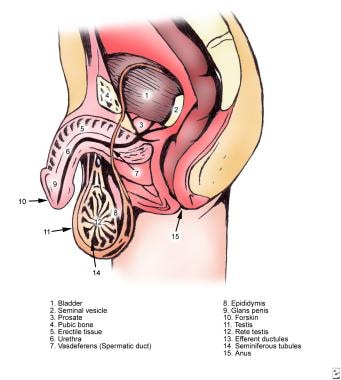
Testis
Blood-Brain Barrier
Rete Testis
Spermatogenesis
Sertoli Cells
Cryptorchidism
Leydig Cells
Blood-Testis Barrier
Spermatids
Spermatozoa
Spermatogonia
Seminiferous Epithelium
Blood-Retinal Barrier
Epididymis
Spermatocytes
Blood-Air Barrier
Testosterone
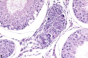
Testicular Neoplasms
Tight Junctions
Communication Barriers
Permeability
Molecular Sequence Data
Infertility, Male
RNA, Messenger
Genitalia, Male
Orchitis
Spermatic Cord Torsion
Tissue Distribution
Occludin
Immunohistochemistry
Capillary Permeability
Amino Acid Sequence
Base Sequence
Rats, Sprague-Dawley
Organ Specificity
Blood-Aqueous Barrier
Sex Differentiation
Reverse Transcriptase Polymerase Chain Reaction
Fertility
Ovary
Sex-Determining Region Y Protein
Zonula Occludens-1 Protein
DNA, Complementary
Seminal Vesicles
In Situ Hybridization
Blood-Nerve Barrier
Follicle Stimulating Hormone
Sertoli Cell Tumor
Cell Membrane Permeability
Gene Expression
Orchiopexy
Gene Expression Regulation, Developmental
Brain
Dibutyl Phthalate
Mice, Inbred C57BL
Cloning, Molecular
Blotting, Northern
Health Services Accessibility
Membrane Proteins
Sperm Motility
Cells, Cultured
Luteinizing Hormone
Epidermis
Seminoma
Sperm Maturation
Claudins
Rats, Wistar
Blotting, Western
Meiosis
Varicocele
Sequence Homology, Amino Acid
Spermatic Cord
Mice, Knockout
Gene Expression Regulation
Inhibins
SOX9 Transcription Factor
Genes, sry
Diethylhexyl Phthalate
Claudin-5
Passage of leptin across the blood-testis barrier. (1/152)
Leptin is a 17-kDa protein, secreted by fat, that controls adiposity and has been proposed to have numerous effects on reproduction in the mouse. To assess whether the effects of leptin on testicular function are direct, we determined whether leptin can cross the murine blood-testis barrier. Multiple time regression analysis showed that a small amount of blood-borne leptin is able to enter the testis but does so by a nonsaturable process. In addition, no significant expression of leptin receptors was found at the Leydig cells or Sertoli cells of the testis. This compares with the presence of a saturable transport system for leptin at the blood-brain barrier and abundant receptors for leptin at the leptomeninges, neurons, and choroid plexus of the central nervous system (CNS). These results support the hypothesis that the effects of leptin on reproductive function are not mediated at the level of the testis but indirectly, probably through the CNS. (+info)Evaluation of cholesteryl ester transfer in the seminiferous tubule cells of immature rats in vivo and in vitro. (2/152)
Sertoli cells and germ cells are separated from the interstitial blood capillaries by an extracellular matrix and the peritubular cells, which constitute a barrier to the movement of plasma lipoproteins. The present study was undertaken to evaluate in vivo and in vitro the high density lipoprotein (HDL) cholesteryl ester transfer from plasma to seminiferous tubule cells in the testis of 30-day-old rats. Firstly, the transfer of HDL cholesteryl oleate from plasma to testicular compartments was evaluated and, secondly, the role of apolipoproteins A-I and E in the uptake of cholesteryl ester by Sertoli cells was investigated. At 2 h after the administration of HDL reconstituted with [3H]cholesteryl ester, dimyristoyl phosphatidylcholine and apolipoproteins, the tissue space in the interstitial cells (740 +/- 60 microliters g-1 cell protein) was fourfold higher than that in the seminiferous tubule cells (170 +/- 10 microliters g-1). Sertoli cells were isolated and incubated with [3H]cholesteryl ester HDL reconstituted with apolipoprotein A-I or E to evaluate the mechanisms of cholesteryl ester influx. At the same apolipoprotein concentration (50 micrograms apolipoprotein ml-1 medium), the uptake of [3H]cholesteryl oleate from phospholipid-apolipoprotein E vesicles was twofold higher than that with phospholipid-apolipoprotein A-I vesicles. The presence of heparin reduced the uptake of cholesteryl ester from apolipoprotein E vesicles but not with apolipoprotein A-I vesicles, indicating that uptake of apolipoprotein A-I vesicles via a secretion of apolipoprotein E by the cells themselves was not involved. These results demonstrate that plasma lipoprotein cholesterol is able to cross the testis lamina propria and that Sertoli cells take up cholesteryl ester for seminiferous tubule cell metabolism mainly via an apolipoprotein E pathway. (+info)Passive immunization with anti-laminin immunoglobulin G modifies the integrity of the seminiferous epithelium and induces arrest of spermatogenesis in the guinea pig. (3/152)
In the testis, the base of the Sertoli cells is in contact with the basement membrane matrix, in which the laminins constitute the major noncollagenous components. We have previously demonstrated that antibodies against a preparation enriched in basement membranes of seminiferous tubules (STBM) or a noncollagenous fraction of STBM passively transferred induced modifications to the basement membranes and focal sloughing of the seminiferous epithelium in the rat. In the present report, we tested the effect of passive immunization with anti-laminin IgG on the limiting membrane of the seminiferous tubules, spermatogenesis, and maintenance of the blood-testis barrier in the adult guinea pig. Rabbit antibodies to laminin 1 (IgG fraction) were injected in adult male guinea pigs (GP). Nonimmunized GP and GP immunized with normal rabbit serum IgG were used as controls. Measurements of variations in the diameter and lumen of the tubules and in the size of individual components of the tubular limiting membrane showed that the highest percentage of tubules with reduced lumen occurred 30 days after passive immunization with anti-laminin, when the limiting membrane was thickest and lesions to the seminiferous epithelium were most severe. The lesions included thickening of the limiting membrane, infolding in the basal lamina, deposits of immune complexes coincident with sloughing of pachytene spermatocytes and spermatids, and vacuolization of the Sertoli cells. Mononuclear cell infiltration of the tubules was rare. Permeability tracer studies revealed that Sertoli cell tight junctions remained impermeable. Fifty and 80 days after treatment, the basement membrane of the tubules and the progression of the spermatogenesis were normal. Passive immunization with anti-laminin IgG provided a valuable experimental model for the in vivo study of the influence of the basement membrane on the issue of spermatogenesis and the integrity of the seminiferous epithelium. (+info)Effect of efferent duct ligation on the function of the blood-testis barrier in rats. (4/152)
The function of the blood-testis barrier has been assessed from the ratio of the Cr-EDTA space in the parenchyma to the measured interstitial volume in the testes of rats at various times after unilateral ligation of the efferent ducts. The barrier remained effective during the phase of fluid accumulation and testicular mass gain, which was linear for at least 24 h, but the testis mass began to decrease between 32 and 40 h after efferent duct ligation, and the Cr-EDTA space at 40 and 48 h after efferent duct ligation exceeded the volume of the interstitial tissue. This finding indicated that, at these times, the barrier to Cr-EDTA, which is normally excluded from the tubules, had broken down and the marker was entering the tubules. Thereafter, the Cr-EDTA space decreased again to be less than the interstitial tissue volume, indicating a restoration of the barrier function, although degeneration of the seminiferous epithelium continued to become more obvious. The present study is the first report of a reversible breakdown of the barrier, but the relevance of the breakdown to the effects on spermatogenesis requires further study. (+info)Testicular damage by microcirculatory disruption and colonization of an immune-privileged site during Borrelia crocidurae infection. (5/152)
The agent of African relapsing fever, Borrelia crocidurae, causes reversible multiple organ damage. We hypothesize that this damage is caused when the spirochete forms aggregate with erythrocytes in vivo, creating rosettes that plug the microcirculatory system. To test this hypothesis, we compared testicular microcirculation over an extended time period in two groups of rats: one experimentally inoculated with B. crocidurae, the other with the nonerythrocyte rosette-forming Borrelia hermsii. In the B. crocidurae group, erythrocyte rosettes formed during spiro-chetemia blocked precapillary blood vessels and reduced the normal pattern of microcirculatory blood flow. After spirochetemia, erythrocyte rosettes disappeared and flow was normalized. Decreased blood flow and focal vascular damage with increased permeability and interstitial bleeding adjacent to the erythrocyte microemboli induced cell death in seminiferous tubules. Interestingly, we found that B. crocidurae could penetrate the tubules and remain in the testis long after the end of spirochetemia, suggesting that the testis can serve as a reservoir for this bacteria in subsequent relapses. The group infected with B. hermsii displayed normal testicular blood flow and vasomotion at all selected time points, and suffered no testicular damage. These results confirmed our hypothesis that the erythrocyte rosettes produce vascular obstruction and are the main cause of histopathology seen in model animal and human infections. (+info)A 22-amino acid synthetic peptide corresponding to the second extracellular loop of rat occludin perturbs the blood-testis barrier and disrupts spermatogenesis reversibly in vivo. (6/152)
When Sertoli cells were cultured in vitro on Matrigel-coated bicameral units, the assembly of the inter-Sertoli tight junction (TJ) permeability barrier correlated with an induction of occludin expression. Inclusion of a 22-amino acid peptide, NH(2)-GSQIYTICSQFYTPGGTGLYVD-COOH, corresponding to residues 209-230 in the second extracellular loop of rat occludin, at 0.2-4 microM into Sertoli cell cultures could perturb the assembly of Sertoli TJs dose-dependently and reversibly. This peptide apparently exerts its effects by interfering with the homotypic interactions of two occludin molecules between adjacent Sertoli cells at the sites of TJs, thereby disrupting TJs, which, in turn, causes a decline in transepithelial electrical resistance across the Sertoli cell epithelium. When similar experiments were performed using a 22-amino acid myotubularin peptide, NH(2)-TKVNERYELCDTYPALLAVPAN-COOH (residues 156-177), no effects on the assembly of inter-Sertoli TJs in vitro were noted. When a single dose of this synthetic occludin peptide was administered to adult rats intratesticularly at 1.5-10 mg/testis, germ cells began to deplete from the seminiferous epithelium within 8-16 days. By 27 days, virtually all tubules were devoid of germ cells. This antispermatogenic effect was reversible, because germ cells progressively repopulated the epithelium thereafter. Treated testes were indistinguishable from normal or control testes by 68 days post-occludin peptide treatment when assessed using histological analysis. In contrast, control rats receiving either no treatment, vehicle alone, or a 22-amino acid synthetic peptide of myotubularin displayed no changes in the testicular morphology at all time points. The occludin peptide-induced germ cell depletion was also accompanied by a disruption of the blood-testis barrier (BTB) when assessed by micropuncture techniques quantifying [(125)I]-BSA in rete testis fluid and seminiferous tubular fluid following i.v. administration of [(125)I]-BSA through the jugular vein. These results illustrate that the occludin peptide-induced disruption of the BTB may possibly affect the underlying adherens junctions, which causes premature release of germ cells from the epithelium and reversible infertility. (+info)Expression of a blood-brain barrier-specific antigen in the reproductive tract of the male rat. (7/152)
The endothelial barrier antigen (EBA) is a protein expressed specifically by the endothelial cells of the rat brain barrier vessels. This antigen has been described as a 'barrier protein' and is used as a marker for the competent blood-brain barrier. A blood-testis barrier has also been described. However, unlike the blood-brain barrier, which is formed by endothelial cells, the blood-testis barrier is formed mainly by the Sertoli cells, which provide an isolated environment for spermatogenic cells within the seminiferous tubules. Testicular blood vessels express the erythroid glucose transporter protein and other markers, which are strongly expressed in brain blood vessels, and may contribute to the blood-testis barrier. This study was carried out to determine whether Sertoli cells or testicular blood vessels express EBA. Tissues of other organs were used as controls for EBA expression. EBA was expressed by the endothelial cells in most microvessels of the testis, and in a few vessels of the epididymis, seminal vesicle, prostate gland, vas deferens and bladder-neck region. Furthermore, EBA was strongly and consistently detected in epithelial cells of the rete testis and dorsolateral prostate gland, and in a few epithelial cells of the ventral prostate gland, the seminal vesicle and the coagulating gland. However, Sertoli cells, which are the main site of the blood-testis barrier, were negative for EBA. In conclusion, EBA may have a wider role in rat tissues than has been previously appreciated. (+info)Cr(V) involvement in the toxicity pathway of testicular damage. (8/152)
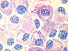
AIM: The functional integrity of the blood-testis barrier (BTB) in male mice exposed to Cr(V) was studied in order to clarify the mechanism underlying testicular injury. METHODS: Adult male mice were subcutaneously injected repeated doses of 8.02 micromol (0.5 ml) of Cr/mouse.day for 5 days. Animals receiving a similar volume of bis(hydroxyethyl)-aminotris(hydroxymethyl)methane buffer (BT) were used as controls. The animals were sacrificed on day 6 and small fragments of seminiferous tubules, approximately 8-10 mm length, were incised and sutured at both ends. They were exposed in vitro to horseradish peroxidase-containing culture medium for 10 minutes. Tissues were then fixed and processed for ultrastructural studies. RESULTS: Controls and Cr(V)-treated group resulted in the uptake of the tracer by Sertoli cells. However, the major finding consisted in the permeability of the BTB only in the Cr(V)-group, as evidenced by the presence of the tracer within the junctions between the neighbouring Sertoli cells. CONCLUSION: The BTB is disrupted in mice submitted to Cr(V). The permeability of the BTB is a crucial feature to be investigated for the understanding of lesions within the seminiferous tubule. (+info)The testis, also known as the testicle, is a male reproductive organ that is part of the endocrine system. It is located in the scrotum, outside of the abdominal cavity. The main function of the testis is to produce sperm and testosterone, the primary male sex hormone.
The testis is composed of many tiny tubules called seminiferous tubules, where sperm are produced. These tubules are surrounded by a network of blood vessels, nerves, and supportive tissues. The sperm then travel through a series of ducts to the epididymis, where they mature and become capable of fertilization.
Testosterone is produced in the Leydig cells, which are located in the interstitial tissue between the seminiferous tubules. Testosterone plays a crucial role in the development and maintenance of male secondary sexual characteristics, such as facial hair, deep voice, and muscle mass. It also supports sperm production and sexual function.
Abnormalities in testicular function can lead to infertility, hormonal imbalances, and other health problems. Regular self-examinations and medical check-ups are recommended for early detection and treatment of any potential issues.
The Blood-Brain Barrier (BBB) is a highly specialized, selective interface between the central nervous system (CNS) and the circulating blood. It is formed by unique endothelial cells that line the brain's capillaries, along with tight junctions, astrocytic foot processes, and pericytes, which together restrict the passage of substances from the bloodstream into the CNS. This barrier serves to protect the brain from harmful agents and maintain a stable environment for proper neural function. However, it also poses a challenge in delivering therapeutics to the CNS, as most large and hydrophilic molecules cannot cross the BBB.
The rete testis is a network of tubules in the male reproductive system that serves as a passageway for sperm to travel from the seminiferous tubules, where sperm are produced, to the epididymis, where they mature. It is located in the mediastinum testis, which is the central part of the testicle.
The rete testis is made up of a series of interconnected tubules that are lined with simple cuboidal epithelial cells. These tubules merge to form larger ducts called efferent ductules, which then connect to the epididymis. The rete testis plays an important role in the transport and maturation of sperm, as well as in the regulation of fluid balance in the male reproductive system.
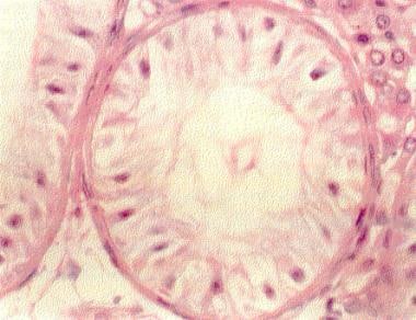
Spermatogenesis is the process by which sperm cells, or spermatozoa, are produced in male organisms. It occurs in the seminiferous tubules of the testes and involves several stages:
1. Spermatocytogenesis: This is the initial stage where diploid spermatogonial stem cells divide mitotically to produce more spermatogonia, some of which will differentiate into primary spermatocytes.
2. Meiosis: The primary spermatocytes undergo meiotic division to form haploid secondary spermatocytes, which then divide again to form haploid spermatids. This process results in the reduction of chromosome number from 46 (diploid) to 23 (haploid).
3. Spermiogenesis: The spermatids differentiate into spermatozoa, undergoing morphological changes such as the formation of a head and tail. During this stage, most of the cytoplasm is discarded, resulting in highly compacted and streamlined sperm cells.
4. Spermation: The final stage where mature sperm are released from the seminiferous tubules into the epididymis for further maturation and storage.
The entire process takes approximately 72-74 days in humans, with continuous production throughout adulthood.
Sertoli cells, also known as sustentacular cells or nurse cells, are specialized cells in the seminiferous tubules of the testis in mammals. They play a crucial role in supporting and nurturing the development of sperm cells (spermatogenesis). Sertoli cells create a microenvironment within the seminiferous tubules that facilitates the differentiation, maturation, and survival of germ cells.
These cells have several essential functions:
1. Blood-testis barrier formation: Sertoli cells form tight junctions with each other, creating a physical barrier called the blood-testis barrier, which separates the seminiferous tubules into basal and adluminal compartments. This barrier protects the developing sperm cells from the immune system and provides an isolated environment for their maturation.
2. Nutrition and support: Sertoli cells provide essential nutrients and growth factors to germ cells, ensuring their proper development and survival. They also engulf and digest residual bodies, which are byproducts of spermatid differentiation.
3. Phagocytosis: Sertoli cells have phagocytic properties, allowing them to remove debris and dead cells within the seminiferous tubules.
4. Hormone metabolism: Sertoli cells express receptors for various hormones, such as follicle-stimulating hormone (FSH), testosterone, and estradiol. They play a role in regulating hormonal signaling within the testis by metabolizing these hormones or producing inhibins, which modulate FSH secretion from the pituitary gland.
5. Regulation of spermatogenesis: Sertoli cells produce and secrete various proteins and growth factors that influence germ cell development and proliferation. They also control the release of mature sperm cells into the epididymis through a process called spermiation.
Cryptorchidism is a medical condition in which one or both of a male infant's testicles fail to descend from the abdomen into the scrotum before birth or within the first year of life. Normally, the testicles descend from the abdomen into the scrotum during fetal development in the second trimester. If the testicles do not descend on their own, medical intervention may be necessary to correct the condition.
Cryptorchidism is a common birth defect, affecting about 3-5% of full-term and 30% of preterm male infants. In most cases, the testicle will descend on its own within the first six months of life. If it does not, treatment may be necessary to prevent complications such as infertility, testicular cancer, and inguinal hernia.
Treatment for cryptorchidism typically involves surgery to bring the testicle down into the scrotum. This procedure is called orchiopexy and is usually performed before the age of 2. In some cases, hormonal therapy may be used as an alternative to surgery. However, this approach has limited success and is generally only recommended in certain situations.
Overall, cryptorchidism is a treatable condition that can help prevent future health problems if addressed early on. Regular check-ups with a pediatrician or healthcare provider can help ensure timely diagnosis and treatment of this condition.
Leydig cells, also known as interstitial cells of Leydig or interstitial cell-stroma, are cells in the testes that produce and release testosterone and other androgens into the bloodstream. They are located in the seminiferous tubules of the testis, near the blood vessels, and are named after Franz Leydig, the German physiologist who discovered them in 1850.
Leydig cells contain cholesterol esters, which serve as precursors for the synthesis of testosterone. They respond to luteinizing hormone (LH) released by the anterior pituitary gland, which stimulates the production and release of testosterone. Testosterone is essential for the development and maintenance of male secondary sexual characteristics, such as facial hair, deep voice, and muscle mass. It also plays a role in sperm production and bone density.
In addition to their endocrine function, Leydig cells have been shown to have non-hormonal functions, including phagocytosis, antigen presentation, and immune regulation. However, these functions are not as well understood as their hormonal roles.
The Blood-Testis Barrier (BTB) is a unique structural and functional feature of the seminiferous epithelium in the testes, which forms a tight junction between adjacent Sertoli cells in the semi-niferous tubules. This barrier selectively restricts the passage of molecules, including potentially harmful substances and immune cells, from the systemic circulation into the adluminal compartment of the seminiferous epithelium where spermatogenesis occurs. This helps to maintain a immunologically privileged microenvironment that is essential for the survival and maturation of developing sperm cells, preventing an immune response against them. The BTB also regulates the movement of molecules required for spermatogenesis, such as nutrients, hormones, and signaling molecules, from the basal compartment to the adluminal compartment.
Spermatids are immature sperm cells that are produced during the process of spermatogenesis in the male testes. They are the product of the final stage of meiosis, where a diploid spermatocyte divides into four haploid spermatids. Each spermatid then undergoes a series of changes, including the development of a tail for motility and the condensation of its nucleus to form a head containing the genetic material. Once this process is complete, the spermatids are considered mature spermatozoa and are capable of fertilizing an egg.
Spermatozoa are the male reproductive cells, or gametes, that are produced in the testes. They are microscopic, flagellated (tail-equipped) cells that are highly specialized for fertilization. A spermatozoon consists of a head, neck, and tail. The head contains the genetic material within the nucleus, covered by a cap-like structure called the acrosome which contains enzymes to help the sperm penetrate the female's egg (ovum). The long, thin tail propels the sperm forward through fluid, such as semen, enabling its journey towards the egg for fertilization.
Spermatogonia are a type of diploid germ cells found in the seminiferous tubules of the testis. They are the stem cells responsible for sperm production (spermatogenesis) in males. There are two types of spermatogonia: A-dark (Ad) and A-pale (Ap). The Ad spermatogonia function as reserve stem cells, while the Ap spermatogonia serve as the progenitor cells that divide to produce type B spermatogonia. Type B spermatogonia then differentiate into primary spermatocytes, which undergo meiosis to form haploid spermatozoa.
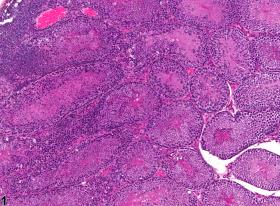
The seminiferous epithelium is a specialized type of epithelial tissue that lines the seminiferous tubules within the testes. It is composed of various cell types, including germ cells in different stages of development (spermatogonia, primary and secondary spermatocytes, spermatids) and supportive cells called Sertoli cells.
The primary function of the seminiferous epithelium is to support sperm production (spermatogenesis). The Sertoli cells provide structural support and nourishment to the developing germ cells, helping them to differentiate into mature spermatozoa (sperm). This process involves a series of complex cellular events, including mitosis, meiosis, and spermiogenesis.
In addition to its role in sperm production, the seminiferous epithelium also plays a crucial part in maintaining the blood-testis barrier, which separates the testicular environment from the systemic circulation. This barrier helps protect developing germ cells from potential immune attacks and maintains an optimal microenvironment for spermatogenesis.
The blood-retinal barrier (BRB) is a specialized physiological barrier in the eye that helps regulate the movement of molecules between the retina and the bloodstream. It is made up of tight junctions between the endothelial cells of retinal blood vessels and between the pigment epithelium cells of the retina, which restrict the paracellular diffusion of solutes.
The BRB plays a crucial role in maintaining the health and function of the retina by preventing harmful substances from entering the retina while allowing essential nutrients and oxygen to reach the retinal tissues. Disruption of the BRB has been implicated in various retinal diseases, including diabetic retinopathy, age-related macular degeneration, and retinal vein occlusion.
The epididymis is a tightly coiled tube located on the upper and posterior portion of the testicle that serves as the site for sperm maturation and storage. It is an essential component of the male reproductive system. The epididymis can be divided into three parts: the head (where newly produced sperm enter from the testicle), the body, and the tail (where mature sperm exit and are stored). Any abnormalities or inflammation in the epididymis may lead to discomfort, pain, or infertility.
Spermatocytes are a type of cell that is involved in the process of spermatogenesis, which is the formation of sperm in the testes. Specifically, spermatocytes are the cells that undergo meiosis, a special type of cell division that results in the production of four haploid daughter cells, each containing half the number of chromosomes as the parent cell.
There are two types of spermatocytes: primary and secondary. Primary spermatocytes are diploid cells that contain 46 chromosomes (23 pairs). During meiosis I, these cells undergo a process called crossing over, in which genetic material is exchanged between homologous chromosomes. After crossing over, the primary spermatocytes divide into two secondary spermatocytes, each containing 23 chromosomes (but still with 23 pairs).
Secondary spermatocytes then undergo meiosis II, which results in the formation of four haploid spermatids. Each spermatid contains 23 single chromosomes and will eventually develop into a mature sperm cell through a process called spermiogenesis.
It's worth noting that spermatocytes are only found in males, as they are specific to the male reproductive system.
I am not aware of a widely recognized or established medical term called "Blood-Air Barrier." It is possible that you may be referring to a concept or phenomenon that goes by a different name, or it could be a term that is specific to certain context or field within medicine.
In general, the terms "blood" and "air" refer to two distinct and separate compartments in the body, and there are various physiological barriers that prevent them from mixing with each other under normal circumstances. For example, the alveolar-capillary membrane in the lungs serves as a barrier that allows for the exchange of oxygen and carbon dioxide between the air in the alveoli and the blood in the capillaries, while preventing the two from mixing together.
If you could provide more context or clarify what you mean by "Blood-Air Barrier," I may be able to provide a more specific answer.
Testosterone is a steroid hormone that belongs to androsten class of hormones. It is primarily secreted by the Leydig cells in the testes of males and, to a lesser extent, by the ovaries and adrenal glands in females. Testosterone is the main male sex hormone and anabolic steroid. It plays a key role in the development of masculine characteristics, such as body hair and muscle mass, and contributes to bone density, fat distribution, red cell production, and sex drive. In females, testosterone contributes to sexual desire and bone health. Testosterone is synthesized from cholesterol and its production is regulated by luteinizing hormone (LH) and follicle-stimulating hormone (FSH).
Testicular diseases refer to a range of conditions that affect the testicles, the male reproductive organs located in the scrotum. These diseases can affect either one or both testicles and may cause pain, swelling, or impact fertility. Here are some examples of testicular diseases:
1. Testicular cancer: A malignant tumor that develops in the testicle. It is a relatively rare cancer but is highly treatable if detected early.
2. Testicular torsion: A surgical emergency that occurs when the spermatic cord, which supplies blood to the testicle, becomes twisted, cutting off the blood flow.
3. Epididymitis: An infection or inflammation of the epididymis, a coiled tube that stores and carries sperm from the testicle.
4. Orchitis: An infection or inflammation of the testicle itself. It can occur on its own or as a complication of mumps.
5. Hydrocele: A fluid-filled sac that forms around the testicle, causing swelling.
6. Varicocele: Enlarged veins in the scrotum that can cause pain and affect fertility.
7. Inguinal hernia: A condition where a portion of the intestine or fat protrudes through a weakened area in the abdominal wall, often appearing as a bulge in the groin or scrotum.
8. Testicular trauma: Injury to the testicle, which can result from accidents, sports injuries, or other causes.
9. Undescended testicles: A condition where one or both testicles fail to descend from the abdomen into the scrotum before birth.
It is essential for men to perform regular self-examinations to check for any unusual lumps, swelling, or pain in the testicles and seek medical attention if they notice any changes.
Testicular neoplasms are abnormal growths or tumors in the testicle that can be benign (non-cancerous) or malignant (cancerous). They are a type of genitourinary cancer, which affects the reproductive and urinary systems. Testicular neoplasms can occur in men of any age but are most commonly found in young adults between the ages of 15 and 40.
Testicular neoplasms can be classified into two main categories: germ cell tumors and non-germ cell tumors. Germ cell tumors, which arise from the cells that give rise to sperm, are further divided into seminomas and non-seminomas. Seminomas are typically slow-growing and have a good prognosis, while non-seminomas tend to grow more quickly and can spread to other parts of the body.
Non-germ cell tumors are less common than germ cell tumors and include Leydig cell tumors, Sertoli cell tumors, and lymphomas. These tumors can have a variety of clinical behaviors, ranging from benign to malignant.
Testicular neoplasms often present as a painless mass or swelling in the testicle. Other symptoms may include a feeling of heaviness or discomfort in the scrotum, a dull ache in the lower abdomen or groin, and breast enlargement (gynecomastia).
Diagnosis typically involves a physical examination, imaging studies such as ultrasound or CT scan, and blood tests to detect tumor markers. Treatment options depend on the type and stage of the neoplasm but may include surgery, radiation therapy, chemotherapy, or a combination of these modalities. Regular self-examinations of the testicles are recommended for early detection and improved outcomes.
Tight junctions, also known as zonula occludens, are specialized types of intercellular junctions that occur in epithelial and endothelial cells. They are located near the apical side of the lateral membranes of adjacent cells, where they form a continuous belt-like structure that seals off the space between the cells.
Tight junctions are composed of several proteins, including occludin, claudins, and junctional adhesion molecules (JAMs), which interact to form a network of strands that create a tight barrier. This barrier regulates the paracellular permeability of ions, solutes, and water, preventing their uncontrolled movement across the epithelial or endothelial layer.
Tight junctions also play an important role in maintaining cell polarity by preventing the mixing of apical and basolateral membrane components. Additionally, they are involved in various signaling pathways that regulate cell proliferation, differentiation, and survival.
Communication barriers in a medical context refer to any factors that prevent or hinder the effective exchange of information between healthcare providers and patients, or among healthcare professionals themselves. These barriers can lead to misunderstandings, errors, and poor patient outcomes. Common communication barriers include:
1. Language differences: When patients and healthcare providers do not speak the same language, it can lead to miscommunication and errors in diagnosis and treatment.
2. Cultural differences: Cultural beliefs and values can affect how patients perceive and communicate their symptoms and concerns, as well as how healthcare providers deliver care.
3. Literacy levels: Low health literacy can make it difficult for patients to understand medical information, follow treatment plans, and make informed decisions about their care.
4. Disability: Patients with hearing or vision impairments, speech disorders, or cognitive impairments may face unique communication challenges that require accommodations and specialized communication strategies.
5. Emotional factors: Patients who are anxious, stressed, or in pain may have difficulty communicating effectively, and healthcare providers may be less likely to listen actively or ask open-ended questions.
6. Power dynamics: Hierarchical relationships between healthcare providers and patients can create power imbalances that discourage patients from speaking up or asking questions.
7. Noise and distractions: Environmental factors such as noise, interruptions, and distractions can make it difficult for patients and healthcare providers to hear, focus, and communicate effectively.
Effective communication is critical in healthcare settings, and addressing communication barriers requires a multifaceted approach that includes training for healthcare providers, language services for limited English proficient patients, and accommodations for patients with disabilities.
In the context of medicine and physiology, permeability refers to the ability of a tissue or membrane to allow the passage of fluids, solutes, or gases. It is often used to describe the property of the capillary walls, which control the exchange of substances between the blood and the surrounding tissues.
The permeability of a membrane can be influenced by various factors, including its molecular structure, charge, and the size of the molecules attempting to pass through it. A more permeable membrane allows for easier passage of substances, while a less permeable membrane restricts the movement of substances.
In some cases, changes in permeability can have significant consequences for health. For example, increased permeability of the blood-brain barrier (a specialized type of capillary that regulates the passage of substances into the brain) has been implicated in a number of neurological conditions, including multiple sclerosis, Alzheimer's disease, and traumatic brain injury.
Sperm count, also known as sperm concentration, is the number of sperm present in a given volume of semen. The World Health Organization (WHO) previously defined a normal sperm count as at least 20 million sperm per milliliter of semen. However, more recent studies suggest that fertility may be affected even when sperm counts are slightly lower than this threshold. It's important to note that sperm count is just one factor among many that can influence male fertility. Other factors, such as sperm motility (the ability of sperm to move properly) and morphology (the shape of the sperm), also play crucial roles in successful conception.
Organ size refers to the volume or physical measurement of an organ in the body of an individual. It can be described in terms of length, width, and height or by using specialized techniques such as imaging studies (like CT scans or MRIs) to determine the volume. The size of an organ can vary depending on factors such as age, sex, body size, and overall health status. Changes in organ size may indicate various medical conditions, including growths, inflammation, or atrophy.
Molecular sequence data refers to the specific arrangement of molecules, most commonly nucleotides in DNA or RNA, or amino acids in proteins, that make up a biological macromolecule. This data is generated through laboratory techniques such as sequencing, and provides information about the exact order of the constituent molecules. This data is crucial in various fields of biology, including genetics, evolution, and molecular biology, allowing for comparisons between different organisms, identification of genetic variations, and studies of gene function and regulation.
Male infertility is a condition characterized by the inability to cause pregnancy in a fertile female. It is typically defined as the failure to achieve a pregnancy after 12 months or more of regular unprotected sexual intercourse.
The causes of male infertility can be varied and include issues with sperm production, such as low sperm count or poor sperm quality, problems with sperm delivery, such as obstructions in the reproductive tract, or hormonal imbalances that affect sperm production. Other factors that may contribute to male infertility include genetic disorders, environmental exposures, lifestyle choices, and certain medical conditions or treatments.
It is important to note that male infertility can often be treated or managed with medical interventions, such as medication, surgery, or assisted reproductive technologies (ART). A healthcare provider can help diagnose the underlying cause of male infertility and recommend appropriate treatment options.
Messenger RNA (mRNA) is a type of RNA (ribonucleic acid) that carries genetic information copied from DNA in the form of a series of three-base code "words," each of which specifies a particular amino acid. This information is used by the cell's machinery to construct proteins, a process known as translation. After being transcribed from DNA, mRNA travels out of the nucleus to the ribosomes in the cytoplasm where protein synthesis occurs. Once the protein has been synthesized, the mRNA may be degraded and recycled. Post-transcriptional modifications can also occur to mRNA, such as alternative splicing and addition of a 5' cap and a poly(A) tail, which can affect its stability, localization, and translation efficiency.
Germ cells are the reproductive cells, also known as sex cells, that combine to form offspring in sexual reproduction. In females, germ cells are called ova or egg cells, and in males, they are called spermatozoa or sperm cells. These cells are unique because they carry half the genetic material necessary for creating new life. They are produced through a process called meiosis, which reduces their chromosome number by half, ensuring that when two germ cells combine during fertilization, the normal diploid number of chromosomes is restored.
"Male genitalia" refers to the reproductive and sexual organs that are typically present in male individuals. These structures include:
1. Testes: A pair of oval-shaped glands located in the scrotum that produce sperm and testosterone.
2. Epididymis: A long, coiled tube that lies on the surface of each testicle where sperm matures and is stored.
3. Vas deferens: A pair of muscular tubes that transport sperm from the epididymis to the urethra.
4. Seminal vesicles: Glands that produce a fluid that mixes with sperm to create semen.
5. Prostate gland: A small gland that surrounds the urethra and produces a fluid that also mixes with sperm to create semen.
6. Bulbourethral glands (Cowper's glands): Two pea-sized glands that produce a lubricating fluid that is released into the urethra during sexual arousal.
7. Urethra: A tube that runs through the penis and carries urine from the bladder out of the body, as well as semen during ejaculation.
8. Penis: The external organ that serves as both a reproductive and excretory organ, expelling both semen and urine.
Orchitis is a medical condition characterized by inflammation of one or both testicles, usually caused by an infection. The most common cause of orchitis is a bacterial infection that spreads from the epididymis, resulting in a condition known as epididymo-orchitis. However, viral infections such as mumps can also lead to orchitis. Symptoms may include sudden and severe pain in the testicle(s), swelling, warmth, redness of the overlying skin, nausea, vomiting, and fever. Treatment typically involves antibiotics for bacterial infections and supportive care for symptom relief. If left untreated, orchitis can lead to complications such as infertility or testicular atrophy.
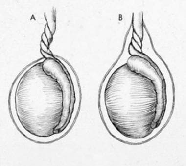
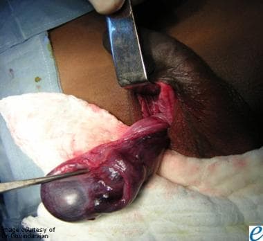
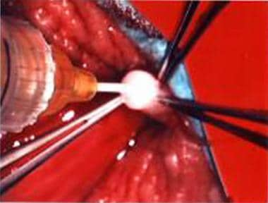
Spermatic cord torsion is a urological emergency that refers to the twisting of the spermatic cord, which contains the vas deferens, blood vessels (testicular artery and pampiniform plexus), nerves, and lymphatics. This twisting results in the compromise of the blood supply to the testicle, leading to potential ischemia, necrosis, and loss of the testicle if not promptly diagnosed and treated.
The spermatic cord torsion mainly affects the pediatric population, particularly newborns and adolescents; however, it can also occur in adults, especially those with a history of an undescended testicle or previous episodes of torsion. The most common presenting symptom is sudden onset of severe scrotal pain, often associated with nausea, vomiting, and fever. A physical examination may reveal swelling, tenderness, and elevation of the affected testicle (known as a high-riding or "bell clapper" testicle). Diagnosis typically involves imaging studies such as ultrasound or Doppler ultrasonography, although in some cases, surgical exploration might be necessary for definitive diagnosis and treatment.
Treatment of spermatic cord torsion usually involves prompt surgical intervention to untwist the spermatic cord and secure the affected testicle to the scrotal wall (orchidopexy) to prevent recurrence. Delayed diagnosis and treatment can lead to severe complications, including loss of the testicle, infertility, and chronic pain.
Sexual maturation is the process of physical development during puberty that leads to the ability to reproduce. This process involves the development of primary and secondary sexual characteristics, changes in hormone levels, and the acquisition of reproductive capabilities. In females, this includes the onset of menstruation and the development of breasts and hips. In males, this includes the deepening of the voice, growth of facial hair, and the production of sperm. Achieving sexual maturation is an important milestone in human development and typically occurs during adolescence.
The scrotum is a part of the external male genitalia. It's a sac-like structure made up of several layers of skin and smooth muscle, which hangs down behind and beneath the penis. The primary function of the scrotum is to maintain the testicles at a temperature slightly lower than the core body temperature, which is optimal for sperm production.
The scrotum contains two compartments, each one housing a testicle. It's located in the pubic region and is usually visible externally. The skin of the scrotum is thin and wrinkled, which allows it to expand and contract depending on the temperature, accommodating the shrinking or swelling of the testicles.
Please note that while I strive to provide accurate information, this definition is intended to be a general overview and should not replace professional medical advice.
Tissue distribution, in the context of pharmacology and toxicology, refers to the way that a drug or xenobiotic (a chemical substance found within an organism that is not naturally produced by or expected to be present within that organism) is distributed throughout the body's tissues after administration. It describes how much of the drug or xenobiotic can be found in various tissues and organs, and is influenced by factors such as blood flow, lipid solubility, protein binding, and the permeability of cell membranes. Understanding tissue distribution is important for predicting the potential effects of a drug or toxin on different parts of the body, and for designing drugs with improved safety and efficacy profiles.
Occludin is a protein that is a component of tight junctions, which are structures that form a barrier between adjacent cells in epithelial and endothelial tissues. Tight junctions help to regulate the movement of molecules between cells and play a crucial role in maintaining the integrity of these tissues.
Occludin is composed of four transmembrane domains, two extracellular loops, and intracellular N- and C-termini. The extracellular loops interact with other tight junction proteins to form the intercellular seal, while the intracellular domains interact with various signaling molecules and cytoskeletal components to regulate the assembly and disassembly of tight junctions.
Mutations in the gene that encodes occludin have been associated with various human diseases, including inflammatory bowel disease, liver cirrhosis, and skin disorders. Additionally, changes in occludin expression and localization have been implicated in the development of cancer and neurological disorders.
Immunohistochemistry (IHC) is a technique used in pathology and laboratory medicine to identify specific proteins or antigens in tissue sections. It combines the principles of immunology and histology to detect the presence and location of these target molecules within cells and tissues. This technique utilizes antibodies that are specific to the protein or antigen of interest, which are then tagged with a detection system such as a chromogen or fluorophore. The stained tissue sections can be examined under a microscope, allowing for the visualization and analysis of the distribution and expression patterns of the target molecule in the context of the tissue architecture. Immunohistochemistry is widely used in diagnostic pathology to help identify various diseases, including cancer, infectious diseases, and immune-mediated disorders.
Capillary permeability refers to the ability of substances to pass through the walls of capillaries, which are the smallest blood vessels in the body. These tiny vessels connect the arterioles and venules, allowing for the exchange of nutrients, waste products, and gases between the blood and the surrounding tissues.
The capillary wall is composed of a single layer of endothelial cells that are held together by tight junctions. The permeability of these walls varies depending on the size and charge of the molecules attempting to pass through. Small, uncharged molecules such as water, oxygen, and carbon dioxide can easily diffuse through the capillary wall, while larger or charged molecules such as proteins and large ions have more difficulty passing through.
Increased capillary permeability can occur in response to inflammation, infection, or injury, allowing larger molecules and immune cells to enter the surrounding tissues. This can lead to swelling (edema) and tissue damage if not controlled. Decreased capillary permeability, on the other hand, can lead to impaired nutrient exchange and tissue hypoxia.
Overall, the permeability of capillaries is a critical factor in maintaining the health and function of tissues throughout the body.
An amino acid sequence is the specific order of amino acids in a protein or peptide molecule, formed by the linking of the amino group (-NH2) of one amino acid to the carboxyl group (-COOH) of another amino acid through a peptide bond. The sequence is determined by the genetic code and is unique to each type of protein or peptide. It plays a crucial role in determining the three-dimensional structure and function of proteins.
A base sequence in the context of molecular biology refers to the specific order of nucleotides in a DNA or RNA molecule. In DNA, these nucleotides are adenine (A), guanine (G), cytosine (C), and thymine (T). In RNA, uracil (U) takes the place of thymine. The base sequence contains genetic information that is transcribed into RNA and ultimately translated into proteins. It is the exact order of these bases that determines the genetic code and thus the function of the DNA or RNA molecule.
Sprague-Dawley rats are a strain of albino laboratory rats that are widely used in scientific research. They were first developed by researchers H.H. Sprague and R.C. Dawley in the early 20th century, and have since become one of the most commonly used rat strains in biomedical research due to their relatively large size, ease of handling, and consistent genetic background.
Sprague-Dawley rats are outbred, which means that they are genetically diverse and do not suffer from the same limitations as inbred strains, which can have reduced fertility and increased susceptibility to certain diseases. They are also characterized by their docile nature and low levels of aggression, making them easier to handle and study than some other rat strains.
These rats are used in a wide variety of research areas, including toxicology, pharmacology, nutrition, cancer, and behavioral studies. Because they are genetically diverse, Sprague-Dawley rats can be used to model a range of human diseases and conditions, making them an important tool in the development of new drugs and therapies.
Organ specificity, in the context of immunology and toxicology, refers to the phenomenon where a substance (such as a drug or toxin) or an immune response primarily affects certain organs or tissues in the body. This can occur due to various reasons such as:
1. The presence of specific targets (like antigens in the case of an immune response or receptors in the case of drugs) that are more abundant in these organs.
2. The unique properties of certain cells or tissues that make them more susceptible to damage.
3. The way a substance is metabolized or cleared from the body, which can concentrate it in specific organs.
For example, in autoimmune diseases, organ specificity describes immune responses that are directed against antigens found only in certain organs, such as the thyroid gland in Hashimoto's disease. Similarly, some toxins or drugs may have a particular affinity for liver cells, leading to liver damage or specific drug interactions.
The blood-aqueous barrier (BAB) is a specialized structure in the eye that helps regulate the exchange of nutrients, oxygen, and waste products between the bloodstream and the anterior chamber of the eye. It is composed of two main components: the nonpigmented epithelial cells of the ciliary body and the endothelial cells of the iris vasculature.
The nonpigmented epithelial cells of the ciliary body form a tight junction that separates the anterior chamber from the ciliary blood vessels, while the endothelial cells lining the iris blood vessels also have tight junctions that restrict the movement of molecules between the blood and the anterior chamber.
The BAB helps maintain the homeostasis of the anterior chamber by controlling the entry of immune cells and preventing the passage of large molecules, toxins, and pathogens from the bloodstream into the eye. Dysfunction of the BAB can lead to various ocular diseases such as uveitis, glaucoma, and age-related macular degeneration.
"Sex differentiation" is a term used in the field of medicine, specifically in reproductive endocrinology and genetics. It refers to the biological development of sexual characteristics that distinguish males from females. This process is regulated by hormones and genetic factors.
There are two main stages of sex differentiation: genetic sex determination and gonadal sex differentiation. Genetic sex determination occurs at fertilization, where the combination of X and Y chromosomes determines the sex of the individual (typically, XX = female and XY = male). Gonadal sex differentiation then takes place during fetal development, where the genetic sex signals the development of either ovaries or testes.
Once the gonads are formed, they produce hormones that drive further sexual differentiation, leading to the development of internal reproductive structures (such as the uterus and fallopian tubes in females, and the vas deferens and seminal vesicles in males) and external genitalia.
It's important to note that while sex differentiation is typically categorized as male or female, there are individuals who may have variations in their sexual development, leading to intersex conditions. These variations can occur at any stage of the sex differentiation process and can result in a range of physical characteristics that do not fit neatly into male or female categories.
Orchiectomy is a surgical procedure where one or both of the testicles are removed. It is also known as castration. This procedure can be performed for various reasons, including the treatment of testicular cancer, prostate cancer, or other conditions that may affect the testicles. It can also be done to reduce levels of male hormones in the body, such as in the case of transgender women undergoing gender affirming surgery. The specific medical definition may vary slightly depending on the context and the extent of the procedure.
Reverse Transcriptase Polymerase Chain Reaction (RT-PCR) is a laboratory technique used in molecular biology to amplify and detect specific DNA sequences. This technique is particularly useful for the detection and quantification of RNA viruses, as well as for the analysis of gene expression.
The process involves two main steps: reverse transcription and polymerase chain reaction (PCR). In the first step, reverse transcriptase enzyme is used to convert RNA into complementary DNA (cDNA) by reading the template provided by the RNA molecule. This cDNA then serves as a template for the PCR amplification step.
In the second step, the PCR reaction uses two primers that flank the target DNA sequence and a thermostable polymerase enzyme to repeatedly copy the targeted cDNA sequence. The reaction mixture is heated and cooled in cycles, allowing the primers to anneal to the template, and the polymerase to extend the new strand. This results in exponential amplification of the target DNA sequence, making it possible to detect even small amounts of RNA or cDNA.
RT-PCR is a sensitive and specific technique that has many applications in medical research and diagnostics, including the detection of viruses such as HIV, hepatitis C virus, and SARS-CoV-2 (the virus that causes COVID-19). It can also be used to study gene expression, identify genetic mutations, and diagnose genetic disorders.
Fertility is the natural ability to conceive or to cause conception of offspring. In humans, it is the capacity of a woman and a man to reproduce through sexual reproduction. For women, fertility usually takes place during their reproductive years, which is from adolescence until menopause. A woman's fertility depends on various factors including her age, overall health, and the health of her reproductive system.
For men, fertility can be affected by a variety of factors such as age, genetics, general health, sexual function, and environmental factors that may affect sperm production or quality. Factors that can negatively impact male fertility include exposure to certain chemicals, radiation, smoking, alcohol consumption, drug use, and sexually transmitted infections (STIs).
Infertility is a common medical condition affecting about 10-15% of couples trying to conceive. Infertility can be primary or secondary. Primary infertility refers to the inability to conceive after one year of unprotected sexual intercourse, while secondary infertility refers to the inability to conceive following a previous pregnancy.
Infertility can be treated with various medical and surgical interventions depending on the underlying cause. These may include medications to stimulate ovulation, intrauterine insemination (IUI), in vitro fertilization (IVF), or surgery to correct anatomical abnormalities.
An ovary is a part of the female reproductive system in which ova or eggs are produced through the process of oogenesis. They are a pair of solid, almond-shaped structures located one on each side of the uterus within the pelvic cavity. Each ovary measures about 3 to 5 centimeters in length and weighs around 14 grams.
The ovaries have two main functions: endocrine (hormonal) function and reproductive function. They produce and release eggs (ovulation) responsible for potential fertilization and development of an embryo/fetus during pregnancy. Additionally, they are essential in the production of female sex hormones, primarily estrogen and progesterone, which regulate menstrual cycles, sexual development, and reproduction.
During each menstrual cycle, a mature egg is released from one of the ovaries into the fallopian tube, where it may be fertilized by sperm. If not fertilized, the egg, along with the uterine lining, will be shed, leading to menstruation.
The Sex-Determining Region Y (SRY) protein is a transcription factor that plays a critical role in male sex determination. It is encoded by the SRY gene, which is located on the Y chromosome in humans and many other mammal species. The primary function of the SRY protein is to initiate the development of the testes during embryonic development.
In the absence of a functional SRY protein, the gonads will develop into ovaries. With a functional SRY protein, the gonads will develop into testes, which then produce androgens, including testosterone, that are necessary for the development of male secondary sexual characteristics. Mutations in the SRY gene can lead to sex reversal, where an individual with a Y chromosome develops as a female due to non-functional or absent SRY protein.
Zonula Occludens-1 (ZO-1) protein is a tight junction (TJ) protein, which belongs to the membrane-associated guanylate kinase (MAGUK) family. It plays a crucial role in the formation and maintenance of tight junctions, which are complex structures that form a barrier between neighboring cells in epithelial and endothelial tissues.
Tight junctions are composed of several proteins, including transmembrane proteins and cytoplasmic plaque proteins. ZO-1 is one of the major cytoplasmic plaque proteins that interact with both transmembrane proteins (such as occludin and claudins) and other cytoskeletal proteins to form a network of protein interactions that maintain the integrity of tight junctions.
ZO-1 has multiple domains, including PDZ domains, SH3 domains, and a guanylate kinase-like domain, which allow it to interact with various binding partners. It is involved in regulating paracellular permeability, cell polarity, and signal transduction pathways that control cell proliferation, differentiation, and survival.
Mutations or dysfunction of ZO-1 protein have been implicated in several human diseases, including inflammatory bowel disease, cancer, and neurological disorders.
Complementary DNA (cDNA) is a type of DNA that is synthesized from a single-stranded RNA molecule through the process of reverse transcription. In this process, the enzyme reverse transcriptase uses an RNA molecule as a template to synthesize a complementary DNA strand. The resulting cDNA is therefore complementary to the original RNA molecule and is a copy of its coding sequence, but it does not contain non-coding regions such as introns that are present in genomic DNA.
Complementary DNA is often used in molecular biology research to study gene expression, protein function, and other genetic phenomena. For example, cDNA can be used to create cDNA libraries, which are collections of cloned cDNA fragments that represent the expressed genes in a particular cell type or tissue. These libraries can then be screened for specific genes or gene products of interest. Additionally, cDNA can be used to produce recombinant proteins in heterologous expression systems, allowing researchers to study the structure and function of proteins that may be difficult to express or purify from their native sources.
The seminal vesicles are a pair of glands located in the male reproductive system, posterior to the urinary bladder and superior to the prostate gland. They are approximately 5 cm long and have a convoluted structure with many finger-like projections called infoldings. The primary function of seminal vesicles is to produce and secrete a significant portion of the seminal fluid, which makes up the bulk of semen along with spermatozoa from the testes and fluids from the prostate gland and bulbourethral glands.
The secretion of the seminal vesicles is rich in fructose, which serves as an energy source for sperm, as well as various proteins, enzymes, vitamins, and minerals that contribute to maintaining the optimal environment for sperm survival, nourishment, and transport. During sexual arousal and ejaculation, the smooth muscles in the walls of the seminal vesicles contract, forcing the stored secretion into the urethra, where it mixes with other fluids before being expelled from the body as semen.
In situ hybridization (ISH) is a molecular biology technique used to detect and localize specific nucleic acid sequences, such as DNA or RNA, within cells or tissues. This technique involves the use of a labeled probe that is complementary to the target nucleic acid sequence. The probe can be labeled with various types of markers, including radioisotopes, fluorescent dyes, or enzymes.
During the ISH procedure, the labeled probe is hybridized to the target nucleic acid sequence in situ, meaning that the hybridization occurs within the intact cells or tissues. After washing away unbound probe, the location of the labeled probe can be visualized using various methods depending on the type of label used.
In situ hybridization has a wide range of applications in both research and diagnostic settings, including the detection of gene expression patterns, identification of viral infections, and diagnosis of genetic disorders.
Electric impedance is a measure of opposition to the flow of alternating current (AC) in an electrical circuit or component, caused by both resistance (ohmic) and reactance (capacitive and inductive). It is expressed as a complex number, with the real part representing resistance and the imaginary part representing reactance. The unit of electric impedance is the ohm (Ω).
In the context of medical devices, electric impedance may be used to measure various physiological parameters, such as tissue conductivity or fluid composition. For example, bioelectrical impedance analysis (BIA) uses electrical impedance to estimate body composition, including fat mass and lean muscle mass. Similarly, electrical impedance tomography (EIT) is a medical imaging technique that uses electric impedance to create images of internal organs and tissues.
The Blood-Nerve Barrier (BNB) is a term that refers to the protective mechanism surrounding the nerves, primarily composed of the endothelial cells that make up the blood vessels within the nerve tissue. These cells are tightly joined together, forming a barrier that restricts the movement of substances from the bloodstream into the nerve tissue. This helps maintain a stable environment for the proper functioning of the nervous system.
The BNB is not as strict as the Blood-Brain Barrier (BBB), which surrounds the brain and has more specialized cells to regulate the entry of substances. Nevertheless, the BNB still plays an essential role in controlling the exchange of molecules between the bloodstream and nerve tissue, protecting the nerves from potentially harmful substances while allowing necessary nutrients to reach the nerve cells.
"Sex determination processes" refer to the series of genetic and biological events that occur during embryonic and fetal development which lead to the development of male or female physical characteristics. In humans, this process is typically determined by the presence or absence of a Y chromosome in the fertilized egg. If the egg has a Y chromosome, it will develop into a male (genetically XY) and if it does not have a Y chromosome, it will develop into a female (genetically XX).
The sex determination process involves the activation and repression of specific genes on the sex chromosomes, which direct the development of the gonads (ovaries or testes) and the production of hormones that influence the development of secondary sexual characteristics. This includes the development of internal and external genitalia, as well as other sex-specific physical traits.
It is important to note that while sex is typically determined by genetics and biology, gender identity is a separate construct that can be self-identified and may not align with an individual's biological sex.
Follicle-Stimulating Hormone (FSH) is a glycoprotein hormone secreted and released by the anterior pituitary gland. In females, it promotes the growth and development of ovarian follicles in the ovary, which ultimately leads to the maturation and release of an egg (ovulation). In males, FSH stimulates the testes to produce sperm. It works in conjunction with luteinizing hormone (LH) to regulate reproductive processes. The secretion of FSH is controlled by the hypothalamic-pituitary-gonadal axis and its release is influenced by the levels of gonadotropin-releasing hormone (GnRH), estrogen, inhibin, and androgens.
Gonads are the reproductive organs that produce gametes (sex cells) and sex hormones. In males, the gonads are the testes, which produce sperm and testosterone. In females, the gonads are the ovaries, which produce eggs and estrogen and progesterone. The development, function, and regulation of the gonads are crucial for reproductive health and fertility.
A Sertoli cell tumor is a rare type of sex-cord stromal tumor that develops in the testicles or, more rarely, in the ovaries. These tumors arise from the Sertoli cells, which are specialized cells within the testicle that help to nurture and protect the developing sperm cells. In the ovary, Sertoli cell tumors are thought to arise from similar cells that are part of the supporting tissue in the ovary.
Sertoli cell tumors can occur in people of any age but are most commonly found in middle-aged adults. They are usually slow-growing and may not cause any symptoms, especially if they are small. However, larger tumors or those that have spread (metastasized) may cause various symptoms depending on their location and size.
Symptoms of a Sertoli cell tumor can include:
* A painless lump or swelling in the testicle or ovary
* Abdominal pain or discomfort
* Bloating or a feeling of fullness in the abdomen
* Changes in bowel habits or urinary frequency
* Pain during sexual intercourse (in women)
* Hormonal imbalances, such as gynecomastia (breast development) in men or menstrual irregularities in women.
Diagnosis of a Sertoli cell tumor typically involves a combination of imaging tests, such as ultrasound, CT scan, or MRI, and blood tests to check for elevated levels of certain hormones that may be produced by the tumor. A biopsy may also be performed to confirm the diagnosis and determine the tumor's grade and stage.
Treatment for Sertoli cell tumors typically involves surgical removal of the tumor, along with any affected lymph nodes or other tissues. Additional treatments, such as radiation therapy or chemotherapy, may be recommended in cases where the tumor has spread or is at a higher risk of recurrence. Regular follow-up care is also important to monitor for any signs of recurrence or new tumors.
Cell membrane permeability refers to the ability of various substances, such as molecules and ions, to pass through the cell membrane. The cell membrane, also known as the plasma membrane, is a thin, flexible barrier that surrounds all cells, controlling what enters and leaves the cell. Its primary function is to protect the cell's internal environment and maintain homeostasis.
The permeability of the cell membrane depends on its structure, which consists of a phospholipid bilayer interspersed with proteins. The hydrophilic (water-loving) heads of the phospholipids face outward, while the hydrophobic (water-fearing) tails face inward, creating a barrier that is generally impermeable to large, polar, or charged molecules.
However, specific proteins within the membrane, called channels and transporters, allow certain substances to cross the membrane. Channels are protein structures that span the membrane and provide a pore for ions or small uncharged molecules to pass through. Transporters, on the other hand, are proteins that bind to specific molecules and facilitate their movement across the membrane, often using energy in the form of ATP.
The permeability of the cell membrane can be influenced by various factors, such as temperature, pH, and the presence of certain chemicals or drugs. Changes in permeability can have significant consequences for the cell's function and survival, as they can disrupt ion balances, nutrient uptake, waste removal, and signal transduction.
Gene expression is the process by which the information encoded in a gene is used to synthesize a functional gene product, such as a protein or RNA molecule. This process involves several steps: transcription, RNA processing, and translation. During transcription, the genetic information in DNA is copied into a complementary RNA molecule, known as messenger RNA (mRNA). The mRNA then undergoes RNA processing, which includes adding a cap and tail to the mRNA and splicing out non-coding regions called introns. The resulting mature mRNA is then translated into a protein on ribosomes in the cytoplasm through the process of translation.
The regulation of gene expression is a complex and highly controlled process that allows cells to respond to changes in their environment, such as growth factors, hormones, and stress signals. This regulation can occur at various stages of gene expression, including transcriptional activation or repression, RNA processing, mRNA stability, and translation. Dysregulation of gene expression has been implicated in many diseases, including cancer, genetic disorders, and neurological conditions.
Orchiopexy is a surgical procedure in which the testicle (or testicles) that have descended into the scrotum incompletely or not at all (undescended or retractile testes) are fixed into their normal position within the scrotum. This procedure is typically performed on boys, often between the ages of 6 and 12 months, to correct cryptorchidism, a condition where one or both testicles fail to descend into the scrotum.
The main goals of orchiopexy are to:
1. Place the testicle in its proper anatomical location within the scrotum.
2. Fix the testicle in a stable position to prevent retractile testes from moving back into the inguinal canal.
3. Preserve the testicular blood supply and innervation, ensuring normal testicular function and development.
4. Lower the risk of testicular torsion (twisting of the spermatic cord) and malignancy in later life.
Orchiopexy can be performed through an open or laparoscopic approach, depending on the location of the undescended testicle(s). The choice of surgical technique depends on factors such as the patient's age, associated conditions, and surgeon's preference.
Developmental gene expression regulation refers to the processes that control the activation or repression of specific genes during embryonic and fetal development. These regulatory mechanisms ensure that genes are expressed at the right time, in the right cells, and at appropriate levels to guide proper growth, differentiation, and morphogenesis of an organism.
Developmental gene expression regulation is a complex and dynamic process involving various molecular players, such as transcription factors, chromatin modifiers, non-coding RNAs, and signaling molecules. These regulators can interact with cis-regulatory elements, like enhancers and promoters, to fine-tune the spatiotemporal patterns of gene expression during development.
Dysregulation of developmental gene expression can lead to various congenital disorders and developmental abnormalities. Therefore, understanding the principles and mechanisms governing developmental gene expression regulation is crucial for uncovering the etiology of developmental diseases and devising potential therapeutic strategies.
The brain is the central organ of the nervous system, responsible for receiving and processing sensory information, regulating vital functions, and controlling behavior, movement, and cognition. It is divided into several distinct regions, each with specific functions:
1. Cerebrum: The largest part of the brain, responsible for higher cognitive functions such as thinking, learning, memory, language, and perception. It is divided into two hemispheres, each controlling the opposite side of the body.
2. Cerebellum: Located at the back of the brain, it is responsible for coordinating muscle movements, maintaining balance, and fine-tuning motor skills.
3. Brainstem: Connects the cerebrum and cerebellum to the spinal cord, controlling vital functions such as breathing, heart rate, and blood pressure. It also serves as a relay center for sensory information and motor commands between the brain and the rest of the body.
4. Diencephalon: A region that includes the thalamus (a major sensory relay station) and hypothalamus (regulates hormones, temperature, hunger, thirst, and sleep).
5. Limbic system: A group of structures involved in emotional processing, memory formation, and motivation, including the hippocampus, amygdala, and cingulate gyrus.
The brain is composed of billions of interconnected neurons that communicate through electrical and chemical signals. It is protected by the skull and surrounded by three layers of membranes called meninges, as well as cerebrospinal fluid that provides cushioning and nutrients.
Dibutyl phthalate (DBP) is a synthetic chemical compound that belongs to a class of chemicals called phthalates. It is a colorless, oily liquid with a mild odor and is widely used as a plasticizer to make plastics more flexible and durable. DBP is commonly added to polyvinyl chloride (PVC) products such as vinyl flooring, wall coverings, shower curtains, and consumer products like cosmetics, personal care products, and cleaning solutions.
In medical terms, DBP has been identified as a reproductive toxicant and endocrine disruptor, which means it can interfere with the body's hormonal system and potentially affect reproductive health. Studies have shown that exposure to DBP during pregnancy may be associated with adverse outcomes such as reduced fetal growth, abnormalities in male reproductive development, and behavioral problems in children.
Therefore, it is important to limit exposure to DBP and other phthalates, especially for pregnant women and young children. Some steps you can take to reduce your exposure include avoiding plastic containers with the recycling codes 3 or 7 (which may contain phthalates), choosing personal care products that are labeled "phthalate-free," and using natural cleaning products whenever possible.
C57BL/6 (C57 Black 6) is an inbred strain of laboratory mouse that is widely used in biomedical research. The term "inbred" refers to a strain of animals where matings have been carried out between siblings or other closely related individuals for many generations, resulting in a population that is highly homozygous at most genetic loci.
The C57BL/6 strain was established in 1920 by crossing a female mouse from the dilute brown (DBA) strain with a male mouse from the black strain. The resulting offspring were then interbred for many generations to create the inbred C57BL/6 strain.
C57BL/6 mice are known for their robust health, longevity, and ease of handling, making them a popular choice for researchers. They have been used in a wide range of biomedical research areas, including studies of cancer, immunology, neuroscience, cardiovascular disease, and metabolism.
One of the most notable features of the C57BL/6 strain is its sensitivity to certain genetic modifications, such as the introduction of mutations that lead to obesity or impaired glucose tolerance. This has made it a valuable tool for studying the genetic basis of complex diseases and traits.
Overall, the C57BL/6 inbred mouse strain is an important model organism in biomedical research, providing a valuable resource for understanding the genetic and molecular mechanisms underlying human health and disease.
In the field of medicine, "time factors" refer to the duration of symptoms or time elapsed since the onset of a medical condition, which can have significant implications for diagnosis and treatment. Understanding time factors is crucial in determining the progression of a disease, evaluating the effectiveness of treatments, and making critical decisions regarding patient care.
For example, in stroke management, "time is brain," meaning that rapid intervention within a specific time frame (usually within 4.5 hours) is essential to administering tissue plasminogen activator (tPA), a clot-busting drug that can minimize brain damage and improve patient outcomes. Similarly, in trauma care, the "golden hour" concept emphasizes the importance of providing definitive care within the first 60 minutes after injury to increase survival rates and reduce morbidity.
Time factors also play a role in monitoring the progression of chronic conditions like diabetes or heart disease, where regular follow-ups and assessments help determine appropriate treatment adjustments and prevent complications. In infectious diseases, time factors are crucial for initiating antibiotic therapy and identifying potential outbreaks to control their spread.
Overall, "time factors" encompass the significance of recognizing and acting promptly in various medical scenarios to optimize patient outcomes and provide effective care.
Molecular cloning is a laboratory technique used to create multiple copies of a specific DNA sequence. This process involves several steps:
1. Isolation: The first step in molecular cloning is to isolate the DNA sequence of interest from the rest of the genomic DNA. This can be done using various methods such as PCR (polymerase chain reaction), restriction enzymes, or hybridization.
2. Vector construction: Once the DNA sequence of interest has been isolated, it must be inserted into a vector, which is a small circular DNA molecule that can replicate independently in a host cell. Common vectors used in molecular cloning include plasmids and phages.
3. Transformation: The constructed vector is then introduced into a host cell, usually a bacterial or yeast cell, through a process called transformation. This can be done using various methods such as electroporation or chemical transformation.
4. Selection: After transformation, the host cells are grown in selective media that allow only those cells containing the vector to grow. This ensures that the DNA sequence of interest has been successfully cloned into the vector.
5. Amplification: Once the host cells have been selected, they can be grown in large quantities to amplify the number of copies of the cloned DNA sequence.
Molecular cloning is a powerful tool in molecular biology and has numerous applications, including the production of recombinant proteins, gene therapy, functional analysis of genes, and genetic engineering.
Northern blotting is a laboratory technique used in molecular biology to detect and analyze specific RNA molecules (such as mRNA) in a mixture of total RNA extracted from cells or tissues. This technique is called "Northern" blotting because it is analogous to the Southern blotting method, which is used for DNA detection.
The Northern blotting procedure involves several steps:
1. Electrophoresis: The total RNA mixture is first separated based on size by running it through an agarose gel using electrical current. This separates the RNA molecules according to their length, with smaller RNA fragments migrating faster than larger ones.
2. Transfer: After electrophoresis, the RNA bands are denatured (made single-stranded) and transferred from the gel onto a nitrocellulose or nylon membrane using a technique called capillary transfer or vacuum blotting. This step ensures that the order and relative positions of the RNA fragments are preserved on the membrane, similar to how they appear in the gel.
3. Cross-linking: The RNA is then chemically cross-linked to the membrane using UV light or heat treatment, which helps to immobilize the RNA onto the membrane and prevent it from washing off during subsequent steps.
4. Prehybridization: Before adding the labeled probe, the membrane is prehybridized in a solution containing blocking agents (such as salmon sperm DNA or yeast tRNA) to minimize non-specific binding of the probe to the membrane.
5. Hybridization: A labeled nucleic acid probe, specific to the RNA of interest, is added to the prehybridization solution and allowed to hybridize (form base pairs) with its complementary RNA sequence on the membrane. The probe can be either a DNA or an RNA molecule, and it is typically labeled with a radioactive isotope (such as ³²P) or a non-radioactive label (such as digoxigenin).
6. Washing: After hybridization, the membrane is washed to remove unbound probe and reduce background noise. The washing conditions (temperature, salt concentration, and detergent concentration) are optimized based on the stringency required for specific hybridization.
7. Detection: The presence of the labeled probe is then detected using an appropriate method, depending on the type of label used. For radioactive probes, this typically involves exposing the membrane to X-ray film or a phosphorimager screen and analyzing the resulting image. For non-radioactive probes, detection can be performed using colorimetric, chemiluminescent, or fluorescent methods.
8. Data analysis: The intensity of the signal is quantified and compared to controls (such as housekeeping genes) to determine the relative expression level of the RNA of interest. This information can be used for various purposes, such as identifying differentially expressed genes in response to a specific treatment or comparing gene expression levels across different samples or conditions.
Health services accessibility refers to the degree to which individuals and populations are able to obtain needed health services in a timely manner. It includes factors such as physical access (e.g., distance, transportation), affordability (e.g., cost of services, insurance coverage), availability (e.g., supply of providers, hours of operation), and acceptability (e.g., cultural competence, language concordance).
According to the World Health Organization (WHO), accessibility is one of the key components of health system performance, along with responsiveness and fair financing. Improving accessibility to health services is essential for achieving universal health coverage and ensuring that everyone has access to quality healthcare without facing financial hardship. Factors that affect health services accessibility can vary widely between and within countries, and addressing these disparities requires a multifaceted approach that includes policy interventions, infrastructure development, and community engagement.
Membrane proteins are a type of protein that are embedded in the lipid bilayer of biological membranes, such as the plasma membrane of cells or the inner membrane of mitochondria. These proteins play crucial roles in various cellular processes, including:
1. Cell-cell recognition and signaling
2. Transport of molecules across the membrane (selective permeability)
3. Enzymatic reactions at the membrane surface
4. Energy transduction and conversion
5. Mechanosensation and signal transduction
Membrane proteins can be classified into two main categories: integral membrane proteins, which are permanently associated with the lipid bilayer, and peripheral membrane proteins, which are temporarily or loosely attached to the membrane surface. Integral membrane proteins can further be divided into three subcategories based on their topology:
1. Transmembrane proteins, which span the entire width of the lipid bilayer with one or more alpha-helices or beta-barrels.
2. Lipid-anchored proteins, which are covalently attached to lipids in the membrane via a glycosylphosphatidylinositol (GPI) anchor or other lipid modifications.
3. Monotopic proteins, which are partially embedded in the membrane and have one or more domains exposed to either side of the bilayer.
Membrane proteins are essential for maintaining cellular homeostasis and are targets for various therapeutic interventions, including drug development and gene therapy. However, their structural complexity and hydrophobicity make them challenging to study using traditional biochemical methods, requiring specialized techniques such as X-ray crystallography, nuclear magnetic resonance (NMR) spectroscopy, and single-particle cryo-electron microscopy (cryo-EM).
Sperm motility is the ability of sperm to move actively and effectively through the female reproductive tract towards the egg for fertilization. It is typically measured as the percentage of moving sperm in a sample, and their progressiveness or velocity. Normal human sperm motility is generally defined as forward progression of at least 25 micrometers per second, with at least 50% of sperm showing progressive motility. Reduced sperm motility, also known as asthenozoospermia, can negatively impact fertility and reproductive outcomes.
"Cells, cultured" is a medical term that refers to cells that have been removed from an organism and grown in controlled laboratory conditions outside of the body. This process is called cell culture and it allows scientists to study cells in a more controlled and accessible environment than they would have inside the body. Cultured cells can be derived from a variety of sources, including tissues, organs, or fluids from humans, animals, or cell lines that have been previously established in the laboratory.
Cell culture involves several steps, including isolation of the cells from the tissue, purification and characterization of the cells, and maintenance of the cells in appropriate growth conditions. The cells are typically grown in specialized media that contain nutrients, growth factors, and other components necessary for their survival and proliferation. Cultured cells can be used for a variety of purposes, including basic research, drug development and testing, and production of biological products such as vaccines and gene therapies.
It is important to note that cultured cells may behave differently than they do in the body, and results obtained from cell culture studies may not always translate directly to human physiology or disease. Therefore, it is essential to validate findings from cell culture experiments using additional models and ultimately in clinical trials involving human subjects.
Reproduction, in the context of biology and medicine, refers to the process by which organisms produce offspring. It is a complex process that involves the creation, development, and growth of new individuals from parent organisms. In sexual reproduction, this process typically involves the combination of genetic material from two parents through the fusion of gametes (sex cells) such as sperm and egg cells. This results in the formation of a zygote, which then develops into a new individual with a unique genetic makeup.
In contrast, asexual reproduction does not involve the fusion of gametes and can occur through various mechanisms such as budding, fragmentation, or parthenogenesis. Asexual reproduction results in offspring that are genetically identical to the parent organism.
Reproduction is a fundamental process that ensures the survival and continuation of species over time. It is also an area of active research in fields such as reproductive medicine, where scientists and clinicians work to understand and address issues related to human fertility, contraception, and genetic disorders.
Luteinizing Hormone (LH) is a glycoprotein hormone, which is primarily produced and released by the anterior pituitary gland. In women, a surge of LH triggers ovulation, the release of an egg from the ovaries during the menstrual cycle. During pregnancy, LH stimulates the corpus luteum to produce progesterone. In men, LH stimulates the testes to produce testosterone. It plays a crucial role in sexual development, reproduction, and maintaining the reproductive system.
The epidermis is the outermost layer of the skin, composed mainly of stratified squamous epithelium. It forms a protective barrier that prevents water loss and inhibits the entry of microorganisms. The epidermis contains no blood vessels, and its cells are nourished by diffusion from the underlying dermis. The bottom-most layer of the epidermis, called the stratum basale, is responsible for generating new skin cells that eventually move up to replace dead cells on the surface. This process of cell turnover takes about 28 days in adults.
The most superficial part of the epidermis consists of dead cells called squames, which are constantly shed and replaced. The exact rate at which this happens varies depending on location; for example, it's faster on the palms and soles than elsewhere. Melanocytes, the pigment-producing cells, are also located in the epidermis, specifically within the stratum basale layer.
In summary, the epidermis is a vital part of our integumentary system, providing not only physical protection but also playing a crucial role in immunity and sensory perception through touch receptors called Pacinian corpuscles.
Seminoma is a type of germ cell tumor that develops in the testicle. It is a malignant tumor, meaning it can spread to other parts of the body if left untreated. Seminomas are typically slow-growing and tend to remain localized to the testicle for a longer period compared to other types of testicular cancer. They usually occur in men between the ages of 25 and 45 but can develop at any age.
Seminomas can be classified into two main subtypes: classical seminoma and spermatocytic seminoma. Classical seminoma is more common and typically responds well to treatment, while spermatocytic seminoma is rarer and tends to have a better prognosis with a lower risk of spreading.
Seminomas are usually treated with surgery to remove the affected testicle (orchiectomy), followed by radiation therapy or chemotherapy to kill any remaining cancer cells. The prognosis for seminoma is generally good, especially when caught and treated early. Regular self-examinations of the testicles can help detect any lumps or abnormalities that may indicate the presence of a seminoma or other type of testicular cancer.
Testicular hormones, also known as androgens, are a type of sex hormone primarily produced in the testes of males. The most important and well-known androgen is testosterone, which plays a crucial role in the development of male reproductive system and secondary sexual characteristics. Testosterone is responsible for the growth and maintenance of male sex organs, such as the testes and prostate, and it also promotes the development of secondary sexual characteristics like facial hair, deep voice, and muscle mass.
Testicular hormones are produced and regulated by a feedback system involving the hypothalamus and pituitary gland in the brain. The hypothalamus produces gonadotropin-releasing hormone (GnRH), which stimulates the pituitary gland to release follicle-stimulating hormone (FSH) and luteinizing hormone (LH). LH stimulates the testes to produce testosterone, while FSH works together with testosterone to promote sperm production.
In addition to their role in male sexual development and function, testicular hormones also have important effects on other bodily functions, such as bone density, muscle mass, red blood cell production, mood, and cognitive function.
Sperm maturation is the process by which spermatids, immature sperm cells produced in meiosis, transform into fully developed spermatozoa capable of fertilization. This complex process occurs in the seminiferous tubules of the testes and includes several stages:
1. **Golfi formation:** The first step involves the spermatids reorganizing their cytoplasm and forming a cap-like structure called the acrosome, which contains enzymes that help the sperm penetrate the egg's outer layers during fertilization.
2. **Flagellum development:** The spermatid also develops a tail (flagellum), enabling it to move independently. This is achieved through the assembly of microtubules and other associated proteins.
3. **Nuclear condensation and elongation:** The sperm's DNA undergoes significant compaction, making the nucleus smaller and more compact. Concurrently, the nucleus elongates and aligns with the flagellum.
4. **Mitochondrial positioning:** Mitochondria, which provide energy for sperm motility, migrate to the midpiece of the sperm, close to the base of the flagellum.
5. **Chromatin packaging:** Histones, proteins that help package DNA in non-sperm cells, are replaced by transition proteins and then protamines, which further compact and protect the sperm's DNA.
6. **Sperm release (spermiation):** The mature sperm is finally released from the supporting Sertoli cells into the lumen of the seminiferous tubule, where it mixes with fluid secreted by the testicular tissue to form seminal plasma.
This entire process takes approximately 64 days in humans.
Claudins are a group of proteins that play a crucial role in the formation and function of tight junctions, which are specialized structures found in the cell membranes of epithelial and endothelial cells. Tight junctions serve as barriers to regulate the paracellular movement of ions, solutes, and water between cells, and claudins are one of the major components that contribute to their selective permeability.
There are over 20 different types of claudins identified in various tissues throughout the body, with each type having a unique structure and function. Claudins can form homotypic or heterotypic interactions with other claudin molecules, allowing for the formation of tight junction strands with varying pore sizes and charge selectivity. This diversity in claudin composition enables the regulation of paracellular transport across different tissues, such as the blood-brain barrier, intestinal epithelium, and renal tubules.
Mutations or dysregulation of claudins have been implicated in several diseases, including cancer, inflammatory bowel disease, and neurological disorders. For example, altered expression levels of specific claudins can contribute to the development of drug resistance in certain types of cancer cells, making them more difficult to treat. Additionally, changes in claudin composition or distribution can disrupt tight junction function, leading to increased permeability and the onset of various pathological conditions.
"Wistar rats" are a strain of albino rats that are widely used in laboratory research. They were developed at the Wistar Institute in Philadelphia, USA, and were first introduced in 1906. Wistar rats are outbred, which means that they are genetically diverse and do not have a fixed set of genetic characteristics like inbred strains.
Wistar rats are commonly used as animal models in biomedical research because of their size, ease of handling, and relatively low cost. They are used in a wide range of research areas, including toxicology, pharmacology, nutrition, cancer, cardiovascular disease, and behavioral studies. Wistar rats are also used in safety testing of drugs, medical devices, and other products.
Wistar rats are typically larger than many other rat strains, with males weighing between 500-700 grams and females weighing between 250-350 grams. They have a lifespan of approximately 2-3 years. Wistar rats are also known for their docile and friendly nature, making them easy to handle and work with in the laboratory setting.
Western blotting is a laboratory technique used in molecular biology to detect and quantify specific proteins in a mixture of many different proteins. This technique is commonly used to confirm the expression of a protein of interest, determine its size, and investigate its post-translational modifications. The name "Western" blotting distinguishes this technique from Southern blotting (for DNA) and Northern blotting (for RNA).
The Western blotting procedure involves several steps:
1. Protein extraction: The sample containing the proteins of interest is first extracted, often by breaking open cells or tissues and using a buffer to extract the proteins.
2. Separation of proteins by electrophoresis: The extracted proteins are then separated based on their size by loading them onto a polyacrylamide gel and running an electric current through the gel (a process called sodium dodecyl sulfate-polyacrylamide gel electrophoresis or SDS-PAGE). This separates the proteins according to their molecular weight, with smaller proteins migrating faster than larger ones.
3. Transfer of proteins to a membrane: After separation, the proteins are transferred from the gel onto a nitrocellulose or polyvinylidene fluoride (PVDF) membrane using an electric current in a process called blotting. This creates a replica of the protein pattern on the gel but now immobilized on the membrane for further analysis.
4. Blocking: The membrane is then blocked with a blocking agent, such as non-fat dry milk or bovine serum albumin (BSA), to prevent non-specific binding of antibodies in subsequent steps.
5. Primary antibody incubation: A primary antibody that specifically recognizes the protein of interest is added and allowed to bind to its target protein on the membrane. This step may be performed at room temperature or 4°C overnight, depending on the antibody's properties.
6. Washing: The membrane is washed with a buffer to remove unbound primary antibodies.
7. Secondary antibody incubation: A secondary antibody that recognizes the primary antibody (often coupled to an enzyme or fluorophore) is added and allowed to bind to the primary antibody. This step may involve using a horseradish peroxidase (HRP)-conjugated or alkaline phosphatase (AP)-conjugated secondary antibody, depending on the detection method used later.
8. Washing: The membrane is washed again to remove unbound secondary antibodies.
9. Detection: A detection reagent is added to visualize the protein of interest by detecting the signal generated from the enzyme-conjugated or fluorophore-conjugated secondary antibody. This can be done using chemiluminescent, colorimetric, or fluorescent methods.
10. Analysis: The resulting image is analyzed to determine the presence and quantity of the protein of interest in the sample.
Western blotting is a powerful technique for identifying and quantifying specific proteins within complex mixtures. It can be used to study protein expression, post-translational modifications, protein-protein interactions, and more. However, it requires careful optimization and validation to ensure accurate and reproducible results.
"Newborn animals" refers to the very young offspring of animals that have recently been born. In medical terminology, newborns are often referred to as "neonates," and they are classified as such from birth until about 28 days of age. During this time period, newborn animals are particularly vulnerable and require close monitoring and care to ensure their survival and healthy development.
The specific needs of newborn animals can vary widely depending on the species, but generally, they require warmth, nutrition, hydration, and protection from harm. In many cases, newborns are unable to regulate their own body temperature or feed themselves, so they rely heavily on their mothers for care and support.
In medical settings, newborn animals may be examined and treated by veterinarians to ensure that they are healthy and receiving the care they need. This can include providing medical interventions such as feeding tubes, antibiotics, or other treatments as needed to address any health issues that arise. Overall, the care and support of newborn animals is an important aspect of animal medicine and conservation efforts.
Meiosis is a type of cell division that results in the formation of four daughter cells, each with half the number of chromosomes as the parent cell. It is a key process in sexual reproduction, where it generates gametes or sex cells (sperm and eggs).
The process of meiosis involves one round of DNA replication followed by two successive nuclear divisions, meiosis I and meiosis II. In meiosis I, homologous chromosomes pair, form chiasma and exchange genetic material through crossing over, then separate from each other. In meiosis II, sister chromatids separate, leading to the formation of four haploid cells. This process ensures genetic diversity in offspring by shuffling and recombining genetic information during the formation of gametes.
A varicocele is defined as an abnormal dilation and tortuosity (twisting or coiling) of the pampiniform plexus, which is a network of veins that surrounds the spermatic cord in the scrotum. This condition is most commonly found on the left side, and it's more prevalent in men of reproductive age.
The dilation of these veins can cause a decrease in the temperature around the testicle, leading to impaired sperm production, reduced sperm quality, and, in some cases, pain or discomfort. Varicoceles are often asymptomatic but may present as a scrotal mass, discomfort, or infertility issues. In severe cases or when accompanied by symptoms, treatment options include surgical ligation (tying off) or embolization of the affected veins to improve testicular function and alleviate symptoms.
Sequence homology, amino acid, refers to the similarity in the order of amino acids in a protein or a portion of a protein between two or more species. This similarity can be used to infer evolutionary relationships and functional similarities between proteins. The higher the degree of sequence homology, the more likely it is that the proteins are related and have similar functions. Sequence homology can be determined through various methods such as pairwise alignment or multiple sequence alignment, which compare the sequences and calculate a score based on the number and type of matching amino acids.
The spermatic cord is a fibrous structure that contains the vas deferens, blood vessels, nerves, and lymphatics, which provide passage for these structures between the abdomen and the scrotum in males. It is covered by several layers of protective sheaths, including the internal spermatic fascia, cremasteric fascia, and external spermatic fascia. The spermatic cord allows the testicles to be located outside the body, which helps maintain a cooler temperature for optimal sperm production.
A "knockout" mouse is a genetically engineered mouse in which one or more genes have been deleted or "knocked out" using molecular biology techniques. This allows researchers to study the function of specific genes and their role in various biological processes, as well as potential associations with human diseases. The mice are generated by introducing targeted DNA modifications into embryonic stem cells, which are then used to create a live animal. Knockout mice have been widely used in biomedical research to investigate gene function, disease mechanisms, and potential therapeutic targets.
'Gene expression regulation' refers to the processes that control whether, when, and where a particular gene is expressed, meaning the production of a specific protein or functional RNA encoded by that gene. This complex mechanism can be influenced by various factors such as transcription factors, chromatin remodeling, DNA methylation, non-coding RNAs, and post-transcriptional modifications, among others. Proper regulation of gene expression is crucial for normal cellular function, development, and maintaining homeostasis in living organisms. Dysregulation of gene expression can lead to various diseases, including cancer and genetic disorders.
Inhibins are a group of protein hormones that play a crucial role in regulating the function of the reproductive system, specifically by inhibiting the production of follicle-stimulating hormone (FSH) in the pituitary gland. They are produced and secreted primarily by the granulosa cells in the ovaries of females and Sertoli cells in the testes of males.
Inhibins consist of two subunits, an alpha subunit, and a beta subunit, which can be further divided into two types: inhibin A and inhibin B. Inhibin A is primarily produced by the granulosa cells of developing follicles in the ovary, while inhibin B is mainly produced by the Sertoli cells in the testes.
By regulating FSH production, inhibins help control the development and maturation of ovarian follicles in females and spermatogenesis in males. Abnormal levels of inhibins have been associated with various reproductive disorders, including polycystic ovary syndrome (PCOS) and certain types of cancer.
SOX9 (SRY-related HMG-box gene 9) is a transcription factor that belongs to the SOX family of proteins, which are characterized by a high mobility group (HMG) box DNA-binding domain. SOX9 plays crucial roles in various developmental processes, including sex determination, chondrogenesis, and neurogenesis.
As a transcription factor, SOX9 binds to specific DNA sequences in the promoter or enhancer regions of its target genes and regulates their expression. In the context of sex determination, SOX9 is essential for the development of Sertoli cells in the male gonad, which are responsible for supporting sperm production. SOX9 also plays a role in maintaining the undifferentiated state of stem cells and promoting cell differentiation in various tissues.
Mutations in the SOX9 gene have been associated with several human genetic disorders, including campomelic dysplasia, a severe skeletal disorder characterized by bowed legs, and sex reversal in individuals with XY chromosomes.
"Hairless mice" is a term used to describe strains of laboratory mice that lack a functional fur coat. This condition is also known as "nude mice." The hairlessness in these mice is caused by a genetic mutation that results in the absence or underdevelopment of hair follicles and a weakened immune system.
Hairless mice are often used in scientific research because their impaired immune systems make them more susceptible to certain diseases, allowing researchers to study the progression and treatment of those conditions in a controlled environment. Additionally, their lack of fur makes it easier to observe and monitor skin conditions and wounds. These mice are also used as models for human diseases such as cancer, AIDS, and autoimmune disorders.
"SRY" (Sex Determining Region Y) is not a gene itself but a specific region on the Y chromosome that contains the genetic information necessary to initiate male sex determination. The SRY region encodes a protein called the testis-determining factor (TDF), which plays a crucial role in the development of the male phenotype by triggering the differentiation of the gonadal ridge into testes.
The SRY gene is typically found only on the Y chromosome and is considered one of the primary genetic factors that distinguish males from females in many mammalian species, including humans. Mutations or abnormalities in the SRY region can lead to sex chromosome-related disorders of sexual development (DSDs), such as Swyer syndrome or XY female disorder of sex development, where individuals with a 46,XY karyotype develop female phenotypes due to the absence or dysfunction of the SRY protein.
Diethylhexyl Phthalate (DEHP) is a type of phthalate compound that is commonly used as a plasticizer, a substance added to plastics to make them more flexible and durable. DEHP is a colorless, oily liquid with an odor similar to oil or benzene. It is soluble in organic solvents but not in water.
DEHP is used primarily in the production of polyvinyl chloride (PVC) plastics, such as flexible tubing, hoses, and medical devices like blood bags and intravenous (IV) lines. DEHP can leach out of these products over time, particularly when they are subjected to heat or other stressors, leading to potential human exposure.
Exposure to DEHP has been linked to a variety of health effects, including reproductive toxicity, developmental and neurological problems, and an increased risk of cancer. As a result, the use of DEHP in certain applications has been restricted or banned in some countries. The medical community is also moving towards using alternative plasticizers that are considered safer for human health.
Claudin-5 is a protein that is a member of the claudin family, which are tight junction proteins. Tight junctions are specialized structures found in epithelial and endothelial cells that help to form a barrier between different cellular compartments. Claudin-5 is specifically expressed in endothelial cells and plays an important role in the formation of tight junctions in the blood-brain barrier, which helps to regulate the movement of molecules between the blood and the brain. Mutations in the gene that encodes claudin-5 have been associated with various neurological disorders.
The intestinal mucosa is the innermost layer of the intestines, which comes into direct contact with digested food and microbes. It is a specialized epithelial tissue that plays crucial roles in nutrient absorption, barrier function, and immune defense. The intestinal mucosa is composed of several cell types, including absorptive enterocytes, mucus-secreting goblet cells, hormone-producing enteroendocrine cells, and immune cells such as lymphocytes and macrophages.
The surface of the intestinal mucosa is covered by a single layer of epithelial cells, which are joined together by tight junctions to form a protective barrier against harmful substances and microorganisms. This barrier also allows for the selective absorption of nutrients into the bloodstream. The intestinal mucosa also contains numerous lymphoid follicles, known as Peyer's patches, which are involved in immune surveillance and defense against pathogens.
In addition to its role in absorption and immunity, the intestinal mucosa is also capable of producing hormones that regulate digestion and metabolism. Dysfunction of the intestinal mucosa can lead to various gastrointestinal disorders, such as inflammatory bowel disease, celiac disease, and food allergies.
 Blood-testis barrier
Stem-cell niche
Nandrolone decanoate
Reproductive immunology
Immunocontraception
Immune privilege
3-MCPD
Testicular immunology
Spermatozoon
Diethylstilbestrol
Ankycorbin
Sertoli cell-only syndrome
Stress granule
Primary testicular diffuse large B-cell lymphoma
Tight junction proteins
Junctional adhesion molecule
Bis(2-ethylhexyl) phthalate
Spermatogenesis
Sperm donation
Algerian mouse
Antisperm antibodies
ICAM-1
Testicular torsion
Sustentacular cell
ICAM2
Sertoli cell
BTB
ABCG2
Index of human sexuality articles
P-glycoprotein
Blood-testis barrier - Wikipedia
 Flutamide induces alterations in the cell-cell junction ultrastructure and reduces the expression of Cx43 at the blood-testis...
GMWatch Home
![Rahamim E[au] - Search Results - PubMed](data:image/png;base64,iVBORw0KGgoAAAANSUhEUgAAABAAAAAQCAMAAAAoLQ9TAAAARVBMVEVHcEwoU45gYmYAUpQAUpRPYGVgYmZLXnJgYmYAUZUAUpRJXnIAUpQAUpRgYmYAUpRgYmZgYmZhYmYAUpQAUpQAUpRgYmaDiPJuAAAAFXRSTlMADOJ+6QewGO8/uTRqtH7GdFJ11p1bCL3TAAAAZUlEQVQYlV2PVw7AIAxDTeney7n/UcsoldX3E+VJOAboEi7MBpHWMs1ADlG8u7UYWauwyZFeRQVPOhG2o+aiwhByJxUx91Jxhje3iJSqGfHuLKI0+0TpXvY1twCOPlFh5pa/++MB0vIOBm+1zaoAAAAASUVORK5CYII=) Rahamim E[au] - Search Results - PubMed
Rahamim E[au] - Search Results - PubMed
Pregnancy, Labor, and Delivery after Ebola Virus Disease and Implications for Infection Control in Obstetric Services, United...
 Block 7 Reproduction Creighton Univ W Xpl Pt 4 - ProProfs Quiz
 High fat diet-induced obesity prolongs critical stages of the spermatogenic cycle in a Ldlr−/−.Leiden mouse model | Scientific...
FIMM Meta Publications | Metabolomics | University of Helsinki
BRS Endocrine Physiology Quiz: Trivia! - ProProfs Quiz
 Sertoli-Cell-Only Syndrome: Practice Essentials, Pathophysiology, Etiology
 Frontiers | The relationship between exposure to phthalate metabolites and adult-onset hypogonadism
 Are You Allergic To Your Own Semen? - Queerty
Latest News
 Incorporating the Use of Silver Diamine Fluoride - Dimensions
 Abraham OSINUBI | Medical Doctor | MBBS MSc MBA PhD FACE FASN FSECAN | University of Lagos, Lagos | Unilag | Department of...
Pharmacology and Toxicology Department Highlights: October 2022 | R. Ken Coit College of Pharmacy
 NOT-HD-20-021: Notice of Special Interest: Emerging Viral Infections and their Impact on the Male and Female Reproductive Tract
astragalus Archives - Pacific Herbs
 Norwegian Drug Control and Drug Discovery Institute NDCDDI AS, Ski, Norway - Articles - Scientific Research Publishing
 Ines Laso-Garcia | IntechOpen
PDF) Orexins and male reproduction
 The actin filament network associated to Sertoli cell ectoplasmic specializations
 Microtubule/Tubulin In Vivo Assay Biochem Kit - from Cytoskeleton, Inc.
 IJMS | Free Full-Text | Health Impact of Silver Nanoparticles: A Review of the Biodistribution and Toxicity Following Various...
Sinan Ozkavukcu - Fingerprint - Discovery - the University of Dundee Research Portal
 Hyperforin modulates MAPK/CCL11 signaling to reduce the inflammatory response of nasal mucosal epithelial cells caused by...
 Steroidogenesis and androgen/estrogen signaling pathways are altered in in vitro matured testicular tissues of prepubertal mice
Spermatogenesis4
- Taniguchi and colleagues reported that while hOvol1 and hOvol2 were detected by reverse transcription-polymerase chain reaction (RT-PCR) in the testes of patients capable of spermatogenesis, they were not detected in those with Sertoli cell-only syndrome. (medscape.com)
- A decline in intratesticular testosterone levels has been highlighted in vitro in 35- to 49-day cultures of 5 d pp mouse testes, i.e. after the end of the first wave of spermatogenesis ( 9 ). (elifesciences.org)
- Immunological tolerance to developmental sperm antigens is not induced in males, because spermatogenesis begins first in the testis on puberty, much after the establishment of tolerance to self-antigens. (benthamscience.com)
- These provide structural and nutritional support for germ cells, regulate spermatogenesis, and maintain the blood-testis barrier. (diva-portal.org)
Sertoli Cells14
- The name "blood-testis barrier" is misleading as it is not a blood-organ barrier in a strict sense, but is formed between Sertoli cells of the seminiferous tubule and isolates the further developed stages of germ cells from the blood. (wikipedia.org)
- The barrier is formed by tight junctions, adherens junctions and gap junctions between the Sertoli cells, which are sustentacular cells (supporting cells) of the seminiferous tubules, and divides the seminiferous tubule into a basal compartment (outer side of the tubule, in contact with blood and lymph) and an endoluminal compartment (inner side of the tubule, isolated from blood and lymph). (wikipedia.org)
- In the testis, Sertoli cells serve as supporting cells and reside as a basal epithelial lining within the seminiferous epithelium. (biomedcentral.com)
- We induced high fat diet (HFD) obesity in low-density lipoprotein receptor knock-out Leiden (Ldlr −/− .Leiden) mice, and studied the morphological structure of the testes and histologically examined the proportion of Sertoli cells, spermatocytes and spermatids in the seminiferous tubules. (nature.com)
- Sertoli-cell-only (SCO) syndrome, also called germ cell aplasia or Del Castillo syndrome, describes a condition of the testes in which only Sertoli cells line the seminiferous tubules. (medscape.com)
- [ 1 ] Sertoli cells help to make up the blood-testis barrier and are responsible assisting with sperm production. (medscape.com)
- This hematoxylin and eosin section of a testis biopsy (400X) demonstrates an individual tubule lined only with Sertoli cells (Sertoli-cell-only [SCO] syndrome). (medscape.com)
- The virus infects Sertoli cells in the testis and causes inflammation leading to structural damage and breakdown of the blood-testis barrier (BTB) in mice. (nih.gov)
- Junctional devices in Sertoli cells conform the blood-testis barrier and play a key role in maturation and differentiation of germ cells. (techscience.com)
- On the other hand, if germ cells are forced to remain attached to the seminiferous epithelium for a period of time longer than necessary to complete their development, they will degenerate and eventually be phagocytosed by Sertoli cells and for this, proper regulation of germ cell migration in the seminiferous epithelium and at blood-testes barrier (abbreviated as BTB) is essential (Ref.1). (proteinlounge.com)
- In the interstitial tissue, Leydig cells synthesize androgens and the insulin-like factor 3 (INSL3) [ 3 ], whereas in the seminiferous tubules, Sertoli cells regulate the nutrients and factors that reach the germ cells by means of the blood-testis barrier. (biomedcentral.com)
- Blood testis barrier is formed by sertoli cells (Sertoli cell barrier). (medicosplexus.com)
- 101 proteins were successfully stained and localized within testis tissue and subcellular locations in Sertoli cells were annotated. (diva-portal.org)
- Proliferation and functional maturation of Sertoli cells, and their relevance to disorders of testis function in adulthood. (medscape.com)
Germ4
- The barrier also protects the germ cells from blood-borne noxious agents, prevents antigenic products of germ cell maturation from entering the circulation and generating an autoimmune response, and may help establish an osmotic gradient that facilitates movement of fluid into the tubular lumen. (wikipedia.org)
- They create a specialized microenvironment to support the germ cell development especially through the formation of the blood-testis barrier (BTB). (biomedcentral.com)
- If the blood-testes barrier, which isolates germ cells from the contact with local immune system, is impaired by some reasons, autoimmunity to sperm antigens could be induced and results in the development of autoimmune orchitis. (benthamscience.com)
- Previous genomics studies on testis tissue from men with KS focused on germ cell loss, while a transcriptomic analysis focused on testicular fibrosis has not yet been performed. (researchportal.be)
Leydig1
- The receptor is also expressed at high levels in the human testis, where it is found in both Sertoli and Leydig cells. (nih.gov)
Cross the blood testes2
- While research into the potential reproductive toxicity of nanoparticle s is still in its infancy, the identified research suggests that nanoparticle s cross the blood testes barrier and deposit in the testes, and that there is potential for adverse effects on sperm cells. (cdc.gov)
- Caffeine can cross the blood-testes barrier and can even be found in semen. (my-fertility-blog.com)
Seminiferous3
- The blood-testis barrier is a physical barrier between the blood vessels and the seminiferous tubules of the animal testes. (wikipedia.org)
- The barrier also prevents passage of cytotoxic agents (bodies or substances that are toxic to cells) into the seminiferous tubules. (wikipedia.org)
- In conclusion, significant interrelations among the β-actin network, the junctional complexes of the blood-testis barrier and the ectoplasmic specializations were detected at different stages of the seminiferous cycle. (techscience.com)
Tissue4
- This study reports useful information on the limits of the organotypic culture of neonatal mouse testes, which has been regarded as an experimental strategy that can be extended to humans in the clinical setting for the conservation and subsequent re-use of testicular tissue. (elifesciences.org)
- Tissue extracts of Mouse Testis (Lane 6) and Mouse Kidney (Lane 7) were electrophoresed using NuPAGE™ 3-8% Tris-Acetate Protein Gel (Product # EA0378BOX). (thermofisher.com)
- In addition, markers for spermatogonia in testis tissue were explored. (researchportal.be)
- The brain tissue/plasma ratio of 0.025 suggested minimal AlbudAb blood-brain barrier penetration. (nih.gov)
Proteins4
- 0.01) and a loss of linear distribution of these proteins at the region of the blood-testis barrier. (biomedcentral.com)
- Moreover, hyperforin promotes the expression of tight junction proteins in endothelial cells after blood-brain barrier injury ( 14 ). (spandidos-publications.com)
- Group treated with Cis showed a decrease in the antioxidant activity and an increase in the reactive oxygen species (ROS), which in turn caused disruption in blood-testis barrier (BTB) proteins in the three different rat ages, and sperm DNA damage in the adult rats compared to control group (p (bvsalud.org)
- ZnO-NPs administration to Cis group manifested a significant decrease in the ROS that restored the BTB proteins, enhanced the architecture of the testis in the three different rat ages, and increased sperm DNA integrity in the adult rats. (bvsalud.org)
Permeability3
- Previous research has shown that lack of sleep has an effect on the blood-brain barrier by increasing its permeability (i.e. that barrier becomes weaker and lets more unwanted "stuff" through to the brain). (testhim.com)
- Their research concluded that lack of sleep did in fact increase permeability of the blood-testis and blood-epididymis barriers, which negatively affected sperm in terms of their motility and viability. (testhim.com)
- An increase in the permeability of the blood-testis barrier allows toxins and other unwanted compounds to enter the testis which cause damage to the production of sperm. (testhim.com)
Morphology2
- Testis morphology was analyzed using light and electron microscopy. (biomedcentral.com)
- Moreover, alterations in the structural and the ultrastructural morphology of the testis were observed compared with the control at 37, 60 and 90 days old rats. (bvsalud.org)
Efferent2
- efferent ductules, rete testis, straight tubules. (topgradeapp.com)
- all waste goes to the proximal convoluted tubule , while blood that will stay in the body goes to the efferent arteriole. (easynotecards.com)
Cytotoxic1
- This review summarizes the current published research on deposition/translocation of nanoparticle s to the testes and male germline cells, and the potential cytotoxic effects. (cdc.gov)
Structural1
- Tight junctions (TJ) are multi-protein complexes that hold epithelial cells together and form structural and functional barriers for maintaining proper biological activities. (biomedcentral.com)
Estradiol1
- The curve shows blood levels of estradiol. (proprofs.com)
Physiological2
- Epithelial cells play a key protective role, since they are the first physiological barrier against allergen infiltration ( 5 ). (spandidos-publications.com)
- P-gp functions physiologically in both excretory and/or protective capacities by regulating concentration gradients of xenobiotics across biological membranes.1 Endogenously, P-gp is expressed at various physiological barriers, such as the blood-brain, blood-placental, and blood-testis barriers, where it prevents entry of exogenous molecules from the blood or lumen into organs. (unscburma.org)
Fertility1
- Chronic sleep loss disrupts blood-testis and blood-epididymis barriers and reduces male fertility. (testhim.com)
Penetrate2
- Steroids penetrate the barrier. (wikipedia.org)
- In fact, I would be astonished if sperm were more likely to penetrate skin than the blood-testis barrier (though the mucus membranes are another possibility, but since there was no thoroughness to the research at all we are stuck speculating). (queerty.com)
Inflammation3
- Silencing of BCL6 reversed the effects of hyperforin on IL‑13‑induced inflammation and barrier damage. (spandidos-publications.com)
- In summary, the present results revealed that hyperforin suppressed IL‑13‑induced nasal epithelial cell inflammation and barrier damage by targeting BCL6/p38 MAPK/CCL11, which may provide promising therapeutic targets for AR. (spandidos-publications.com)
- Thus, the present study aimed to explore the functional roles of hyperforin on IL-13-stimulated nasal epithelial cells and discuss the mechanism by which hyperforin influences nasal epithelial cell viability, inflammation and barrier damage. (spandidos-publications.com)
Occur1
- The researchers hypothesised that as the blood-testis and blood-epididymis barriers share similar characteristics to the blood-brain barrier, they expected the same issue to occur in the testis and epididymis. (testhim.com)
Sperm production1
- The testis is a complex organ responsible for sperm production and hormone synthesis. (diva-portal.org)
Protein expression1
- Present study was designed to establish a causal connection between changes in the cell-cell junction protein expression at the blood-testis barrier and alterations in the adult rat testis histology following an anti-androgen flutamide exposure. (biomedcentral.com)
Functional1
- Male hypogonadism is defined as a disorder associated with decreased functional activity of the testes, with decreased production of sexual hormones ( 2 ). (frontiersin.org)
Arterial3
- Which of the following is the main arterial supply to the testis? (topgradeapp.com)
- Predominant arterial blood supply of stomach is through Left gastric artery. (medicosplexus.com)
- Sildenafil used to treat pulmonary arterial hypertension (high blood pressure in the pulmonary artery). (who.int)
Epithelial cells3
- Th2 cytokines also induce the breakdown of the epithelial barrier in allergic diseases, and epithelial cells release cytokines to promote Th2 immune response ( 6 ). (spandidos-publications.com)
- Barrier functions of TJ were examined in wild-type or DUSP3-deficient lung epithelial cells. (biomedcentral.com)
- Normal epithelial cells, particularly in simple epithelium, have apical and basal-lateral polarity and form barrier against environmental challenges. (biomedcentral.com)
Reproductive2
- Three articles pertained to deposition/ translocation of nanoparticle s in the testes, two pertained to cytotoxicity of nanoparticle s on male germline cells, and one study assessed deposition and bioaccumulation of nanoparticle s in the testes, and potential for adverse reproductive outcomes in successive offspring. (cdc.gov)
- Cortisol increases blood sugar levels and enhances the brains use of glucose, whilst also suppressing our immune system and reproductive system. (testhim.com)
Differentiation1
- The evidence that the culture of testicular fragments of 6.5-day-old mouse testes does not allow optimal differentiation of steroidogenic cells is compelling and should enable further optimizations in the future. (elifesciences.org)
Vessels2
- You may as well say that a person could be allergic to their own blood because the immune system does not have access to the inner walls of the blood vessels. (queerty.com)
- A very large dose of ionizing radiation can also damage the heart and blood vessels (cardiovascular system), brain, and skin. (msdmanuals.com)
Semen1
- Their analysis showed that Viramune better penetrated the blood-testes barrier than other NNRTIs or protease inhibitors, and was detected at higher concentrations in semen than the other anti-HIV meds. (hivplusmag.com)
Hyperforin2
- The results revealed that hyperforin inhibited IL‑13‑induced nasal epithelial inflammatory cytokine release and repressed the damage to the cellular barrier from IL‑13 stimulation. (spandidos-publications.com)
- In addition, hyperforin has been revealed to have anti-inflammatory properties and is resistant to barrier damage. (spandidos-publications.com)
Exposure1
- Blood concentration of volatile organic compounds in a nonoccupationally exposed US population and in groups with suspected exposure. (cdc.gov)
Endothelial1
- The red blood cell as a mediator of endothelial dysfunction in patients with familial hypercholesterolemia and dyslipidemia. (helsinki.fi)
Methods2
- We found that, in addition to methodological challenges, a variety of implementation barriers are expected in relation to the uptake of phylodynamic methods for HIV surveillance. (cdc.gov)
- We aimed to describe the demographic and disease profile of patients presenting with infection requiring surgical management, describe determinants of patients' health-seeking behaviour, and identify barriers to care.Methods: A prospective descriptive questionnaire-based study was conducted at Edenvale General Hospital between February 2014 and October 2016. (bvsalud.org)
Production2
- Determining volatile organic compounds in human blood from a large sample production by using purge and trap gas chromatography-mass spectrometry. (cdc.gov)
- Large doses of ionizing radiation can cause acute illness by reducing the production of blood cells and damaging the digestive tract. (msdmanuals.com)
Function3
- Therefore, preservation of the epithelial barrier function can reduce the type 2 immune response caused by the influx of harmful environmental molecules, including allergens, which is considered as a promising therapeutic strategy for AR therapy ( 7 ). (spandidos-publications.com)
- Poor sleep tends to lead to lower mood, disturbances in blood sugar levels, poor digestion, poor cognitive function and so much more. (testhim.com)
- HA650 trade name] are at an increased risk for severe and potentially fatal liver adverse reactions, and may require blood tests to monitor liver function. (who.int)
Levels3
- Blood levels of which substance are described by curve B? (proprofs.com)
- Moreover, our recent transcriptomic analysis reveals a decline in mRNA levels of Cyp17a1 , encoding a steroidogenic enzyme, in 4- to 30-day cultures of 6 d pp mouse testes ( 10 ). (elifesciences.org)
- Furthermore, we found that the expression of the testosterone synthesis-related genes LHCGR (luteinizing hormone/choriogonadotropin receptor) and Hsd3b6 (3 beta- and steroid delta-isomerase 6) was significantly downregulated in Cd-treated testes, but these genes maintained similar expression levels in letrozole-treated testes as those in the control group. (bvsalud.org)
Cells1
- When the blood-testes barrier is breached, and sperm enters the bloodstream, the immune system mounts an autoimmune response against the sperm, since the immune system has not been tolerized against the unique sperm antigens that are only expressed by these cells. (wikipedia.org)
High1
- Low blood [Ca2+] and high blood [phosphate] are consistent with hypoparathyroidism. (proprofs.com)
Human1
- The development of the human blood-CSF-brain barrier. (cdc.gov)
Male1
- Measurement of serum anti-Müllerian hormone (AMH) has become increasingly widespread since it provides information about the activity of the male gonad without the need for dynamic tests, and also reflects the action of FSH and androgens within the testis. (biomedcentral.com)
Enters1
- Formation of urine Blood leaves the heart via the aorta and enters the renal artery where it flows into the interlobar arteries. (easynotecards.com)
Brain1
- Sympathetic control of blood supply is least to Brain (mainly by metabolic control). (medicosplexus.com)
Urine1
- Trace the flow of blood and the flow of filtrate/urine through the kidney. (easynotecards.com)
Damage1
- Epithelial cell barrier damage was measured using western blotting and immunofluorescence staining. (spandidos-publications.com)
Muscle1
- Slowly increasing ratios in muscle, testis, pancreas, and spleen reflected either slow AlbudAb penetration and/or 89 Zr residualization in these organs. (nih.gov)
Studies1
- Testicular biopsy is indicated in azoospermic men with a normal-sized testis and normal findings on hormonal studies to evaluate for ductal obstruction, to further evaluate idiopathic infertility, and to retrieve sperm. (medscape.com)
Control1
- Testes from 90-day-old control and flutamide-exposed rats were used for all analyses. (biomedcentral.com)
Supply1
- Blood supply to medulla includes Anterior spinal artery, Posterior inferior cerebellar artery but does not include Superior cerebellar. (medicosplexus.com)
Vaginal2
- In females, ZIKV RNA has been detected in cervical mucus and vaginal secretions after its clearance from blood. (nih.gov)
- When reviewing personal protective equipment, the hospital staff did importantly point out the often imperfect adherence to the use of PPE during labor and delivery, so they expressed concern over the patient's history of Ebola virus disease because large volumes of blood and amniotic fluid are often encountered in typical, uncomplicated vaginal deliveries. (cdc.gov)
Immune3
- The anti-sperm antibodies generated by the immune system can bind to various antigenic sites on the surface of the developing sperm within the testes. (wikipedia.org)
- b) The immune system has no more access to the surface of one's skin than to sperm within the testis. (queerty.com)
- One hypothesis is that the testes may harbor SARS-CoV-2 in men, providing it sanctuary from the immune system, beyond the BTB. (nih.gov)
Result1
- The blood-testes barrier can be damaged by trauma to the testes (including torsion or impact), by surgery or as a result of vasectomy. (wikipedia.org)