Biomechanical Phenomena
Stress, Mechanical
Weight-Bearing
Finite Element Analysis
Tensile Strength
Compressive Strength
Range of Motion, Articular
Models, Anatomic
Bone Plates
Elastic Modulus
Internal Fixators
Lumbar Vertebrae
Materials Testing
Models, Biological
Tendons
Bony Callus
Movement
Spinal Fusion
Computer Simulation
Tibia
Fracture Fixation, Internal
Intervertebral Disc
Torque
Vocal Cords
Equipment Failure Analysis
Cartilage, Articular
Pressure
Patellar Ligament
Fracture Healing
Anterior Cruciate Ligament
Rotation
Thoracic Vertebrae
Tendon Injuries
Bone and Bones
Mechanotransduction, Cellular
Prostheses and Implants
Torsion, Mechanical
Cervical Vertebrae
Pliability
Bone Density
Ankle Joint
Raynaud Disease
Phonation
Tissue Engineering
Ligaments
Joint Instability
Orthopedic Fixation Devices
Foot
Collagen
Ligaments, Articular
Muscle, Skeletal
Tibial Fractures
Shear Strength
Orthopedic Procedures
Orthotic Devices
Suture Techniques
Sclera
Back Injuries
Biomedical Engineering
Locomotion
Models, Animal
Zygapophyseal Joint
Bone Wires
Biocompatible Materials
Lifting
Walking
Suture Anchors
Compliance
Tissue Scaffolds
Polymethyl Methacrylate
Cornea
Total Disc Replacement
Postural Balance
Cumulative Trauma Disorders
Pronation
Titanium
Connective Tissue
Temporomandibular Joint
Electromyography
Osseointegration
Reconstructive Surgical Procedures
Video Recording
Hip
Bone Cements
Imaging, Three-Dimensional
Viscosity
Disease Models, Animal
Temporomandibular Joint Disc
Fracture Fixation, Intramedullary
Fibrocartilage
Extracellular Matrix
Fibrillar Collagens
Back
Hip Joint
Dinosaurs
Osteoporosis
Analysis of Variance
Corneal Topography
Models, Cardiovascular
Mandible
Elastic Tissue
Keratoconus
Intervertebral Disc Degeneration
Bone Substitutes
Adaptation, Physiological
Joints
Bone Nails
Rotator Cuff
Dental Implants
Foot Deformities
No-Reflow Phenomenon
Vibration
Bone Remodeling
Task Performance and Analysis
Dental Stress Analysis
Periodontal Ligament
Sacrum
Shoes
Fibula
Hardness
Swine
Running
Muscle Contraction
External Fixators
Metatarsal Bones
Numerical Analysis, Computer-Assisted
Deltoid Muscle
Algorithms
Aging
Elastin
Microscopy, Acoustic
Nonlinear Dynamics
Tonometry, Ocular
Bone Regeneration
Microscopy, Polarization
Fracture Fixation
Spinal Diseases
Shoulder
Rats, Sprague-Dawley
Biological Evolution
Reproducibility of Results
Microscopy, Electron, Scanning
Radius
Periosteum
Models, Theoretical
Femur Head
Cells, Cultured
Sports Equipment
Sheep
Olecranon Process
Sus scrofa
Cartilage
Soccer
Microscopy, Atomic Force
Diskectomy
Rabbits
Cerebellar Purkinje cell simple spike discharge encodes movement velocity in primates during visuomotor arm tracking. (1/12618)
Pathophysiological, lesion, and electrophysiological studies suggest that the cerebellar cortex is important for controlling the direction and speed of movement. The relationship of cerebellar Purkinje cell discharge to the control of arm movement parameters, however, remains unclear. The goal of this study was to examine how movement direction and speed and their interaction-velocity-modulate Purkinje cell simple spike discharge in an arm movement task in which direction and speed were independently controlled. The simple spike discharge of 154 Purkinje cells was recorded in two monkeys during the performance of two visuomotor tasks that required the animals to track targets that moved in one of eight directions and at one of four speeds. Single-parameter regression analyses revealed that a large proportion of cells had discharge modulation related to movement direction and speed. Most cells with significant directional tuning, however, were modulated at one speed, and most cells with speed-related discharge were modulated along one direction; this suggested that the patterns of simple spike discharge were not adequately described by single-parameter models. Therefore, a regression surface was fitted to the data, which showed that the discharge could be tuned to specific direction-speed combinations (preferred velocities). The overall variability in simple spike discharge was well described by the surface model, and the velocities corresponding to maximal and minimal discharge rates were distributed uniformly throughout the workspace. Simple spike discharge therefore appears to integrate information about both the direction and speed of arm movements, thereby encoding movement velocity. (+info)Flow-mediated vasodilation and distensibility of the brachial artery in renal allograft recipients. (2/12618)
BACKGROUND: Alterations of large artery function and structure are frequently observed in renal allograft recipients. However, endothelial function has not yet been assessed in this population. METHODS: Flow-mediated vasodilation is a useful index of endothelial function. We measured the diameter and distensibility of the brachial artery at rest using high-resolution ultrasound and Doppler frequency analysis of vessel wall movements in the M mode. Thereafter, changes in brachial artery diameter were measured during reactive hyperemia (after 4 min of forearm occlusion) in 16 cyclosporine-treated renal allograft recipients and 16 normal controls of similar age and sex ratio. Nitroglycerin-mediated vasodilation was measured to assess endothelium-independent vasodilation. Brachial artery blood pressure was measured using an automatic sphygmomanometer, and brachial artery flow was estimated using pulsed Doppler. RESULTS: Distensibility was reduced in renal allograft recipients (5.31 +/- 0. 74 vs. 9.10 +/- 0.94 x 10⁻³/kPa, P = 0.003, mean +/- sem), while the brachial artery diameter at rest was higher (4.13 +/- 0.14 vs. 3.25 +/- 0.14 mm, P < 0.001). Flow-mediated vasodilation was significantly reduced in renal allograft recipients (0.13 +/- 0.08 vs. 0.60 +/- 0.08 mm or 3 +/- 2 vs. 19 +/- 3%, both P < 0.001). However, nitroglycerin-mediated vasodilation was similar in renal allograft recipients and controls (0.76 +/- 0.10 vs. 0.77 +/- 0.09 mm, NS, or 19 +/- 3 vs. 22 +/- 2%, NS). There were no significant differences in brachial artery flow at rest and during reactive hyperemia between both groups. The impairments of flow-mediated vasodilation and distensibility in renal allograft recipients remained significant after correction for serum cholesterol, creatinine, parathyroid hormone concentrations, end-diastolic diameter, as well as blood pressure levels, and were also present in eight renal allograft recipients not treated with cyclosporine. Flow-mediated vasodilation was not related to distensibility in either group. CONCLUSIONS: The results show impaired endothelial function and reduced brachial artery distensibility in renal allograft recipients. The impairments of flow-mediated vasodilation and distensibility are not attributable to a diminished brachial artery vasodilator capacity, because endothelium-independent vasodilation was preserved in renal allograft recipients. (+info)Phase reversal of biomechanical functions and muscle activity in backward pedaling. (3/12618)
Computer simulations of pedaling have shown that a wide range of pedaling tasks can be performed if each limb has the capability of executing six biomechanical functions, which are arranged into three pairs of alternating antagonistic functions. An Ext/Flex pair accelerates the limb into extension or flexion, a Plant/Dorsi pair accelerates the foot into plantarflexion or dorsiflexion, and an Ant/Post pair accelerates the foot anteriorly or posteriorly relative to the pelvis. Because each biomechanical function (i.e., Ext, Flex, Plant, Dorsi, Ant, or Post) contributes to crank propulsion during a specific region in the cycle, phasing of a muscle is hypothesized to be a consequence of its ability to contribute to one or more of the biomechanical functions. Analysis of electromyogram (EMG) patterns has shown that this biomechanical framework assists in the interpretation of muscle activity in healthy and hemiparetic subjects during forward pedaling. Simulations show that backward pedaling can be produced with a phase shift of 180 degrees in the Ant/Post pair. No phase shifts in the Ext/Flex and Plant/Dorsi pairs are then necessary. To further test whether this simple yet biomechanically viable strategy may be used by the nervous system, EMGs from 7 muscles in 16 subjects were measured during backward as well as forward pedaling. As predicted, phasing in vastus medialis (VM), tibialis anterior (TA), medial gastrocnemius (MG), and soleus (SL) were unaffected by pedaling direction, with VM and SL contributing to Ext, MG to Plant, and TA to Dorsi. In contrast, phasing in biceps femoris (BF) and semimembranosus (SM) were affected by pedaling direction, as predicted, compatible with their contribution to the directionally sensitive Post function. Phasing of rectus femoris (RF) was also affected by pedaling direction; however, its ability to contribute to the directionally sensitive Ant function may only be expressed in forward pedaling. RF also contributed significantly to the directionally insensitive Ext function in both forward and backward pedaling. Other muscles also appear to have contributed to more than one function, which was especially evident in backward pedaling (i.e. , BF, SM, MG, and TA to Flex). We conclude that the phasing of only the Ant and Post biomechanical functions are directionally sensitive. Further, we suggest that task-dependent modulation of the expression of the functions in the motor output provides this biomechanics-based neural control scheme with the capability to execute a variety of lower limb tasks, including walking. (+info)Kinetic and thermodynamic aspects of lipid translocation in biological membranes. (4/12618)
A theoretical analysis of the lipid translocation in cellular bilayer membranes is presented. We focus on an integrative model of active and passive transport processes determining the asymmetrical distribution of the major lipid components between the monolayers. The active translocation of the aminophospholipids phosphatidylserine and phosphatidylethanolamine is mathematically described by kinetic equations resulting from a realistic ATP-dependent transport mechanism. Concerning the passive transport of the aminophospholipids as well as of phosphatidylcholine, sphingomyelin, and cholesterol, two different approaches are used. The first treatment makes use of thermodynamic flux-force relationships. Relevant forces are transversal concentration differences of the lipids as well as differences in the mechanical states of the monolayers due to lateral compressions. Both forces, originating primarily from the operation of an aminophospholipid translocase, are expressed as functions of the lipid compositions of the two monolayers. In the case of mechanical forces, lipid-specific parameters such as different molecular surface areas and compression force constants are taken into account. Using invariance principles, it is shown how the phenomenological coefficients depend on the total lipid amounts. In a second approach, passive transport is analyzed in terms of kinetic mechanisms of carrier-mediated translocation, where mechanical effects are incorporated into the translocation rate constants. The thermodynamic as well as the kinetic approach are applied to simulate the time-dependent redistribution of the lipid components in human red blood cells. In the thermodynamic model the steady-state asymmetrical lipid distribution of erythrocyte membranes is simulated well under certain parameter restrictions: 1) the time scales of uncoupled passive transbilayer movement must be different among the lipid species; 2) positive cross-couplings of the passive lipid fluxes are needed, which, however, may be chosen lipid-unspecifically. A comparison of the thermodynamic and the kinetic approaches reveals that antiport mechanisms for passive lipid movements may be excluded. Simulations with kinetic symport mechanisms are in qualitative agreement with experimental data but show discrepancies in the asymmetrical distribution for sphingomyelin. (+info)A pilot study on the human body vibration induced by low frequency noise. (5/12618)
To understand the basic characteristics of the human body vibration induced by low frequency noise and to use it to evaluate the effects on health, we designed a measuring method with a miniature accelerometer and carried out preliminary measurements. Vibration was measured on the chest and abdomen of 6 male subjects who were exposed to pure tones in the frequency range of 20 to 50 Hz, where the method we designed was proved to be sensitive enough to detect vibration on the body surface. The level and rate of increase with frequency of the vibration turned out to be higher on the chest than on the abdomen. This difference was considered to be due to the mechanical structure of the human body. It also turned out that the measured noise-induced vibration negatively correlated with the subject's BMI (Body Mass Index), which suggested that the health effects of low frequency noise depended not only on the mechanical structure but also on the physical constitution of the human body. (+info)Morphology and mechanics of tongue movement in the African pig-nosed frog Hemisus marmoratum: a muscular hydrostatic model. (6/12618)
The goal of this study was to investigate morphological adaptations associated with hydrostatic elongation of the tongue during feeding in the African pig-nosed frog Hemisus marmoratum. Whereas previous studies had suggested that the tongue of H. marmoratum elongates hydraulically, the anatomical observations reported here favour a muscular hydrostatic mechanism of tongue elongation. H. marmoratum possesses a previously undescribed compartment of the m. genioglossus (m. genioglossus dorsoventralis), which is intrinsic to the tongue and whose muscle fibres are oriented perpendicular to the long axis of the tongue. On the basis of the arrangement and orientation of muscle fibres in the m. genioglossus and m. hyoglossus, we propose a muscular hydrostatic model of tongue movement in which contraction of the m. genioglossus dorsoventralis, together with unfolding of the intrinsic musculature of the tongue, results in a doubling in tongue length. Electron micrographs of sarcomeres from resting and elongated tongues show that no special adaptations of the sarcomeres are necessary to accommodate the observed doubling in tongue length during feeding. Rather, the sarcomeres of the m. genioglossus longitudinalis are strikingly similar to those of anuran limb muscles. The ability to elongate the tongue hydrostatically, conferred by the presence of the m. genioglossus dorsoventralis, is associated with the appearance of several novel aspects of feeding behaviour in H. marmoratum. These include the ability to protract the tongue slowly, thereby increasing capture success, and the ability to aim the tongue in azimuth and elevation relative to the head. Compared with other frogs, the muscular hydrostatic system of H. marmoratum allows more precise, localized and diverse tongue movements. This may explain why the m. genioglossus of H. marmoratum is composed of a larger number of motor units than that of other frogs. (+info)The role of ventral medial wall motor areas in bimanual co-ordination. A combined lesion and activation study. (7/12618)
Two patients with midline tumours and disturbances of bimanual co-ordination as the presenting symptoms were examined. Both reported difficulties whenever the two hands had to act together simultaneously, whereas they had no problems with unimanual dexterity or the use of both hands sequentially. In the first patient the lesion was confined to the cingulate gyrus; in the second it also invaded the corpus callosum and the supplementary motor area. Kinematic analysis of bimanual in-phase and anti-phase movements revealed an impairment of both the temporal adjustment between the hands and the independence of movements between the two hands. A functional imaging study in six volunteers, who performed the same bimanual in-phase and anti-phase tasks, showed strong activations of midline areas including the cingulate and ventral supplementary motor area. The prominent activation of the ventral medial wall motor areas in the volunteers in conjunction with the bimanual co-ordination disorder in the two patients with lesions compromising their function is evidence for their pivotal role in bimanual co-ordination. (+info)Experimental assessment of proximal stent-graft (InterVascular) fixation in human cadaveric infrarenal aortas. (8/12618)
OBJECTIVES: This paper investigates the radial deformation load of an aortic endoluminal prosthesis and determines the longitudinal load required to cause migration in a human cadaveric aorta of the endoprosthesis. DESIGN AND METHODS: The endovascular prosthesis under investigation was a 24 mm diameter, nitinol, self-expanding aortoaortic device (InterVascular, Clearwater, Florida, U.S.A.). Initially, a motorised digital force gauge developed an incremental load which was applied to the ends of five stent-grafts, to a maximum of 10 mm (42%) compression. Secondly, using a simple bench model, each ends of four stent-grafts were deployed into 10 cadaveric experimental aneurysm necks and a longitudinal load applied to effect distraction. RESULTS: Increasing load produced increasing percentage deformation of the stent-grafts. The mean longitudinal distraction load for an aneurysm neck of 20 mm was 409 g (200-480 g), for 15 mm was 277 g (130-410 g) and for 10 mm was 218 g (130-340 g). The aneurysm diameter and aortic calcification had p values of 0.002 and 0.047, respectively, while the p value for aneurysm neck length was less than 0.00001. CONCLUSIONS: These results suggest that there is a theoretical advantage of oversizing an aortic prosthesis and that sufficient anchorage is achieved in an aortic neck of 10 mm to prevent migration when fully deployed. (+info)Biomechanics is the application of mechanical laws to living structures and systems, particularly in the field of medicine and healthcare. A biomechanical phenomenon refers to a observable event or occurrence that involves the interaction of biological tissues or systems with mechanical forces. These phenomena can be studied at various levels, from the molecular and cellular level to the tissue, organ, and whole-body level.
Examples of biomechanical phenomena include:
1. The way that bones and muscles work together to produce movement (known as joint kinematics).
2. The mechanical behavior of biological tissues such as bone, cartilage, tendons, and ligaments under various loads and stresses.
3. The response of cells and tissues to mechanical stimuli, such as the way that bone tissue adapts to changes in loading conditions (known as Wolff's law).
4. The biomechanics of injury and disease processes, such as the mechanisms of joint injury or the development of osteoarthritis.
5. The use of mechanical devices and interventions to treat medical conditions, such as orthopedic implants or assistive devices for mobility impairments.
Understanding biomechanical phenomena is essential for developing effective treatments and prevention strategies for a wide range of medical conditions, from musculoskeletal injuries to neurological disorders.
Mechanical stress, in the context of physiology and medicine, refers to any type of force that is applied to body tissues or organs, which can cause deformation or displacement of those structures. Mechanical stress can be either external, such as forces exerted on the body during physical activity or trauma, or internal, such as the pressure changes that occur within blood vessels or other hollow organs.
Mechanical stress can have a variety of effects on the body, depending on the type, duration, and magnitude of the force applied. For example, prolonged exposure to mechanical stress can lead to tissue damage, inflammation, and chronic pain. Additionally, abnormal or excessive mechanical stress can contribute to the development of various musculoskeletal disorders, such as tendinitis, osteoarthritis, and herniated discs.
In order to mitigate the negative effects of mechanical stress, the body has a number of adaptive responses that help to distribute forces more evenly across tissues and maintain structural integrity. These responses include changes in muscle tone, joint positioning, and connective tissue stiffness, as well as the remodeling of bone and other tissues over time. However, when these adaptive mechanisms are overwhelmed or impaired, mechanical stress can become a significant factor in the development of various pathological conditions.
"Weight-bearing" is a term used in the medical field to describe the ability of a body part or limb to support the weight or pressure exerted upon it, typically while standing, walking, or performing other physical activities. In a clinical setting, healthcare professionals often use the term "weight-bearing exercise" to refer to physical activities that involve supporting one's own body weight, such as walking, jogging, or climbing stairs. These exercises can help improve bone density, muscle strength, and overall physical function, particularly in individuals with conditions affecting the bones, joints, or muscles.
In addition, "weight-bearing" is also used to describe the positioning of a body part during medical imaging studies, such as X-rays or MRIs. For example, a weight-bearing X-ray of the foot or ankle involves taking an image while the patient stands on the affected limb, allowing healthcare providers to assess any alignment or stability issues that may not be apparent in a non-weight-bearing position.
I'm sorry for any confusion, but "Finite Element Analysis" (FEA) is not a medical term. It is a computational technique used in engineering and physical sciences. FEA is a computerized method for predicting how a product reacts to real-world forces, vibration, heat, fluid flow, and other physical effects. It's a way that engineers can simulate the performance of a product or system before it is built, which can help reduce costs, improve quality, and shorten the development time.
However, in a medical context, FEA might be used in the field of biomechanical engineering to analyze the mechanical behavior of biological systems, such as bones, joints, or soft tissues, under various loads and conditions. This can help researchers and clinicians better understand the mechanisms of injury, disease, or the effects of treatment, and develop more effective prevention, diagnostic, or therapeutic strategies.
A cadaver is a deceased body that is used for medical research or education. In the field of medicine, cadavers are often used in anatomy lessons, surgical training, and other forms of medical research. The use of cadavers allows medical professionals to gain a deeper understanding of the human body and its various systems without causing harm to living subjects. Cadavers may be donated to medical schools or obtained through other means, such as through consent of the deceased or their next of kin. It is important to handle and treat cadavers with respect and dignity, as they were once living individuals who deserve to be treated with care even in death.
Tensile strength is a material property that measures the maximum amount of tensile (pulling) stress that a material can withstand before failure, such as breaking or fracturing. It is usually measured in units of force per unit area, such as pounds per square inch (psi) or pascals (Pa). In the context of medical devices or biomaterials, tensile strength may be used to describe the mechanical properties of materials used in implants, surgical tools, or other medical equipment. High tensile strength is often desirable in these applications to ensure that the material can withstand the stresses and forces it will encounter during use.
In medicine, elasticity refers to the ability of a tissue or organ to return to its original shape after being stretched or deformed. This property is due to the presence of elastic fibers in the extracellular matrix of the tissue, which can stretch and recoil like rubber bands.
Elasticity is an important characteristic of many tissues, particularly those that are subjected to repeated stretching or compression, such as blood vessels, lungs, and skin. For example, the elasticity of the lungs allows them to expand and contract during breathing, while the elasticity of blood vessels helps maintain normal blood pressure by allowing them to expand and constrict in response to changes in blood flow.
In addition to its role in normal physiology, elasticity is also an important factor in the diagnosis and treatment of various medical conditions. For example, decreased elasticity in the lungs can be a sign of lung disease, while increased elasticity in the skin can be a sign of aging or certain genetic disorders. Medical professionals may use techniques such as pulmonary function tests or skin biopsies to assess elasticity and help diagnose these conditions.
Compressive strength is a measure of the maximum compressive load that a material or structure can withstand before failure or deformation. It is typically expressed in units of pressure, such as pounds per square inch (psi) or megapascals (MPa). Compressive strength is an important property in the design and analysis of structures and materials, as it helps to ensure their safety and durability under compressive loads.
In medical terminology, compressive strength may refer to the ability of biological tissues, such as bone or cartilage, to withstand compressive forces without deforming or failing. For example, osteoporosis is a condition characterized by reduced bone density and compressive strength, which can increase the risk of fractures in affected individuals. Similarly, degenerative changes in articular cartilage can lead to decreased compressive strength and joint pain or stiffness.
Articular Range of Motion (AROM) is a term used in physiotherapy and orthopedics to describe the amount of movement available in a joint, measured in degrees of a circle. It refers to the range through which synovial joints can actively move without causing pain or injury. AROM is assessed by measuring the degree of motion achieved by active muscle contraction, as opposed to passive range of motion (PROM), where the movement is generated by an external force.
Assessment of AROM is important in evaluating a patient's functional ability and progress, planning treatment interventions, and determining return to normal activities or sports participation. It is also used to identify any restrictions in joint mobility that may be due to injury, disease, or surgery, and to monitor the effectiveness of rehabilitation programs.
Bone screws are medical devices used in orthopedic and trauma surgery to affix bone fracture fragments or to attach bones to other bones or to metal implants such as plates, rods, or artificial joints. They are typically made of stainless steel or titanium alloys and have a threaded shaft that allows for purchase in the bone when tightened. The head of the screw may have a hexagonal or star-shaped design to allow for precise tightening with a screwdriver. Bone screws come in various shapes, sizes, and designs, including fully threaded, partially threaded, cannulated (hollow), and headless types, depending on their intended use and location in the body.
Anatomic models are three-dimensional representations of body structures used for educational, training, or demonstration purposes. They can be made from various materials such as plastic, wax, or rubber and may depict the entire body or specific regions, organs, or systems. These models can be used to provide a visual aid for understanding anatomy, physiology, and pathology, and can be particularly useful in situations where actual human specimens are not available or practical to use. They may also be used for surgical planning and rehearsal, as well as in medical research and product development.
Bone plates are medical devices used in orthopedic surgery to stabilize and hold together fractured or broken bones during the healing process. They are typically made of surgical-grade stainless steel, titanium, or other biocompatible materials. The plate is shaped to fit the contour of the bone and is held in place with screws that are inserted through the plate and into the bone on either side of the fracture. This provides stability and alignment to the broken bones, allowing them to heal properly. Bone plates can be used to treat a variety of fractures, including those that are complex or unstable. After healing is complete, the bone plate may be left in place or removed, depending on the individual's needs and the surgeon's recommendation.
The Elastic Modulus, also known as Young's modulus, is a measure of the stiffness of a material. It is defined as the ratio of stress (force per unit area) to strain (partial deformation or change in length per unit length) in the elastic range of deformation of a material. In other words, it measures how much a material will deform (change in length or size) when subjected to a given amount of force. A higher elastic modulus indicates that a material is stiffer and less likely to deform, while a lower elastic modulus indicates that a material is more flexible and will deform more easily. The elastic modulus is typically expressed in units of Pascals (Pa) or Gigapascals (GPa).
Internal fixators are medical devices that are implanted into the body through surgery to stabilize and hold broken or fractured bones in the correct position while they heal. These devices can be made from various materials, such as metal (stainless steel or titanium) or bioabsorbable materials. Internal fixators can take many forms, including plates, screws, rods, nails, wires, or cages, depending on the type and location of the fracture.
The main goal of using internal fixators is to promote bone healing by maintaining accurate reduction and alignment of the fractured bones, allowing for early mobilization and rehabilitation. This can help reduce the risk of complications such as malunion, nonunion, or deformity. Internal fixators are typically removed once the bone has healed, although some bioabsorbable devices may not require a second surgery for removal.
It is important to note that while internal fixators provide stability and support for fractured bones, they do not replace the need for proper immobilization, protection, or rehabilitation during the healing process. Close follow-up with an orthopedic surgeon is essential to ensure appropriate healing and address any potential complications.
The lumbar vertebrae are the five largest and strongest vertebrae in the human spine, located in the lower back region. They are responsible for bearing most of the body's weight and providing stability during movement. The lumbar vertebrae have a characteristic shape, with a large body in the front, which serves as the main weight-bearing structure, and a bony ring in the back, formed by the pedicles, laminae, and processes. This ring encloses and protects the spinal cord and nerves. The lumbar vertebrae are numbered L1 to L5, starting from the uppermost one. They allow for flexion, extension, lateral bending, and rotation movements of the trunk.
I'm happy to help! However, I must clarify that "materials testing" is not a medical term per se. It is a term used in various fields, including engineering and materials science, to describe the process of subjecting materials to different tests to evaluate their properties and performance under various conditions.
In the context of medical devices or implants, materials testing may refer to the evaluation of the physical and mechanical properties of materials used in their construction. These tests can include assessments of strength, durability, biocompatibility, and other factors that are critical to ensuring the safety and efficacy of medical devices.
Medical device manufacturers must comply with regulatory standards for materials testing to ensure that their products meet specific requirements for performance, safety, and quality. These standards may vary depending on the type of device, its intended use, and the country or region in which it will be marketed and sold.
Biological models, also known as physiological models or organismal models, are simplified representations of biological systems, processes, or mechanisms that are used to understand and explain the underlying principles and relationships. These models can be theoretical (conceptual or mathematical) or physical (such as anatomical models, cell cultures, or animal models). They are widely used in biomedical research to study various phenomena, including disease pathophysiology, drug action, and therapeutic interventions.
Examples of biological models include:
1. Mathematical models: These use mathematical equations and formulas to describe complex biological systems or processes, such as population dynamics, metabolic pathways, or gene regulation networks. They can help predict the behavior of these systems under different conditions and test hypotheses about their underlying mechanisms.
2. Cell cultures: These are collections of cells grown in a controlled environment, typically in a laboratory dish or flask. They can be used to study cellular processes, such as signal transduction, gene expression, or metabolism, and to test the effects of drugs or other treatments on these processes.
3. Animal models: These are living organisms, usually vertebrates like mice, rats, or non-human primates, that are used to study various aspects of human biology and disease. They can provide valuable insights into the pathophysiology of diseases, the mechanisms of drug action, and the safety and efficacy of new therapies.
4. Anatomical models: These are physical representations of biological structures or systems, such as plastic models of organs or tissues, that can be used for educational purposes or to plan surgical procedures. They can also serve as a basis for developing more sophisticated models, such as computer simulations or 3D-printed replicas.
Overall, biological models play a crucial role in advancing our understanding of biology and medicine, helping to identify new targets for therapeutic intervention, develop novel drugs and treatments, and improve human health.
The femur is the medical term for the thigh bone, which is the longest and strongest bone in the human body. It connects the hip bone to the knee joint and plays a crucial role in supporting the weight of the body and allowing movement during activities such as walking, running, and jumping. The femur is composed of a rounded head, a long shaft, and two condyles at the lower end that articulate with the tibia and patella to form the knee joint.
Gait is a medical term used to describe the pattern of movement of the limbs during walking or running. It includes the manner or style of walking, including factors such as rhythm, speed, and step length. A person's gait can provide important clues about their physical health and neurological function, and abnormalities in gait may indicate the presence of underlying medical conditions, such as neuromuscular disorders, orthopedic problems, or injuries.
A typical human gait cycle involves two main phases: the stance phase, during which the foot is in contact with the ground, and the swing phase, during which the foot is lifted and moved forward in preparation for the next step. The gait cycle can be further broken down into several sub-phases, including heel strike, foot flat, midstance, heel off, and toe off.
Gait analysis is a specialized field of study that involves observing and measuring a person's gait pattern using various techniques, such as video recordings, force plates, and motion capture systems. This information can be used to diagnose and treat gait abnormalities, improve mobility and function, and prevent injuries.
A tendon is the strong, flexible band of tissue that connects muscle to bone. It helps transfer the force produced by the muscle to allow various movements of our body parts. Tendons are made up of collagen fibers arranged in parallel bundles and have a poor blood supply, making them prone to injuries and slow to heal. Examples include the Achilles tendon, which connects the calf muscle to the heel bone, and the patellar tendon, which connects the kneecap to the shinbone.
The spine, also known as the vertebral column, is a complex structure in the human body that is part of the axial skeleton. It is composed of 33 individual vertebrae (except in some people where there are fewer due to fusion of certain vertebrae), intervertebral discs, facet joints, ligaments, muscles, and nerves.
The spine has several important functions:
1. Protection: The spine protects the spinal cord, which is a major component of the nervous system, by enclosing it within a bony canal.
2. Support: The spine supports the head and upper body, allowing us to maintain an upright posture and facilitating movement of the trunk and head.
3. Movement: The spine enables various movements such as flexion (bending forward), extension (bending backward), lateral flexion (bending sideways), and rotation (twisting).
4. Weight-bearing: The spine helps distribute weight and pressure evenly across the body, reducing stress on individual vertebrae and other structures.
5. Blood vessel and nerve protection: The spine protects vital blood vessels and nerves that pass through it, including the aorta, vena cava, and spinal nerves.
The spine is divided into five regions: cervical (7 vertebrae), thoracic (12 vertebrae), lumbar (5 vertebrae), sacrum (5 fused vertebrae), and coccyx (4 fused vertebrae, also known as the tailbone). Each region has unique characteristics that allow for specific functions and adaptations to the body's needs.
Bony callus is a medical term that refers to the specialized tissue that forms in response to a bone fracture. It is a crucial part of the natural healing process, as it helps to stabilize and protect the broken bone while it mends.
When a bone is fractured, the body responds by initiating an inflammatory response, which triggers the production of various cells and signaling molecules that promote healing. As part of this process, specialized cells called osteoblasts begin to produce new bone tissue at the site of the fracture. This tissue is initially soft and pliable, allowing it to bridge the gap between the broken ends of the bone.
Over time, this soft callus gradually hardens and calcifies, forming a bony callus that helps to stabilize the fracture and provide additional support as the bone heals. The bony callus is typically composed of a mixture of woven bone (which is less organized than normal bone) and more structured lamellar bone (which is similar in structure to normal bone).
As the bone continues to heal, the bony callus may be gradually remodeled and reshaped by osteoclasts, which are specialized cells that break down and remove excess or unwanted bone tissue. This process helps to restore the bone's original shape and strength, allowing it to function normally again.
It is worth noting that excessive bony callus formation can sometimes lead to complications, such as stiffness, pain, or decreased range of motion in the affected limb. In some cases, surgical intervention may be necessary to remove or reduce the size of the bony callus and promote proper healing.
I'm sorry for any confusion, but "mechanical processes" is not a specific medical term. However, in a broader scientific context, mechanical processes refer to actions or events that involve forces and the movement or conversion of energy. This could include physical movements or manipulations of the body or bodily systems, such as surgical procedures, or the functioning of mechanical medical devices like pacemakers. If you have a more specific context in mind, I'd be happy to help further!
X-ray microtomography, often referred to as micro-CT, is a non-destructive imaging technique used to visualize and analyze the internal structure of objects with high spatial resolution. It is based on the principles of computed tomography (CT), where multiple X-ray images are acquired at different angles and then reconstructed into cross-sectional slices using specialized software. These slices can be further processed to create 3D visualizations, allowing researchers and clinicians to examine the internal structure and composition of samples in great detail. Micro-CT is widely used in materials science, biology, medicine, and engineering for various applications such as material characterization, bone analysis, and defect inspection.
The knee joint, also known as the tibiofemoral joint, is the largest and one of the most complex joints in the human body. It is a synovial joint that connects the thighbone (femur) to the shinbone (tibia). The patella (kneecap), which is a sesamoid bone, is located in front of the knee joint and helps in the extension of the leg.
The knee joint is made up of three articulations: the femorotibial joint between the femur and tibia, the femoropatellar joint between the femur and patella, and the tibiofibular joint between the tibia and fibula. These articulations are surrounded by a fibrous capsule that encloses the synovial membrane, which secretes synovial fluid to lubricate the joint.
The knee joint is stabilized by several ligaments, including the medial and lateral collateral ligaments, which provide stability to the sides of the joint, and the anterior and posterior cruciate ligaments, which prevent excessive forward and backward movement of the tibia relative to the femur. The menisci, which are C-shaped fibrocartilaginous structures located between the femoral condyles and tibial plateaus, also help to stabilize the joint by absorbing shock and distributing weight evenly across the articular surfaces.
The knee joint allows for flexion, extension, and a small amount of rotation, making it essential for activities such as walking, running, jumping, and sitting.
In the context of medicine and healthcare, "movement" refers to the act or process of changing physical location or position. It involves the contraction and relaxation of muscles, which allows for the joints to move and the body to be in motion. Movement can also refer to the ability of a patient to move a specific body part or limb, which is assessed during physical examinations. Additionally, "movement" can describe the progression or spread of a disease within the body.
Spinal fusion is a surgical procedure where two or more vertebrae in the spine are fused together to create a solid bone. The purpose of this procedure is to restrict movement between the fused vertebrae, which can help reduce pain and stabilize the spine. This is typically done using bone grafts or bone graft substitutes, along with hardware such as rods, screws, or cages to hold the vertebrae in place while they heal together. The procedure may be recommended for various spinal conditions, including degenerative disc disease, spinal stenosis, spondylolisthesis, scoliosis, or fractures.
Posture is the position or alignment of body parts supported by the muscles, especially the spine and head in relation to the vertebral column. It can be described as static (related to a stationary position) or dynamic (related to movement). Good posture involves training your body to stand, walk, sit, and lie in positions where the least strain is placed on supporting muscles and ligaments during movement or weight-bearing activities. Poor posture can lead to various health issues such as back pain, neck pain, headaches, and respiratory problems.
A computer simulation is a process that involves creating a model of a real-world system or phenomenon on a computer and then using that model to run experiments and make predictions about how the system will behave under different conditions. In the medical field, computer simulations are used for a variety of purposes, including:
1. Training and education: Computer simulations can be used to create realistic virtual environments where medical students and professionals can practice their skills and learn new procedures without risk to actual patients. For example, surgeons may use simulation software to practice complex surgical techniques before performing them on real patients.
2. Research and development: Computer simulations can help medical researchers study the behavior of biological systems at a level of detail that would be difficult or impossible to achieve through experimental methods alone. By creating detailed models of cells, tissues, organs, or even entire organisms, researchers can use simulation software to explore how these systems function and how they respond to different stimuli.
3. Drug discovery and development: Computer simulations are an essential tool in modern drug discovery and development. By modeling the behavior of drugs at a molecular level, researchers can predict how they will interact with their targets in the body and identify potential side effects or toxicities. This information can help guide the design of new drugs and reduce the need for expensive and time-consuming clinical trials.
4. Personalized medicine: Computer simulations can be used to create personalized models of individual patients based on their unique genetic, physiological, and environmental characteristics. These models can then be used to predict how a patient will respond to different treatments and identify the most effective therapy for their specific condition.
Overall, computer simulations are a powerful tool in modern medicine, enabling researchers and clinicians to study complex systems and make predictions about how they will behave under a wide range of conditions. By providing insights into the behavior of biological systems at a level of detail that would be difficult or impossible to achieve through experimental methods alone, computer simulations are helping to advance our understanding of human health and disease.
The tibia, also known as the shin bone, is the larger of the two bones in the lower leg and part of the knee joint. It supports most of the body's weight and is a major insertion point for muscles that flex the foot and bend the leg. The tibia articulates with the femur at the knee joint and with the fibula and talus bone at the ankle joint. Injuries to the tibia, such as fractures, are common in sports and other activities that put stress on the lower leg.
Fracture fixation, internal, is a surgical procedure where a fractured bone is fixed using metal devices such as plates, screws, or rods that are implanted inside the body. This technique helps to maintain the alignment and stability of the broken bone while it heals. The implants may be temporarily or permanently left inside the body, depending on the nature and severity of the fracture. Internal fixation allows for early mobilization and rehabilitation, which can result in a faster recovery and improved functional outcome.
An intervertebral disc is a fibrocartilaginous structure found between the vertebrae of the spinal column in humans and other animals. It functions as a shock absorber, distributes mechanical stress during weight-bearing activities, and allows for varying degrees of mobility between adjacent vertebrae.
The disc is composed of two parts: the annulus fibrosus, which forms the tough, outer layer; and the nucleus pulposus, which is a gel-like substance in the center that contains proteoglycans and water. The combination of these components provides the disc with its unique ability to distribute forces and allow for movement.
The intervertebral discs are essential for the normal functioning of the spine, providing stability, flexibility, and protection to the spinal cord and nerves. However, they can also be subject to degeneration and injury, which may result in conditions such as herniated discs or degenerative disc disease.
"Torque" is not a term that has a specific medical definition. It is a physical concept used in the fields of physics and engineering, referring to a twisting force that causes rotation around an axis. However, in certain medical contexts, such as in discussions of spinal or joint biomechanics, the term "torque" may be used to describe a rotational force applied to a body part. But generally speaking, "torque" is not a term commonly used in medical terminology.
Bite force refers to the amount of force or pressure that can be exerted by the teeth and jaw when biting down or clenching together. It is a measure of an individual's maximum biting strength, typically expressed in units such as pounds (lb) or newtons (N). Bite force is an important factor in various biological and medical contexts, including oral health, nutrition, and the study of animal behavior and evolution.
In humans, bite force can vary widely depending on factors such as age, sex, muscle strength, and dental health. On average, a healthy adult human male may have a maximum bite force of around 150-200 pounds (670-890 newtons), while an adult female may have a bite force of around 100-130 pounds (445-578 newtons). However, these values can vary significantly from person to person.
Abnormalities in bite force can be indicative of various medical conditions or injuries, such as temporomandibular joint disorders (TMD), muscle weakness, or neurological disorders affecting the facial muscles. Assessing and measuring bite force may also be useful in evaluating the effectiveness of dental treatments or appliances, such as dentures or orthodontic devices.
Vocal cords, also known as vocal folds, are specialized bands of muscle, membrane, and connective tissue located within the larynx (voice box). They are essential for speech, singing, and other sounds produced by the human voice. The vocal cords vibrate when air from the lungs is passed through them, creating sound waves that vary in pitch and volume based on the tension, length, and mass of the vocal cords. These sound waves are then further modified by the resonance chambers of the throat, nose, and mouth to produce speech and other vocalizations.
Equipment Failure Analysis is a process of identifying the cause of failure in medical equipment or devices. This involves a systematic examination and evaluation of the equipment, its components, and operational history to determine why it failed. The analysis may include physical inspection, chemical testing, and review of maintenance records, as well as assessment of design, manufacturing, and usage factors that may have contributed to the failure.
The goal of Equipment Failure Analysis is to identify the root cause of the failure, so that corrective actions can be taken to prevent similar failures in the future. This is important in medical settings to ensure patient safety and maintain the reliability and effectiveness of medical equipment.
Articular cartilage is the smooth, white tissue that covers the ends of bones where they come together to form joints. It provides a cushion between bones and allows for smooth movement by reducing friction. Articular cartilage also absorbs shock and distributes loads evenly across the joint, protecting the bones from damage. It is avascular, meaning it does not have its own blood supply, and relies on the surrounding synovial fluid for nutrients. Over time, articular cartilage can wear down or become damaged due to injury or disease, leading to conditions such as osteoarthritis.
In medical terms, pressure is defined as the force applied per unit area on an object or body surface. It is often measured in millimeters of mercury (mmHg) in clinical settings. For example, blood pressure is the force exerted by circulating blood on the walls of the arteries and is recorded as two numbers: systolic pressure (when the heart beats and pushes blood out) and diastolic pressure (when the heart rests between beats).
Pressure can also refer to the pressure exerted on a wound or incision to help control bleeding, or the pressure inside the skull or spinal canal. High or low pressure in different body systems can indicate various medical conditions and require appropriate treatment.
The patellar ligament, also known as the patellar tendon, is a strong band of tissue that connects the bottom part of the kneecap (patella) to the top part of the shinbone (tibia). This ligament plays a crucial role in enabling the extension and straightening of the leg during activities such as walking, running, and jumping. Injuries to the patellar ligament, such as tendonitis or tears, can cause pain and difficulty with mobility.
Fracture healing is the natural process by which a broken bone repairs itself. When a fracture occurs, the body responds by initiating a series of biological and cellular events aimed at restoring the structural integrity of the bone. This process involves the formation of a hematoma (a collection of blood) around the fracture site, followed by the activation of inflammatory cells that help to clean up debris and prepare the area for repair.
Over time, specialized cells called osteoblasts begin to lay down new bone matrix, or osteoid, along the edges of the broken bone ends. This osteoid eventually hardens into new bone tissue, forming a bridge between the fracture fragments. As this process continues, the callus (a mass of newly formed bone and connective tissue) gradually becomes stronger and more compact, eventually remodeling itself into a solid, unbroken bone.
The entire process of fracture healing can take several weeks to several months, depending on factors such as the severity of the injury, the patient's age and overall health, and the location of the fracture. In some cases, medical intervention may be necessary to help promote healing or ensure proper alignment of the bone fragments. This may include the use of casts, braces, or surgical implants such as plates, screws, or rods.
The Anterior Cruciate Ligament (ACL) is a major stabilizing ligament in the knee. It is one of the four strong bands of tissue that connect the bones of the knee joint together. The ACL runs diagonally through the middle of the knee and helps to control the back and forth motion of the knee, as well as provide stability to the knee joint. Injuries to the ACL often occur during sports or physical activities that involve sudden stops, changes in direction, or awkward landings.
In the context of medicine, particularly in anatomy and physiology, "rotation" refers to the movement of a body part around its own axis or the long axis of another structure. This type of motion is three-dimensional and can occur in various planes. A common example of rotation is the movement of the forearm bones (radius and ulna) around each other during pronation and supination, which allows the hand to be turned palm up or down. Another example is the rotation of the head during mastication (chewing), where the mandible moves in a circular motion around the temporomandibular joint.
The thoracic vertebrae are the 12 vertebrae in the thoracic region of the spine, which is the portion between the cervical and lumbar regions. These vertebrae are numbered T1 to T12, with T1 being closest to the skull and T12 connecting to the lumbar region.
The main function of the thoracic vertebrae is to provide stability and support for the chest region, including protection for the vital organs within, such as the heart and lungs. Each thoracic vertebra has costal facets on its sides, which articulate with the heads of the ribs, forming the costovertebral joints. This connection between the spine and the ribcage allows for a range of movements while maintaining stability.
The thoracic vertebrae have a unique structure compared to other regions of the spine. They are characterized by having long, narrow bodies, small bony processes, and prominent spinous processes that point downwards. This particular shape and orientation of the thoracic vertebrae contribute to their role in limiting excessive spinal movement and providing overall trunk stability.
Tendon injuries, also known as tendinopathies, refer to the damage or injury of tendons, which are strong bands of tissue that connect muscles to bones. Tendon injuries typically occur due to overuse or repetitive motion, causing micro-tears in the tendon fibers. The most common types of tendon injuries include tendinitis, which is inflammation of the tendon, and tendinosis, which is degeneration of the tendon's collagen.
Tendon injuries can cause pain, swelling, stiffness, and limited mobility in the affected area. The severity of the injury can vary from mild discomfort to severe pain that makes it difficult to move the affected joint. Treatment for tendon injuries may include rest, ice, compression, elevation (RICE) therapy, physical therapy, medication, or in some cases, surgery. Preventing tendon injuries involves warming up properly before exercise, using proper form and technique during physical activity, gradually increasing the intensity and duration of workouts, and taking regular breaks to rest and recover.
"Bone" is the hard, dense connective tissue that makes up the skeleton of vertebrate animals. It provides support and protection for the body's internal organs, and serves as a attachment site for muscles, tendons, and ligaments. Bone is composed of cells called osteoblasts and osteoclasts, which are responsible for bone formation and resorption, respectively, and an extracellular matrix made up of collagen fibers and mineral crystals.
Bones can be classified into two main types: compact bone and spongy bone. Compact bone is dense and hard, and makes up the outer layer of all bones and the shafts of long bones. Spongy bone is less dense and contains large spaces, and makes up the ends of long bones and the interior of flat and irregular bones.
The human body has 206 bones in total. They can be further classified into five categories based on their shape: long bones, short bones, flat bones, irregular bones, and sesamoid bones.
Cellular mechanotransduction is the process by which cells convert mechanical stimuli into biochemical signals, resulting in changes in cell behavior and function. This complex process involves various molecular components, including transmembrane receptors, ion channels, cytoskeletal proteins, and signaling molecules. Mechanical forces such as tension, compression, or fluid flow can activate these components, leading to alterations in gene expression, protein synthesis, and cell shape or movement. Cellular mechanotransduction plays a crucial role in various physiological processes, including tissue development, homeostasis, and repair, as well as in pathological conditions such as fibrosis and cancer progression.
Prostheses: Artificial substitutes or replacements for missing body parts, such as limbs, eyes, or teeth. They are designed to restore the function, appearance, or mobility of the lost part. Prosthetic devices can be categorized into several types, including:
1. External prostheses: Devices that are attached to the outside of the body, like artificial arms, legs, hands, and feet. These may be further classified into:
a. Cosmetic or aesthetic prostheses: Primarily designed to improve the appearance of the affected area.
b. Functional prostheses: Designed to help restore the functionality and mobility of the lost limb.
2. Internal prostheses: Implanted artificial parts that replace missing internal organs, bones, or tissues, such as heart valves, hip joints, or intraocular lenses.
Implants: Medical devices or substances that are intentionally placed inside the body to replace or support a missing or damaged biological structure, deliver medication, monitor physiological functions, or enhance bodily functions. Examples of implants include:
1. Orthopedic implants: Devices used to replace or reinforce damaged bones, joints, or cartilage, such as knee or hip replacements.
2. Cardiovascular implants: Devices that help support or regulate heart function, like pacemakers, defibrillators, and artificial heart valves.
3. Dental implants: Artificial tooth roots that are placed into the jawbone to support dental prostheses, such as crowns, bridges, or dentures.
4. Neurological implants: Devices used to stimulate nerves, brain structures, or spinal cord tissues to treat various neurological conditions, like deep brain stimulators for Parkinson's disease or cochlear implants for hearing loss.
5. Ophthalmic implants: Artificial lenses that are placed inside the eye to replace a damaged or removed natural lens, such as intraocular lenses used in cataract surgery.
Equipment design, in the medical context, refers to the process of creating and developing medical equipment and devices, such as surgical instruments, diagnostic machines, or assistive technologies. This process involves several stages, including:
1. Identifying user needs and requirements
2. Concept development and brainstorming
3. Prototyping and testing
4. Design for manufacturing and assembly
5. Safety and regulatory compliance
6. Verification and validation
7. Training and support
The goal of equipment design is to create safe, effective, and efficient medical devices that meet the needs of healthcare providers and patients while complying with relevant regulations and standards. The design process typically involves a multidisciplinary team of engineers, clinicians, designers, and researchers who work together to develop innovative solutions that improve patient care and outcomes.
Mechanical torsion in a medical context refers to the twisting or rotational deformation of a body or structure due to an applied torque or force. This can occur in various biological structures, such as blood vessels, intestines, or muscles, leading to impaired function, pain, or even tissue necrosis if severe or prolonged.
For example, in the case of the gastrointestinal tract, torsion can cause a segment of the bowel to twist around its own axis, cutting off blood flow and causing ischemia or necrosis. This is a surgical emergency that requires prompt intervention to prevent further complications. Similarly, in the eye, torsion can refer to the rotation of the eyeball within the orbit, which can cause double vision or other visual disturbances.
The cervical vertebrae are the seven vertebrae that make up the upper part of the spine, also known as the neck region. They are labeled C1 to C7, with C1 being closest to the skull and C7 connecting to the thoracic vertebrae in the chest region. The cervical vertebrae have unique structures to allow for a wide range of motion in the neck while also protecting the spinal cord and providing attachment points for muscles and ligaments.
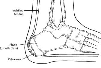
The Achilles tendon, also known as the calcaneal tendon, is a strong band of tissue that connects the calf muscles to the heel bone (calcaneus). It plays a crucial role in enabling activities such as walking, running, and jumping by facilitating the movement of the foot downward, which is called plantar flexion. Injuries to the Achilles tendon, such as tendinitis or ruptures, can be quite painful and impact mobility.
In the context of medicine, particularly in physical therapy and rehabilitation, "pliability" refers to the quality or state of being flexible or supple. It describes the ability of tissues, such as muscles or fascia (connective tissue), to stretch, deform, and adapt to forces applied upon them without resistance or injury. Improving pliability can help enhance range of motion, reduce muscle stiffness, promote circulation, and alleviate pain. Techniques like soft tissue mobilization, myofascial release, and stretching are often used to increase pliability in clinical settings.
Bone density refers to the amount of bone mineral content (usually measured in grams) in a given volume of bone (usually measured in cubic centimeters). It is often used as an indicator of bone strength and fracture risk. Bone density is typically measured using dual-energy X-ray absorptiometry (DXA) scans, which provide a T-score that compares the patient's bone density to that of a young adult reference population. A T-score of -1 or above is considered normal, while a T-score between -1 and -2.5 indicates osteopenia (low bone mass), and a T-score below -2.5 indicates osteoporosis (porous bones). Regular exercise, adequate calcium and vitamin D intake, and medication (if necessary) can help maintain or improve bone density and prevent fractures.
The ankle joint, also known as the talocrural joint, is the articulation between the bones of the lower leg (tibia and fibula) and the talus bone in the foot. It is a synovial hinge joint that allows for dorsiflexion and plantarflexion movements, which are essential for walking, running, and jumping. The ankle joint is reinforced by strong ligaments on both sides to provide stability during these movements.
Raynaud's disease, also known as Raynaud's phenomenon or syndrome, is a condition that affects the blood vessels, particularly in the fingers and toes. It is characterized by episodes of vasospasm (constriction) of the small digital arteries and arterioles, which can be triggered by cold temperatures or emotional stress. This results in reduced blood flow to the affected areas, causing them to become pale or white and then cyanotic (blue) due to the accumulation of deoxygenated blood. As the episode resolves, the affected areas may turn red as blood flow returns, sometimes accompanied by pain, numbness, or tingling sensations.
Raynaud's disease can be primary, meaning it occurs without an underlying medical condition, or secondary, which is associated with connective tissue disorders, autoimmune diseases, or other health issues such as carpal tunnel syndrome, vibration tool usage, or smoking. Primary Raynaud's is more common and tends to be less severe than secondary Raynaud's.
Treatment for Raynaud's disease typically involves avoiding triggers, keeping the body warm, and using medications to help dilate blood vessels and improve circulation. In some cases, lifestyle modifications and smoking cessation may also be recommended to manage symptoms and prevent progression of the condition.
Phonation is the process of sound production in speech, singing, or crying. It involves the vibration of the vocal folds (also known as the vocal cords) in the larynx, which is located in the neck. When air from the lungs passes through the vibrating vocal folds, it causes them to vibrate and produce sound waves. These sound waves are then shaped into speech sounds by the articulatory structures of the mouth, nose, and throat.
Phonation is a critical component of human communication and is used in various forms of verbal expression, such as speaking, singing, and shouting. It requires precise control of the muscles that regulate the tension, mass, and length of the vocal folds, as well as the air pressure and flow from the lungs. Dysfunction in phonation can result in voice disorders, such as hoarseness, breathiness, or loss of voice.
Musculoskeletal physiological phenomena refer to the various functions, processes, and responses that occur in the musculoskeletal system. This system includes the muscles, bones, joints, cartilages, tendons, ligaments, and other connective tissues that work together to support the body's structure, enable movement, and protect vital organs.
Musculoskeletal physiological phenomena can be categorized into several areas:
1. Muscle contraction and relaxation: This involves the conversion of chemical energy into mechanical energy through the sliding of actin and myosin filaments in muscle fibers, leading to muscle shortening or lengthening.
2. Bone homeostasis: This includes the maintenance of bone mass, density, and strength through a balance between bone formation by osteoblasts and bone resorption by osteoclasts.
3. Joint movement and stability: The movement of joints is enabled by the interaction between muscles, tendons, ligaments, and articular cartilage, while stability is maintained through the passive tension provided by ligaments and the active contraction of muscles.
4. Connective tissue repair and regeneration: This involves the response of tissues such as tendons, ligaments, and muscles to injury or damage, including inflammation, cell proliferation, and matrix remodeling.
5. Neuromuscular control: The coordination of muscle activity through the integration of sensory information from proprioceptors (e.g., muscle spindles, Golgi tendon organs) and motor commands from the central nervous system.
6. Skeletal development and growth: This includes the processes of bone formation, mineralization, and modeling during fetal development and childhood, as well as the maintenance of bone mass and strength throughout adulthood.
7. Aging and degeneration: The progressive decline in musculoskeletal function and structure with age, including sarcopenia (loss of muscle mass), osteoporosis (brittle bones), and joint degeneration (osteoarthritis).
Understanding these physiological phenomena is essential for the diagnosis, treatment, and prevention of musculoskeletal disorders and injuries.
Tissue engineering is a branch of biomedical engineering that combines the principles of engineering, materials science, and biological sciences to develop functional substitutes for damaged or diseased tissues and organs. It involves the creation of living, three-dimensional structures that can restore, maintain, or improve tissue function. This is typically accomplished through the use of cells, scaffolds (biodegradable matrices), and biologically active molecules. The goal of tissue engineering is to develop biological substitutes that can ultimately restore normal function and structure in damaged tissues or organs.
Ligaments are bands of dense, fibrous connective tissue that surround joints and provide support, stability, and limits the range of motion. They are made up primarily of collagen fibers arranged in a parallel pattern to withstand tension and stress. Ligaments attach bone to bone, and their function is to prevent excessive movement that could cause injury or dislocation.
There are two main types of ligaments: extracapsular and intracapsular. Extracapsular ligaments are located outside the joint capsule and provide stability to the joint by limiting its range of motion. Intracapsular ligaments, on the other hand, are found inside the joint capsule and help maintain the alignment of the joint surfaces.
Examples of common ligaments in the body include the anterior cruciate ligament (ACL) and posterior cruciate ligament (PCL) in the knee, the medial collateral ligament (MCL) and lateral collateral ligament (LCL) in the elbow, and the coracoacromial ligament in the shoulder.
Injuries to ligaments can occur due to sudden trauma or overuse, leading to sprains, strains, or tears. These injuries can cause pain, swelling, bruising, and limited mobility, and may require medical treatment such as immobilization, physical therapy, or surgery.
Joint instability is a condition characterized by the loss of normal joint function and increased risk of joint injury due to impaired integrity of the supporting structures, such as ligaments, muscles, or cartilage. This can result in excessive movement or laxity within the joint, leading to decreased stability and increased susceptibility to dislocations or subluxations. Joint instability may cause pain, swelling, and limited range of motion, and it can significantly impact a person's mobility and quality of life. It is often caused by trauma, degenerative conditions, or congenital abnormalities and may require medical intervention, such as physical therapy, bracing, or surgery, to restore joint stability.
Orthopedic fixation devices are medical implants used in orthopedic surgery to provide stability and promote the healing of fractured or broken bones, as well as joints or spinal segments. These devices can be internal or external and include a variety of products such as:
1. Intramedullary nails: Long rods that are inserted into the center of a bone to stabilize fractures in long bones like the femur or tibia.
2. Plates and screws: Metal plates are attached to the surface of a bone with screws to hold the fragments together while they heal.
3. Screws: Used alone or in combination with other devices, they can be used to stabilize small fractures or to fix implants like total joint replacements.
4. Wires: Used to hold bone fragments together, often in conjunction with other devices.
5. External fixators: A external frame attached to the bones using pins or wires that is placed outside the skin to provide stability and alignment of fractured bones.
6. Spinal fixation devices: These include pedicle screws, rods, hooks, and plates used to stabilize spinal fractures or deformities.
7. Orthopedic staples: Small metal staples used to stabilize small bone fragments or for joint fusion.
The choice of orthopedic fixation device depends on the location and severity of the injury or condition being treated. The primary goal of these devices is to provide stability, promote healing, and restore function.
In medical terms, the foot is the part of the lower limb that is distal to the leg and below the ankle, extending from the tarsus to the toes. It is primarily responsible for supporting body weight and facilitating movement through push-off during walking or running. The foot is a complex structure made up of 26 bones, 33 joints, and numerous muscles, tendons, ligaments, and nerves that work together to provide stability, balance, and flexibility. It can be divided into three main parts: the hindfoot, which contains the talus and calcaneus (heel) bones; the midfoot, which includes the navicular, cuboid, and cuneiform bones; and the forefoot, which consists of the metatarsals and phalanges that form the toes.
Collagen is the most abundant protein in the human body, and it is a major component of connective tissues such as tendons, ligaments, skin, and bones. Collagen provides structure and strength to these tissues and helps them to withstand stretching and tension. It is made up of long chains of amino acids, primarily glycine, proline, and hydroxyproline, which are arranged in a triple helix structure. There are at least 16 different types of collagen found in the body, each with slightly different structures and functions. Collagen is important for maintaining the integrity and health of tissues throughout the body, and it has been studied for its potential therapeutic uses in various medical conditions.
A femoral fracture is a medical term that refers to a break in the thigh bone, which is the longest and strongest bone in the human body. The femur extends from the hip joint to the knee joint and is responsible for supporting the weight of the upper body and allowing movement of the lower extremity. Femoral fractures can occur due to various reasons such as high-energy trauma, low-energy trauma in individuals with weak bones (osteoporosis), or as a result of a direct blow to the thigh.
Femoral fractures can be classified into different types based on their location, pattern, and severity. Some common types of femoral fractures include:
1. Transverse fracture: A break that occurs straight across the bone.
2. Oblique fracture: A break that occurs at an angle across the bone.
3. Spiral fracture: A break that occurs in a helical pattern around the bone.
4. Comminuted fracture: A break that results in multiple fragments of the bone.
5. Open or compound fracture: A break in which the bone pierces through the skin.
6. Closed or simple fracture: A break in which the bone does not pierce through the skin.
Femoral fractures can cause severe pain, swelling, bruising, and difficulty walking or bearing weight on the affected leg. Diagnosis typically involves a physical examination, medical history, and imaging tests such as X-rays or CT scans. Treatment may involve surgical intervention, including the use of metal rods, plates, or screws to stabilize the bone, followed by rehabilitation and physical therapy to restore mobility and strength.
Articular ligaments, also known as fibrous ligaments, are bands of dense, fibrous connective tissue that connect and stabilize bones to each other at joints. They help to limit the range of motion of a joint and provide support, preventing excessive movement that could cause injury. Articular ligaments are composed mainly of collagen fibers arranged in a parallel pattern, making them strong and flexible. They have limited blood supply and few nerve endings, which makes them less prone to injury but also slower to heal if damaged. Examples of articular ligaments include the anterior cruciate ligament (ACL) and posterior cruciate ligament (PCL) in the knee joint, and the medial collateral ligament (MCL) and lateral collateral ligament (LCL) in the elbow joint.
Skeletal muscle, also known as striated or voluntary muscle, is a type of muscle that is attached to bones by tendons or aponeuroses and functions to produce movements and support the posture of the body. It is composed of long, multinucleated fibers that are arranged in parallel bundles and are characterized by alternating light and dark bands, giving them a striped appearance under a microscope. Skeletal muscle is under voluntary control, meaning that it is consciously activated through signals from the nervous system. It is responsible for activities such as walking, running, jumping, and lifting objects.
A tibial fracture is a medical term that refers to a break in the shin bone, which is called the tibia. The tibia is the larger of the two bones in the lower leg and is responsible for supporting much of your body weight. Tibial fractures can occur in various ways, such as from high-energy trauma like car accidents or falls, or from low-energy trauma in individuals with weakened bones due to osteoporosis or other medical conditions.
Tibial fractures can be classified into different types based on the location, pattern, and severity of the break. Some common types of tibial fractures include:
1. Transverse fracture: A straight break that goes across the bone.
2. Oblique fracture: A diagonal break that slopes across the bone.
3. Spiral fracture: A break that spirals around the bone, often caused by twisting or rotational forces.
4. Comminuted fracture: A break where the bone is shattered into multiple pieces.
5. Open fracture: A break in which the bone pierces through the skin, increasing the risk of infection.
6. Closed fracture: A break in which the bone does not pierce through the skin.
Tibial fractures can cause symptoms such as pain, swelling, bruising, deformity, and difficulty walking or bearing weight on the affected leg. Treatment for tibial fractures may include immobilization with a cast or brace, surgery to realign and stabilize the bone with plates, screws, or rods, and rehabilitation to restore strength, mobility, and function to the injured limb.
Shear strength is a property of a material that describes its ability to withstand forces that cause internal friction and sliding of one portion of the material relative to another. In the context of human tissues, shear strength is an important factor in understanding how tissues respond to various stresses and strains, such as those experienced during physical activities or injuries.
For example, in the case of bones, shear strength is a critical factor in determining their ability to resist fractures under different types of loading conditions. Similarly, in soft tissues like ligaments and tendons, shear strength plays a crucial role in maintaining the integrity of these structures during movement and preventing excessive deformation or injury.
It's worth noting that measuring the shear strength of human tissues can be challenging due to their complex structure and anisotropic properties. As such, researchers often use specialized techniques and equipment to quantify these properties under controlled conditions in the lab.
In the field of medicine, "time factors" refer to the duration of symptoms or time elapsed since the onset of a medical condition, which can have significant implications for diagnosis and treatment. Understanding time factors is crucial in determining the progression of a disease, evaluating the effectiveness of treatments, and making critical decisions regarding patient care.
For example, in stroke management, "time is brain," meaning that rapid intervention within a specific time frame (usually within 4.5 hours) is essential to administering tissue plasminogen activator (tPA), a clot-busting drug that can minimize brain damage and improve patient outcomes. Similarly, in trauma care, the "golden hour" concept emphasizes the importance of providing definitive care within the first 60 minutes after injury to increase survival rates and reduce morbidity.
Time factors also play a role in monitoring the progression of chronic conditions like diabetes or heart disease, where regular follow-ups and assessments help determine appropriate treatment adjustments and prevent complications. In infectious diseases, time factors are crucial for initiating antibiotic therapy and identifying potential outbreaks to control their spread.
Overall, "time factors" encompass the significance of recognizing and acting promptly in various medical scenarios to optimize patient outcomes and provide effective care.
Orthopedic procedures are surgical or nonsurgical methods used to treat musculoskeletal conditions, including injuries, deformities, or diseases of the bones, joints, muscles, ligaments, and tendons. These procedures can range from simple splinting or casting to complex surgeries such as joint replacements, spinal fusions, or osteotomies (cutting and repositioning bones). The primary goal of orthopedic procedures is to restore function, reduce pain, and improve the quality of life for patients.
Orthotic devices are custom-made or prefabricated appliances designed to align, support, prevent deformity, or improve the function of movable body parts. They are frequently used in the treatment of various musculoskeletal disorders, such as foot and ankle conditions, knee problems, spinal alignment issues, and hand or wrist ailments. These devices can be adjustable or non-adjustable and are typically made from materials like plastic, metal, leather, or fabric. They work by redistributing forces across joints, correcting alignment, preventing unwanted movements, or accommodating existing deformities. Examples of orthotic devices include ankle-foot orthoses, knee braces, back braces, wrist splints, and custom-made foot insoles.
Suture techniques refer to the various methods used by surgeons to sew or stitch together tissues in the body after an injury, trauma, or surgical incision. The main goal of suturing is to approximate and hold the edges of the wound together, allowing for proper healing and minimizing scar formation.
There are several types of suture techniques, including:
1. Simple Interrupted Suture: This is one of the most basic suture techniques where the needle is passed through the tissue at a right angle, creating a loop that is then tightened to approximate the wound edges. Multiple stitches are placed along the length of the incision or wound.
2. Continuous Locking Suture: In this technique, the needle is passed continuously through the tissue in a zigzag pattern, with each stitch locking into the previous one. This creates a continuous line of sutures that provides strong tension and support to the wound edges.
3. Running Suture: Similar to the continuous locking suture, this technique involves passing the needle continuously through the tissue in a straight line. However, instead of locking each stitch, the needle is simply passed through the previous loop before being tightened. This creates a smooth and uninterrupted line of sutures that can be easily removed after healing.
4. Horizontal Mattress Suture: In this technique, two parallel stitches are placed horizontally across the wound edges, creating a "mattress" effect that provides additional support and tension to the wound. This is particularly useful in deep or irregularly shaped wounds.
5. Vertical Mattress Suture: Similar to the horizontal mattress suture, this technique involves placing two parallel stitches vertically across the wound edges. This creates a more pronounced "mattress" effect that can help reduce tension and minimize scarring.
6. Subcuticular Suture: In this technique, the needle is passed just below the surface of the skin, creating a smooth and barely visible line of sutures. This is particularly useful in cosmetic surgery or areas where minimizing scarring is important.
The choice of suture technique depends on various factors such as the location and size of the wound, the type of tissue involved, and the patient's individual needs and preferences. Proper suture placement and tension are crucial for optimal healing and aesthetic outcomes.
The sclera is the tough, white, fibrous outer coating of the eye in humans and other vertebrates, covering about five sixths of the eyeball's surface. It provides protection for the delicate inner structures of the eye and maintains its shape. The sclera is composed mainly of collagen and elastic fiber, making it strong and resilient. Its name comes from the Greek word "skleros," which means hard.
Back injuries refer to damages or traumas that affect the structures of the back, including the muscles, nerves, ligaments, bones, and other tissues. These injuries can occur due to various reasons such as sudden trauma (e.g., falls, accidents), repetitive stress, or degenerative conditions. Common types of back injuries include strains, sprains, herniated discs, fractured vertebrae, and spinal cord injuries. Symptoms may vary from mild discomfort to severe pain, numbness, tingling, or weakness, depending on the severity and location of the injury. Treatment options range from conservative measures like physical therapy and medication to surgical intervention in severe cases.
Biomedical engineering is a field that combines engineering principles and design concepts with medical and biological sciences to develop solutions to healthcare challenges. It involves the application of engineering methods to analyze, understand, and solve problems in biology and medicine, with the goal of improving human health and well-being. Biomedical engineers may work on a wide range of projects, including developing new medical devices, designing artificial organs, creating diagnostic tools, simulating biological systems, and optimizing healthcare delivery systems. They often collaborate with other professionals such as doctors, nurses, and scientists to develop innovative solutions that meet the needs of patients and healthcare providers.
Locomotion, in a medical context, refers to the ability to move independently and change location. It involves the coordinated movement of the muscles, bones, and nervous system that enables an individual to move from one place to another. This can include walking, running, jumping, or using assistive devices such as wheelchairs or crutches. Locomotion is a fundamental aspect of human mobility and is often assessed in medical evaluations to determine overall health and functioning.
An animal model in medicine refers to the use of non-human animals in experiments to understand, predict, and test responses and effects of various biological and chemical interactions that may also occur in humans. These models are used when studying complex systems or processes that cannot be easily replicated or studied in human subjects, such as genetic manipulation or exposure to harmful substances. The choice of animal model depends on the specific research question being asked and the similarities between the animal's and human's biological and physiological responses. Examples of commonly used animal models include mice, rats, rabbits, guinea pigs, and non-human primates.
In the context of medicine and physiology, acceleration refers to the process of increasing or quickening a function or process. For example, heart rate acceleration is an increase in the speed at which the heart beats. It can also refer to the rate at which something increases, such as the acceleration of muscle strength during rehabilitation. In physics terms, acceleration refers to the rate at which an object changes its velocity, but this definition is not typically used in a medical context.
A zygapophyseal joint, also known as a facet joint, is a type of synovial joint that connects the articulating processes of adjacent vertebrae in the spine. These joints are formed by the superior and inferior articular processes of the vertebral bodies and are covered with hyaline cartilage. They allow for smooth movement between the vertebrae, providing stability and limiting excessive motion while allowing flexibility in the spine. The zygapophyseal joints are supported by a capsule and ligaments that help to maintain their alignment and restrict abnormal movements. These joints can become sources of pain and discomfort when they become inflamed or damaged due to conditions such as arthritis, degenerative disc disease, or injury.
I'm not aware of a medical term called "bone wires." The term "wiring" is used in orthopedic surgery to describe the use of metal wire to hold bones or fractures in place during healing. However, I couldn't find any specific medical definition or term related to "bone wires." It may be a colloquialism, a term used in a specific context, or a term from science fiction. If you could provide more context about where you encountered this term, I might be able to give a more accurate answer.
Biocompatible materials are non-toxic and non-reacting substances that can be used in medical devices, tissue engineering, and drug delivery systems without causing harm or adverse reactions to living tissues or organs. These materials are designed to mimic the properties of natural tissues and are able to integrate with biological systems without being rejected by the body's immune system.
Biocompatible materials can be made from a variety of substances, including metals, ceramics, polymers, and composites. The specific properties of these materials, such as their mechanical strength, flexibility, and biodegradability, are carefully selected to meet the requirements of their intended medical application.
Examples of biocompatible materials include titanium used in dental implants and joint replacements, polyethylene used in artificial hips, and hydrogels used in contact lenses and drug delivery systems. The use of biocompatible materials has revolutionized modern medicine by enabling the development of advanced medical technologies that can improve patient outcomes and quality of life.
Wound healing is a complex and dynamic process that occurs after tissue injury, aiming to restore the integrity and functionality of the damaged tissue. It involves a series of overlapping phases: hemostasis, inflammation, proliferation, and remodeling.
1. Hemostasis: This initial phase begins immediately after injury and involves the activation of the coagulation cascade to form a clot, which stabilizes the wound and prevents excessive blood loss.
2. Inflammation: Activated inflammatory cells, such as neutrophils and monocytes/macrophages, infiltrate the wound site to eliminate pathogens, remove debris, and release growth factors that promote healing. This phase typically lasts for 2-5 days post-injury.
3. Proliferation: In this phase, various cell types, including fibroblasts, endothelial cells, and keratinocytes, proliferate and migrate to the wound site to synthesize extracellular matrix (ECM) components, form new blood vessels (angiogenesis), and re-epithelialize the wounded area. This phase can last up to several weeks depending on the size and severity of the wound.
4. Remodeling: The final phase of wound healing involves the maturation and realignment of collagen fibers, leading to the restoration of tensile strength in the healed tissue. This process can continue for months to years after injury, although the tissue may never fully regain its original structure and function.
It is important to note that wound healing can be compromised by several factors, including age, nutrition, comorbidities (e.g., diabetes, vascular disease), and infection, which can result in delayed healing or non-healing chronic wounds.
In a medical context, "lifting" generally refers to the act or process of physically moving or supporting an object or person with the use of one's muscles, particularly those in the back, legs, and arms. However, I am a large language model, not a doctor, and this definition is based on general usage rather than specific medical terminology.
Improper lifting techniques can lead to injuries, especially musculoskeletal ones. For this reason, healthcare professionals often provide guidelines for safe lifting, emphasizing the importance of maintaining a wide base of support, keeping the object close to the body, using leg muscles instead of the back, and avoiding twisting or bending at the waist.
In some cases, "lifting" may also refer to medical procedures where a surgeon raises a part of the body during surgery, but this is more commonly referred to as "elevation."
Prosthesis design is a specialized field in medical device technology that involves creating and developing artificial substitutes to replace a missing body part, such as a limb, tooth, eye, or internal organ. The design process typically includes several stages: assessment of the patient's needs, selection of appropriate materials, creation of a prototype, testing and refinement, and final fabrication and fitting of the prosthesis.
The goal of prosthesis design is to create a device that functions as closely as possible to the natural body part it replaces, while also being comfortable, durable, and aesthetically pleasing for the patient. The design process may involve collaboration between medical professionals, engineers, and designers, and may take into account factors such as the patient's age, lifestyle, occupation, and overall health.
Prosthesis design can be highly complex, particularly for advanced devices such as robotic limbs or implantable organs. These devices often require sophisticated sensors, actuators, and control systems to mimic the natural functions of the body part they replace. As a result, prosthesis design is an active area of research and development in the medical field, with ongoing efforts to improve the functionality, comfort, and affordability of these devices for patients.
Medical science often defines and describes "walking" as a form of locomotion or mobility where an individual repeatedly lifts and sets down each foot to move forward, usually bearing weight on both legs. It is a complex motor activity that requires the integration and coordination of various systems in the human body, including the musculoskeletal, neurological, and cardiovascular systems.
Walking involves several components such as balance, coordination, strength, and endurance. The ability to walk independently is often used as a measure of functional mobility and overall health status. However, it's important to note that the specific definition of walking may vary depending on the context and the medical or scientific field in question.
A suture anchor is a medical device used in surgical procedures, particularly in orthopedic and cardiovascular surgeries. It is typically made of biocompatible materials such as metal (titanium or absorbable steel) or polymer (absorbable or non-absorbable). The suture anchor serves to attach a suture to bone securely, providing a stable fixation point for soft tissue reattachment or repair.
Suture anchors come in various shapes and sizes, including screws, hooks, or buttons, designed to fit specific surgical needs. Surgeons insert the anchor into a predrilled hole in the bone, and then pass the suture through the eyelet or loop of the anchor. Once the anchor is securely in place, the surgeon can tie the suture to attach tendons, ligaments, or other soft tissues to the bone.
The use of suture anchors has revolutionized many surgical procedures by providing a more reliable and less invasive method for reattaching soft tissues to bones compared to traditional methods such as drill holes and staples.
A rupture, in medical terms, refers to the breaking or tearing of an organ, tissue, or structure in the body. This can occur due to various reasons such as trauma, injury, increased pressure, or degeneration. A ruptured organ or structure can lead to serious complications, including internal bleeding, infection, and even death, if not treated promptly and appropriately. Examples of ruptures include a ruptured appendix, ruptured eardrum, or a ruptured disc in the spine.
In medical terms, compliance refers to the degree to which a patient follows the recommendations or instructions of their healthcare provider. This may include taking prescribed medications as directed, following a treatment plan, making lifestyle changes, or attending follow-up appointments. Good compliance is essential for achieving the best possible health outcomes and can help prevent complications or worsening of medical conditions. Factors that can affect patient compliance include forgetfulness, lack of understanding of the instructions, cost of medications or treatments, and side effects of medications. Healthcare providers can take steps to improve patient compliance by providing clear and concise instructions, discussing potential barriers to compliance, and involving patients in their care plan.
Tissue scaffolds, also known as bioactive scaffolds or synthetic extracellular matrices, refer to three-dimensional structures that serve as templates for the growth and organization of cells in tissue engineering and regenerative medicine. These scaffolds are designed to mimic the natural extracellular matrix (ECM) found in biological tissues, providing a supportive environment for cell attachment, proliferation, differentiation, and migration.
Tissue scaffolds can be made from various materials, including naturally derived biopolymers (e.g., collagen, alginate, chitosan, hyaluronic acid), synthetic polymers (e.g., polycaprolactone, polylactic acid, poly(lactic-co-glycolic acid)), or a combination of both. The choice of material depends on the specific application and desired properties, such as biocompatibility, biodegradability, mechanical strength, and porosity.
The primary functions of tissue scaffolds include:
1. Cell attachment: Providing surfaces for cells to adhere, spread, and form stable focal adhesions.
2. Mechanical support: Offering a structural framework that maintains the desired shape and mechanical properties of the engineered tissue.
3. Nutrient diffusion: Ensuring adequate transport of nutrients, oxygen, and waste products throughout the scaffold to support cell survival and function.
4. Guided tissue growth: Directing the organization and differentiation of cells through spatial cues and biochemical signals.
5. Biodegradation: Gradually degrading at a rate that matches tissue regeneration, allowing for the replacement of the scaffold with native ECM produced by the cells.
Tissue scaffolds have been used in various applications, such as wound healing, bone and cartilage repair, cardiovascular tissue engineering, and neural tissue regeneration. The design and fabrication of tissue scaffolds are critical aspects of tissue engineering, aiming to create functional substitutes for damaged or diseased tissues and organs.
Polymethyl methacrylate (PMMA) is a type of synthetic resin that is widely used in the medical field due to its biocompatibility and versatility. It is a transparent, rigid, and lightweight material that can be easily molded into different shapes and forms. Here are some of the medical definitions of PMMA:
1. A biocompatible acrylic resin used in various medical applications such as bone cement, intraocular lenses, dental restorations, and drug delivery systems.
2. A type of synthetic material that is used as a bone cement to fix prosthetic joint replacements and vertebroplasty for the treatment of spinal fractures.
3. A transparent and shatter-resistant material used in the manufacture of medical devices such as intravenous (IV) fluid bags, dialyzer housings, and oxygenators.
4. A drug delivery system that can be used to administer drugs locally or systemically, such as intraocular sustained-release drug implants for the treatment of chronic eye diseases.
5. A component of dental restorations such as fillings, crowns, and bridges due to its excellent mechanical properties and esthetic qualities.
Overall, PMMA is a versatile and valuable material in the medical field, with numerous applications that take advantage of its unique properties.
The cornea is the clear, dome-shaped surface at the front of the eye. It plays a crucial role in focusing vision. The cornea protects the eye from harmful particles and microorganisms, and it also serves as a barrier against UV light. Its transparency allows light to pass through and get focused onto the retina. The cornea does not contain blood vessels, so it relies on tears and the fluid inside the eye (aqueous humor) for nutrition and oxygen. Any damage or disease that affects its clarity and shape can significantly impact vision and potentially lead to blindness if left untreated.
Total disc replacement (TDR), also known as total disc arthroplasty, is a surgical procedure in which the damaged or degenerated intervertebral disc in the spine is removed and replaced with an artificial device. The primary goal of this procedure is to maintain motion within the spinal segment while alleviating pain and other symptoms caused by the damaged disc.
The artificial disc, typically made from materials such as metal or polymer, is designed to replicate the natural movement and function of a healthy intervertebral disc. The surgery can be performed at various levels of the spine, including cervical (neck) and lumbar (lower back), depending on the location of the damaged disc.
TDR is generally considered for patients with degenerative disc disease who have not responded to non-surgical treatments such as physical therapy or pain management. The potential benefits of TDR over traditional spinal fusion surgery include preserving motion, reducing the risk of adjacent segment degeneration, and potentially faster recovery times. However, as with any surgical procedure, there are risks involved, including infection, implant wear, dislocation, or subsidence (sinking of the implant into the bone). It is essential to discuss these potential risks and benefits with a qualified medical professional before making a decision about undergoing TDR surgery.
Postural balance is the ability to maintain, achieve, or restore a state of equilibrium during any posture or activity. It involves the integration of sensory information (visual, vestibular, and proprioceptive) to control and adjust body position in space, thereby maintaining the center of gravity within the base of support. This is crucial for performing daily activities and preventing falls, especially in older adults and individuals with neurological or orthopedic conditions.
Cumulative Trauma Disorders (CTDs) are a group of conditions that result from repeated exposure to biomechanical stressors, often related to work activities. These disorders can affect the muscles, tendons, nerves, and joints, leading to symptoms such as pain, numbness, tingling, weakness, and reduced range of motion.
CTDs are also known as repetitive strain injuries (RSIs) or overuse injuries. They occur when there is a mismatch between the demands placed on the body and its ability to recover from those demands. Over time, this imbalance can lead to tissue damage and inflammation, resulting in chronic pain and functional limitations.
Examples of CTDs include carpal tunnel syndrome, tendonitis, epicondylitis (tennis elbow), rotator cuff injuries, and trigger finger. Prevention strategies for CTDs include proper ergonomics, workstation design, body mechanics, taking regular breaks to stretch and rest, and performing exercises to strengthen and condition the affected muscles and joints.
In medical terms, the knee is referred to as the largest and one of the most complex joints in the human body. It is a hinge joint that connects the thigh bone (femur) to the shin bones (tibia and fibula), enabling movements like flexion, extension, and a small amount of rotation. The knee also contains several other components such as menisci, ligaments, tendons, and bursae, which provide stability, cushioning, and protection during movement.
Pronation is a term used in the medical field, particularly in the study of human biomechanics and orthopedics. It refers to the normal motion that occurs in the subtalar joint of the foot, which allows the foot to adapt to various surfaces and absorb shock during walking or running.
During pronation, the arch of the foot collapses, and the heel rolls inward, causing the forefoot to rotate outward. This motion helps distribute the forces of impact evenly across the foot and lower limb, reducing stress on individual structures and providing stability during weight-bearing activities.
However, excessive pronation can lead to biomechanical issues and increase the risk of injuries such as plantar fasciitis, shin splints, and knee pain. Proper assessment and management of foot mechanics, including orthotics or physical therapy interventions, may be necessary to address excessive pronation and related conditions.
Knee injuries refer to damages or harm caused to the structures surrounding or within the knee joint, which may include the bones (femur, tibia, and patella), cartilage (meniscus and articular cartilage), ligaments (ACL, PCL, MCL, and LCL), tendons (patellar and quadriceps), muscles, bursae, and other soft tissues. These injuries can result from various causes, such as trauma, overuse, degeneration, or sports-related activities. Symptoms may include pain, swelling, stiffness, instability, reduced range of motion, and difficulty walking or bearing weight on the affected knee. Common knee injuries include fractures, dislocations, meniscal tears, ligament sprains or ruptures, and tendonitis. Proper diagnosis and treatment are crucial to ensure optimal recovery and prevent long-term complications.
Titanium is not a medical term, but rather a chemical element (symbol Ti, atomic number 22) that is widely used in the medical field due to its unique properties. Medically, it is often referred to as a biocompatible material used in various medical applications such as:
1. Orthopedic implants: Titanium and its alloys are used for making joint replacements (hips, knees, shoulders), bone plates, screws, and rods due to their high strength-to-weight ratio, excellent corrosion resistance, and biocompatibility.
2. Dental implants: Titanium is also commonly used in dental applications like implants, crowns, and bridges because of its ability to osseointegrate, or fuse directly with bone tissue, providing a stable foundation for replacement teeth.
3. Cardiovascular devices: Titanium alloys are used in the construction of heart valves, pacemakers, and other cardiovascular implants due to their non-magnetic properties, which prevent interference with magnetic resonance imaging (MRI) scans.
4. Medical instruments: Due to its resistance to corrosion and high strength, titanium is used in the manufacturing of various medical instruments such as surgical tools, needles, and catheters.
In summary, Titanium is a chemical element with unique properties that make it an ideal material for various medical applications, including orthopedic and dental implants, cardiovascular devices, and medical instruments.
Connective tissue is a type of biological tissue that provides support, strength, and protection to various structures in the body. It is composed of cells called fibroblasts, which produce extracellular matrix components such as collagen, elastin, and proteoglycans. These components give connective tissue its unique properties, including tensile strength, elasticity, and resistance to compression.
There are several types of connective tissue in the body, each with its own specific functions and characteristics. Some examples include:
1. Loose or Areolar Connective Tissue: This type of connective tissue is found throughout the body and provides cushioning and support to organs and other structures. It contains a large amount of ground substance, which allows for the movement and gliding of adjacent tissues.
2. Dense Connective Tissue: This type of connective tissue has a higher concentration of collagen fibers than loose connective tissue, making it stronger and less flexible. Dense connective tissue can be further divided into two categories: regular (or parallel) and irregular. Regular dense connective tissue, such as tendons and ligaments, has collagen fibers that run parallel to each other, providing great tensile strength. Irregular dense connective tissue, such as the dermis of the skin, has collagen fibers arranged in a more haphazard pattern, providing support and flexibility.
3. Adipose Tissue: This type of connective tissue is primarily composed of fat cells called adipocytes. Adipose tissue serves as an energy storage reservoir and provides insulation and cushioning to the body.
4. Cartilage: A firm, flexible type of connective tissue that contains chondrocytes within a matrix of collagen and proteoglycans. Cartilage is found in various parts of the body, including the joints, nose, ears, and trachea.
5. Bone: A specialized form of connective tissue that consists of an organic matrix (mainly collagen) and an inorganic mineral component (hydroxyapatite). Bone provides structural support to the body and serves as a reservoir for calcium and phosphate ions.
6. Blood: Although not traditionally considered connective tissue, blood does contain elements of connective tissue, such as plasma proteins and leukocytes (white blood cells). Blood transports nutrients, oxygen, hormones, and waste products throughout the body.
The humerus is the long bone in the upper arm that extends from the shoulder joint (glenohumeral joint) to the elbow joint. It articulates with the glenoid cavity of the scapula to form the shoulder joint and with the radius and ulna bones at the elbow joint. The proximal end of the humerus has a rounded head that provides for movement in multiple planes, making it one of the most mobile joints in the body. The greater and lesser tubercles are bony prominences on the humeral head that serve as attachment sites for muscles that move the shoulder and arm. The narrow shaft of the humerus provides stability and strength for weight-bearing activities, while the distal end forms two articulations: one with the ulna (trochlea) and one with the radius (capitulum). Together, these structures allow for a wide range of motion in the shoulder and elbow joints.
The temporomandibular joint (TMJ) is the articulation between the mandible (lower jaw) and the temporal bone of the skull. It's a complex joint that involves the movement of two bones, several muscles, and various ligaments. The TMJ allows for movements like rotation and translation, enabling us to open and close our mouth, chew, speak, and yawn. Dysfunction in this joint can lead to temporomandibular joint disorders (TMD), which can cause pain, discomfort, and limited jaw movement.
Electromyography (EMG) is a medical diagnostic procedure that measures the electrical activity of skeletal muscles during contraction and at rest. It involves inserting a thin needle electrode into the muscle to record the electrical signals generated by the muscle fibers. These signals are then displayed on an oscilloscope and may be heard through a speaker.
EMG can help diagnose various neuromuscular disorders, such as muscle weakness, numbness, or pain, and can distinguish between muscle and nerve disorders. It is often used in conjunction with other diagnostic tests, such as nerve conduction studies, to provide a comprehensive evaluation of the nervous system.
EMG is typically performed by a neurologist or a physiatrist, and the procedure may cause some discomfort or pain, although this is usually minimal. The results of an EMG can help guide treatment decisions and monitor the progression of neuromuscular conditions over time.
A spinal fracture, also known as a vertebral compression fracture, is a break in one or more bones (vertebrae) of the spine. This type of fracture often occurs due to weakened bones caused by osteoporosis, but it can also result from trauma such as a car accident or a fall.
In a spinal fracture, the front part of the vertebra collapses, causing the height of the vertebra to decrease, while the back part of the vertebra remains intact. This results in a wedge-shaped deformity of the vertebra. Multiple fractures can lead to a hunched forward posture known as kyphosis or dowager's hump.
Spinal fractures can cause pain, numbness, tingling, or weakness in the back, legs, or arms, depending on the location and severity of the fracture. In some cases, spinal cord compression may occur, leading to more severe symptoms such as paralysis or loss of bladder and bowel control.
Osseointegration is a direct structural and functional connection between living bone and the surface of an implant. It's a process where the bone grows in and around the implant, which is typically made of titanium or another biocompatible material. This process provides a solid foundation for dental prosthetics, such as crowns, bridges, or dentures, or for orthopedic devices like artificial limbs. The success of osseointegration depends on various factors, including the patient's overall health, the quality and quantity of available bone, and the surgical technique used for implant placement.
Reconstructive surgical procedures are a type of surgery aimed at restoring the form and function of body parts that are defective or damaged due to various reasons such as congenital abnormalities, trauma, infection, tumors, or disease. These procedures can involve the transfer of tissue from one part of the body to another, manipulation of bones, muscles, and tendons, or use of prosthetic materials to reconstruct the affected area. The goal is to improve both the physical appearance and functionality of the body part, thereby enhancing the patient's quality of life. Examples include breast reconstruction after mastectomy, cleft lip and palate repair, and treatment of severe burns.
I apologize, but "video recording" is not a term that has a specific medical definition. It generally refers to the process of capturing and storing moving visual images on electronic media, which is used in various medical fields for different purposes such as surgical training, telemedicine consultations, or monitoring patient conditions. However, there is no unique medical meaning associated with this term.
The menisci are crescent-shaped fibrocartilaginous structures located in the knee joint. There are two menisci in each knee: the medial meniscus and the lateral meniscus. The tibial menisci, also known as the medial and lateral menisci, are named according to their location in the knee joint. They lie on the top surface of the tibia (shin bone) and provide shock absorption, stability, and lubrication to the knee joint.
The tibial menisci have a complex shape, with a wider outer portion called the peripheral rim and a narrower inner portion called the central portion or root attachment. The menisci are attached to the bones of the knee joint by ligaments and have a rich blood supply in their outer portions, which helps in healing after injury. However, the inner two-thirds of the menisci have a poor blood supply, making them more prone to degeneration and less likely to heal after injury.
Damage to the tibial menisci can occur due to trauma or degenerative changes, leading to symptoms such as pain, swelling, stiffness, and limited mobility of the knee joint. Treatment for meniscal injuries may include physical therapy, bracing, or surgery, depending on the severity and location of the injury.
The patella, also known as the kneecap, is a sesamoid bone located at the front of the knee joint. It is embedded in the tendon of the quadriceps muscle and serves to protect the knee joint and increase the leverage of the extensor mechanism, allowing for greater extension force of the lower leg. The patella moves within a groove on the femur called the trochlea during flexion and extension of the knee.
In medical terms, the hip is a ball-and-socket joint where the rounded head of the femur (thigh bone) fits into the cup-shaped socket, also known as the acetabulum, of the pelvis. This joint allows for a wide range of movement in the lower extremities and supports the weight of the upper body during activities such as walking, running, and jumping. The hip joint is surrounded by strong ligaments, muscles, and tendons that provide stability and enable proper functioning.
Bone cements are medical-grade materials used in orthopedic and trauma surgery to fill gaps between bone surfaces and implants, such as artificial joints or screws. They serve to mechanically stabilize the implant and provide a smooth, load-bearing surface. The two most common types of bone cement are:
1. Polymethylmethacrylate (PMMA) cement: This is a two-component system consisting of powdered PMMA and liquid methyl methacrylate monomer. When mixed together, they form a dough-like consistency that hardens upon exposure to air. PMMA cement has been widely used for decades in joint replacement surgeries, such as hip or knee replacements.
2. Calcium phosphate (CP) cement: This is a two-component system consisting of a powdered CP compound and an aqueous solution. When mixed together, they form a paste that hardens through a chemical reaction at body temperature. CP cement has lower mechanical strength compared to PMMA but demonstrates better biocompatibility, bioactivity, and the ability to resorb over time.
Both types of bone cements have advantages and disadvantages, and their use depends on the specific surgical indication and patient factors.
A bone fracture is a medical condition in which there is a partial or complete break in the continuity of a bone due to external or internal forces. Fractures can occur in any bone in the body and can vary in severity from a small crack to a shattered bone. The symptoms of a bone fracture typically include pain, swelling, bruising, deformity, and difficulty moving the affected limb. Treatment for a bone fracture may involve immobilization with a cast or splint, surgery to realign and stabilize the bone, or medication to manage pain and prevent infection. The specific treatment approach will depend on the location, type, and severity of the fracture.
Three-dimensional (3D) imaging in medicine refers to the use of technologies and techniques that generate a 3D representation of internal body structures, organs, or tissues. This is achieved by acquiring and processing data from various imaging modalities such as X-ray computed tomography (CT), magnetic resonance imaging (MRI), ultrasound, or confocal microscopy. The resulting 3D images offer a more detailed visualization of the anatomy and pathology compared to traditional 2D imaging techniques, allowing for improved diagnostic accuracy, surgical planning, and minimally invasive interventions.
In 3D imaging, specialized software is used to reconstruct the acquired data into a volumetric model, which can be manipulated and viewed from different angles and perspectives. This enables healthcare professionals to better understand complex anatomical relationships, detect abnormalities, assess disease progression, and monitor treatment response. Common applications of 3D imaging include neuroimaging, orthopedic surgery planning, cancer staging, dental and maxillofacial reconstruction, and interventional radiology procedures.
Viscosity is a physical property of a fluid that describes its resistance to flow. In medical terms, viscosity is often discussed in relation to bodily fluids such as blood or synovial fluid (found in joints). The unit of measurement for viscosity is the poise, although it is more commonly expressed in millipascals-second (mPa.s) in SI units. Highly viscous fluids flow more slowly than less viscous fluids. Changes in the viscosity of bodily fluids can have significant implications for health and disease; for example, increased blood viscosity has been associated with cardiovascular diseases, while decreased synovial fluid viscosity can contribute to joint pain and inflammation in conditions like osteoarthritis.
Animal disease models are specialized animals, typically rodents such as mice or rats, that have been genetically engineered or exposed to certain conditions to develop symptoms and physiological changes similar to those seen in human diseases. These models are used in medical research to study the pathophysiology of diseases, identify potential therapeutic targets, test drug efficacy and safety, and understand disease mechanisms.
The genetic modifications can include knockout or knock-in mutations, transgenic expression of specific genes, or RNA interference techniques. The animals may also be exposed to environmental factors such as chemicals, radiation, or infectious agents to induce the disease state.
Examples of animal disease models include:
1. Mouse models of cancer: Genetically engineered mice that develop various types of tumors, allowing researchers to study cancer initiation, progression, and metastasis.
2. Alzheimer's disease models: Transgenic mice expressing mutant human genes associated with Alzheimer's disease, which exhibit amyloid plaque formation and cognitive decline.
3. Diabetes models: Obese and diabetic mouse strains like the NOD (non-obese diabetic) or db/db mice, used to study the development of type 1 and type 2 diabetes, respectively.
4. Cardiovascular disease models: Atherosclerosis-prone mice, such as ApoE-deficient or LDLR-deficient mice, that develop plaque buildup in their arteries when fed a high-fat diet.
5. Inflammatory bowel disease models: Mice with genetic mutations affecting intestinal barrier function and immune response, such as IL-10 knockout or SAMP1/YitFc mice, which develop colitis.
Animal disease models are essential tools in preclinical research, but it is important to recognize their limitations. Differences between species can affect the translatability of results from animal studies to human patients. Therefore, researchers must carefully consider the choice of model and interpret findings cautiously when applying them to human diseases.
The temporomandibular joint (TMJ) disc is a small, thin piece of fibrocartilaginous tissue located within the TMJ, which is the joint that connects the mandible (jawbone) to the temporal bone of the skull. The disc acts as a cushion and allows for smooth movement of the jaw during activities such as eating, speaking, and yawning. It divides the joint into two compartments: the upper and lower compartments.
The TMJ disc is composed of several types of tissue, including collagen fibers, elastin fibers, and a small number of cells called fibroblasts. The disc's unique structure allows it to withstand the forces generated during jaw movement and helps to distribute these forces evenly across the joint.
The TMJ disc can become damaged or displaced due to various factors such as trauma, teeth grinding (bruxism), or degenerative joint diseases like osteoarthritis. This can lead to temporomandibular disorders (TMDs) characterized by pain, stiffness, and limited jaw movement.
Osteotomy is a surgical procedure in which a bone is cut to shorten, lengthen, or change its alignment. It is often performed to correct deformities or to realign bones that have been damaged by trauma or disease. The bone may be cut straight across (transverse osteotomy) or at an angle (oblique osteotomy). After the bone is cut, it can be realigned and held in place with pins, plates, or screws until it heals. This procedure is commonly performed on bones in the leg, such as the femur or tibia, but can also be done on other bones in the body.
Intramedullary fracture fixation is a surgical technique used to stabilize and align bone fractures. In this procedure, a metal rod or nail is inserted into the marrow cavity (intramedullary canal) of the affected bone, spanning the length of the fracture. The rod is then secured to the bone using screws or other fixation devices on either side of the fracture. This provides stability and helps maintain proper alignment during the healing process.
The benefits of intramedullary fixation include:
1. Load sharing: The intramedullary rod shares some of the load bearing capacity with the bone, which can help reduce stress on the healing bone.
2. Minimal soft tissue dissection: Since the implant is inserted through the medullary canal, there is less disruption to the surrounding muscles, tendons, and ligaments compared to other fixation methods.
3. Biomechanical stability: Intramedullary fixation provides rotational and bending stiffness, which helps maintain proper alignment of the fracture fragments during healing.
4. Early mobilization: Patients with intramedullary fixation can often begin weight bearing and rehabilitation exercises earlier than those with other types of fixation, leading to faster recovery times.
Common indications for intramedullary fracture fixation include long bone fractures in the femur, tibia, humerus, and fibula, as well as certain pelvic and spinal fractures. However, the choice of fixation method depends on various factors such as patient age, fracture pattern, location, and associated injuries.
Fibrocartilage is a type of tough, dense connective tissue that contains both collagen fibers and cartilaginous matrix. It is composed of fibroblasts embedded in a extracellular matrix rich in collagen types I and II, proteoglycans and elastin. Fibrocartilage is found in areas of the body where strong, flexible support is required, such as intervertebral discs, menisci (knee cartilage), labrum (shoulder and hip cartilage) and pubic symphysis. It has both the elasticity and flexibility of cartilage and the strength and durability of fibrous tissue. Fibrocartilage can withstand high compressive loads and provides cushioning, shock absorption and stability to the joints and spine.
The extracellular matrix (ECM) is a complex network of biomolecules that provides structural and biochemical support to cells in tissues and organs. It is composed of various proteins, glycoproteins, and polysaccharides, such as collagens, elastin, fibronectin, laminin, and proteoglycans. The ECM plays crucial roles in maintaining tissue architecture, regulating cell behavior, and facilitating communication between cells. It provides a scaffold for cell attachment, migration, and differentiation, and helps to maintain the structural integrity of tissues by resisting mechanical stresses. Additionally, the ECM contains various growth factors, cytokines, and chemokines that can influence cellular processes such as proliferation, survival, and differentiation. Overall, the extracellular matrix is essential for the normal functioning of tissues and organs, and its dysregulation can contribute to various pathological conditions, including fibrosis, cancer, and degenerative diseases.
Fibrillar collagens are a type of collagen that form rope-like fibrils in the extracellular matrix of connective tissues. They are composed of three polypeptide chains, called alpha chains, which are coiled together in a triple helix structure. The most common types of fibrillar collagens are Type I, II, III, V, and XI. These collagens provide strength and support to tissues such as tendons, ligaments, skin, and bones. They also play important roles in the regulation of cell behavior and tissue development. Mutations in genes encoding fibrillar collagens can lead to a variety of connective tissue disorders, including osteogenesis imperfecta, Ehlers-Danlos syndrome, and Marfan syndrome.
The term "back" is a common word used to describe the large posterior part of the body of a human or an animal, which extends from the neck to the pelvis and contains the spine, spinal cord, ribs, muscles, and other various tissues. In medical terms, the back is also known as the dorsal region. It provides support, protection, and mobility for the body, allowing us to stand upright, bend, twist, and perform various physical activities. The back is susceptible to various injuries, disorders, and conditions, such as back pain, strains, sprains, herniated discs, scoliosis, and arthritis, among others.
The hip joint, also known as the coxal joint, is a ball-and-socket type synovial joint that connects the femur (thigh bone) to the pelvis. The "ball" is the head of the femur, while the "socket" is the acetabulum, a concave surface on the pelvic bone.
The hip joint is surrounded by a strong fibrous capsule and is reinforced by several ligaments, including the iliofemoral, ischiofemoral, and pubofemoral ligaments. The joint allows for flexion, extension, abduction, adduction, medial and lateral rotation, and circumduction movements, making it one of the most mobile joints in the body.
The hip joint is also supported by various muscles, including the gluteus maximus, gluteus medius, gluteus minimus, iliopsoas, and other hip flexors and extensors. These muscles provide stability and strength to the joint, allowing for weight-bearing activities such as walking, running, and jumping.
The lumbosacral region is the lower part of the back where the lumbar spine (five vertebrae in the lower back) connects with the sacrum (a triangular bone at the base of the spine). This region is subject to various conditions such as sprains, strains, herniated discs, and degenerative disorders that can cause pain and discomfort. It's also a common site for surgical intervention when non-surgical treatments fail to provide relief.
In medical terms, the jaw is referred to as the mandible (in humans and some other animals), which is the lower part of the face that holds the lower teeth in place. It's a large, horseshoe-shaped bone that forms the lower jaw and serves as a attachment point for several muscles that are involved in chewing and moving the lower jaw.
In addition to the mandible, the upper jaw is composed of two bones known as the maxillae, which fuse together at the midline of the face to form the upper jaw. The upper jaw holds the upper teeth in place and forms the roof of the mouth, as well as a portion of the eye sockets and nasal cavity.
Together, the mandible and maxillae allow for various functions such as speaking, eating, and breathing.
Dinosaurs are a group of reptiles that were the dominant terrestrial vertebrates for over 160 million years, from the late Triassic period until the end of the Cretaceous period. They first appeared approximately 230 million years ago and went extinct around 65 million years ago.
Dinosaurs are characterized by their upright stance, with legs positioned directly under their bodies, and a wide range of body sizes and shapes. Some dinosaurs were enormous, such as the long-necked sauropods that could reach lengths of over 100 feet, while others were small and agile.
Dinosaurs are classified into two main groups: the saurischians (lizard-hipped) and the ornithischians (bird-hipped). The saurischians include both the large carnivorous theropods, such as Tyrannosaurus rex, and the long-necked sauropods. The ornithischians were primarily herbivores and included a diverse array of species, such as the armored ankylosaurs and the horned ceratopsians.
Despite their extinction, dinosaurs have left a lasting impact on our planet and continue to be a source of fascination for people of all ages. The study of dinosaurs, known as paleontology, has shed light on many aspects of Earth's history and the evolution of life on our planet.
Osteoporosis is a systemic skeletal disease characterized by low bone mass, deterioration of bone tissue, and disruption of bone architecture, leading to increased risk of fractures, particularly in the spine, wrist, and hip. It mainly affects older people, especially postmenopausal women, due to hormonal changes that reduce bone density. Osteoporosis can also be caused by certain medications, medical conditions, or lifestyle factors such as smoking, alcohol abuse, and a lack of calcium and vitamin D in the diet. The diagnosis is often made using bone mineral density testing, and treatment may include medication to slow bone loss, promote bone formation, and prevent fractures.
Prosthesis implantation is a surgical procedure where an artificial device or component, known as a prosthesis, is placed inside the body to replace a missing or damaged body part. The prosthesis can be made from various materials such as metal, plastic, or ceramic and is designed to perform the same function as the original body part.
The implantation procedure involves making an incision in the skin to create a pocket where the prosthesis will be placed. The prosthesis is then carefully positioned and secured in place using screws, cement, or other fixation methods. In some cases, tissue from the patient's own body may be used to help anchor the prosthesis.
Once the prosthesis is in place, the incision is closed with sutures or staples, and the area is bandaged. The patient will typically need to undergo rehabilitation and physical therapy to learn how to use the new prosthesis and regain mobility and strength.
Prosthesis implantation is commonly performed for a variety of reasons, including joint replacement due to arthritis or injury, dental implants to replace missing teeth, and breast reconstruction after mastectomy. The specific procedure and recovery time will depend on the type and location of the prosthesis being implanted.
Analysis of Variance (ANOVA) is a statistical technique used to compare the means of two or more groups and determine whether there are any significant differences between them. It is a way to analyze the variance in a dataset to determine whether the variability between groups is greater than the variability within groups, which can indicate that the groups are significantly different from one another.
ANOVA is based on the concept of partitioning the total variance in a dataset into two components: variance due to differences between group means (also known as "between-group variance") and variance due to differences within each group (also known as "within-group variance"). By comparing these two sources of variance, ANOVA can help researchers determine whether any observed differences between groups are statistically significant, or whether they could have occurred by chance.
ANOVA is a widely used technique in many areas of research, including biology, psychology, engineering, and business. It is often used to compare the means of two or more experimental groups, such as a treatment group and a control group, to determine whether the treatment had a significant effect. ANOVA can also be used to compare the means of different populations or subgroups within a population, to identify any differences that may exist between them.
Corneal topography is a non-invasive medical imaging technique used to create a detailed map of the surface curvature of the cornea, which is the clear, dome-shaped surface at the front of the eye. This procedure provides valuable information about the shape and condition of the cornea, helping eye care professionals assess various eye conditions such as astigmatism, keratoconus, and other corneal abnormalities. It can also be used in contact lens fitting, refractive surgery planning, and post-surgical evaluation.
Cardiovascular models are simplified representations or simulations of the human cardiovascular system used in medical research, education, and training. These models can be physical, computational, or mathematical and are designed to replicate various aspects of the heart, blood vessels, and blood flow. They can help researchers study the structure and function of the cardiovascular system, test new treatments and interventions, and train healthcare professionals in diagnostic and therapeutic techniques.
Physical cardiovascular models may include artificial hearts, blood vessels, or circulation systems made from materials such as plastic, rubber, or silicone. These models can be used to study the mechanics of heart valves, the effects of different surgical procedures, or the impact of various medical devices on blood flow.
Computational and mathematical cardiovascular models use algorithms and equations to simulate the behavior of the cardiovascular system. These models may range from simple representations of a single heart chamber to complex simulations of the entire circulatory system. They can be used to study the electrical activity of the heart, the biomechanics of blood flow, or the distribution of drugs in the body.
Overall, cardiovascular models play an essential role in advancing our understanding of the human body and improving patient care.
The mandible, also known as the lower jaw, is the largest and strongest bone in the human face. It forms the lower portion of the oral cavity and plays a crucial role in various functions such as mastication (chewing), speaking, and swallowing. The mandible is a U-shaped bone that consists of a horizontal part called the body and two vertical parts called rami.
The mandible articulates with the skull at the temporomandibular joints (TMJs) located in front of each ear, allowing for movements like opening and closing the mouth, protrusion, retraction, and side-to-side movement. The mandible contains the lower teeth sockets called alveolar processes, which hold the lower teeth in place.
In medical terminology, the term "mandible" refers specifically to this bone and its associated structures.
Elastic tissue is a type of connective tissue found in the body that is capable of returning to its original shape after being stretched or deformed. It is composed mainly of elastin fibers, which are protein molecules with a unique structure that allows them to stretch and recoil. Elastic tissue is found in many areas of the body, including the lungs, blood vessels, and skin, where it provides flexibility and resilience.
The elastin fibers in elastic tissue are intertwined with other types of connective tissue fibers, such as collagen, which provide strength and support. The combination of these fibers allows elastic tissue to stretch and recoil efficiently, enabling organs and tissues to function properly. For example, the elasticity of lung tissue allows the lungs to expand and contract during breathing, while the elasticity of blood vessels helps maintain blood flow and pressure.
Elastic tissue can become less flexible and resilient with age or due to certain medical conditions, such as emphysema or Marfan syndrome. This can lead to a variety of health problems, including respiratory difficulties, cardiovascular disease, and skin sagging.
In medical terms, sutures are specialized surgical threads made from various materials such as absorbable synthetic or natural fibers, or non-absorbable materials like nylon or silk. They are used to approximate and hold together the edges of a wound or incision in the skin or other tissues during the healing process. Sutures come in different sizes, types, and shapes, each designed for specific uses and techniques depending on the location and type of tissue being sutured. Properly placed sutures help to promote optimal healing, minimize scarring, and reduce the risk of infection or other complications.
Keratoconus is a degenerative non-inflammatory disorder of the eye, primarily affecting the cornea. It is characterized by a progressive thinning and steepening of the central or paracentral cornea, causing it to assume a conical shape. This results in irregular astigmatism, myopia, and scattering of light leading to blurred vision, visual distortions, and sensitivity to glare. The exact cause of keratoconus is unknown, but it may be associated with genetics, eye rubbing, and certain medical conditions. It typically starts in the teenage years and progresses into the third or fourth decade of life. Treatment options include glasses, contact lenses, cross-linking, and corneal transplantation in advanced cases.
Intervertebral disc degeneration is a physiological and biochemical process that occurs in the spinal discs, which are located between each vertebra in the spine. These discs act as shock absorbers and allow for movement and flexibility of the spine.
The degenerative process involves changes in the structure and composition of the disc, including loss of water content, decreased production of proteoglycans (which help to maintain the disc's elasticity), and disorganization of the collagen fibers that make up the disc's outer layer (annulus fibrosus). These changes can lead to a decrease in the disc's height and mobility, as well as the development of tears or cracks in the annulus fibrosus.
In advanced stages of degeneration, the disc may herniate or bulge outward, causing pressure on nearby nerves and potentially leading to pain, numbness, tingling, or weakness in the affected area. It's worth noting that while intervertebral disc degeneration is a normal part of aging, certain factors such as injury, smoking, obesity, and repetitive stress can accelerate the process.
In medical terms, lubrication refers to the application of a slippery substance or fluid to reduce friction and facilitate smooth movement between two surfaces. This is particularly relevant in the context of human anatomy, where lubrication plays a crucial role in various bodily functions. For instance, the mucous membranes that line body cavities such as the mouth, vagina, and rectum secrete fluids to provide lubrication for easy movement of tissues and foreign substances (like food or during sexual intercourse). Similarly, synovial fluid, a viscous substance found in joints, provides lubrication that enables smooth articulation between bones. Artificial lubricants may also be used in medical procedures to facilitate the insertion and movement of medical devices such as catheters or endoscopes.
Intraocular pressure (IOP) is the fluid pressure within the eye, specifically within the anterior chamber, which is the space between the cornea and the iris. It is measured in millimeters of mercury (mmHg). The aqueous humor, a clear fluid that fills the anterior chamber, is constantly produced and drained, maintaining a balance that determines the IOP. Normal IOP ranges from 10-21 mmHg, with average values around 15-16 mmHg. Elevated IOP is a key risk factor for glaucoma, a group of eye conditions that can lead to optic nerve damage and vision loss if not treated promptly and effectively. Regular monitoring of IOP is essential in diagnosing and managing glaucoma and other ocular health issues.
Bone transplantation, also known as bone grafting, is a surgical procedure in which bone or bone-like material is transferred from one part of the body to another or from one person to another. The graft may be composed of cortical (hard outer portion) bone, cancellous (spongy inner portion) bone, or a combination of both. It can be taken from different sites in the same individual (autograft), from another individual of the same species (allograft), or from an animal source (xenograft). The purpose of bone transplantation is to replace missing bone, provide structural support, and stimulate new bone growth. This procedure is commonly used in orthopedic, dental, and maxillofacial surgeries to repair bone defects caused by trauma, tumors, or congenital conditions.
Bone substitutes are materials that are used to replace missing or damaged bone in the body. They can be made from a variety of materials, including natural bone from other parts of the body or from animals, synthetic materials, or a combination of both. The goal of using bone substitutes is to provide structural support and promote the growth of new bone tissue.
Bone substitutes are often used in dental, orthopedic, and craniofacial surgery to help repair defects caused by trauma, tumors, or congenital abnormalities. They can also be used to augment bone volume in procedures such as spinal fusion or joint replacement.
There are several types of bone substitutes available, including:
1. Autografts: Bone taken from another part of the patient's body, such as the hip or pelvis.
2. Allografts: Bone taken from a deceased donor and processed to remove any cells and infectious materials.
3. Xenografts: Bone from an animal source, typically bovine or porcine, that has been processed to remove any cells and infectious materials.
4. Synthetic bone substitutes: Materials such as calcium phosphate ceramics, bioactive glass, and polymer-based materials that are designed to mimic the properties of natural bone.
The choice of bone substitute material depends on several factors, including the size and location of the defect, the patient's medical history, and the surgeon's preference. It is important to note that while bone substitutes can provide structural support and promote new bone growth, they may not have the same strength or durability as natural bone. Therefore, they may not be suitable for all applications, particularly those that require high load-bearing capacity.
The ankle, also known as the talocrural region, is the joint between the leg and the foot. It is a synovial hinge joint that allows for dorsiflexion and plantarflexion movements. The ankle is composed of three bones: the tibia and fibula of the lower leg, and the talus of the foot. The bottom portion of the tibia and fibula, called the malleoli, form a mortise that surrounds and articulates with the talus.
The ankle joint is strengthened by several ligaments, including the medial (deltoid) ligament and lateral ligament complex. The ankle also contains important nerves and blood vessels that provide sensation and circulation to the foot.
Damage to the ankle joint, such as sprains or fractures, can result in pain, swelling, and difficulty walking. Proper care and rehabilitation are essential for maintaining the health and function of the ankle joint.
Chondrocytes are the specialized cells that produce and maintain the extracellular matrix of cartilage tissue. They are responsible for synthesizing and secreting the collagen fibers, proteoglycans, and other components that give cartilage its unique properties, such as elasticity, resiliency, and resistance to compression. Chondrocytes are located within lacunae, or small cavities, in the cartilage matrix, and they receive nutrients and oxygen through diffusion from the surrounding tissue fluid. They are capable of adapting to changes in mechanical stress by modulating the production and organization of the extracellular matrix, which allows cartilage to withstand various loads and maintain its structural integrity. Chondrocytes play a crucial role in the development, maintenance, and repair of cartilaginous tissues throughout the body, including articular cartilage, costal cartilage, and growth plate cartilage.
Physiological adaptation refers to the changes or modifications that occur in an organism's biological functions or structures as a result of environmental pressures or changes. These adaptations enable the organism to survive and reproduce more successfully in its environment. They can be short-term, such as the constriction of blood vessels in response to cold temperatures, or long-term, such as the evolution of longer limbs in animals that live in open environments.
In the context of human physiology, examples of physiological adaptation include:
1. Acclimatization: The process by which the body adjusts to changes in environmental conditions, such as altitude or temperature. For example, when a person moves to a high-altitude location, their body may produce more red blood cells to compensate for the lower oxygen levels, leading to improved oxygen delivery to tissues.
2. Exercise adaptation: Regular physical activity can lead to various physiological adaptations, such as increased muscle strength and endurance, enhanced cardiovascular function, and improved insulin sensitivity.
3. Hormonal adaptation: The body can adjust hormone levels in response to changes in the environment or internal conditions. For instance, during prolonged fasting, the body releases stress hormones like cortisol and adrenaline to help maintain energy levels and prevent muscle wasting.
4. Sensory adaptation: Our senses can adapt to different stimuli over time. For example, when we enter a dark room after being in bright sunlight, it takes some time for our eyes to adjust to the new light level. This process is known as dark adaptation.
5. Aging-related adaptations: As we age, various physiological changes occur that help us adapt to the changing environment and maintain homeostasis. These include changes in body composition, immune function, and cognitive abilities.
The skull is the bony structure that encloses and protects the brain, the eyes, and the ears. It is composed of two main parts: the cranium, which contains the brain, and the facial bones. The cranium is made up of several fused flat bones, while the facial bones include the upper jaw (maxilla), lower jaw (mandible), cheekbones, nose bones, and eye sockets (orbits).
The skull also provides attachment points for various muscles that control chewing, moving the head, and facial expressions. Additionally, it contains openings for blood vessels, nerves, and the spinal cord to pass through. The skull's primary function is to protect the delicate and vital structures within it from injury and trauma.
A joint is the location at which two or more bones make contact. They are constructed to allow movement and provide support and stability to the body during motion. Joints can be classified in several ways, including structure, function, and the type of tissue that forms them. The three main types of joints based on structure are fibrous (or fixed), cartilaginous, and synovial (or diarthrosis). Fibrous joints do not have a cavity and have limited movement, while cartilaginous joints allow for some movement and are connected by cartilage. Synovial joints, the most common and most movable type, have a space between the articular surfaces containing synovial fluid, which reduces friction and wear. Examples of synovial joints include hinge, pivot, ball-and-socket, saddle, and condyloid joints.
I believe you are referring to "bone pins" or "bone nails" rather than "bone nails." These terms are used in the medical field to describe surgical implants made of metal or biocompatible materials that are used to stabilize and hold together fractured bones during the healing process. They can also be used in spinal fusion surgery to provide stability and promote bone growth between vertebrae.
Bone pins or nails typically have a threaded or smooth shaft, with a small diameter that allows them to be inserted into the medullary canal of long bones such as the femur or tibia. They may also have a head or eyelet on one end that allows for attachment to external fixation devices or other surgical instruments.
The use of bone pins and nails has revolutionized orthopedic surgery, allowing for faster healing times, improved stability, and better functional outcomes for patients with fractures or spinal deformities.
The rotator cuff is a group of four muscles and their tendons that attach to the shoulder blade (scapula) and help stabilize and move the shoulder joint. These muscles are the supraspinatus, infraspinatus, teres minor, and subscapularis. The rotator cuff helps to keep the head of the humerus (upper arm bone) centered in the glenoid fossa (shoulder socket), providing stability during shoulder movements. It also allows for rotation and elevation of the arm. Rotator cuff injuries or conditions, such as tears or tendinitis, can cause pain and limit shoulder function.
In medical terms, the thumb is referred to as "pollex" and it's the first digit of the hand, located laterally to the index finger. It's opposable, meaning it can move opposite to the other fingers, allowing for powerful gripping and precise manipulation. The thumb contains two phalanges bones - the distal and proximal - and is connected to the hand by the carpometacarpal joint, which provides a wide range of motion.
Dental implants are artificial tooth roots that are surgically placed into the jawbone to replace missing or extracted teeth. They are typically made of titanium, a biocompatible material that can fuse with the bone over time in a process called osseointegration. Once the implant has integrated with the bone, a dental crown, bridge, or denture can be attached to it to restore function and aesthetics to the mouth.
Dental implants are a popular choice for tooth replacement because they offer several advantages over traditional options like dentures or bridges. They are more stable and comfortable, as they do not rely on adjacent teeth for support and do not slip or move around in the mouth. Additionally, dental implants can help to preserve jawbone density and prevent facial sagging that can occur when teeth are missing.
The process of getting dental implants typically involves several appointments with a dental specialist called a prosthodontist or an oral surgeon. During the first appointment, the implant is placed into the jawbone, and the gum tissue is stitched closed. Over the next few months, the implant will fuse with the bone. Once this process is complete, a second surgery may be necessary to expose the implant and attach an abutment, which connects the implant to the dental restoration. Finally, the crown, bridge, or denture is attached to the implant, providing a natural-looking and functional replacement for the missing tooth.
The torso refers to the central part of the human body, which is composed of the spine, ribcage, and the abdomen. It does not include the head, neck, arms, or legs. In anatomical terms, it is often used to describe the area between the neck and the pelvis.
Foot deformities refer to abnormal changes in the structure and/or alignment of the bones, joints, muscles, ligaments, or tendons in the foot, leading to a deviation from the normal shape and function of the foot. These deformities can occur in various parts of the foot, such as the toes, arch, heel, or ankle, and can result in pain, difficulty walking, and reduced mobility. Some common examples of foot deformities include:
1. Hammertoes: A deformity where the toe bends downward at the middle joint, resembling a hammer.
2. Mallet toes: A condition where the end joint of the toe is bent downward, creating a mallet-like shape.
3. Claw toes: A combination of both hammertoes and mallet toes, causing all three joints in the toe to bend abnormally.
4. Bunions: A bony bump that forms on the inside of the foot at the base of the big toe, caused by the misalignment of the big toe joint.
5. Tailor's bunion (bunionette): A similar condition to a bunion but occurring on the outside of the foot, at the base of the little toe.
6. Flat feet (pes planus): A condition where the arch of the foot collapses, causing the entire sole of the foot to come into contact with the ground when standing or walking.
7. High arches (pes cavus): An excessively high arch that doesn't provide enough shock absorption and can lead to pain and instability.
8. Cavus foot: A condition characterized by a very high arch and tight heel cord, often leading to an imbalance in the foot structure and increased risk of ankle injuries.
9. Haglund's deformity: A bony enlargement on the back of the heel, which can cause pain and irritation when wearing shoes.
10. Charcot foot: A severe deformity that occurs due to nerve damage in the foot, leading to weakened bones, joint dislocations, and foot collapse.
Foot deformities can be congenital (present at birth) or acquired (develop later in life) due to various factors such as injury, illness, poor footwear, or abnormal biomechanics. Proper diagnosis, treatment, and management are essential for maintaining foot health and preventing further complications.
The "no-reflow" phenomenon is a term used in the medical field, particularly in interventional cardiology and neurology. It refers to the inability to restore blood flow to an organ or tissue despite successful removal of the obstruction in the blood vessel that supplies it. This can occur during procedures such as angioplasty and stenting, where the opening of a narrowed or blocked artery is attempted.
The no-reflow phenomenon is thought to be caused by several factors, including damage to the blood vessel walls, formation of microthrombi (small blood clots), and spasm of the blood vessels. This can lead to further tissue damage and poor clinical outcomes, such as reduced organ function or even death of the tissue in extreme cases.
In the context of cardiology, the no-reflow phenomenon is often seen during percutaneous coronary intervention (PCI) procedures, where the goal is to open up a blocked artery in the heart (coronary artery) to improve blood flow to the heart muscle. Despite successful restoration of blood flow through the use of balloons and stents, some areas of the heart muscle may not receive adequate blood flow due to the no-reflow phenomenon.
In neurology, the no-reflow phenomenon can occur during procedures aimed at restoring blood flow to the brain, such as mechanical thrombectomy for acute ischemic stroke. The presence of the no-reflow phenomenon in this context has been associated with worse clinical outcomes and increased risk of disability or death.
In the context of medicine and physiology, vibration refers to the mechanical oscillation of a physical body or substance with a periodic back-and-forth motion around an equilibrium point. This motion can be produced by external forces or internal processes within the body.
Vibration is often measured in terms of frequency (the number of cycles per second) and amplitude (the maximum displacement from the equilibrium position). In clinical settings, vibration perception tests are used to assess peripheral nerve function and diagnose conditions such as neuropathy.
Prolonged exposure to whole-body vibration or hand-transmitted vibration in certain occupational settings can also have adverse health effects, including hearing loss, musculoskeletal disorders, and vascular damage.
Mastication is the medical term for the process of chewing food. It's the first step in digestion, where food is broken down into smaller pieces by the teeth, making it easier to swallow and further digest. The act of mastication involves not only the physical grinding and tearing of food by the teeth but also the mixing of the food with saliva, which contains enzymes that begin to break down carbohydrates. This process helps to enhance the efficiency of digestion and nutrient absorption in the subsequent stages of the digestive process.
Bone remodeling is the normal and continuous process by which bone tissue is removed from the skeleton (a process called resorption) and new bone tissue is formed (a process called formation). This ongoing cycle allows bones to repair microdamage, adjust their size and shape in response to mechanical stress, and maintain mineral homeostasis. The cells responsible for bone resorption are osteoclasts, while the cells responsible for bone formation are osteoblasts. These two cell types work together to maintain the structural integrity and health of bones throughout an individual's life.
During bone remodeling, the process can be divided into several stages:
1. Activation: The initiation of bone remodeling is triggered by various factors such as microdamage, hormonal changes, or mechanical stress. This leads to the recruitment and activation of osteoclast precursor cells.
2. Resorption: Osteoclasts attach to the bone surface and create a sealed compartment called a resorption lacuna. They then secrete acid and enzymes that dissolve and digest the mineralized matrix, creating pits or cavities on the bone surface. This process helps remove old or damaged bone tissue and releases calcium and phosphate ions into the bloodstream.
3. Reversal: After resorption is complete, the osteoclasts undergo apoptosis (programmed cell death), and mononuclear cells called reversal cells appear on the resorbed surface. These cells prepare the bone surface for the next stage by cleaning up debris and releasing signals that attract osteoblast precursors.
4. Formation: Osteoblasts, derived from mesenchymal stem cells, migrate to the resorbed surface and begin producing a new organic matrix called osteoid. As the osteoid mineralizes, it forms a hard, calcified structure that gradually replaces the resorbed bone tissue. The osteoblasts may become embedded within this newly formed bone as they differentiate into osteocytes, which are mature bone cells responsible for maintaining bone homeostasis and responding to mechanical stress.
5. Mineralization: Over time, the newly formed bone continues to mineralize, becoming stronger and more dense. This process helps maintain the structural integrity of the skeleton and ensures adequate calcium storage.
Throughout this continuous cycle of bone remodeling, hormones, growth factors, and mechanical stress play crucial roles in regulating the balance between resorption and formation. Disruptions to this delicate equilibrium can lead to various bone diseases, such as osteoporosis, where excessive resorption results in weakened bones and increased fracture risk.
'Task Performance and Analysis' is not a commonly used medical term, but it can be found in the field of rehabilitation medicine and ergonomics. It refers to the process of evaluating and understanding how a specific task is performed, in order to identify any physical or cognitive demands placed on an individual during the performance of that task. This information can then be used to inform the design of interventions, such as workplace modifications or rehabilitation programs, aimed at improving task performance or reducing the risk of injury.
In a medical context, task performance and analysis may be used in the assessment and treatment of individuals with disabilities or injuries, to help them return to work or other activities of daily living. The analysis involves breaking down the task into its component parts, observing and measuring the physical and cognitive demands of each part, and evaluating the individual's ability to perform those demands. Based on this analysis, recommendations may be made for modifications to the task or the environment, training or education, or assistive devices that can help the individual perform the task more safely and efficiently.
Overall, task performance and analysis is a valuable tool in promoting safe and effective task performance, reducing the risk of injury, and improving functional outcomes for individuals with disabilities or injuries.
Dental stress analysis is a method used in dentistry to evaluate the amount and distribution of forces that act upon teeth and surrounding structures during biting, chewing, or other functional movements. This analysis helps dental professionals identify areas of excessive stress or strain that may lead to dental problems such as tooth fracture, mobility, or periodontal (gum) disease. By identifying these areas, dentists can develop treatment plans to reduce the risk of dental issues and improve overall oral health.
Dental stress analysis typically involves the use of specialized equipment, such as strain gauges, T-scan occlusal analysis systems, or finite element analysis software, to measure and analyze the forces that act upon teeth during various functional movements. The results of the analysis can help dentists determine the best course of treatment, which may include adjusting the bite, restoring damaged teeth with crowns or fillings, or fabricating custom-made oral appliances to redistribute the forces evenly across the dental arch.
Overall, dental stress analysis is an important tool in modern dentistry that helps dental professionals diagnose and treat dental problems related to occlusal (bite) forces, ensuring optimal oral health and function for their patients.
The periodontal ligament, also known as the "PDL," is the soft tissue that connects the tooth root to the alveolar bone within the dental alveolus (socket). It consists of collagen fibers organized into groups called principal fibers and accessory fibers. These fibers are embedded into both the cementum of the tooth root and the alveolar bone, providing shock absorption during biting and chewing forces, allowing for slight tooth movement, and maintaining the tooth in its position within the socket.
The periodontal ligament plays a crucial role in the health and maintenance of the periodontium, which includes the gingiva (gums), cementum, alveolar bone, and the periodontal ligament itself. Inflammation or infection of the periodontal ligament can lead to periodontal disease, potentially causing tooth loss if not treated promptly and appropriately.
The sacrum is a triangular-shaped bone in the lower portion of the human vertebral column, located between the lumbar spine and the coccyx (tailbone). It forms through the fusion of several vertebrae during fetal development. The sacrum's base articulates with the fifth lumbar vertebra, while its apex connects with the coccyx.
The sacrum plays an essential role in supporting the spine and transmitting weight from the upper body to the pelvis and lower limbs. It also serves as an attachment site for various muscles and ligaments. The sacral region is often a focus in medical and chiropractic treatments due to its importance in spinal stability, posture, and overall health.
I'm sorry for any confusion, but "shoes" are not a medical term. Shoes are items of footwear intended to protect and comfort the feet. They typically have a durable sole that provides protection from sharp objects, hot surfaces, and the effects of moisture. The upper part of a shoe can be made from various materials such as leather, plastic, or textiles, and is designed to provide coverage and support for the foot.
If you have any questions related to medical terminology or health-related topics, I'd be happy to help!
The fibula is a slender bone located in the lower leg of humans and other vertebrates. It runs parallel to the larger and more robust tibia, and together they are known as the bones of the leg or the anterior tibial segment. The fibula is the lateral bone in the leg, positioned on the outside of the tibia.
In humans, the fibula extends from the knee joint proximally to the ankle joint distally. Its proximal end, called the head of the fibula, articulates with the lateral condyle of the tibia and forms part of the inferior aspect of the knee joint. The narrowed portion below the head is known as the neck of the fibula.
The shaft of the fibula, also called the body of the fibula, is a long, thin structure that descends from the neck and serves primarily for muscle attachment rather than weight-bearing functions. The distal end of the fibula widens to form the lateral malleolus, which is an important bony landmark in the ankle region. The lateral malleolus articulates with the talus bone of the foot and forms part of the ankle joint.
The primary functions of the fibula include providing attachment sites for muscles that act on the lower leg, ankle, and foot, as well as contributing to the stability of the ankle joint through its articulation with the talus bone. Fractures of the fibula can occur due to various injuries, such as twisting or rotational forces applied to the ankle or direct trauma to the lateral aspect of the lower leg.
In the context of medical terminology, "hardness" is not a term that has a specific or standardized definition. It may be used in various ways to describe the firmness or consistency of a tissue, such as the hardness of an artery or tumor, but it does not have a single authoritative medical definition.
In some cases, healthcare professionals may use subjective terms like "hard," "firm," or "soft" to describe their tactile perception during a physical examination. For example, they might describe the hardness of an enlarged liver or spleen by comparing it to the feel of their knuckles when gently pressed against the abdomen.
However, in other contexts, healthcare professionals may use more objective measures of tissue stiffness or elasticity, such as palpation durometry or shear wave elastography, which provide quantitative assessments of tissue hardness. These techniques can be useful for diagnosing and monitoring conditions that affect the mechanical properties of tissues, such as liver fibrosis or cancer.
Therefore, while "hardness" may be a term used in medical contexts to describe certain physical characteristics of tissues, it does not have a single, universally accepted definition.
"Swine" is a common term used to refer to even-toed ungulates of the family Suidae, including domestic pigs and wild boars. However, in a medical context, "swine" often appears in the phrase "swine flu," which is a strain of influenza virus that typically infects pigs but can also cause illness in humans. The 2009 H1N1 pandemic was caused by a new strain of swine-origin influenza A virus, which was commonly referred to as "swine flu." It's important to note that this virus is not transmitted through eating cooked pork products; it spreads from person to person, mainly through respiratory droplets produced when an infected person coughs or sneezes.
A comminuted fracture is a type of bone break where the bone is shattered into three or more pieces. This type of fracture typically occurs after high-energy trauma, such as a car accident or a fall from a great height. Commminuted fractures can also occur in bones that are weakened by conditions like osteoporosis or cancer. Because of the severity and complexity of comminuted fractures, they often require extensive treatment, which may include surgery to realign and stabilize the bone fragments using metal screws, plates, or rods.
I couldn't find a specific medical definition for "running" as an exercise or physical activity. However, in a medical or clinical context, running usually refers to the act of moving at a steady speed by lifting and setting down each foot in turn, allowing for a faster motion than walking. It is often used as a form of exercise, recreation, or transportation.
Running can be described medically in terms of its biomechanics, physiological effects, and potential health benefits or risks. For instance, running involves the repetitive movement of the lower extremities, which can lead to increased heart rate, respiratory rate, and metabolic demand, ultimately improving cardiovascular fitness and burning calories. However, it is also associated with potential injuries such as runner's knee, shin splints, or plantar fasciitis, especially if proper precautions are not taken.
It is important to note that before starting any new exercise regimen, including running, individuals should consult their healthcare provider, particularly those with pre-existing medical conditions or concerns about their ability to engage in physical activity safely.
Muscle contraction is the physiological process in which muscle fibers shorten and generate force, leading to movement or stability of a body part. This process involves the sliding filament theory where thick and thin filaments within the sarcomeres (the functional units of muscles) slide past each other, facilitated by the interaction between myosin heads and actin filaments. The energy required for this action is provided by the hydrolysis of adenosine triphosphate (ATP). Muscle contractions can be voluntary or involuntary, and they play a crucial role in various bodily functions such as locomotion, circulation, respiration, and posture maintenance.
An external fixator is a type of orthopedic device used in the treatment of severe fractures or deformities of bones. It consists of an external frame that is attached to the bone with pins or wires that pass through the skin and into the bone. This provides stability to the injured area while allowing for alignment and adjustment of the bone during the healing process.
External fixators are typically used in cases where traditional casting or internal fixation methods are not feasible, such as when there is extensive soft tissue damage, infection, or when a limb needs to be gradually stretched or shortened. They can also be used in reconstructive surgery for bone defects or deformities.
The external frame of the fixator is made up of bars and clamps that are adjustable, allowing for precise positioning and alignment of the bones. The pins or wires that attach to the bone are carefully inserted through small incisions in the skin, and are held in place by the clamps on the frame.
External fixators can be used for a period of several weeks to several months, depending on the severity of the injury and the individual's healing process. During this time, the patient may require regular adjustments and monitoring by an orthopedic surgeon or other medical professional. Once the bone has healed sufficiently, the external fixator can be removed in a follow-up procedure.
The metatarsal bones are a group of five long bones in the foot that connect the tarsal bones in the hindfoot to the phalanges in the forefoot. They are located between the tarsal and phalangeal bones and are responsible for forming the arch of the foot and transmitting weight-bearing forces during walking and running. The metatarsal bones are numbered 1 to 5, with the first metatarsal being the shortest and thickest, and the fifth metatarsal being the longest and thinnest. Each metatarsal bone has a base, shaft, and head, and they articulate with each other and with the surrounding bones through joints. Any injury or disorder affecting the metatarsal bones can cause pain and difficulty in walking or standing.
I'm sorry for any confusion, but "Numerical Analysis, Computer-Assisted" is not a commonly used medical term or concept. Numerical analysis is a branch of mathematics dealing with the approximation of problems by numerical values and the use of algorithms to solve these problems. It can be used in various fields, including medicine, for example, in modeling biological systems or analyzing medical data. However, "computer-assisted" generally refers to the use of computers to aid in a task, which is not exclusive to numerical analysis.
If you have any questions related to medicine or healthcare, I would be happy to try and help answer them!
Tooth movement, in a dental and orthodontic context, refers to the physical change in position or alignment of one or more teeth within the jaw bone as a result of controlled forces applied through various orthodontic appliances such as braces, aligners, or other orthodontic devices. The purposeful manipulation of these forces encourages the periodontal ligament (the tissue that connects the tooth to the bone) to remodel, allowing the tooth to move gradually over time into the desired position. This process is crucial in achieving proper bite alignment, correcting malocclusions, and enhancing overall oral function and aesthetics.
The deltoid muscle is a large, triangular-shaped muscle that covers the shoulder joint. It is responsible for shoulder abduction (raising the arm away from the body), flexion (lifting the arm forward), and extension (pulling the arm backward). The muscle is divided into three sections: the anterior deltoid, which lies on the front of the shoulder and is responsible for flexion and internal rotation; the middle deltoid, which lies on the side of the shoulder and is responsible for abduction; and the posterior deltoid, which lies on the back of the shoulder and is responsible for extension and external rotation. Together, these muscles work to provide stability and mobility to the shoulder joint.
Artificial limbs, also known as prosthetics, are artificial substitutes that replace a part or all of an absent extremity or limb. They are designed to restore the function, mobility, and appearance of the lost limb as much as possible. Artificial limbs can be made from various materials such as wood, plastic, metal, or carbon fiber, and they can be custom-made to fit the individual's specific needs and measurements.
Prosthetic limbs can be categorized into two main types: cosmetic and functional. Cosmetic prosthetics are designed to look like natural limbs and are primarily used to improve the appearance of the person. Functional prosthetics, on the other hand, are designed to help the individual perform specific tasks and activities. They may include features such as hooks, hands, or specialized feet that can be used for different purposes.
Advances in technology have led to the development of more sophisticated artificial limbs, including those that can be controlled by the user's nervous system, known as bionic prosthetics. These advanced prosthetic devices can provide a greater degree of mobility and control for the user, allowing them to perform complex movements and tasks with ease.
An algorithm is not a medical term, but rather a concept from computer science and mathematics. In the context of medicine, algorithms are often used to describe step-by-step procedures for diagnosing or managing medical conditions. These procedures typically involve a series of rules or decision points that help healthcare professionals make informed decisions about patient care.
For example, an algorithm for diagnosing a particular type of heart disease might involve taking a patient's medical history, performing a physical exam, ordering certain diagnostic tests, and interpreting the results in a specific way. By following this algorithm, healthcare professionals can ensure that they are using a consistent and evidence-based approach to making a diagnosis.
Algorithms can also be used to guide treatment decisions. For instance, an algorithm for managing diabetes might involve setting target blood sugar levels, recommending certain medications or lifestyle changes based on the patient's individual needs, and monitoring the patient's response to treatment over time.
Overall, algorithms are valuable tools in medicine because they help standardize clinical decision-making and ensure that patients receive high-quality care based on the latest scientific evidence.
Aging is a complex, progressive and inevitable process of bodily changes over time, characterized by the accumulation of cellular damage and degenerative changes that eventually lead to increased vulnerability to disease and death. It involves various biological, genetic, environmental, and lifestyle factors that contribute to the decline in physical and mental functions. The medical field studies aging through the discipline of gerontology, which aims to understand the underlying mechanisms of aging and develop interventions to promote healthy aging and extend the human healthspan.
Elastin is a protein that provides elasticity to tissues and organs, allowing them to resume their shape after stretching or contracting. It is a major component of the extracellular matrix in many tissues, including the skin, lungs, blood vessels, and ligaments. Elastin fibers can stretch up to 1.5 times their original length and then return to their original shape due to the unique properties of this protein. The elastin molecule is made up of cross-linked chains of the protein tropoelastin, which are produced by cells called fibroblasts and then assembled into larger elastin fibers by enzymes called lysyl oxidases. Elastin has a very long half-life, with some estimates suggesting that it can remain in the body for up to 70 years or more.
Acoustic microscopy is a non-invasive imaging technique that uses sound waves to visualize and analyze the structure and properties of various materials, including biological samples. In the context of medical diagnostics and research, acoustic microscopy can be used to examine tissues, cells, and cellular components with high resolution, providing valuable information about their mechanical and physical properties.
In acoustic microscopy, high-frequency sound waves are focused onto a sample using a transducer. The interaction between the sound waves and the sample generates echoes, which contain information about the sample's internal structure and properties. These echoes are then recorded and processed to create an image of the sample.
Acoustic microscopy offers several advantages over other imaging techniques, such as optical microscopy or electron microscopy. For example, it does not require staining or labeling of samples, which can be time-consuming and potentially damaging. Additionally, acoustic microscopy can provide high-resolution images of samples in their native state, allowing researchers to study the effects of various treatments or interventions on living cells and tissues.
In summary, acoustic microscopy is a non-invasive imaging technique that uses sound waves to visualize and analyze the structure and properties of biological samples with high resolution, providing valuable information for medical diagnostics and research.
"Nonlinear dynamics is a branch of mathematics and physics that deals with the study of systems that exhibit nonlinear behavior, where the output is not directly proportional to the input. In the context of medicine, nonlinear dynamics can be used to model complex biological systems such as the human cardiovascular system or the brain, where the interactions between different components can lead to emergent properties and behaviors that are difficult to predict using traditional linear methods. Nonlinear dynamic models can help to understand the underlying mechanisms of these systems, make predictions about their behavior, and develop interventions to improve health outcomes."
Ocular tonometry is a medical test used to measure the pressure inside the eye, also known as intraocular pressure (IOP). This test is an essential part of diagnosing and monitoring glaucoma, a group of eye conditions that can cause vision loss and blindness due to damage to the optic nerve from high IOP.
The most common method of ocular tonometry involves using a tonometer device that gently touches the front surface of the eye (cornea) with a small probe or prism. The device measures the amount of force required to flatten the cornea slightly, which correlates with the pressure inside the eye. Other methods of ocular tonometry include applanation tonometry, which uses a small amount of fluorescein dye and a blue light to measure the IOP, and rebound tonometry, which uses a lightweight probe that briefly touches the cornea and then bounces back to determine the IOP.
Regular ocular tonometry is important for detecting glaucoma early and preventing vision loss. It is typically performed during routine eye exams and may be recommended more frequently for individuals at higher risk of developing glaucoma, such as those with a family history of the condition or certain medical conditions like diabetes.
Bone regeneration is the biological process of new bone formation that occurs after an injury or removal of a portion of bone. This complex process involves several stages, including inflammation, migration and proliferation of cells, matrix deposition, and mineralization, leading to the restoration of the bone's structure and function.
The main cells involved in bone regeneration are osteoblasts, which produce new bone matrix, and osteoclasts, which resorb damaged or old bone tissue. The process is tightly regulated by various growth factors, hormones, and signaling molecules that promote the recruitment, differentiation, and activity of these cells.
Bone regeneration can occur naturally in response to injury or surgical intervention, such as fracture repair or dental implant placement. However, in some cases, bone regeneration may be impaired due to factors such as age, disease, or trauma, leading to delayed healing or non-union of the bone. In these situations, various strategies and techniques, including the use of bone grafts, scaffolds, and growth factors, can be employed to enhance and support the bone regeneration process.
Polarized light microscopy is a type of microscopy that uses polarized light to enhance contrast and reveal unique optical properties in specimens. In this technique, a polarizing filter is placed under the light source, which polarizes the light as it passes through. The specimen is then illuminated with this linearly polarized light. As the light travels through the specimen, its plane of polarization may be altered due to birefringence, a property of certain materials that causes the light to split into two separate rays with different refractive indices.
A second polarizing filter, called an analyzer, is placed in the light path between the objective and the eyepiece. The orientation of this filter can be adjusted to either allow or block the transmission of light through the microscope. When the polarizer and analyzer are aligned perpendicularly, no light will pass through if the specimen does not exhibit birefringence. However, if the specimen has birefringent properties, it will cause the plane of polarization to rotate, allowing some light to pass through the analyzer and create a contrasting image.
Polarized light microscopy is particularly useful for observing structures in minerals, crystals, and certain biological materials like collagen fibers, muscle proteins, and starch granules. It can also be used to study stress patterns in plastics and other synthetic materials.
Fracture fixation is a surgical procedure in orthopedic trauma surgery where a fractured bone is stabilized using various devices and techniques to promote proper healing and alignment. The goal of fracture fixation is to maintain the broken bone ends in correct anatomical position and length, allowing for adequate stability during the healing process.
There are two main types of fracture fixation:
1. Internal fixation: In this method, metal implants like plates, screws, or intramedullary rods are inserted directly into the bone to hold the fragments in place. These implants can be either removed or left in the body once healing is complete, depending on the type and location of the fracture.
2. External fixation: This technique involves placing pins or screws through the skin and into the bone above and below the fracture site. These pins are then connected to an external frame that maintains alignment and stability. External fixators are typically used when there is significant soft tissue damage, infection, or when internal fixation is not possible due to the complexity of the fracture.
The choice between internal and external fixation depends on various factors such as the type and location of the fracture, patient's age and overall health, surgeon's preference, and potential complications. Both methods aim to provide a stable environment for bone healing while minimizing the risk of malunion, nonunion, or deformity.
Spinal diseases refer to a range of medical conditions that affect the spinal column, which is made up of vertebrae (bones), intervertebral discs, facet joints, nerves, ligaments, and muscles. These diseases can cause pain, discomfort, stiffness, numbness, weakness, or even paralysis, depending on the severity and location of the condition. Here are some examples of spinal diseases:
1. Degenerative disc disease: This is a condition where the intervertebral discs lose their elasticity and height, leading to stiffness, pain, and decreased mobility.
2. Herniated disc: This occurs when the inner material of the intervertebral disc bulges or herniates out through a tear in the outer layer, causing pressure on the spinal nerves and resulting in pain, numbness, tingling, or weakness in the affected area.
3. Spinal stenosis: This is a narrowing of the spinal canal or the neural foramen (the openings where the spinal nerves exit the spinal column), which can cause pressure on the spinal cord or nerves and result in pain, numbness, tingling, or weakness.
4. Scoliosis: This is a curvature of the spine that can occur in children or adults, leading to an abnormal posture, back pain, and decreased lung function.
5. Osteoarthritis: This is a degenerative joint disease that affects the facet joints in the spine, causing pain, stiffness, and decreased mobility.
6. Ankylosing spondylitis: This is a chronic inflammatory disease that affects the spine and sacroiliac joints, leading to pain, stiffness, and fusion of the vertebrae.
7. Spinal tumors: These are abnormal growths that can occur in the spinal column, which can be benign or malignant, causing pain, neurological symptoms, or even paralysis.
8. Infections: Bacterial or viral infections can affect the spine, leading to pain, fever, and other systemic symptoms.
9. Trauma: Fractures, dislocations, or sprains of the spine can occur due to accidents, falls, or sports injuries, causing pain, neurological deficits, or even paralysis.
In anatomical terms, the shoulder refers to the complex joint of the human body that connects the upper limb to the trunk. It is formed by the union of three bones: the clavicle (collarbone), scapula (shoulder blade), and humerus (upper arm bone). The shoulder joint is a ball-and-socket type of synovial joint, allowing for a wide range of movements such as flexion, extension, abduction, adduction, internal rotation, and external rotation.
The shoulder complex includes not only the glenohumeral joint but also other structures that contribute to its movement and stability, including:
1. The acromioclavicular (AC) joint: where the clavicle meets the acromion process of the scapula.
2. The coracoclavicular (CC) ligament: connects the coracoid process of the scapula to the clavicle, providing additional stability to the AC joint.
3. The rotator cuff: a group of four muscles (supraspinatus, infraspinatus, teres minor, and subscapularis) that surround and reinforce the shoulder joint, contributing to its stability and range of motion.
4. The biceps tendon: originates from the supraglenoid tubercle of the scapula and passes through the shoulder joint, helping with flexion, supination, and stability.
5. Various ligaments and capsular structures that provide additional support and limit excessive movement in the shoulder joint.
The shoulder is a remarkable joint due to its wide range of motion, but this also makes it susceptible to injuries and disorders such as dislocations, subluxations, sprains, strains, tendinitis, bursitis, and degenerative conditions like osteoarthritis. Proper care, exercise, and maintenance are essential for maintaining shoulder health and function throughout one's life.
In medical terms, the arm refers to the upper limb of the human body, extending from the shoulder to the wrist. It is composed of three major bones: the humerus in the upper arm, and the radius and ulna in the lower arm. The arm contains several joints, including the shoulder joint, elbow joint, and wrist joint, which allow for a wide range of motion. The arm also contains muscles, blood vessels, nerves, and other soft tissues that are essential for normal function.
Sprague-Dawley rats are a strain of albino laboratory rats that are widely used in scientific research. They were first developed by researchers H.H. Sprague and R.C. Dawley in the early 20th century, and have since become one of the most commonly used rat strains in biomedical research due to their relatively large size, ease of handling, and consistent genetic background.
Sprague-Dawley rats are outbred, which means that they are genetically diverse and do not suffer from the same limitations as inbred strains, which can have reduced fertility and increased susceptibility to certain diseases. They are also characterized by their docile nature and low levels of aggression, making them easier to handle and study than some other rat strains.
These rats are used in a wide variety of research areas, including toxicology, pharmacology, nutrition, cancer, and behavioral studies. Because they are genetically diverse, Sprague-Dawley rats can be used to model a range of human diseases and conditions, making them an important tool in the development of new drugs and therapies.
Biological evolution is the change in the genetic composition of populations of organisms over time, from one generation to the next. It is a process that results in descendants differing genetically from their ancestors. Biological evolution can be driven by several mechanisms, including natural selection, genetic drift, gene flow, and mutation. These processes can lead to changes in the frequency of alleles (variants of a gene) within populations, resulting in the development of new species and the extinction of others over long periods of time. Biological evolution provides a unifying explanation for the diversity of life on Earth and is supported by extensive evidence from many different fields of science, including genetics, paleontology, comparative anatomy, and biogeography.
Reproducibility of results in a medical context refers to the ability to obtain consistent and comparable findings when a particular experiment or study is repeated, either by the same researcher or by different researchers, following the same experimental protocol. It is an essential principle in scientific research that helps to ensure the validity and reliability of research findings.
In medical research, reproducibility of results is crucial for establishing the effectiveness and safety of new treatments, interventions, or diagnostic tools. It involves conducting well-designed studies with adequate sample sizes, appropriate statistical analyses, and transparent reporting of methods and findings to allow other researchers to replicate the study and confirm or refute the results.
The lack of reproducibility in medical research has become a significant concern in recent years, as several high-profile studies have failed to produce consistent findings when replicated by other researchers. This has led to increased scrutiny of research practices and a call for greater transparency, rigor, and standardization in the conduct and reporting of medical research.
Scanning electron microscopy (SEM) is a type of electron microscopy that uses a focused beam of electrons to scan the surface of a sample and produce a high-resolution image. In SEM, a beam of electrons is scanned across the surface of a specimen, and secondary electrons are emitted from the sample due to interactions between the electrons and the atoms in the sample. These secondary electrons are then detected by a detector and used to create an image of the sample's surface topography. SEM can provide detailed images of the surface of a wide range of materials, including metals, polymers, ceramics, and biological samples. It is commonly used in materials science, biology, and electronics for the examination and analysis of surfaces at the micro- and nanoscale.
The radius is one of the two bones in the forearm in humans and other vertebrates. In humans, it runs from the lateral side of the elbow to the thumb side of the wrist. It is responsible for rotation of the forearm and articulates with the humerus at the elbow and the carpals at the wrist. Any medical condition or injury that affects the radius can impact the movement and function of the forearm and hand.
The periosteum is a highly vascularized and innervated tissue that surrounds the outer surface of bones, except at the articular surfaces. It consists of two layers: an outer fibrous layer containing blood vessels, nerves, and fibroblasts; and an inner cellular layer called the cambium or osteogenic layer, which contains progenitor cells capable of bone formation and repair.
The periosteum plays a crucial role in bone growth, remodeling, and healing by providing a source of osteoprogenitor cells and blood supply. It also contributes to the sensation of pain in response to injury or inflammation of the bone. Additionally, the periosteum can respond to mechanical stress by activating bone formation, making it an essential component in orthopedic treatments such as distraction osteogenesis.
The term "Theoretical Models" is used in various scientific fields, including medicine, to describe a representation of a complex system or phenomenon. It is a simplified framework that explains how different components of the system interact with each other and how they contribute to the overall behavior of the system. Theoretical models are often used in medical research to understand and predict the outcomes of diseases, treatments, or public health interventions.
A theoretical model can take many forms, such as mathematical equations, computer simulations, or conceptual diagrams. It is based on a set of assumptions and hypotheses about the underlying mechanisms that drive the system. By manipulating these variables and observing the effects on the model's output, researchers can test their assumptions and generate new insights into the system's behavior.
Theoretical models are useful for medical research because they allow scientists to explore complex systems in a controlled and systematic way. They can help identify key drivers of disease or treatment outcomes, inform the design of clinical trials, and guide the development of new interventions. However, it is important to recognize that theoretical models are simplifications of reality and may not capture all the nuances and complexities of real-world systems. Therefore, they should be used in conjunction with other forms of evidence, such as experimental data and observational studies, to inform medical decision-making.
The femoral head is the rounded, ball-like top portion of the femur (thigh bone) that fits into the hip socket (acetabulum) to form the hip joint. It has a smooth, articular cartilage surface that allows for smooth and stable articulation with the pelvis. The femoral head is connected to the femoral neck, which is a narrower section of bone that angles downward and leads into the shaft of the femur. Together, the femoral head and neck provide stability and range of motion to the hip joint.
"Cells, cultured" is a medical term that refers to cells that have been removed from an organism and grown in controlled laboratory conditions outside of the body. This process is called cell culture and it allows scientists to study cells in a more controlled and accessible environment than they would have inside the body. Cultured cells can be derived from a variety of sources, including tissues, organs, or fluids from humans, animals, or cell lines that have been previously established in the laboratory.
Cell culture involves several steps, including isolation of the cells from the tissue, purification and characterization of the cells, and maintenance of the cells in appropriate growth conditions. The cells are typically grown in specialized media that contain nutrients, growth factors, and other components necessary for their survival and proliferation. Cultured cells can be used for a variety of purposes, including basic research, drug development and testing, and production of biological products such as vaccines and gene therapies.
It is important to note that cultured cells may behave differently than they do in the body, and results obtained from cell culture studies may not always translate directly to human physiology or disease. Therefore, it is essential to validate findings from cell culture experiments using additional models and ultimately in clinical trials involving human subjects.
I'm sorry for any confusion, but "Sports Equipment" is not a term that typically has a medical definition. It generally refers to the physical items or gear used in sports and athletic activities, such as balls, bats, helmets, protective pads, etc. If you're looking for a term related to sports that has a medical definition, I'd be happy to help with that!
I apologize, but "sheep" is not a term that has a medical definition. It is a common animal with the scientific name Ovis aries. If you have any medical or health-related questions, I would be happy to try and help answer those for you.
The olecranon process is a bony prominence and the tip of the ulna bone, which forms the point of the elbow. It serves as an attachment site for several muscles and tendons, including the triceps brachii muscle, and provides structure to the back of the elbow joint. The olecranon process also articulates with the humerus bone to form the hinge joint that allows for extension and flexion of the forearm.
'Sus scrofa' is the scientific name for the wild boar, a species of suid that is native to much of Eurasia and North Africa. It is not a medical term or concept. If you have any questions related to medical terminology or health-related topics, I would be happy to help with those instead!
Cartilage is a type of connective tissue that is found throughout the body in various forms. It is made up of specialized cells called chondrocytes, which are embedded in a firm, flexible matrix composed of collagen fibers and proteoglycans. This unique structure gives cartilage its characteristic properties of being both strong and flexible.
There are three main types of cartilage in the human body: hyaline cartilage, elastic cartilage, and fibrocartilage.
1. Hyaline cartilage is the most common type and is found in areas such as the articular surfaces of bones (where they meet to form joints), the nose, trachea, and larynx. It has a smooth, glassy appearance and provides a smooth, lubricated surface for joint movement.
2. Elastic cartilage contains more elastin fibers than hyaline cartilage, which gives it greater flexibility and resilience. It is found in structures such as the external ear and parts of the larynx and epiglottis.
3. Fibrocartilage has a higher proportion of collagen fibers and fewer chondrocytes than hyaline or elastic cartilage. It is found in areas that require high tensile strength, such as the intervertebral discs, menisci (found in joints like the knee), and the pubic symphysis.
Cartilage plays a crucial role in supporting and protecting various structures within the body, allowing for smooth movement and providing a cushion between bones to absorb shock and prevent wear and tear. However, cartilage has limited capacity for self-repair and regeneration, making damage or degeneration of cartilage tissue a significant concern in conditions such as osteoarthritis.
I'm sorry for any confusion, but "soccer" is not a medical term. It is the common name for the sport also known as football in many parts of the world. The official name of the sport in the United States and Canada is "soccer," which helps distinguish it from other forms of football that involve carrying the ball, such as American football or Canadian football.
If you have any medical questions or terms you'd like defined, I'd be happy to help!
Atomic Force Microscopy (AFM) is a type of microscopy that allows visualization and measurement of surfaces at the atomic level. It works by using a sharp probe, called a tip, that is mounted on a flexible cantilever. The tip is brought very close to the surface of the sample and as the sample is scanned, the forces between the tip and the sample cause the cantilever to deflect. This deflection is measured and used to generate a topographic map of the surface with extremely high resolution, often on the order of fractions of a nanometer. AFM can be used to study both conductive and non-conductive samples, and can operate in various environments, including air and liquid. It has applications in fields such as materials science, biology, and chemistry.
Diskectomy is a surgical procedure in which all or part of an intervertebral disc (the cushion between two vertebrae) is removed. This procedure is typically performed to alleviate pressure on nerve roots or the spinal cord caused by a herniated or degenerative disc. In a diskectomy, the surgeon accesses the damaged disc through an incision in the back or neck and removes the portion of the disc that is causing the compression. This can help to relieve pain, numbness, tingling, or weakness in the affected limb. Diskectomy may be performed as an open surgery or using minimally invasive techniques, depending on the individual case.
I believe there may be some confusion in your question. "Rabbits" is a common name used to refer to the Lagomorpha species, particularly members of the family Leporidae. They are small mammals known for their long ears, strong legs, and quick reproduction.
However, if you're referring to "rabbits" in a medical context, there is a term called "rabbit syndrome," which is a rare movement disorder characterized by repetitive, involuntary movements of the fingers, resembling those of a rabbit chewing. It is also known as "finger-chewing chorea." This condition is usually associated with certain medications, particularly antipsychotics, and typically resolves when the medication is stopped or adjusted.
"Animal Flight" is not a medical term per se, but it is a concept that is studied in the field of comparative physiology and biomechanics, which are disciplines related to medicine. Animal flight refers to the ability of certain animal species to move through the air by flapping their wings or other appendages. This mode of locomotion is most commonly associated with birds, bats, and insects, but some mammals such as flying squirrels and sugar gliders are also capable of gliding through the air.
The study of animal flight involves understanding the biomechanics of how animals generate lift and propulsion, as well as the physiological adaptations that allow them to sustain flight. For example, birds have lightweight skeletons and powerful chest muscles that enable them to flap their wings rapidly and generate lift. Bats, on the other hand, use a more complex system of membranes and joints to manipulate their wings and achieve maneuverability in flight.
Understanding animal flight has important implications for the design of aircraft and other engineering systems, as well as for our broader understanding of how animals have evolved to adapt to their environments.
A "torsion abnormality" is not a standard medical term, but I believe you are asking about torsional deformities or abnormalities related to torsion. Torsion refers to a twisting force or movement that can cause structures to rotate around their long axis. In the context of medical definitions:
Torsional abnormality could refer to a congenital or acquired condition where anatomical structures, such as blood vessels, muscles, tendons, or bones, are twisted or rotated in an abnormal way. This can lead to various complications depending on the structure involved and the degree of torsion.
For instance, in congenital torsional deformities of long bones (like tibia or femur), the rotation of the bone axis can cause issues with gait, posture, and joint function. In some cases, this may require surgical intervention to correct the abnormality.
In the context of vascular torsion abnormalities, such as mesenteric torsion, it could lead to bowel ischemia due to the twisting of blood vessels that supply the intestines. This can be a surgical emergency and requires immediate intervention to restore blood flow and prevent further damage.
It's essential to consult with a medical professional for a precise diagnosis and treatment options if you or someone else experiences symptoms related to torsional abnormalities.
The elbow joint, also known as the cubitus joint, is a hinge joint that connects the humerus bone of the upper arm to the radius and ulna bones of the forearm. It allows for flexion and extension movements of the forearm, as well as some degree of rotation. The main articulation occurs between the trochlea of the humerus and the trochlear notch of the ulna, while the radial head of the radius also contributes to the joint's stability and motion. Ligaments, muscles, and tendons surround and support the elbow joint, providing strength and protection during movement.
A humeral fracture is a medical term that refers to a break in the humerus bone, which is the long bone located in the upper arm that runs from the shoulder to the elbow. Humeral fractures can occur anywhere along the length of the bone and can vary in severity, from small hairline cracks to complete breaks that separate the bone into several pieces.
These types of fractures can be caused by a variety of factors, including trauma, falls, sports injuries, or repetitive stress injuries. Symptoms of a humeral fracture may include pain, swelling, bruising, deformity, limited mobility, and difficulty moving the arm.
Humeral fractures are typically diagnosed through physical examination, medical history, and imaging tests such as X-rays or CT scans. Treatment options for humeral fractures depend on the severity and location of the break, and may include immobilization with a sling or cast, surgery to realign and stabilize the bone with plates, screws, or rods, or physical therapy to help restore mobility and strength to the arm.
Athletic injuries are damages or injuries to the body that occur while participating in sports, physical activities, or exercise. These injuries can be caused by a variety of factors, including:
1. Trauma: Direct blows, falls, collisions, or crushing injuries can cause fractures, dislocations, contusions, lacerations, or concussions.
2. Overuse: Repetitive motions or stress on a particular body part can lead to injuries such as tendonitis, stress fractures, or muscle strains.
3. Poor technique: Using incorrect form or technique during exercise or sports can put additional stress on muscles, joints, and ligaments, leading to injury.
4. Inadequate warm-up or cool-down: Failing to properly prepare the body for physical activity or neglecting to cool down afterwards can increase the risk of injury.
5. Lack of fitness or flexibility: Insufficient strength, endurance, or flexibility can make individuals more susceptible to injuries during sports and exercise.
6. Environmental factors: Extreme weather conditions, poor field or court surfaces, or inadequate equipment can contribute to the risk of athletic injuries.
Common athletic injuries include ankle sprains, knee injuries, shoulder dislocations, tennis elbow, shin splints, and concussions. Proper training, warm-up and cool-down routines, use of appropriate protective gear, and attention to technique can help prevent many athletic injuries.
X-ray computed tomography (CT or CAT scan) is a medical imaging method that uses computer-processed combinations of many X-ray images taken from different angles to produce cross-sectional (tomographic) images (virtual "slices") of the body. These cross-sectional images can then be used to display detailed internal views of organs, bones, and soft tissues in the body.
The term "computed tomography" is used instead of "CT scan" or "CAT scan" because the machines take a series of X-ray measurements from different angles around the body and then use a computer to process these data to create detailed images of internal structures within the body.
CT scanning is a noninvasive, painless medical test that helps physicians diagnose and treat medical conditions. CT imaging provides detailed information about many types of tissue including lung, bone, soft tissue and blood vessels. CT examinations can be performed on every part of the body for a variety of reasons including diagnosis, surgical planning, and monitoring of therapeutic responses.
In computed tomography (CT), an X-ray source and detector rotate around the patient, measuring the X-ray attenuation at many different angles. A computer uses this data to construct a cross-sectional image by the process of reconstruction. This technique is called "tomography". The term "computed" refers to the use of a computer to reconstruct the images.
CT has become an important tool in medical imaging and diagnosis, allowing radiologists and other physicians to view detailed internal images of the body. It can help identify many different medical conditions including cancer, heart disease, lung nodules, liver tumors, and internal injuries from trauma. CT is also commonly used for guiding biopsies and other minimally invasive procedures.
In summary, X-ray computed tomography (CT or CAT scan) is a medical imaging technique that uses computer-processed combinations of many X-ray images taken from different angles to produce cross-sectional images of the body. It provides detailed internal views of organs, bones, and soft tissues in the body, allowing physicians to diagnose and treat medical conditions.
I believe you may be looking for the term "human factors engineering" or "ergonomics," as there is no widely recognized medical definition for "human engineering." Human factors engineering is a multidisciplinary field that focuses on the design and integration of systems, products, and environments to optimize human well-being and overall system performance. This includes considering human capabilities, limitations, and characteristics in the design process to ensure safe, efficient, and effective interactions between humans and technology.
Cranial sutures are the fibrous joints that connect and hold together the bones of the skull (cranium) in humans and other animals. These sutures provide flexibility for the skull during childbirth and growth, allowing the skull to expand as the brain grows in size, especially during infancy and early childhood.
There are several cranial sutures in the human skull, including:
1. The sagittal suture, which runs along the midline of the skull, connecting the two parietal bones.
2. The coronal suture, which connects the frontal bone to the two parietal bones.
3. The lambdoid suture, which connects the occipital bone to the two parietal bones.
4. The squamosal suture, which connects the temporal bone to the parietal bone.
5. The frontosphenoidal and sphenoethmoidal sutures, which connect the frontal bone, sphenoid bone, and ethmoid bone in the anterior cranial fossa.
These sutures are typically made up of a specialized type of connective tissue called Sharpey's fibers, which interdigitate with each other to form a strong yet flexible joint. Over time, as the skull bones fully fuse together, these sutures become less prominent and eventually ossify (turn into bone). In some cases, abnormalities in cranial suture development or fusion can lead to medical conditions such as craniosynostosis.
The lunate bone is a carpal bone located in the wrist, more specifically in the proximal row of carpals. It is shaped like a crescent moon, hence the name "lunate" which is derived from the Latin word "luna" meaning moon. The lunate bone articulates with the radius bone in the forearm and forms part of the wrist joint. It also articulates with the triquetral bone proximally, and the scaphoid and capitate bones distally. The blood supply to the lunate bone is mainly derived from the dorsal carpal branch of the radial artery, making it susceptible to avascular necrosis (Kienböck's disease) in case of trauma or reduced blood flow.
I'm sorry for any confusion, but "friction" is not a term that has a specific medical definition. Friction is a physical concept that describes the force that resists the sliding of one surface over another. It is commonly described as the "roughness" between two surfaces. While friction can have effects on various biological processes and medical devices, it does not have a unique medical meaning in and of itself.
Supination is a term used in the medical field, particularly in the study of anatomy and orthopedics. It refers to the external rotation of the forearm so that the palm faces forward or upward. This motion allows for the hand to be in a position to perform actions such as lifting, holding, or throwing objects. It's also used to describe the movement of the foot when it rolls outward, which is important for normal walking and running gait. Abnormal supination can lead to issues with mobility and pain in the affected limb.
 Tremor
Graphology
Blood bank
Embryonic differentiation waves
Blood transfusion
P. S. Ayyaswamy
Contact guidance
James E. Cutting
Elephant communication
Patterns in nature
Outline of academic disciplines
University of Campinas School of Mechanical Engineering
Biomechanics
Vascular remodelling in the embryo
Illusions of self-motion
Sheldon Weinbaum
Automatic gain control
Bonobo
Unilateral training
David Williamson Shaffer
The Dome: Ground Zero
Undulatory locomotion
Voltes V
Neon Genesis Evangelion
Magnetic resonance velocimetry
Haken-Kelso-Bunz model
Stewart platform
Dry needling
Victoria Arbour
Outline of biology
 Experimental impact injury to the cervical spine: relating motion of the head and the mechanism of injury
A biomechanical analysis of straight and hexagonal barbell deadlifts using submaximal loads
 Overuse Injury: Practice Essentials, Pathophysiology, Epidemiology
 Our People - Biomechanical Engineering | Centre for Bioengineering and Biotechnology | University of Waterloo
Anterior cruciate ligament injury risk factors in football - The Journal of Sports Medicine and Physical Fitness 2019 October...
 A Biomechanical Comparison of Back and Front Squats in Healt... : The Journal of Strength & Conditioning Research
 Magiran | Annals of Applied Sport Science، Volume:10 Issue: 4, Winter 2022
![Proceeding] Biomechanics ~BioMECH 2003~](data:image/png;base64,iVBORw0KGgoAAAANSUhEUgAAABAAAAAQCAYAAAAf8/9hAAABF0lEQVQ4jZ1TPWsCQRScOzkRgrERuyAiiBj8B+/PWFoLC/4VK0GwsBR/go3LBRPCXiO5PpgUKojVTYrjRO8jek61PGbmzb7dZyED/mDA/XQKp9k8116XSyvOSxQ0wCeRVIERYZYRAGAFMCL9h02vl+RogBq4KY6nsQHgvV4nAETRc8PN2f0yhQ0AQan0WPdiEfZ2NqNdreJlNMpvcDjAthwHPB7xMx7n1genU3jQdz5fHOtaLdQ8MkQjwt/5PNR8tFrUAH2l7jIxInTL5WtulOKWiRGhrxS3i0WS5wLUhQKjmfhKce95NCKMfuq60eBXv38WJ5birVIhdzsAQJCSoO15eO500pfpEt+TCY0IP7tdbobDzGv9ASx7iFuKF5JvAAAAAElFTkSuQmCC) Proceeding] Biomechanics ~BioMECH 2003~
Proceeding] Biomechanics ~BioMECH 2003~
 Portal Regional da BVS
Tremor - Wikipedia
 Henrik Jörntell - Fingerprint
- Lund University
Henrik Jörntell - Fingerprint
- Lund University
Mechanotransduction, Cellular | Profiles RNS
![Bournemouth University Research Online [BURO] - Key Factors Related to Short Course 100 m Breaststroke Performance.](data:image/png;base64,iVBORw0KGgoAAAANSUhEUgAAABAAAAAQCAYAAAAf8/9hAAAB2klEQVQ4jc2Su2pUURSGv7XPPnOOceLkrpIEFdKojYKFNj6BxCAI2tkpCFGxCZjOFwgipNEmIFpY2Qha2AppDGqwCuKgkpmERIYkk8zea1nMCczkBfSHH9aFdftZ8K8hDI+fAoYAK2ItYJWG1rbuVtxuZByoDswth/2in/5BBpwFtjwwA9wEFBAgAKuU3dOPNV6fH/CzZrK8Pn3u8+CTT+8ASGwUeAYsO6AH6C0oQBk4jfB4ZrF5MdFsBE3vo/46wK++e0fkkN2R3Lzkhus4ZwW4AkwDO0D/xh4TLpaMUNomlFoAlCynZBfINCXTrgYpMFbQA7WhUvIthlQsplhM26JliuSKZGaSGb6jwQngRYf/Yep45UsMaecQyLXL7UzuAEvAV2AXmJxf2bhtwYsFjwXfvUHBgxpMApeBt0BPo6VTm005LNErIfXrs5eEnpiQRyGPkGvsPKFcFAswAbAd9Q8xqRt+yNSdiUsnb0nv3jHbS/pQMUyqBzVYKGwBWs0QXw64bC0EHqFJBZWHJIqkgkW3idobD9SBH7QfCSAWsVeYPu/3qdZDHEHlGkgFQRH7Lc7mYzNdFIbHB2k/0z4UaAAN6lUDqF294VAZQ92oBddC5fvR9wtr/Bf4C4qbu6NaMsgvAAAAAElFTkSuQmCC) Bournemouth University Research Online [BURO] - Key Factors Related to Short Course 100 m Breaststroke Performance.
Bournemouth University Research Online [BURO] - Key Factors Related to Short Course 100 m Breaststroke Performance.
 Entropy | Free Full-Text | Coupling between Blood Pressure and Subarachnoid Space Width Oscillations during Slow Breathing
 Workplace Use of Back Belts
 Medical Science Monitor | Biomechanical Comparison of Integrated Fixation Cage Versus Anterior Cervical Plate and Cage in...
 How to Quit Running, Part 1 - iRunFar
Chiroindex.org - Index To Chiropractic Literature
BVS Brasil
 WTS database | WHO FCTC
Kelsey, Megan | Colorado PROFILES
 Three-dimensional magnetic resonance imaging of fetal head molding and brain shape changes during the second stage of labor |...
 Tuning T cell traits and functions with biomechanical materials | ScienceDaily
 DLR - Institute of Robotics and Mechatronics - M-Runners
 Deformation of a Circular Elastic Tube between Two Parallel Bars: Quasi-Analytical Geometrical Ring Models
NIOSHTIC-2 Search Results - Full View
Quantifying the competing relationship between durability and kinematics of total knee replacements using multiobjective design...
Newly Identified Gait Patterns in Patients With Multiple Sclerosis May Be Related to Push-off Quality<...
Séminaires PMMH
 SSHRC brings research in music to Acfas congress 2017
Biomechanics1
- To better understand the biomechanics of the club release phenomenon that is happening in Henrik Stenson's downswing action, it is better to study DTL capture images. (perfectgolfswingreview.net)
Vitro biomechanical1
- OBJECTIVE: The first in vitro biomechanical investigation comparing the immediate and postcyclical rigidities of thoracic translaminar versus pedicle screws in posterior constructs crossing the cervicothoracic junction (CTJ). (johnshopkins.edu)
Bone3
- This phenomenon, known as pedicle screw "plowing," has been described in previous biomechanical studies, which demonstrated that repeated craniocaudal stress forces in cadaveric bone can result in pedicle screw movement in bone, loss of screw purchase, and decreased axial pullout resistance. (medscape.com)
- The reduced bone turnover induced by ALN, documented histomorphometrically could be at the origin of this phenomenon. (fluoridealert.org)
- Together with the earlier SAXS data these results may explain in part the increase in bone density and the improvement of biomechanical properties observed after ALN treatment in animals and in osteoporotic patients. (fluoridealert.org)
Biophysics1
- At QT 34, the caption was changed from Biophysics to Biophysical phenomena . (nih.gov)
Soft tissues1
- Due to limitations in existing biomechanical simulate the response of soft tissues connecting the arms, models of spinal loading, however, it has been difficult to head, neck, trunk and legs. (cdc.gov)
Vascular2
- For sickle cell disease, her lab applies innovative engineering approaches and technologies to better understand conditions that contribute to vaso-occlusion, a hallmark of the disease, and the relationship between inflammation, vascular remodeling, and vascular biomechanical abnormalities. (nih.gov)
- An idiopathic vascular disorder characterized by bilateral Raynaud phenomenon, the abrupt onset of digital paleness or CYANOSIS in response to cold exposure or stress. (lookformedical.com)
Clinical2
- Exploration of human pulmonary artery endothelial cell (EC) as a prototypical biomechanical system has important pathophysiologic relevance because this cell type plays a key role in the development of a wide variety of clinical conditions. (nih.gov)
- Zurück zum Zitat Bull A, Amis AA (1998) The pivot-shift phenomenon: a clinical and biomechanical perspective. (springermedizin.de)
Flexion1
- The outcome measures were lower extremity biomechanical parameters: sagittal plane flexion angles, dynamic knee valgus and vertical ground reaction force. (advrehab.org)
Physical2
- By studying performer-instrument interactions and the physical and biomechanical phenomena involved in musical performance, her research is aimed at finding new approaches to instrumental learning. (gc.ca)
- A well-studied set of biomechanical (physical) stressors contributes to the development of MSDs. (cdc.gov)
Mechanical1
- To write a realistic story about " how you think the clubhead gets on the other side of the hands " in a professional golfer's downswing, the storyteller must not only describe the club release phenomenon in terms of the path of clubhead motion, but he must also describe the biomechanical and mechanical actions that cause, and accompany, the club release phenomenon. (perfectgolfswingreview.net)
Trunk4
- There are insufficient dam indicating that typical industrial back belts significantly reduce the biomechanical loading of the trunk during manual lifting. (cdc.gov)
- PURPOSE: To compare the trunk biomechanical characteristics between the sit-to-stand and stand-to-sit performed at self-selected and fast speeds in stroke survivors and healthy-matched controls. (bvsalud.org)
- CONCLUSIONS: These results confirm specific trunk biomechanical characteristics between sit-to-stand and stand-to-sit in stroke survivors and healthy-matched controls. (bvsalud.org)
- Implications for RehabilitationSpecific biomechanical characteristics between the sit-to-stand and stand-to-sit were confirmed in stroke survivors and healthy-matched controls at both speeds.Fast speeds showed differences that were not observed at self-selected speeds.Trunk biomechanical characteristics must be carefully evaluated and should be considered in rehabilitation programs that aim to improve sit-to-stand and stand-to-sit performance. (bvsalud.org)
Modeling1
- Biomechanical modeling that enables simulation of physics and physiology-based lung deformation has therefore been generating interest of recent. (scirp.org)
Occupations1
- In order to estimate the loading on the spine due to whole body vibration (WBV) to low back pain (LBP), it jarring and jolting, we developed a specialized multi-body was noted that operators of heavy equipment in occupations biomechanical model of the human skeletal system. (cdc.gov)
Mechanisms1
- While the mechanisms by which biomechanical exposures can cause MSDs are well described, we are still learning how job stress, a general body reaction mediated through the central nervous system, can contribute to MSD development. (cdc.gov)
Deformation3
- Representative load-deformation curves for a cervical vertebral specimen obtained from a canine cadaver during biomechanical evaluation to create a dorsal bending moment. (avma.org)
- A biomechanical model is developed and validated for breathing-induced deformation of human lung. (scirp.org)
- Seyfi, B. , Santhanam, A. and Ilegbusi, O. (2016) A Biomechanical Model of Human Lung Deformation Utilizing Patient-Specific Elastic Property. (scirp.org)
Parameters1
- Then, using the lab's unique software program called FlyWalker , the researchers can extract various biomechanical parameters of walking in time and space. (nih.gov)
Cells1
- Biomechanical properties of cells are important determinants of cell behavior and organ function in normal and disease states. (nih.gov)
Injuries1
- Musculoskeletal disorders (MSDs), cumulative trauma disorders (CTDs), repetitive strain injuries (RSIs) and other similar names all refer to the same phenomena--disorders of the musculoskeletal system, affecting joints, muscles, tendons, ligaments, cartilage, nerves and so on. (cdc.gov)
Study3
- A 4-body spinal model is more efficient than a 17 model can employ as many bodies as needed to study a segment model for obtaining gross-motion simulation, given phenomena. (cdc.gov)
- The present study applies a biomechanical model utilizing human subject-specific lung properties and assesses model accuracy by comparison of prediction with those derived from image registration. (scirp.org)
- The goal of our study is to develop a biomechanical model of sacroiliac range of motion. (asme.org)
Exposure2
- This paper describes the development of a multi-body biomechanical model that can be used to assess the risk of low back disorders due to occupational exposure to jarring and jolting from operation of heavy mobile equipment (e.g., trucks, haulers, graders, tractors, etc. (cdc.gov)
- The risk experienced by any given individual depends on the intensity of the biomechanical and psychosocial exposures and the duration of the exposure. (cdc.gov)
Researchers1
- Dismissed by some as a myth, researchers are increasingly convinced of the existence of this strange phenomenon, which to date apparently is prevalent only with British made Automotive paraphernalia. (jag-lovers.org)
Effects1
- Caution in interpreting the results of studies that evaluated the effects of belt use on predictions of biomechanical loading of the spine. (cdc.gov)
Motion2
Development1
- However, there is no standard biomechanical testing procedure for SI fusion devices, although such a protocol would benefit further product development and comparison testing. (asme.org)
Medical1
- Include here only general works on physics, mathematics, and engineering as applied to physiological and medical phenomena. (nih.gov)
Control1
- Treinta ratas albinas Wistar macho, de tres meses de edad y con un peso de 250-300 g, se dividieron en cuatro grupos: diabetes, Ang 1-7, diabetes más Ang 1-7 y control. (bvsalud.org)