Barium Sulfate
Feeding difficulties and foregut dysmotility in Noonan's syndrome. (1/389)
PURPOSE: Noonan's syndrome is a common dysmorphic syndrome in which failure to thrive and gastrointestinal symptoms are frequent but poorly understood. DESIGN: Twenty five children with Noonan's syndrome were investigated by contrast radiology, pH monitoring, surface electrogastrography (EGG), and antroduodenal manometry (ADM). RESULTS: Sixteen had poor feeding and symptoms of gastrointestinal dysfunction. All 16 required tube feeding. Seven of 25 had symptoms of foregut dysmotility and gastro-oesophageal reflux. In the most symptomatic children (four of seven) EGG showed fasting frequency gradient loss along the stomach fundus and pylorus with antral postprandial frequency loss. ADM showed shortened fasting cycle length, with abnormal phase III and shortened postprandial activity containing phasic contractions. IMPLICATIONS: Gastroduodenal motor activity was reminiscent of 32-35 week preterm patterns. The feeding difficulties appear to resolve as gut motility matures. In Noonan's syndrome, feeding problems appear to be the result of delayed gastrointestinal motor development. (+info)Colorectal cancer: risk factors and recommendations for early detection. (2/389)
Spurred by mounting evidence that the detection and treatment of early-stage colorectal cancers and adenomatous polyps can reduce mortality, Medicare and some other payors recently authorized reimbursement for colorectal cancer screening in persons at average risk for this malignancy. A collaborative group of experts convened by the U.S. Agency for Health Care Policy and Research has recommended screening for average-risk persons over the age of 50 years using one of the following techniques: fecal occult blood testing each year, flexible sigmoidoscopy every five years, fecal occult blood testing every year combined with flexible sigmoidoscopy every five years, double-contrast barium enema every five to 10 years or colonoscopy every 10 years. Screening of persons with risk factors should begin at an earlier age, depending on the family history of colorectal cancer or polyps. These recommendations augment the colorectal cancer screening guidelines of the American Academy of Family physicians. Recent advances in genetic research have made it possible to identify persons at high risk for colorectal cancer because of an inherited predisposition to develop this malignancy. These patients require aggressive screening, usually by lower endoscopy performed at an early age. In some patients, genetic testing can guide screening and may be cost-effective. (+info)Physician intention to recommend complete diagnostic evaluation in colorectal cancer screening. (3/389)
Primary care physicians (PCPs) often do not recommend complete diagnostic evaluation (CDE; i.e., diagnostic colonoscopy or the combination of flexible sigmoidoscopy and barium enema X-ray procedures) for patients with an abnormal screening fecal occult blood test (FOBT+) result. Information is needed to understand why PCPs do not recommend CDE. In the spring of 1994, a telephone survey was carried out using a random sample of 520 PCPs in Pennsylvania or New Jersey who had patients that were targeted for an FOBT screening program. Survey data were obtained from 363 (70%) PCPs on physician practice characteristics; personal background; perceptions concerning FOBT screening, CDE performance, and patient behavior; social influence related to CDE; and intention to recommend CDE for FOBT+ patients. Physician CDE intention scores were distributed as follows: low (22%), moderate (51%), and high (27%). Multivariate analyses demonstrate that physician board certification status, time in practice, belief in CDE efficacy, and belief that CDE is standard practice were positively associated with CDE intention, whereas concern about CDE-related costs was negatively associated with CDE intention. Among physicians in larger practices, perceived FOBT screening efficacy was negatively associated with CDE intention, and belief in the benefit of CDE was positively associated with outcome. There is substantial variability in CDE intention among PCPs. Physician perceptions about FOBT screening and follow-up are associated with CDE intention, are likely to influence CDE performance, and may be amenable to educational intervention. Additional research is needed to evaluate the impact of educational interventions on CDE intention and performance. (+info)Cervical osteoarthropathy: an unusual cause of dysphagia. (4/389)
PRESENTATION: A 72-year-old man complained of progressive dysphagia for solids associated with a sensation of foreign body in his throat for 2 years. A barium swallow showed a bridging osteophyte between C4 and C5 vertebrae indenting the oesophagus posteriorly and displacing it anteriorly. OUTCOME: He refused surgical intervention and was given dietary advice. After 6 months, his weight was steady and he was able to swallow semi-solid food without difficulty. (+info)The risks of screening: data from the Nottingham randomised controlled trial of faecal occult blood screening for colorectal cancer. (5/389)
AIMS: To determine the harm that ensues from faecal occult blood (FOB) screening for colorectal cancer. METHODS: 150 251 people were randomly allocated either to receive biennial Haemoccult FOB tests (n =75 253) or not to be contacted (n=74 998). Study group patients returning positive tests were offered colonic investigation; 1774 underwent complete investigation of the colon. RESULTS: There was no significant difference in the stage at presentation of interval versus control group cancers. Survival in the interval cancer group was significantly prolonged compared with the control group. Sensitivity for colonoscopy or flexible sigmoidoscopy and double contrast barium enema (DCBE) was 96.7%. There were no complications of DCBE but seven (0.5%) complications of colonoscopy, of which six required surgical intervention. There were no colonoscopy related deaths. No patients without colorectal cancer died within 30 days of colonic investigation. Five patients died within 30 days of surgery for screen detected colorectal neoplasia and a further two died without having surgery. Six patients died after 30 days but within two years of surgery for screen detected benign adenomas or stage A cancers; in all cases the cause of death was not related to colorectal cancer. CONCLUSIONS: There was investigation related morbidity but no mortality and little to support overdiagnosis bias. The group returning falsely negative tests had a better outcome compared with the whole control group. There is a negative side to any screening programme but mortality reduction in this and other trials suggests that a national programme of colorectal cancer screening should be given consideration. (+info)Lumbar hernia: a rare cause of large bowel obstruction. (6/389)
We describe a 70-year-old woman presenting with large bowel obstruction secondary to incarceration of the mid descending colon within a lumbar hernia. This was diagnosed on barium enema and successfully treated surgically. (+info)Audit of the investigation of iron deficiency anaemia in a district general hospital, with sample guidelines for future practice. (7/389)
Iron deficiency anaemia commonly presents in patients who are asymptomatic. In the absence of published guidelines the search for a cause in such cases is frequently uncoordinated, and risks delay in the diagnosis of pathologies requiring urgent attention. This audit was undertaken to determine how thoroughly patients referred to the gastrointestinal unit in a district general hospital between 1990 and 1995 had been investigated, and to draw up guidelines for future practice on the basis of its results. From the case notes of 334 patients examined endoscopically for anaemia 126 were identified as having both proved iron deficiency and a lack of clinical pointers to its cause. The percentage and details of diagnoses made during initial study and a median follow up period of 28 months were ascertained, together with the certified diagnoses of patients who had died. A cause of iron deficiency was identified in 48 (38%) of patients, 22 with cancer. Ten others received a diagnosis during follow up, of whom three died from the condition to which their anaemia had been attributed. Death certificates supplied diagnoses of potential relevance in three further cases. The main gaps in endoscopic coverage consisted of omitting duodenal biopsy or colonoscopy after negative upper gastrointestinal endoscopy. Moreover, diagnosis of certain extraintestinal pathologies, including cancers, was sometimes delayed for lack of liaison between gastroenterologists and other specialists. These and other points have been addressed in the guidelines now proposed. (+info)Abdominal aortic aneurysm: the role of clinical examination and opportunistic detection. (8/389)
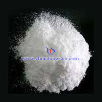
OBJECTIVES: to investigate the method of discovery of abdominal aortic aneurysms (AAA) in a district general hospital setting. DESIGN: retrospective study. MATERIALS AND METHODS: we analysed 198 patients with an AAA who presented to our unit over a 3-year period. The method of initial diagnosis, size of the AAA and whether this was palpable, irrespective of the method of detection, were recorded. RESULTS: ninety-five (48%) were discovered clinically, 74 (37.4%) during a radiological investigation, and 29 (14.6%) at laparotomy. Of the 74 AAAs first detected radiologically, subsequent physical examination showed that 28 (37.8%) were in fact palpable and missed at presentation. The average size of those discovered clinically (6. 48+/-1.32 cm) was larger than those found radiologically (5.37+/-1. 44 cm, p<0.001) or at operation (5.43+/-1.48 cm, p=0.039). The average diameter of the palpable AAAs was also greater than that of the non-palpable AAAs (6.42+/-1.24 cm vs. 4.86+/-1.38 cm, p<0.001). CONCLUSIONS: opportunistic detection of a clinically unsuspected aneurysm during clinical examination or investigation for another reason is the most common way the diagnosis of an AAA is made. Almost half of the aneurysms were diagnosed clinically, but physical examination also missed more than a third of those detected radiologically. Despite technological advancement, clinical examination still plays a paramount role in the detection of AAAs. Larger AAAs are usually palpable and more likely to be detected on clinical examination. (+info)Barium sulfate is a medication that is commonly used as a contrast material in medical imaging procedures, such as X-rays and CT scans. It works by coating the inside of the digestive tract, making it visible on an X-ray or CT scan and allowing doctors to see detailed images of the stomach, intestines, and other parts of the digestive system.
Barium sulfate is a white, chalky powder that is mixed with water to create a thick, milky liquid. It is generally safe and does not cause significant side effects when used in medical imaging procedures. However, it should not be taken by individuals who have a known allergy to barium or who have certain digestive conditions, such as obstructions or perforations of the bowel.
It's important to note that while barium sulfate is an important tool for medical diagnosis, it is not a treatment for any medical condition and should only be used under the direction of a healthcare professional.
Contrast media are substances that are administered to a patient in order to improve the visibility of internal body structures or processes in medical imaging techniques such as X-rays, CT scans, MRI scans, and ultrasounds. These media can be introduced into the body through various routes, including oral, rectal, or intravenous administration.
Contrast media work by altering the appearance of bodily structures in imaging studies. For example, when a patient undergoes an X-ray examination, contrast media can be used to highlight specific organs, tissues, or blood vessels, making them more visible on the resulting images. In CT and MRI scans, contrast media can help to enhance the differences between normal and abnormal tissues, allowing for more accurate diagnosis and treatment planning.
There are several types of contrast media available, each with its own specific properties and uses. Some common examples include barium sulfate, which is used as a contrast medium in X-ray studies of the gastrointestinal tract, and iodinated contrast media, which are commonly used in CT scans to highlight blood vessels and other structures.
While contrast media are generally considered safe, they can sometimes cause adverse reactions, ranging from mild symptoms such as nausea or hives to more serious complications such as anaphylaxis or kidney damage. As a result, it is important for healthcare providers to carefully evaluate each patient's medical history and individual risk factors before administering contrast media.
Barium is a naturally occurring, silvery-white metallic chemical element with the symbol Ba and atomic number 56. In medical terms, barium is commonly used as a contrast agent in radiology, particularly in X-ray examinations such as an upper GI series or barium enema. The barium sulfate powder is mixed with water to create a liquid or thick paste that is swallowed or inserted through the rectum. This provides a white coating on the inside lining of the digestive tract, allowing it to be seen more clearly on X-ray images and helping doctors diagnose various conditions such as ulcers, tumors, or inflammation.
It's important to note that barium is not absorbed by the body and does not cause any harm when used in medical imaging procedures. However, if it is accidentally inhaled or aspirated into the lungs during administration, it can cause chemical pneumonitis, a potentially serious condition. Therefore, it should only be administered under the supervision of trained medical professionals.
 Barium sulfate
 Barium Sulfate: MedlinePlus Drug Information
 DailyMed - Search Results for BARIUM SULFATE
 How Oxalate Affects Barium Sulfate Crystallization - Science Trends
 KP259 - Sulphate Precipitant Barium chloride crystals
 Barium sulfate mass and molar concentrations
 barium sulfate grinding equipment
 Varibar® Honey Barium Sulfate
 Natural Barium Sulfate, barium sulfate
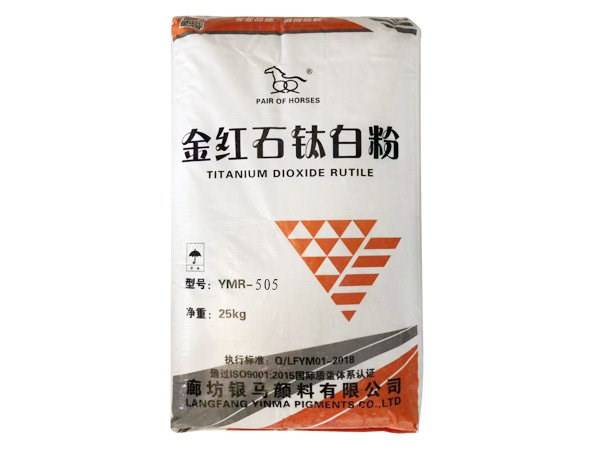
 ODP97 Barium Sulfate Coating Material
 What is Barium Sulfate? (with pictures)
 Barium Sulphate - GP LLC
 Barium Sulphate | Nandu Chemicals
 Barium Sulphate HDPE bottle - Vital Minerals
 Potassium Powder, Barium Sulphate Powder Manufacturers
 Barium Sulphate For Soil Testing | Vickers Laboratories
 Precipitated Barium Sulphate in Coatings - Hoyonn Blog
 NSN: 6505-00-419-8457 (BARIUM SULFATE,DIAG) - ArmyProperty.com
 Barium Sulphate Powder - Barium Sulphate Manufacturer from Delhi
 Symptom or Signs as Drug Side Effect
 CAS 7727-43-7 Dull Matting Barium Sulphate Paint , Barium Sulfate Coating


![Green Glue Alternative Noiseproofing Compounds [2023]](https://soundproofsilence.com/wp-content/uploads/2020/09/best-Green-Glue-alternatives.jpg)