Autonomic Dysreflexia
Spinal Cord Injuries
Quadriplegia
Autonomic Nervous System Diseases
Reflex, Abnormal
Intracranial Hemorrhage, Hypertensive
Paraplegia
Autonomic Fibers, Preganglionic
Reflex
Galvanic Skin Response
Thoracic Vertebrae
Afferent Pathways
Adrenergic alpha-Antagonists
Cervical Vertebrae
Receptors, Adrenergic, alpha
Management of life-threatening autonomic hyper-reflexia using magnesium sulphate in a patient with a high spinal cord injury in the intensive care unit. (1/58)
We report the successful use of i.v. magnesium sulphate to control life-threatening autonomic hyper-reflexia associated with chronic spinal cord injury in the intensive care environment. A 37-yr-old, male was admitted to the intensive care unit with a diagnosis of septic shock and acute renal failure secondary to pyelonephritis. He had been found unresponsive at home following a 2-day history of pyrexia and purulent discharge from his suprapubic catheter. He had sustained a T5 spinal cord transection 20 yr previously. Initial management included assisted ventilation, fluid resuscitation, vasopressor support, and continuous veno-venous haemofiltration. The sepsis was treated with antibiotic therapy and percutaneous nephrostomy drainage of the pyonephrosis. On the fifth day, the patient developed profuse diarrhoea. This was associated with paroxysms of systemic hypertension and diaphoresis, his arterial pressure rising on occasion to 240/140 mm Hg. A diagnosis of autonomic hyper-reflexia was made and a bolus dose of magnesium sulphate 5 g was administered over 15 min followed by an infusion of 1-2 g h(-1). There was an almost immediate decrease in the severity and frequency of the hypertensive episodes. There were no adverse cardiac effects associated with the administration of magnesium, only a slight decrease in minute ventilation as the plasma level approached the upper end of the therapeutic range (2-4 mmol litre(-1)). In view of the beneficial effects observed in this case we advocate further research into the use of magnesium sulphate in the treatment or prevention of autonomic hyper-reflexia secondary to chronic spinal cord injury in the intensive care unit. (+info)TENS attenuates response to colon distension in paraplegic and quadriplegic rats. (2/58)
Individuals with spinal cord injuries above thoracic level 6 experience episodic bouts of life-threatening hypertension as part of a condition termed autonomic dysreflexia (AD). The hypertension can be caused by stimulation of the skin, distension of the urinary bladder or colon, and/or muscle spasms. Transcutaneous electrical nerve stimulation (TENS) may reduce the severity of AD because TENS has been used to inhibit second-order neurons in the dorsal horn. Therefore, we tested the hypothesis that TENS attenuates the hemodynamic responses to colon distension. Eleven Wistar rats underwent spinal cord transection between thoracic vertebrae 4 and 5 (paraplegic, n = 6) or between cervical vertebra 7 and thoracic vertebra 1 (quadriplegic, n = 5). After recovery, all rats were instrumented with a radiotelemetry device for recording arterial pressure. Subsequently, the hemodynamic responses to graded colon distension were determined before and during TENS. During TENS the hemodynamic responses to colon distension were significantly attenuated. Thus TENS may be a preventive approach to reduce the severity of AD in paraplegic and quadriplegic individuals. (+info)Long-term result of Memokath urethral sphincter stent in spinal cord injury patients. (3/58)
BACKGROUND: Memokath urethral sphincter stents are used to facilitate bladder emptying in patients with spinal cord injury, but long term follow-up has not been reported. METHODS: Case series of ten men with spinal cord injury who underwent insertion of Memokath stents and were followed for up to nine years. RESULTS: Within four years, the stent had to be removed in nine out of ten patients because of: extensive mucosal proliferation causing obstruction to the lumen of the stent; stone around the proximal end of the stent, incomplete bladder emptying, and recurrent urinary infections; migration of the stent into the bladder related to digital evacuation of bowels; large residual urine; concretions within the stent causing obstruction to flow of urine, and partial blockage of the stent causing frequent episodes of autonomic dysreflexia. In one patient the stent continued to function satisfactorily after nine years. CONCLUSIONS: The Memokath stent has a role as a temporary measure for treatment of detrusor-sphincter dyssynergia in selected SCI patients who do not get recurrent urinary infection and do not require manual evacuation of bowels. (+info)Rehabilitation medicine: 1. Autonomic dysreflexia. (4/58)
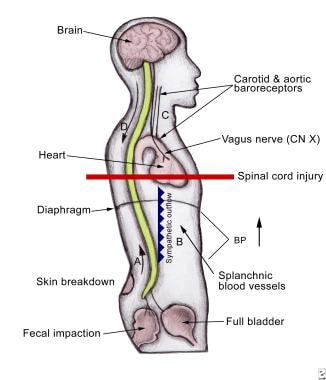
Autonomic dysreflexia is an acute syndrome of excessive, uncontrolled sympathetic output that can occur in patients who have had an injury to the spinal cord (generally at or above the sixth thoracic neurologic level). It is caused by spinal reflex mechanisms that remain intact despite the patient's injury, leading to hypertension. This review describes the clinical features of autonomic dysreflexia, its common causes (most frequently stimulation of the lower urinary tract) and a recommended approach to treatment. The condition can nearly always be managed successfully, but prompt recognition is essential--without treatment there may be dire consequences, including death. (+info)Tail arteries from chronically spinalized rats have potentiated responses to nerve stimulation in vitro. (5/58)
Patients with severe spinal cord lesions that damage descending autonomic pathways generally have low resting arterial pressure but bladder or colon distension or unheeded injuries may elicit a life-threatening hypertensive episode. Such episodes (known as autonomic dysreflexia) are thought to result from the loss of descending baroreflex inhibition and/or plasticity within the spinal cord. However, it is not clear whether changes in the periphery contribute to the exaggerated reflex vasoconstriction. The effects of spinal transection at T7-8 on nerve- and agonist-evoked contractions of the rat tail artery were investigated in vitro. Isometric contractions of arterial segments were recorded and responses of arteries from spinalized animals ('spinalized arteries') and age-matched and sham-operated controls were compared. Two and eight weeks after transection, nerve stimulation at 0.1-10 Hz produced contractions of greater force and duration in spinalized arteries. At both stages, the alpha-adrenoceptor antagonists prazosin (10 nm) and idazoxan (0.1 microm) produced less blockade of nerve-evoked contraction in spinalized arteries. Two weeks after transection, spinalized arteries were supersensitive to the alpha(1)-adrenoceptor agonist phenylephrine, and the alpha(2)-adrenoceptor agonist, clonidine, but 8 weeks after transection, spinalized arteries were supersensitive only to clonidine. Contractions of spinalized arteries elicited by 60 mm K(+) were larger and decayed more slowly at both stages. These findings demonstrate that spinal transection markedly increases nerve-evoked contractions and this can, in part, be accounted for by increased reactivity of the vascular smooth muscle to vasoconstrictor agents. This hyper-reactivity may contribute to the genesis of autonomic dysreflexia in patients. (+info)Transient blockade of the CD11d/CD18 integrin reduces secondary damage after spinal cord injury, improving sensory, autonomic, and motor function. (6/58)
The early inflammatory response to spinal cord injury (SCI) causes significant secondary damage. Strategies that nonselectively suppress inflammation have not improved outcomes after SCI, perhaps because inflammation has both adverse and beneficial effects after SCI. We have shown that the selective, time-limited action of a monoclonal antibody (mAb) to the CD11d subunit of the CD11d/CD18 integrin, delivered intravenously during the first 48 hr after SCI in rats, markedly decreases the infiltration of neutrophils and delays the entry of hematogenous monocyte-macrophages into the injured cord. We hypothesized that this targeted strategy would lead to neuroprotection and improved neurological outcomes. In this study the development of chronic pain was detected in rats by assessing mechanical allodynia on the trunk and hindpaws 2 weeks to 3 months after a clinically relevant clip-compression SCI at the twelfth thoracic segment. The anti-CD11d mAb treatment reduced this pain by half. Motor performance also improved as rats were able to plantar-place their hindpaws and use them for weight support instead of sweeping movements only. Improved cardiovascular outcome was shown after SCI at the fourth thoracic segment by significant decreases in autonomic dysreflexia. Locomotor performance was also improved. These functional changes correlated with significantly greater amounts and increased organization of myelin and neurofilament near the lesion. The improved neurological recovery after the specific reduction of early inflammation after SCI demonstrates that this selective strategy increases tissue at the injury site and improves its functional capacity. This early neuroprotective treatment would be an ideal foundation for building later cell-based therapies. (+info)Autonomic dysreflexia during sperm retrieval in spinal cord injury: influence of lesion level and sildenafil citrate. (7/58)
Autonomic dysreflexia (AD) can occur during penile vibratory stimulation in men with spinal cord injury, but this is variable, and the association with lesion level is unclear. The purpose of this study was to characterize the cardiovascular responses to penile vibratory stimulation in men with spinal cord injury. We hypothesized that those with cervical injuries would demonstrate a greater degree of AD compared with men with thoracic injuries. We also questioned whether the rise in blood pressure could be attenuated by sildenafil citrate. Participants were classified as having cervical (n = 8) or thoracic (n = 5) injuries. While in a supine position, subjects were instrumented with an ECG, and arterial blood pressure was determined beat by beat. Subjects reported to the laboratory twice and received an oral dose of sildenafil citrate (25-100 mg) or no medication. Penile vibratory stimulation was performed using a handheld vibrator to the point of ejaculation. At ejaculation during the nonmedicated trials, the cervical group had a significant decrease in heart rate (-5-10 beats/min) and increase in mean arterial blood pressure (+70-90 mmHg) relative to resting conditions, whereas the thoracic group had significant increases in both heart rate (+8-15 beats/min) and mean arterial pressure (+25-30 mmHg). Sildenafil citrate had no effect on the change in heart rate or mean arterial pressure in either group. In summary, men with cervical injuries had more pronounced AD during penile vibratory stimulation than men with thoracic injuries. Administration of sildenafil citrate had no effect on heart rate or blood pressure during penile vibratory stimulation in men with spinal cord injury. (+info)Autonomic dysreflexia: a medical emergency. (8/58)
Autonomic dysreflexia is an important clinical diagnosis that requires prompt treatment to avoid devastating complications. The condition may present itself to all members of medical and surgical specialties, who may not be accustomed to treating it. It is the clinician's responsibility to have a basic understanding of the pathophysiology of the condition and the simple steps required to treat it. (+info)Autonomic dysreflexia is a medical condition that primarily affects individuals with spinal cord injuries at level T6 or above. It is characterized by an overactive autonomic nervous system response, leading to potentially life-threatening symptoms. This occurs when there is a stimulus below the level of the spinal cord injury that triggers a reflexive sympathetic nervous system response, causing a rapid and significant increase in blood pressure and heart rate.
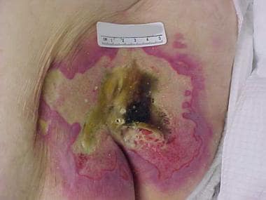
Common triggers for autonomic dysreflexia include bladder distention, bowel distension or constipation, skin irritation, pressure sores, infection, or sexual activity. Symptoms of autonomic dysreflexia may include severe headaches, sweating above the level of injury, flushing or pallor, goosebumps, nasal congestion, and blurred vision. If left untreated, it can lead to seizures, stroke, or even cardiac arrest.
Management of autonomic dysreflexia involves identifying and removing the underlying trigger, as well as managing symptoms through medications such as antihypertensives, and monitoring vital signs closely. Prevention strategies include regular bladder and bowel management, skin checks, and prompt treatment of infections or other potential triggers.
Spinal cord injuries (SCI) refer to damage to the spinal cord that results in a loss of function, such as mobility or feeling. This injury can be caused by direct trauma to the spine or by indirect damage resulting from disease or degeneration of surrounding bones, tissues, or blood vessels. The location and severity of the injury on the spinal cord will determine which parts of the body are affected and to what extent.
The effects of SCI can range from mild sensory changes to severe paralysis, including loss of motor function, autonomic dysfunction, and possible changes in sensation, strength, and reflexes below the level of injury. These injuries are typically classified as complete or incomplete, depending on whether there is any remaining function below the level of injury.
Immediate medical attention is crucial for spinal cord injuries to prevent further damage and improve the chances of recovery. Treatment usually involves immobilization of the spine, medications to reduce swelling and pressure, surgery to stabilize the spine, and rehabilitation to help regain lost function. Despite advances in treatment, SCI can have a significant impact on a person's quality of life and ability to perform daily activities.
Quadriplegia, also known as tetraplegia, is a medical condition characterized by paralysis affecting all four limbs and the trunk of the body. It results from damage to the cervical spinal cord, typically at levels C1-C8, which controls signals to the muscles in the arms, hands, trunk, legs, and pelvic organs. The extent of quadriplegia can vary widely, ranging from weakness to complete loss of movement and sensation below the level of injury. Other symptoms may include difficulty breathing, bowel and bladder dysfunction, and sexual dysfunction. The severity and prognosis depend on the location and extent of the spinal cord injury.
The Autonomic Nervous System (ANS) is a part of the nervous system that controls involuntary actions, such as heart rate, digestion, respiratory rate, pupillary response, urination, and sexual arousal. It consists of two subdivisions: the sympathetic and parasympathetic nervous systems, which generally have opposing effects and maintain homeostasis in the body.
Autonomic Nervous System Diseases (also known as Autonomic Disorders or Autonomic Neuropathies) refer to a group of conditions that affect the functioning of the autonomic nervous system. These diseases can cause damage to the nerves that control automatic functions, leading to various symptoms and complications.
Autonomic Nervous System Diseases can be classified into two main categories:
1. Primary Autonomic Nervous System Disorders: These are conditions that primarily affect the autonomic nervous system without any underlying cause. Examples include:
* Pure Autonomic Failure (PAF): A rare disorder characterized by progressive loss of autonomic nerve function, leading to symptoms such as orthostatic hypotension, urinary retention, and constipation.
* Multiple System Atrophy (MSA): A degenerative neurological disorder that affects both the autonomic nervous system and movement coordination. Symptoms may include orthostatic hypotension, urinary incontinence, sexual dysfunction, and Parkinsonian features like stiffness and slowness of movements.
* Autonomic Neuropathy associated with Parkinson's Disease: Some individuals with Parkinson's disease develop autonomic symptoms such as orthostatic hypotension, constipation, and urinary dysfunction due to the degeneration of autonomic nerves.
2. Secondary Autonomic Nervous System Disorders: These are conditions that affect the autonomic nervous system as a result of an underlying cause or disease. Examples include:
* Diabetic Autonomic Neuropathy: A complication of diabetes mellitus that affects the autonomic nerves, leading to symptoms such as orthostatic hypotension, gastroparesis (delayed gastric emptying), and sexual dysfunction.
* Autoimmune-mediated Autonomic Neuropathies: Conditions like Guillain-Barré syndrome or autoimmune autonomic ganglionopathy can cause autonomic symptoms due to the immune system attacking the autonomic nerves.
* Infectious Autonomic Neuropathies: Certain infections, such as HIV or Lyme disease, can lead to autonomic dysfunction as a result of nerve damage.
* Toxin-induced Autonomic Neuropathy: Exposure to certain toxins, like heavy metals or organophosphate pesticides, can cause autonomic neuropathy.
Autonomic nervous system disorders can significantly impact a person's quality of life and daily functioning. Proper diagnosis and management are crucial for improving symptoms and preventing complications. Treatment options may include lifestyle modifications, medications, and in some cases, devices or surgical interventions.
An abnormal reflex in a medical context refers to an involuntary and exaggerated response or lack of response to a stimulus that is not expected in the normal physiological range. These responses can be indicative of underlying neurological disorders or damage to the nervous system. Examples include hyperreflexia (overactive reflexes) and hyporeflexia (underactive reflexes). The assessment of reflexes is an important part of a physical examination, as it can provide valuable information about the functioning of the nervous system.
Intracranial hemorrhage, hypertensive is a type of intracranial hemorrhage that occurs due to the rupture of blood vessels in the brain as a result of chronic high blood pressure (hypertension). It is also known as hypertensive intracerebral hemorrhage.
Hypertension can weaken and damage the walls of the small arteries and arterioles in the brain over time, making them more susceptible to rupture. When these blood vessels burst, they cause bleeding into the surrounding brain tissue, forming a hematoma that can compress and damage brain cells.
Intracranial hemorrhage, hypertensive is a medical emergency that requires immediate treatment. Symptoms may include sudden severe headache, weakness or numbness in the face or limbs, difficulty speaking or understanding speech, vision changes, loss of balance or coordination, and altered level of consciousness.
The diagnosis of intracranial hemorrhage, hypertensive is typically made through imaging tests such as computed tomography (CT) or magnetic resonance imaging (MRI) scans. Treatment may involve medications to reduce blood pressure, surgery to remove the hematoma, and supportive care to manage complications such as brain swelling or seizures.
Paraplegia is a medical condition characterized by partial or complete loss of motor function and sensation in the lower extremities, typically affecting both legs. This results from damage to the spinal cord, often due to trauma such as accidents, falls, or gunshot wounds, or from diseases like spina bifida, polio, or tumors. The specific area and extent of the injury on the spinal cord determine the severity and location of paralysis. Individuals with paraplegia may require assistive devices for mobility, such as wheelchairs, and may face various health challenges, including pressure sores, urinary tract infections, and chronic pain.
Preganglionic autonomic fibers are the nerve fibers that originate from neurons located in the brainstem and spinal cord, and synapse with postganglionic neurons in autonomic ganglia. These preganglionic fibers release acetylcholine as a neurotransmitter to activate the postganglionic neurons, which then innervate effector organs such as smooth muscle, cardiac muscle, and glands.
The autonomic nervous system is divided into two main subdivisions: the sympathetic and parasympathetic systems. The preganglionic fibers of the sympathetic nervous system originate from the lateral horn of the spinal cord from levels T1 to L2/L3, while those of the parasympathetic nervous system originate from cranial nerves III, VII, IX, and X, as well as sacral segments S2 to S4.
Preganglionic fibers are generally longer than postganglionic fibers, and their cell bodies are located in the central nervous system. They are responsible for transmitting signals from the CNS to the peripheral autonomic ganglia, where they synapse with postganglionic neurons that innervate target organs.
A reflex is an automatic, involuntary and rapid response to a stimulus that occurs without conscious intention. In the context of physiology and neurology, it's a basic mechanism that involves the transmission of nerve impulses between neurons, resulting in a muscle contraction or glandular secretion.
Reflexes are important for maintaining homeostasis, protecting the body from harm, and coordinating movements. They can be tested clinically to assess the integrity of the nervous system, such as the knee-j jerk reflex, which tests the function of the L3-L4 spinal nerve roots and the sensitivity of the stretch reflex arc.
Galvanic Skin Response (GSR), also known as Electrodermal Activity (EDA), is a physiological response that reflects the activation of the sympathetic nervous system. It measures changes in the electrical properties of the skin, which are influenced by the sweat gland activity. GSR is often used as an indicator of emotional arousal or psychological stress in various research and clinical settings.
Doxazosin is an antihypertensive drug, which belongs to the class of medications called alpha-1 receptor blockers. It works by relaxing the muscles in the blood vessels, which helps to lower blood pressure and improve blood flow. Doxazosin is primarily used to treat high blood pressure (hypertension) and benign prostatic hyperplasia (BPH), a condition characterized by an enlarged prostate gland that can cause urinary symptoms such as difficulty in beginning the flow of urine, weak stream, and frequent urination.
The medical definition of Doxazosin is:
Doxazosin mesylate - A selective alpha-1 adrenergic receptor blocker used in the treatment of hypertension and benign prostatic hyperplasia (BPH). It works by relaxing the smooth muscle in blood vessels, which lowers blood pressure and improves blood flow. Doxazosin may also be used off-label for other indications such as Raynaud's phenomenon or painful bladder syndrome. The drug is available in oral tablet form and is typically taken once daily. Common side effects include dizziness, lightheadedness, and headache.
The rectum is the lower end of the digestive tract, located between the sigmoid colon and the anus. It serves as a storage area for feces before they are eliminated from the body. The rectum is about 12 cm long in adults and is surrounded by layers of muscle that help control defecation. The mucous membrane lining the rectum allows for the detection of stool, which triggers the reflex to have a bowel movement.
Urodynamics is a medical test that measures the function and performance of the lower urinary tract, which includes the bladder, urethra, and sphincters. It involves the use of specialized equipment to record measurements such as bladder pressure, urine flow rate, and residual urine volume. The test can help diagnose various urinary problems, including incontinence, urinary retention, and overactive bladder.
During the test, a small catheter is inserted into the bladder through the urethra to measure bladder pressure while filling it with sterile water or saline solution. Another catheter may be placed in the rectum to record abdominal pressure. The patient is then asked to urinate, and the flow rate and any leaks are recorded.
Urodynamics can help identify the underlying cause of urinary symptoms and guide treatment decisions. It is often recommended for patients with complex or persistent urinary problems that have not responded to initial treatments.
The thoracic vertebrae are the 12 vertebrae in the thoracic region of the spine, which is the portion between the cervical and lumbar regions. These vertebrae are numbered T1 to T12, with T1 being closest to the skull and T12 connecting to the lumbar region.
The main function of the thoracic vertebrae is to provide stability and support for the chest region, including protection for the vital organs within, such as the heart and lungs. Each thoracic vertebra has costal facets on its sides, which articulate with the heads of the ribs, forming the costovertebral joints. This connection between the spine and the ribcage allows for a range of movements while maintaining stability.
The thoracic vertebrae have a unique structure compared to other regions of the spine. They are characterized by having long, narrow bodies, small bony processes, and prominent spinous processes that point downwards. This particular shape and orientation of the thoracic vertebrae contribute to their role in limiting excessive spinal movement and providing overall trunk stability.
Afferent pathways, also known as sensory pathways, refer to the neural connections that transmit sensory information from the peripheral nervous system to the central nervous system (CNS), specifically to the brain and spinal cord. These pathways are responsible for carrying various types of sensory information, such as touch, temperature, pain, pressure, vibration, hearing, vision, and taste, to the CNS for processing and interpretation.
The afferent pathways begin with sensory receptors located throughout the body, which detect changes in the environment and convert them into electrical signals. These signals are then transmitted via afferent neurons, also known as sensory neurons, to the spinal cord or brainstem. Within the CNS, the information is further processed and integrated with other neural inputs before being relayed to higher cognitive centers for conscious awareness and response.
Understanding the anatomy and physiology of afferent pathways is essential for diagnosing and treating various neurological conditions that affect sensory function, such as neuropathies, spinal cord injuries, and brain disorders.
Blood pressure is the force exerted by circulating blood on the walls of the blood vessels. It is measured in millimeters of mercury (mmHg) and is given as two figures:
1. Systolic pressure: This is the pressure when the heart pushes blood out into the arteries.
2. Diastolic pressure: This is the pressure when the heart rests between beats, allowing it to fill with blood.
Normal blood pressure for adults is typically around 120/80 mmHg, although this can vary slightly depending on age, sex, and other factors. High blood pressure (hypertension) is generally considered to be a reading of 130/80 mmHg or higher, while low blood pressure (hypotension) is usually defined as a reading below 90/60 mmHg. It's important to note that blood pressure can fluctuate throughout the day and may be affected by factors such as stress, physical activity, and medication use.
Adrenergic alpha-antagonists, also known as alpha-blockers, are a class of medications that block the effects of adrenaline and noradrenaline at alpha-adrenergic receptors. These receptors are found in various tissues throughout the body, including the smooth muscle of blood vessels, the heart, the genitourinary system, and the eyes.
When alpha-blockers bind to these receptors, they prevent the activation of the sympathetic nervous system, which is responsible for the "fight or flight" response. This results in a relaxation of the smooth muscle, leading to vasodilation (widening of blood vessels), decreased blood pressure, and increased blood flow.
Alpha-blockers are used to treat various medical conditions, such as hypertension (high blood pressure), benign prostatic hyperplasia (enlarged prostate), pheochromocytoma (a rare tumor of the adrenal gland), and certain types of glaucoma.
Examples of alpha-blockers include doxazosin, prazosin, terazosin, and tamsulosin. Side effects of alpha-blockers may include dizziness, lightheadedness, headache, weakness, and orthostatic hypotension (a sudden drop in blood pressure upon standing).
The cervical vertebrae are the seven vertebrae that make up the upper part of the spine, also known as the neck region. They are labeled C1 to C7, with C1 being closest to the skull and C7 connecting to the thoracic vertebrae in the chest region. The cervical vertebrae have unique structures to allow for a wide range of motion in the neck while also protecting the spinal cord and providing attachment points for muscles and ligaments.
Adrenergic receptors are a type of G protein-coupled receptor that bind and respond to catecholamines, such as epinephrine (adrenaline) and norepinephrine (noradrenaline). Alpha adrenergic receptors (α-ARs) are a subtype of adrenergic receptors that are classified into two main categories: α1-ARs and α2-ARs.
The activation of α1-ARs leads to the activation of phospholipase C, which results in an increase in intracellular calcium levels and the activation of various signaling pathways that mediate diverse physiological responses such as vasoconstriction, smooth muscle contraction, and cell proliferation.
On the other hand, α2-ARs are primarily located on presynaptic nerve terminals where they function to inhibit the release of neurotransmitters, including norepinephrine. The activation of α2-ARs also leads to the inhibition of adenylyl cyclase and a decrease in intracellular cAMP levels, which can mediate various physiological responses such as sedation, analgesia, and hypotension.
Overall, α-ARs play important roles in regulating various physiological functions, including cardiovascular function, mood, and cognition, and are also involved in the pathophysiology of several diseases, such as hypertension, heart failure, and neurodegenerative disorders.
The colon, also known as the large intestine, is a part of the digestive system in humans and other vertebrates. It is an organ that eliminates waste from the body and is located between the small intestine and the rectum. The main function of the colon is to absorb water and electrolytes from digested food, forming and storing feces until they are eliminated through the anus.
The colon is divided into several regions, including the cecum, ascending colon, transverse colon, descending colon, sigmoid colon, rectum, and anus. The walls of the colon contain a layer of muscle that helps to move waste material through the organ by a process called peristalsis.
The inner surface of the colon is lined with mucous membrane, which secretes mucus to lubricate the passage of feces. The colon also contains a large population of bacteria, known as the gut microbiota, which play an important role in digestion and immunity.
 Autonomic dysreflexia
F3 (classification)
F1 (classification)
Nifedipine
F2 (classification)
F4 (classification)
Spinal shock
Boosting (doping)
Sexuality after spinal cord injury
Hyperreflexia
Ganglionic blocker
Mitrofanoff procedure
Winter Paralympic Games
Cheating at the Paralympic Games
Disability and women's health
Spinal cord injury
Pressure ulcer
AD (disambiguation)
Mecamylamine
List of MeSH codes (C10)
Catastrophic injury
Doping
Hyperhidrosis
Tetraplegia
Autonomic dysreflexia - Wikipedia
 Autonomic dysreflexia: MedlinePlus Medical Encyclopedia
 Autonomic Dysreflexia in wheelchair tennis athletes
 Autonomic Dysreflexia in Spinal Cord Injury: Practice Essentials, Pathophysiology, Causes of Autonomic Dysreflexia
 Knowledge of autonomic dysreflexia in the emergency department | Emergency Medicine Journal
 Spinal Cord Autonomic Dysreflexia - Neurologic Disorders - Merck Manuals Professional Edition
 Reversing Maladaptive Plasticity to Cure Autonomic Dysreflexia after Spinal Cord Injury
 Automated Detection of Symptomatic Autonomic Dysreflexia through Multimodal Sensing - IEEE Journal of Translational Engineering...
 Featured Article: Targeted ablation of sympathetic neurons reduces ventricular arrhythmias and autonomic dysreflexia - Advanced...
 What is Autonomic Dysreflexia? | Answers | Spinal Cord Injury Zone!
Spinal Cord Autonomic Dysreflexia - Neurologic Disorders - MSD Manual Professional Edition
 CE Activity | Autonomic Dysreflexia | Nurses
 Early Pregabalin Treatment Suppresses Autonomic Dysreflexia Following Spinal Cord Injury in Rats
 Autonomic Dysreflexia
Stichworte Silent autonomic dysreflexia
 Autonomic Dysreflexia | Triumph Foundation
Autonomic Dysreflexia | Profiles RNS
 Cardiac Effects of Recurring Autonomic Dysreflexia
Autonomic Dysreflexia - Pathogenesis and clinical findings | Calgary Guide
ABC of Autonomic Dysreflexia | in Spinal Cord Injury
 Consumer Fact Sheets
 What Causes Night Sweats in Men? 10 Causes, When to See Doctor
Leslie, Stephen W | Creighton University
 Bladder distension: An unusual cause of reflux of blood and hemodynamic changes (autonomic dysreflexia) during endovascular...
 Persistent Night Sweats: Diagnostic Evaluation | AAFP
 Table of Contents - April 13, 2004, 170 (8) | CMAJ
 Provisional Mortality by Multiple Cause of Death
 Free Occupational Therapy Flashcards about Spinal Cord Info 2
 Dr. Rajul Parikh, MD, Clinical Neurophysiologist - Orange Park, FL | Sharecare
 SCInfo Blog | ICORD | Page 7
Hyperreflexia1
- Autonomic Dysreflexia (AD) , also known as Hyperreflexia, is a potentially dangerous complication of spinal cord injury (SCI). (spinalcordinjuryzone.com)
Pathophysiology2
- This activity reviews the causes, pathophysiology, presentation, and treatment of autonomic dysreflexia and highlights the role of the interprofessional team in its management. (statpearls.com)
- Describe the pathophysiology of autonomic dysreflexia. (statpearls.com)
Nervous12
- Autonomic dysreflexia is an abnormal, overreaction of the involuntary (autonomic) nervous system to stimulation. (medlineplus.gov)
- Autonomic dysreflexia is a disorder of autonomic nervous system dysregulation that occurs in patients with a spinal cord injury and that can result in life-threatening hypertension. (merckmanuals.com)
- Dysregulation of the autonomic nervous system leads to an uncoordinated sympathetic response that may result in a potentially life-threatening hypertensive episode when there is a noxious stimulus below the level of the spinal cord injury. (statpearls.com)
- The nervous system responds via the autonomic nervous system, which results in dilation of the blood vessels, this in turn lowers the blood pressure and the body circulation remains in a safe and static state. (ashfordstpeters.info)
- Patients with a spinal cord injury located at Thoracic spinal level 6 (T6) the ability of the autonomic nervous system to respond normally is affected. (ashfordstpeters.info)
- What does the parasympathetic division of the autonomic nervous system control? (toptenid.com)
- Though the threats that modern humans face are not large predators, the autonomic nervous system is adapted to this type of stimulus. (toptenid.com)
- Homeostasis, remember, is a word that Walter Cannon coined, and what does the autonomic nervous system have to do with homeostasis? (toptenid.com)
- Acute AD is a reaction of the autonomic (involuntary) nervous system to overstimulation. (triumph-foundation.org)
- The bladder and urethra are innervated by 3 sets of peripheral nerves arising from the autonomic nervous system (ANS) and somatic nervous system. (medscape.com)
- Dysautonomia is when your autonomic nervous system doesn't work properly, usually due to an underlying condition. (healthline.com)
- Your autonomic nervous system (ANS) is responsible for functions that your body does without you having to think about them, such as pumping blood, digesting food, and breathing in and out. (healthline.com)
Sixth thoracic2
- Briefly, autonomic dysreflexia develops in individuals with a neurologic level of SCI at or above the sixth thoracic vertebral level (T6). (medscape.com)
- Persons with a spinal cord injury (SCI) above the sixth thoracic vertebrae commonly experience autonomic dysreflexia (AD), 90 percent of individuals with this level of injury are susceptible to AD which is associated with an increase in sympathetic nerve activity. (purdue.edu)
Bladder6
- Thus, assessment of autonomic dysreflexia in patients with known causative factors may include palpation of the bladder and bowel and can also include bladder scan. (wikipedia.org)
- Chemodenervation of the bladder using onabotulinumtoxinA has been shown to help prevent autonomic dysreflexia when used appropriately (ie, when all other measures to prevent it have been ineffective). (merckmanuals.com)
- The most common causes of autonomic dysreflexia seen in patients with spinal cord injury are impaction in the bowels and distention in case of the bladder. (triumph-foundation.org)
- Autonomic dysreflexia due to distended bladder is well known. (ruralneuropractice.com)
- Martinez also developed a severe headache and recognized that he was experiencing autonomic dysreflexia, which is often caused by an overly full bladder. (theblaze.com)
- Contraindications include a high-pressure neurogenic bladder, a history of autonomic dysreflexia, a febrile UTI, and situations following any procedure or trauma where there is a possibility for urinary extravasation and urinary drainage is needed for optimal tissue repair, such as TURBT, bladder repair or trauma, or radical prostatectomy. (medgadget.com)
Peripheral1
- Complications associated with autonomic dysreflexia result directly from sustained, severe peripheral hypertension. (medscape.com)
Hypertension7
- Autonomic dysreflexia (AD) is a potentially fatal medical emergency classically characterized by uncontrolled hypertension and cardiac arrhythmia. (wikipedia.org)
- Some of the common signs and symptoms during an episode of autonomic dysreflexia include headache, sweating, flushing, hypertension, slow or fast heart rate, goosebumps and pupillary constriction. (usta.com)
- Autonomic dysreflexia is a potentially dangerous and, in rare cases, lethal clinical syndrome that develops in individuals with spinal cord injury (SCI), resulting in acute, uncontrolled hypertension. (medscape.com)
- Autonomic dysreflexia causes an imbalanced reflex sympathetic discharge, leading to potentially life-threatening hypertension. (medscape.com)
- Autonomic dysreflexia should be suspected in a patient with a spinal cord injury above the level of T6, severe hypertension, and increased sympathetic activity, especially if provoked by distention of a hollow viscus. (merckmanuals.com)
- Autonomic dysreflexia occurs after a spinal cord injury and can result in life-threatening hypertension. (merckmanuals.com)
- Autonomic dysreflexia AD is a potential life threatening condition characterized as episodic vascular hypertension often with bradycardia that develops in most people with a spinal cord injury SCI above thoracic spinal level T5. (dtic.mil)
Spinal cord injury pat1
- Autonomic dysreflexia: An important cardiovascular complication in spinal cord injury patients. (nih.gov)
Symptoms2
- For this newsletter topic, we will help define autonomic dysreflexia, discuss signs and symptoms, management and also describe boosting . (usta.com)
- Symptoms of autonomic dysreflexia are variable, intermittent. (merckmanuals.com)
Occur3
- The first episode of autonomic dysreflexia may occur weeks to years after spinal cord injury takes place, but most people at risk (92%) develop their first episode within the first year after injury. (wikipedia.org)
- Cord injury is usually above the T6 level, with dysreflexia unlikely to occur after an injury below the T10 level. (merckmanuals.com)
- Autonomic dysreflexia can occur in susceptible individuals up to 40 times per day. (statpearls.com)
Disorders1
- Autonomic disorders and their management. (medlineplus.gov)
Occurs1
- Objective: Autonomic Dysreflexia (AD) is a potentially life-threatening syndrome which occurs in individuals with higher level spinal cord injuries (SCI). (embs.org)
Episodes1
- Additionally, the body does not respond to pain as it normally would and one can be prone to episodes of autonomic dysreflexia . (usta.com)
Sympathetic1
- Immunostaining for substance P revealed a significantly higher density in both the dorsal horn and central autonomic area in iPGB animals when compared to saline-treated and uninjured animals, indicating a possible mechanism of sympathetic inhibition following iPGB treatment. (dal.ca)
Treatment1
- Treatment of autonomic dysreflexia requires close monitoring of vital signs. (merckmanuals.com)
Management1
- Summarize the management options for autonomic dysreflexia. (statpearls.com)
Patients3
- Clinicians should suspect autonomic dysreflexia if patients have a T6 or higher spinal cord lesion and report headache. (merckmanuals.com)
- Outline the importance of improving care coordination among interprofessional team members to improve outcomes for patients affected by autonomic dysreflexia. (statpearls.com)
- Following spinal cord injury (SCI), up to 70% of patients develop a condition known as autonomic dysreflexia (AD). (dal.ca)
Commonly1
- Autonomic dysreflexia is not a commonly encountered condition. (usta.com)
Severe1
- The consequences of uncontrolled spikes in blood pressure, known as autonomic dysreflexia (AD), after spinal cord injury (SCI) are extremely severe. (abcofad.ca)
Stroke1
- Autonomic Dysreflexia (AD) is characterised by a sudden rise in blood pressure which may lead onto a cerebral haemorrhage (stroke) and even death. (ashfordstpeters.info)
Condition3
- Autonomic dysreflexia is a condition that emerges after a spinal cord injury, usually when the damage has occurred above the T6 level. (statpearls.com)
- Which patient below is at most risk for developing a condition called autonomic dysreflexia? (toptenid.com)
- Autonomic Dysreflexia (AD) is a potentially life threatening condition which can be considered a medical emergency requiring immediate attention. (triumph-foundation.org)
Definition1
- An elevation of 20 mm Hg over baseline systolic blood pressure, with a potential source below the neurological level of injury, meets the current definition of dysreflexia. (wikipedia.org)
Blood pressure1
- As a result the autonomic system cannot lower the blood pressure in response to the pain or discomfort below the level of the spinal cord injury. (ashfordstpeters.info)
Common1
- The most common cause of autonomic dysreflexia (AD) is spinal cord injury. (medlineplus.gov)
People2
- This graph shows the total number of publications written about "Autonomic Dysreflexia" by people in this website by year, and whether "Autonomic Dysreflexia" was a major or minor topic of these publications. (sdsu.edu)
- Below are the most recent publications written about "Autonomic Dysreflexia" by people in Profiles. (sdsu.edu)
Require1
- Conclusion Due to the potentially serious complications of autonomic dysreflexia, staff require teaching on autonomic dysreflexia accompanied by permanent reminders in the form of posters. (bmj.com)
Medical2
Care1
- If you are a pre-hospital care worker and need to find out more about Autonomic Dysreflexia (AD), this free online course was created with you in mind. (abcofad.ca)
Study1
- Method The study design was a prospective questionnaire, which was completed by 91 staff in the spinal unit and emergency department in Christchurch, who then undertook a teaching session on autonomic dysreflexia. (bmj.com)
Injury4
- What is Autonomic Dysreflexia and how does it affect spinal cord injury? (spinalcordinjuryzone.com)
- With this type of injury he is also at high risk from autonomic dysreflexia. (justgiving.com)
- Understanding the complex effects of spinal cord injury, including autonomic dysreflexia, is essential to effective trial advocacy for clients whose injuries involve paraplegia or quadriplegia. (atlantainjurylawblog.com)
- John Bethea , PhD, a professor and department head of biology in the College of Arts and Sciences, and Veronica Tom, PhD, an associate professor of neurobiology and anatomy in the College of Medicine, were awarded a $2,497,558 grant from the National Institutes of Health for the project "Soluble TNFa in the Development of Autonomic Dysreflexia after Spinal Cord Injury. (drexel.edu)
Risk1
- Which of the following clients is at highest risk for autonomic dysreflexia? (toptenid.com)