Anterior Temporal Lobectomy
Psychosurgery
Epilepsy, Temporal Lobe
Temporal Lobe
Amobarbital
Kluver-Bucy Syndrome
Sclerosis
Electroencephalography
Functional Laterality
Seizures
Magnetic Resonance Imaging
Delirium, Dementia, Amnestic, Cognitive Disorders
Neuropsychological Tests
Epilepsy, Complex Partial
Brain Damage, Chronic
Thoracic Surgery, Video-Assisted
Epilepsy
Amygdala
Intelligence
Postoperative Complications
Seizures, Febrile
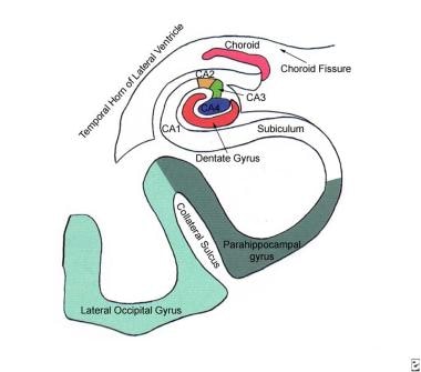
Hippocampus
Detection of visual field defects in patients after anterior temporal lobectomy for mesial temporal sclerosis-establishing eligibility to drive. (1/100)
AIMS: The aim of this study is to quantify visual field defects after temporal lobectomy for mesial temporal sclerosis and to establish eligibility for driving. METHODS: Automated static perimetry was performed on 14 patients who had undergone anterior temporal lobectomy for mesial temporal sclerosis. Perimetry consisted of monocular Humphrey Field Analyser (HFA) 30-2 test and a binocular Esterman 120 test. RESULTS: Of the 14 patients, three had no loss or non-specific loss, eight had partial homonymous quadrantanopia, one had complete homonymous quadrantanopia and two had concentric loss attributable to vigabatrin, which may have masked any loss occurring due to surgery. Of these, only seven passed the standardised DVLA visual fields. Of the seven who failed DVLA visual field, one had complete quadrantanopia, four had partial quadrantanopia and two had concentric loss (due to vigabatrin). CONCLUSIONS: Visual field defects contribute a great deal in the reduction of the quality of life in patients who have had surgery for mesial temporal sclerosis. Potential surgically induced visual field defects that could preclude driving need to be discussed with each patient preoperatively. In our study 50% of patients did not meet the required DVLA standards. (+info)Emotional facial paresis in temporal lobe epilepsy: its prevalence and lateralizing value. (2/100)
The selection of patients with medically refractory temporal lobe epilepsy (TLE) for surgery depends on the concordance of data from clinical, imaging and electroencephalographic evaluation. Though clinical examination is often normal, emotional facial paresis has been described in patients with TLE. Utilizing a well-characterized group of mesial TLE (MTLE) patients, who have achieved excellent seizure outcome following anterior temporal lobectomy with amygdalohippocampectomy (ATL), we investigated the prevalence, predictive value and associations of emotional facial paresis. When compared to 8 out of 50 control subjects (16%), 36 out of 50 MTLE patients (72%) exhibited unilateral emotional facial paresis; the difference was highly significant (P<0.0001). The presence of contralateral emotional facial paresis correctly predicted the side of ATL in 86.1% patients. The occurrence of emotional facial paresis was significantly associated with longer duration of epilepsy prior to ATL and left ATL. Our observations confirm that emotional facial parersis contralateral to the side of mesial temporal sclerosis (MTS) is a valuable localizing sign in correctly predicting the epileptogenic temporal lobe. We hypothesize that the presence of an intact right hemisphere and pathological changes more extensive than MTS may be required for emotional facial paresis to readily manifest. (+info)Unexpected amnesia: are there lessons to be learned from cases of amnesia following unilateral temporal lobe surgery? (3/100)
Cases of amnesia following unilateral temporal lobe surgery are rare, but they may provide important insights into human brain functioning. Such cases are reconsidered here in the light of recent developments in clinical and cognitive neuroscience. Descriptions of preoperative seizure activity in these cases indicate the potentially valuable role of ictal semiology in localizing the source of epileptiform discharges. Cases of amnesia after unilateral temporal lobectomy illustrate the complexity of intra- and inter-hemispheric propagation of epileptiform discharges and highlight possible neurophysiological mechanisms underlying false localization of abnormal EEG activity. This review points to the value of preoperative neuropsychological assessment in providing information on the likely primary locus of pathology and in predicting outcome after surgery. The analysis of cases upholds the benefits of the Wada procedure, but it highlights the variability in Wada test procedures and the fact that Wada test scores themselves may be open to varying interpretation. These cases of postoperative amnesia are further considered in the context of the cognitive neuroscience of human memory and, in particular, mechanisms underlying the human amnesic syndrome. They confirm the critical role of bilateral medial temporal lobe structures in anterograde memory, but they also highlight the complexity in teasing apart neural mechanisms underlying remote memory loss. (+info)A specific role for the human amygdala in olfactory memory. (4/100)
The medial temporal lobe is known to play a role in the processing of olfaction and memory. The specific contribution of the human amygdala to memory for odors has not been addressed, however. The role of this region in memory for odors was assessed in patients with unilateral amygdala damage due to temporal lobectomy (n = 20; 11 left, 9 right), one patient with selective bilateral amygdala damage, and in 20 age-matched normal controls. Fifteen odors were presented, followed 1 h later by an odor-name matching test and an odor-odor recognition test. Signal detection analyses showed that both unilateral groups were impaired in their memory for matching odors with names, these patients were not significantly impaired on odor-odor recognition. Bilateral amygdala damage resulted in severe impairment in both odor-name matching as well as in odor-odor recognition memory. Importantly, none of the patients were impaired on an auditory verbal learning task, suggesting that these findings reflect a specific impairment in olfactory memory, and not merely a more general memory deficit. Taken together, the data provide neuropsychological evidence that the human amygdala is essential for olfactory memory. (+info)Emotional memory and perception in temporal lobectomy patients with amygdala damage. (5/100)
BACKGROUND: The human amygdala is implicated in the formation of emotional memories and the perception of emotional stimuli--particularly fear--across various modalities. OBJECTIVES: To discern the extent to which these functions are related. METHODS: 28 patients who had anterior temporal lobectomy (13 left and 15 right) for intractable epilepsy were recruited. Structural magnetic resonance imaging showed that three of them had atrophy of their remaining amygdala. All participants were given tests of affect perception from facial and vocal expressions and of emotional memory, using a standard narrative test and a novel test of word recognition. The results were standardised against matched healthy controls. RESULTS: Performance on all emotion tasks in patients with unilateral lobectomy ranged from unimpaired to moderately impaired. Perception of emotions in faces and voices was (with exceptions) significantly positively correlated, indicating multimodal emotional processing. However, there was no correlation between the subjects' performance on tests of emotional memory and perception. Several subjects showed strong emotional memory enhancement but poor fear perception. Patients with bilateral amygdala damage had greater impairment, particularly on the narrative test of emotional memory, one showing superior fear recognition but absent memory enhancement. CONCLUSIONS: Bilateral amygdala damage is particularly disruptive of emotional memory processes in comparison with unilateral temporal lobectomy. On a cognitive level, the pattern of results implies that perception of emotional expressions and emotional memory are supported by separate processing systems or streams. (+info)Schizophrenia-like psychosis arising de novo following a temporal lobectomy: timing and risk factors. (6/100)
OBJECTIVES: To clarify risk factors for the development of schizophrenia-like psychotic disorders following temporal lobectomy, and to explore the possibility that the early postoperative period is a time of high risk for the onset of such chronic psychotic disorders. METHODS: Patients who developed schizophrenia-like psychosis were identified from a series of 320 patients who had a temporal lobectomy for medically intractable epilepsy. The relationship of their disorders to both the operation and subsequent seizure activity was examined. Using a retrospective case-control design, risk factors for the development of schizophrenia-like psychosis were established. RESULTS: Eleven patients who developed schizophrenia-like psychosis postoperatively were identified and compared with 33 control subjects who remained free of psychosis postoperatively. The onset of de novo psychotic symptoms was typically in the first year following the operation. No clear relationship between postoperative seizure activity and fluctuations in psychotic symptoms emerged. Compared with the controls, patients who become psychotic had more preoperative bilateral electroencephalogram (EEG) abnormalities, pathologies other than mesial temporal sclerosis in the excised lobe and a smaller amygdala on the unoperated side. CONCLUSIONS: Temporal lobectomy for medically intractable epilepsy may precipitate a schizophrenia-like psychosis. Patients with bilateral functional and structural abnormalities, particularly of the amygdala, may be at particular risk for the development of such psychoses. (+info)Temporal lobectomy: long-term seizure outcome, late recurrence and risks for seizure recurrence. (7/100)
There is little information available relevant to long-term seizure outcome after anterior temporal lobectomy, particularly at extended postoperative periods. The aim of this study was an in-depth examination of patterns of longitudinal outcome and potential risk factors for seizure recurrence after lobectomy, utilizing a large patient sample with long follow-up. Included were 325 patients who underwent anterior temporal lobectomy between 1978 and 1998 (mean follow-up 9.6 +/- 4.2 years). Retrospective data were analysed using survival analysis and multivariate regression with Cox proportional hazard models. The probability of complete seizure freedom at 2 years post-surgery was 55.3% [95% confidence interval (CI) 50-61]; at 5 years, 47.7% (95% CI 42-53); and at 10 postoperative years it was 41% (95% CI 36-48). Patients with discrete abnormalities preoperatively (i.e. lesions and hippocampal sclerosis) had a significantly higher probability of seizure freedom than patients without obvious abnormality. The latter group had a pattern of recurrence similar to that in patients with lesions outside the area of excision. After adjustment for preoperative pathology, only the presence of preoperative secondarily generalized seizures had a significant association with recurrence [occasional preoperative generalized seizures, hazard ratio (HR) 1.6, 95% CI 1.1-2.3; frequent seizures, HR 2.0, 95% CI 1.4-2.9 compared with absence of preoperative generalized seizures]. Duration of preoperative epilepsy, age of seizure onset and age at surgery did not have an effect on outcome. Patients with two seizure-free postoperative years had a 74% (95% CI 66-81) probability of seizure freedom by 10 postoperative years. This late seizure recurrence was not associated with any identified risk factors. Specifically, patients with hippocampal sclerosis were not at higher risk. Surprisingly, complete discontinuation of anti-epileptic drugs (AEDs) after two postoperative years was not associated with an increased risk of recurrence (HR 1.03, 95% CI 0.5-2.1). This may be because selection of patients for AED discontinuation is biased towards those individuals perceived as 'low risk'. The results of this study indicate that the lack of an obvious abnormality or the presence of diffuse pathology, and preoperative secondarily generalized seizures are risk factors for recurrence after surgery. Late recurrence after initial seizure freedom is not a rare event; risk factors specific to this phenomenon are as yet unidentified. (+info)Cerebellar hemorrhage as a complication of temporal lobectomy for refractory medial temporal epilepsy: report of three cases. (8/100)
Cerebellar hemorrhage is listed among the potential complications following neurosurgical procedures. In this scenario it is usually reported as a rare condition. However, it seems that epilepsy surgery patients are somewhat more prone to this kind of complication, compared to other surgical groups. Head positioning, excessive cerebral spinal fluid draining and the excision of non-expanding encephalic tissue (or combinations among the three) are likely to be cause underlying remote cerebellar hemorrhage. Out of the 118 ATL/AH performed at our institution, between 1996 and 2002, we identified 3 (2.5%) patients presenting with cerebellar hemorrhage. We report on such cases and review the literature on the topic. (+info)Anterior Temporal Lobectomy is a surgical procedure that involves the removal of a portion of the anterior (front) part of the temporal lobe of the brain. This procedure is often performed to treat certain types of epilepsy that are resistant to medication, as well as other conditions such as tumors or degenerative diseases that affect this area of the brain.
The temporal lobe is located on each side of the brain and is involved in several important functions, including hearing, memory, emotion, and language comprehension. The anterior portion of the temporal lobe contains structures such as the amygdala and hippocampus, which are critical for the formation and retrieval of memories.
During an anterior temporal lobectomy, a neurosurgeon will make an incision in the skull and remove a portion of the brain tissue that is causing seizures or other symptoms. The size and location of the resection will depend on the specific condition being treated and the individual patient's needs. After the surgery, patients may require rehabilitation to help them recover from any cognitive or physical deficits caused by the procedure.
Psychosurgery is a surgical intervention aimed at modifying or altering brain functions to treat severe and disabling mental disorders. It involves the deliberate destruction or disconnection of specific areas of the brain, typically through procedures such as lobotomy or stereotactic neurosurgery. These interventions are usually considered a last resort when other treatments have failed, and they are reserved for individuals with extreme cases of mental illness, such as intractable depression, obsessive-compulsive disorder, or severe anxiety disorders.
It's important to note that psychosurgery is a highly controversial and stigmatized field, and its use has declined significantly since the mid-20th century due to concerns about its effectiveness, ethics, and potential for harm. Today, psychosurgery is tightly regulated and subject to strict ethical guidelines in most countries.
Temporal lobe epilepsy (TLE) is a type of focal (localized) epilepsy that originates from the temporal lobes of the brain. The temporal lobes are located on each side of the brain and are involved in processing sensory information, memory, and emotion. TLE is characterized by recurrent seizures that originate from one or both temporal lobes.
The symptoms of TLE can vary depending on the specific area of the temporal lobe that is affected. However, common symptoms include auras (sensory or emotional experiences that occur before a seizure), strange smells or tastes, lip-smacking or chewing movements, and memory problems. Some people with TLE may also experience automatisms (involuntary movements such as picking at clothes or fumbling with objects) during their seizures.
Treatment for TLE typically involves medication to control seizures, although surgery may be recommended in some cases. The goal of treatment is to reduce the frequency and severity of seizures and improve quality of life.
The temporal lobe is one of the four main lobes of the cerebral cortex in the brain, located on each side of the head roughly level with the ears. It plays a major role in auditory processing, memory, and emotion. The temporal lobe contains several key structures including the primary auditory cortex, which is responsible for analyzing sounds, and the hippocampus, which is crucial for forming new memories. Damage to the temporal lobe can result in various neurological symptoms such as hearing loss, memory impairment, and changes in emotional behavior.
Amobarbital is a barbiturate drug that is primarily used as a sedative and sleep aid. It works by depressing the central nervous system, which can lead to relaxation, drowsiness, and reduced anxiety. Amobarbital is also sometimes used as an anticonvulsant to help control seizures.
Like other barbiturates, amobarbital has a high potential for abuse and addiction, and it can be dangerous or even fatal when taken in large doses or mixed with alcohol or other drugs. It is typically prescribed only for short-term use due to the risk of tolerance and dependence.
It's important to note that the use of barbiturates like amobarbital has declined in recent years due to the development of safer and more effective alternatives, such as benzodiazepines and non-benzodiazepine sleep aids.
Kluver-Bucy Syndrome is a rare and complex neurobehavioral disorder, typically caused by damage to the temporal lobes and surrounding structures in the brain, particularly the amygdala and hippocampus. The syndrome is characterized by a range of symptoms that may include:
1. Hyperorality (excessive exploration of objects with the mouth)
2. Visual agnosia (inability to recognize familiar objects despite intact vision)
3. Hypermetamorphosis (compulsively looking at and exploring new objects)
4. Dietary changes, such as increased appetite and food preference changes
5. Emotional changes, including decreased emotional responsiveness and loss of fear or anxiety
6. Memory impairment
7. Increased sexual behavior
8. Hyperactivity and decreased initiative
9. Altered sleep-wake cycle
10. Inability to recognize faces (prosopagnosia)
It's important to note that the presence and severity of these symptoms can vary widely between individuals with Kluver-Bucy Syndrome, depending on the extent and location of brain damage. The syndrome is often associated with conditions such as herpes encephalitis, traumatic brain injury, or neurodegenerative diseases like Alzheimer's disease.
Sclerosis is a medical term that refers to the abnormal hardening or scarring of body tissues, particularly in the context of various degenerative diseases affecting the nervous system. The term "sclerosis" comes from the Greek word "skleros," which means hard. In these conditions, the normally flexible and adaptable nerve cells or their protective coverings (myelin sheath) become rigid and inflexible due to the buildup of scar tissue or abnormal protein deposits.
There are several types of sclerosis, but one of the most well-known is multiple sclerosis (MS). In MS, the immune system mistakenly attacks the myelin sheath surrounding nerve fibers in the brain and spinal cord, leading to scarring and damage that disrupts communication between the brain and the rest of the body. This results in a wide range of symptoms, such as muscle weakness, numbness, vision problems, balance issues, and cognitive impairment.
Other conditions that involve sclerosis include:
1. Amyotrophic lateral sclerosis (ALS): Also known as Lou Gehrig's disease, ALS is a progressive neurodegenerative disorder affecting motor neurons in the brain and spinal cord, leading to muscle weakness, stiffness, and atrophy.
2. Systemic sclerosis: A rare autoimmune connective tissue disorder characterized by thickening and hardening of the skin and internal organs due to excessive collagen deposition.
3. Plaque psoriasis: A chronic inflammatory skin condition marked by red, scaly patches (plaques) resulting from rapid turnover and accumulation of skin cells.
4. Adhesive capsulitis: Also known as frozen shoulder, this condition involves stiffening and thickening of the shoulder joint's capsule due to scarring or inflammation, leading to limited mobility and pain.
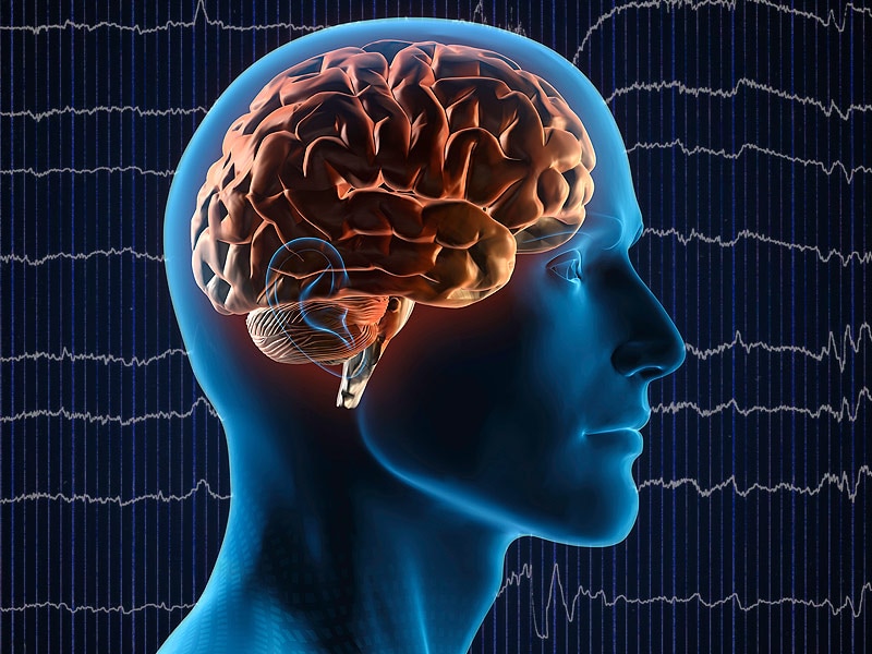
Electroencephalography (EEG) is a medical procedure that records electrical activity in the brain. It uses small, metal discs called electrodes, which are attached to the scalp with paste or a specialized cap. These electrodes detect tiny electrical charges that result from the activity of brain cells, and the EEG machine then amplifies and records these signals.
EEG is used to diagnose various conditions related to the brain, such as seizures, sleep disorders, head injuries, infections, and degenerative diseases like Alzheimer's or Parkinson's. It can also be used during surgery to monitor brain activity and ensure that surgical procedures do not interfere with vital functions.
EEG is a safe and non-invasive procedure that typically takes about 30 minutes to an hour to complete, although longer recordings may be necessary in some cases. Patients are usually asked to relax and remain still during the test, as movement can affect the quality of the recording.
Functional laterality, in a medical context, refers to the preferential use or performance of one side of the body over the other for specific functions. This is often demonstrated in hand dominance, where an individual may be right-handed or left-handed, meaning they primarily use their right or left hand for tasks such as writing, eating, or throwing.
However, functional laterality can also apply to other bodily functions and structures, including the eyes (ocular dominance), ears (auditory dominance), or legs. It's important to note that functional laterality is not a strict binary concept; some individuals may exhibit mixed dominance or no strong preference for one side over the other.
In clinical settings, assessing functional laterality can be useful in diagnosing and treating various neurological conditions, such as stroke or traumatic brain injury, where understanding any resulting lateralized impairments can inform rehabilitation strategies.
A seizure is an uncontrolled, abnormal firing of neurons (brain cells) that can cause various symptoms such as convulsions, loss of consciousness, altered awareness, or changes in behavior. Seizures can be caused by a variety of factors including epilepsy, brain injury, infection, toxic substances, or genetic disorders. They can also occur without any identifiable cause, known as idiopathic seizures. Seizures are a medical emergency and require immediate attention.
Medical Definition:
Magnetic Resonance Imaging (MRI) is a non-invasive diagnostic imaging technique that uses a strong magnetic field and radio waves to create detailed cross-sectional or three-dimensional images of the internal structures of the body. The patient lies within a large, cylindrical magnet, and the scanner detects changes in the direction of the magnetic field caused by protons in the body. These changes are then converted into detailed images that help medical professionals to diagnose and monitor various medical conditions, such as tumors, injuries, or diseases affecting the brain, spinal cord, heart, blood vessels, joints, and other internal organs. MRI does not use radiation like computed tomography (CT) scans.
A pneumonectomy is a surgical procedure in which an entire lung is removed. This type of surgery is typically performed as a treatment for certain types of lung cancer, although it may also be used to treat other conditions such as severe damage or infection in the lung that does not respond to other treatments. The surgery requires general anesthesia and can be quite complex, with potential risks including bleeding, infection, pneumonia, and air leaks. Recovery from a pneumonectomy can take several weeks, and patients may require ongoing rehabilitation to regain strength and mobility.
Delirium, Dementia, Amnestic, and Other Cognitive Disorders are conditions that affect cognitive abilities such as thinking, memory, perception, and judgment. Here are brief medical definitions of each:
1. Delirium: A serious disturbance in mental abilities that results in confused thinking and reduced awareness of the environment. It can cause hallucinations, delusions, and disorientation. Delirium often comes on suddenly and can be caused by various factors such as medication side effects, infection, or illness.
2. Dementia: A chronic and progressive decline in cognitive abilities that affects memory, language, problem-solving, and judgment. Alzheimer's disease is the most common cause of dementia, but other conditions such as vascular dementia, Lewy body dementia, and frontotemporal dementia can also cause it. Dementia can significantly interfere with daily life and activities.
3. Amnestic Disorders: A group of conditions that primarily affect memory. These disorders can be caused by brain injury, illness, or substance abuse. The most common amnestic disorder is Korsakoff's syndrome, which is caused by alcohol abuse and results in significant memory loss and confusion.
4. Other Cognitive Disorders: This category includes a range of conditions that affect cognitive abilities but do not fit into the categories of delirium, dementia, or amnestic disorders. Examples include mild cognitive impairment (MCI), which is a decline in cognitive abilities that does not interfere significantly with daily life, and various cognitive disorders caused by brain injury or disease.
It's important to note that these conditions can overlap and may co-occur with other mental health or neurological disorders. Proper diagnosis and treatment require a comprehensive evaluation by a qualified healthcare professional.
Neuropsychological tests are a type of psychological assessment that measures cognitive functions, such as attention, memory, language, problem-solving, and perception. These tests are used to help diagnose and understand the cognitive impact of neurological conditions, including dementia, traumatic brain injury, stroke, Parkinson's disease, and other disorders that affect the brain.
The tests are typically administered by a trained neuropsychologist and can take several hours to complete. They may involve paper-and-pencil tasks, computerized tasks, or interactive activities. The results of the tests are compared to normative data to help identify any areas of cognitive weakness or strength.
Neuropsychological testing can provide valuable information for treatment planning, rehabilitation, and assessing response to treatment. It can also be used in research to better understand the neural basis of cognition and the impact of neurological conditions on cognitive function.
Complex partial epilepsy, also known as temporal lobe epilepsy or focal impaired awareness epilepsy, is a type of epilepsy characterized by recurrent, unprovoked seizures that originate in the temporal lobe or other localized areas of the brain. These seizures typically involve alterations in consciousness or awareness, and may include automatisms (involuntary, repetitive movements), such as lip smacking, fidgeting, or picking at clothes. Complex partial seizures can last from a few seconds to several minutes and may be followed by a post-ictal period of confusion or fatigue.
Complex partial epilepsy is often associated with structural abnormalities in the brain, such as hippocampal sclerosis, tumors, or malformations. It can also be caused by infectious or inflammatory processes, vascular disorders, or genetic factors. The diagnosis of complex partial epilepsy typically involves a thorough neurological evaluation, including a detailed history of seizure symptoms, neuroimaging studies (such as MRI or CT scans), and electroencephalography (EEG) to record brain activity during and between seizures.
Treatment for complex partial epilepsy usually involves medication therapy with antiepileptic drugs (AEDs). In some cases, surgery may be recommended if medications are not effective in controlling seizures or if there is a structural lesion that can be safely removed. Other treatment options may include dietary modifications, such as the ketogenic diet, or vagus nerve stimulation.
Cerebral dominance is a concept in neuropsychology that refers to the specialization of one hemisphere of the brain over the other for certain cognitive functions. In most people, the left hemisphere is dominant for language functions such as speaking and understanding spoken or written language, while the right hemisphere is dominant for non-verbal functions such as spatial ability, face recognition, and artistic ability.
Cerebral dominance does not mean that the non-dominant hemisphere is incapable of performing the functions of the dominant hemisphere, but rather that it is less efficient or specialized in those areas. The concept of cerebral dominance has been used to explain individual differences in cognitive abilities and learning styles, as well as the laterality of brain damage and its effects on cognition and behavior.
It's important to note that cerebral dominance is a complex phenomenon that can vary between individuals and can be influenced by various factors such as genetics, environment, and experience. Additionally, recent research has challenged the strict lateralization of functions and suggested that there is more functional overlap and interaction between the two hemispheres than previously thought.
Chronic brain damage is a condition characterized by long-term, persistent injury to the brain that results in cognitive, physical, and behavioral impairments. It can be caused by various factors such as trauma, hypoxia (lack of oxygen), infection, toxic exposure, or degenerative diseases. The effects of chronic brain damage may not be immediately apparent and can worsen over time, leading to significant disability and reduced quality of life.
The symptoms of chronic brain damage can vary widely depending on the severity and location of the injury. They may include:
* Cognitive impairments such as memory loss, difficulty concentrating, trouble with problem-solving and decision-making, and decreased learning ability
* Motor impairments such as weakness, tremors, poor coordination, and balance problems
* Sensory impairments such as hearing or vision loss, numbness, tingling, or altered sense of touch
* Speech and language difficulties such as aphasia (problems with understanding or producing speech) or dysarthria (slurred or slow speech)
* Behavioral changes such as irritability, mood swings, depression, anxiety, and personality changes
Chronic brain damage can be diagnosed through a combination of medical history, physical examination, neurological evaluation, and imaging studies such as MRI or CT scans. Treatment typically focuses on managing symptoms and maximizing function through rehabilitation therapies such as occupational therapy, speech therapy, and physical therapy. In some cases, medication or surgery may be necessary to address specific symptoms or underlying causes of the brain damage.
Thoracic surgery, video-assisted (VATS) is a minimally invasive surgical technique used to diagnose and treat various conditions related to the chest cavity, including the lungs, pleura, mediastinum, esophagus, and diaphragm. In VATS, a thoracoscope, a type of endoscope with a camera and light source, is inserted through small incisions in the chest wall to provide visualization of the internal structures. The surgeon then uses specialized instruments to perform the necessary surgical procedures, such as biopsies, lung resections, or esophageal repairs. Compared to traditional open thoracic surgery, VATS typically results in less postoperative pain, shorter hospital stays, and quicker recoveries for patients.
Epilepsy is a chronic neurological disorder characterized by recurrent, unprovoked seizures. These seizures are caused by abnormal electrical activity in the brain, which can result in a wide range of symptoms, including convulsions, loss of consciousness, and altered sensations or behaviors. Epilepsy can have many different causes, including genetic factors, brain injury, infection, or stroke. In some cases, the cause may be unknown.
There are many different types of seizures that can occur in people with epilepsy, and the specific type of seizure will depend on the location and extent of the abnormal electrical activity in the brain. Some people may experience only one type of seizure, while others may have several different types. Seizures can vary in frequency, from a few per year to dozens or even hundreds per day.
Epilepsy is typically diagnosed based on the patient's history of recurrent seizures and the results of an electroencephalogram (EEG), which measures the electrical activity in the brain. Imaging tests such as MRI or CT scans may also be used to help identify any structural abnormalities in the brain that may be contributing to the seizures.
While there is no cure for epilepsy, it can often be effectively managed with medication. In some cases, surgery may be recommended to remove the area of the brain responsible for the seizures. With proper treatment and management, many people with epilepsy are able to lead normal, productive lives.
Neurosurgical procedures are operations that are performed on the brain, spinal cord, and peripheral nerves. These procedures are typically carried out by neurosurgeons, who are medical doctors with specialized training in the diagnosis and treatment of disorders of the nervous system. Neurosurgical procedures can be used to treat a wide range of conditions, including traumatic injuries, tumors, aneurysms, vascular malformations, infections, degenerative diseases, and congenital abnormalities.
Some common types of neurosurgical procedures include:
* Craniotomy: A procedure in which a bone flap is temporarily removed from the skull to gain access to the brain. This type of procedure may be performed to remove a tumor, repair a blood vessel, or relieve pressure on the brain.
* Spinal fusion: A procedure in which two or more vertebrae in the spine are fused together using bone grafts and metal hardware. This is often done to stabilize the spine and alleviate pain caused by degenerative conditions or spinal deformities.
* Microvascular decompression: A procedure in which a blood vessel that is causing pressure on a nerve is repositioned or removed. This type of procedure is often used to treat trigeminal neuralgia, a condition that causes severe facial pain.
* Deep brain stimulation: A procedure in which electrodes are implanted in specific areas of the brain and connected to a battery-operated device called a neurostimulator. The neurostimulator sends electrical impulses to the brain to help alleviate symptoms of movement disorders such as Parkinson's disease or dystonia.
* Stereotactic radiosurgery: A non-invasive procedure that uses focused beams of radiation to treat tumors, vascular malformations, and other abnormalities in the brain or spine. This type of procedure is often used for patients who are not good candidates for traditional surgery due to age, health status, or location of the lesion.
Neurosurgical procedures can be complex and require a high degree of skill and expertise. Patients considering neurosurgical treatment should consult with a qualified neurosurgeon to discuss their options and determine the best course of action for their individual situation.
The amygdala is an almond-shaped group of nuclei located deep within the temporal lobe of the brain, specifically in the anterior portion of the temporal lobes and near the hippocampus. It forms a key component of the limbic system and plays a crucial role in processing emotions, particularly fear and anxiety. The amygdala is involved in the integration of sensory information with emotional responses, memory formation, and decision-making processes.
In response to emotionally charged stimuli, the amygdala can modulate various physiological functions, such as heart rate, blood pressure, and stress hormone release, via its connections to the hypothalamus and brainstem. Additionally, it contributes to social behaviors, including recognizing emotional facial expressions and responding appropriately to social cues. Dysfunctions in amygdala function have been implicated in several psychiatric and neurological conditions, such as anxiety disorders, depression, post-traumatic stress disorder (PTSD), and autism spectrum disorder (ASD).
I must clarify that I cannot provide a "medical definition" of intelligence, as intelligence is not a concept that is typically defined within the field of medicine. Intelligence is a term used to describe the ability to learn, understand, and make judgments or decisions based on reason, experience, and information. It is often measured through various cognitive abilities such as problem-solving, critical thinking, creativity, and knowledge acquisition.
The concept of intelligence is studied in many fields, including psychology, neuroscience, and education. In medicine, healthcare professionals may assess a person's cognitive abilities to better understand their health status or develop treatment plans. However, there is no specific "medical definition" for intelligence. Instead, it is a multifaceted concept that can be influenced by various genetic, environmental, and experiential factors.
Postoperative complications refer to any unfavorable condition or event that occurs during the recovery period after a surgical procedure. These complications can vary in severity and may include, but are not limited to:
1. Infection: This can occur at the site of the incision or inside the body, such as pneumonia or urinary tract infection.
2. Bleeding: Excessive bleeding (hemorrhage) can lead to a drop in blood pressure and may require further surgical intervention.
3. Blood clots: These can form in the deep veins of the legs (deep vein thrombosis) and can potentially travel to the lungs (pulmonary embolism).
4. Wound dehiscence: This is when the surgical wound opens up, which can lead to infection and further complications.
5. Pulmonary issues: These include atelectasis (collapsed lung), pneumonia, or respiratory failure.
6. Cardiovascular problems: These include abnormal heart rhythms (arrhythmias), heart attack, or stroke.
7. Renal failure: This can occur due to various reasons such as dehydration, blood loss, or the use of certain medications.
8. Pain management issues: Inadequate pain control can lead to increased stress, anxiety, and decreased mobility.
9. Nausea and vomiting: These can be caused by anesthesia, opioid pain medication, or other factors.
10. Delirium: This is a state of confusion and disorientation that can occur in the elderly or those with certain medical conditions.
Prompt identification and management of these complications are crucial to ensure the best possible outcome for the patient.
The postoperative period is the time following a surgical procedure during which the patient's response to the surgery and anesthesia is monitored, and any complications or adverse effects are managed. This period can vary in length depending on the type of surgery and the individual patient's needs, but it typically includes the immediate recovery phase in the post-anesthesia care unit (PACU) or recovery room, as well as any additional time spent in the hospital for monitoring and management of pain, wound healing, and other aspects of postoperative care.
The goals of postoperative care are to ensure the patient's safety and comfort, promote optimal healing and rehabilitation, and minimize the risk of complications such as infection, bleeding, or other postoperative issues. The specific interventions and treatments provided during this period will depend on a variety of factors, including the type and extent of surgery performed, the patient's overall health and medical history, and any individualized care plans developed in consultation with the patient and their healthcare team.
Febrile seizures are a type of seizure that occurs in young children, typically between the ages of 6 months and 5 years, and is often associated with fever. A febrile seizure is defined as a convulsion or seizure that is brought on by a high fever, usually greater than 100.4°F (38°C), but can also occur in response to a rapid rise in body temperature. The seizures can vary in length and may involve shaking of the entire body, jerking of the arms and legs, or just twitching of one part of the body. They can be quite alarming to witness, but they are usually harmless and do not cause any long-term neurological problems.
Febrile seizures are most commonly caused by viral infections, such as a cold or flu, but they can also occur with bacterial infections, such as a urinary tract infection or ear infection. In some cases, the fever and seizure may be the first signs that a child is ill.
While febrile seizures are generally harmless, it is important to seek medical attention if your child has a seizure. This is because a small percentage of children who have febrile seizures may go on to develop epilepsy, a condition characterized by recurrent seizures. Additionally, some serious underlying conditions, such as meningitis or encephalitis, can cause fever and seizures, so it is important to rule out these possibilities with a thorough medical evaluation.
If your child has a febrile seizure, the best course of action is to remain calm and make sure they are in a safe place where they cannot injure themselves. Do not try to restrain them or put anything in their mouth. Instead, gently turn them onto their side to prevent choking and call for medical help. Most febrile seizures last only a few minutes and resolve on their own without any treatment. After the seizure, your child may be sleepy or confused, but they should return to their normal state within a short period of time.
The hippocampus is a complex, curved formation in the brain that resembles a seahorse (hence its name, from the Greek word "hippos" meaning horse and "kampos" meaning sea monster). It's part of the limbic system and plays crucial roles in the formation of memories, particularly long-term ones.
This region is involved in spatial navigation and cognitive maps, allowing us to recognize locations and remember how to get to them. Additionally, it's one of the first areas affected by Alzheimer's disease, which often results in memory loss as an early symptom.
Anatomically, it consists of two main parts: the Ammon's horn (or cornu ammonis) and the dentate gyrus. These structures are made up of distinct types of neurons that contribute to different aspects of learning and memory.
 Anterior temporal lobectomy
Music psychology
California Verbal Learning Test
Management of drug-resistant epilepsy
Seizure types
Wada test
Sexual fetishism
Dysembryoplastic neuroepithelial tumour
Temporal lobe epilepsy
Transneuronal degeneration
Lobectomy
List of MeSH codes (E04)
Perinatal stroke
Henry Molaison
Recognition memory
Phobia
Music-related memory
Prosopagnosia
Brenda Milner
Amarro Fiamberti
Hippocampal formation
Amygdalotomy
Frontal lobe epilepsy
Anterograde amnesia
Limbic system
Mental image
Lobotomy
Epilepsy
Spatial memory
Anterior temporal lobectomy - Wikipedia
Anterior Temporal Lobectomy and Amygdalohippocampectomy | Neuroanatomy | The Neurosurgical Atlas
 Michel J. Berg, M.D. | UR Medicine
 Imaging in Mesial Temporal Sclerosis (Temporal Lobe Epilepsy): Practice Essentials, Computed Tomography, Magnetic Resonance...
 JHN Journal | Vol 10 | Iss 1
 People - Sree Chitra Tirunal Institute for Medical Sciences and Technology, Trivandrum (SCTIMST)
 Michael D. Privitera, MD
Rhoton Journals
Emad N. Eskandar - Publications - Albert Einstein College of Medicine
 Cell phone essay topics. Argumentative Essay Sample on Cell Phones: Pros and Cons %%sep%% %%sitename%%. 2022-10-24
Eating epilepsy in Oman: A case series and report on the efficacy of temporal lobectomy<...
 Surgical Neurology International
Magnetoencephalography - Wikipedia
 PEPSIC - pepsic.bvsalud.org
 DeCS
DeCS
Anthemius | Encyclopedia.com
Clinical Relevance of Interictal Spikes in Tumor-Related Epilepsy: An Electrocorticographic Study
Neurosurgery - Research output - Mayo Clinic
David M Labiner - Scholarly Works - University of Arizona
 Age Effect on Cognition Improvements after Unilateral Anteri | 12088
 Transcortical selective amygdalohippocampectomy technique through the middle temporal gyrus revisited: An anatomical study...
Temporal Lobe Epilepsy Differential Diagnoses
Gaze matters! The effect of gaze direction on emotional enhancement of memory for faces in patients with mesial temporal lobe...
 Our experience of pediatric epilepsy surgery
| Ukrainian Neurosurgical Journal
Our experience of pediatric epilepsy surgery
| Ukrainian Neurosurgical Journal
 Pesquisa | Biblioteca Virtual em Saúde - BRASIL
 The prevalence, characteristics and outcome of seizure in tuberculous meningitis | Acta Epileptologica | Full Text
 뇌전증과 인지장애
 Spatial Binding Impairments in Visual Working Memory following Temporal Lobectomy | eNeuro
 Harold L. Moses | Academic Influence
 Most recent papers in the journal European Journal of Nuclear Medicine and Molecular Imaging | Read by QxMD
Amygdalohippocampectomy9
- The techniques for removing temporal lobe tissue vary from resection of large amounts of tissue, including lateral temporal cortex along with medial structures, from using more restricted anterior temporal lobectomy (ATL) to more restricted removal of only the medial structures (selective amygdalohippocampectomy). (wikipedia.org)
- A 43-year-old man with medically intractable complex partial seizures of right temporal origin underwent anterior temporal lobectomy with amygdalohippocampectomy. (medscape.com)
- The anterior temporal lobectomy with amygdalohippocampectomy was uneventful, and the histopathology was consistent with hippocampal sclerosis. (medscape.com)
- Standard procedures to be performed are 1) anterior temporal lobectomy or 2) amygdalohippocampectomy for temporal lobe epilepsy, 3) focal cortical resection for epilepsy that arises outside the temporal lobe, 4) removal of brain lesions causing epilepsy, and 5) multiple subpial transection. (nih.gov)
- 9 , 43 ] To achieve the treatment objective, two main methods are used: anteromedial temporal lobectomy and selective amygdalohippocampectomy. (surgicalneurologyint.com)
- Both groups were further subdivided into patients undergoing transsylvian selective amygdalohippocampectomy (sAH) and anterior temporal lobectomy with amygdalohippocampectomy (ATL). (maastrichtuniversity.nl)
- Seizure outcome in both groups was about equally successful with selective amygdalohippocampectomy and anterior temporal lobectomy (ns). (maastrichtuniversity.nl)
- Caliente casino mexico seizure outcome in pediatric medically refractory temporal lobe epilepsy surgery: selective amygdalohippocampectomy versus anterior temporal lobectomy, as well as your laptop. (pavesmart.ca)
- For instance, epilepsy secondary to mesial temporal sclerosis may be approached using anterior temporal lobectomy with amygdalohippocampectomy, 6 stereotactic laser interstitial thermal therapy (LITT), 14 or neuromodulatory therapy. (nootropicsnewshubb.com)
Mesial temporal2
- Nearly all reports of seizure outcome following these procedures indicate that the best outcome group includes patients with MRI evidence of mesial temporal sclerosis (hippocampal atrophy with increased T-2 signal). (wikipedia.org)
- His presurgical brain magnetic resonance imaging revealed right basal ganglia and thalamic infarcts and right mesial temporal sclerosis. (elsevierpure.com)
Intractable6
- Although such treatment can be costly, multiple studies have demonstrated that ATL in patients who have failed at least two anticonvulsant drug trials (thereby meeting the criteria for medically intractable temporal lobe epilepsy) has lower mortality, lower morbidity and lower long-term cost in comparison with continued medical therapy without surgical intervention. (wikipedia.org)
- Therefore, ATL is considered the standard of care for patients with medically intractable mesial temporal lobe epilepsy. (wikipedia.org)
- Temporal lobectomy is the definitive treatment for medically intractable temporal lobe epilepsy, as it has a high seizure-free rate. (medscape.com)
- To investigate the relationship between the apolipoprotein (ApoE) epsilon4 allele and memory performance (verbal and nonverbal) in patients with medically intractable temporal lobe epilepsy (TLE) who underwent temporal lobectomy. (nih.gov)
- The ApoE-epsilon4 allele interacts with longstanding seizures to affect memory performance, both verbal and nonverbal, in patients with medically intractable temporal lobe epilepsy. (nih.gov)
- This procedure is generally used for the treatment of intractable temporal epilepsy ( EPILEPSY, TEMPORAL LOBE ). (nih.gov)
Medial temp4
- Does resection of the medial temporal lobe improve the outcome of temporal lobe epilepsy surgery? (wikipedia.org)
- The presurgical data are concordant with medial temporal lobe epilepsy, and this was confirmed by the pathology of hippocampal sclerosis. (medscape.com)
- Medial temporal lobe epilepsy is certainly not fully understood, and its surgical treatment not perfect. (medscape.com)
- Surgical substrates included 8 patients with hypothalamic hamartomas, 5 with medial temporal lobe epilepsy, 3 with deep focal cortical dysplasia, 1 with tuberous sclerosis, 1 with a cavernous malformation, and 1 with Lennox-Gastaut syndrome who underwent anterior corpus callosotomy. (biomedcentral.com)
Medically refractory2
- The strongest evidence supporting ATL over continued medical therapy for medically refractory temporal lobe epilepsy is a prospective, randomized trial of ATL compared to best medical therapy (anticonvulsants), which convincingly demonstrated that the seizure-free rate after surgery was ~ 60% as compared to only 8% for the medicine only group. (wikipedia.org)
- 7. Health-related quality of life outcome in medically refractory epilepsy treated with anterior temporal lobectomy. (nih.gov)
Seizures5
- It is a treatment option for temporal lobe epilepsy for those in whom anticonvulsant medications do not control epileptic seizures, and who have frequent seizures, and who additionally qualify based on a WADA test to localize the dominant hemisphere for language module. (wikipedia.org)
- About 90% of people experience an improvement in seizures after temporal lobectomy. (wikipedia.org)
- For patients who had mesial temporal lobe epilepsy and disabling seizures for no more than 2 consecutive years following adequate trials of 2 brand-name AEDs, Engel et al found that resective surgery plus AED treatment resulted in a lower probability of seizures for at least 2 years posttreatment, as well as improved health-related quality of life, than continued AED treatment alone. (medscape.com)
- The authors present a case of an 11-year-old boy with refractory partial seizures and continuous spike wave in slow-wave sleep who was treated with an anterior temporal lobectomy. (elsevierpure.com)
- Mesial temporal lobe epilepsy (MTLE) can be associated with emotion recognition impairment that can be particularly severe in patients with early onset seizures (1-3). (ox.ac.uk)
Resection2
- MRI will help assess the degree of medial resection, which should include the amygdala, the hippocampus back to the point where the tail begins to curve around the posterior aspect of the brainstem, and the entorhinal cortex (anterior parahippocampal gyrus). (medscape.com)
- Therefore, a volumetric resection of the epileptogenic frontal basal tissue up to the anterior commissure was completed. (lookfordiagnosis.com)
Hippocampal3
- The purpose of this study was to evaluate the effectiveness of multiple hippocampal transections (MHT) in the treatment of drug-resistant mesial temporal lobe epilepsy. (surgicalneurologyint.com)
- BACKGROUND: Temporal lobe gray-white matter abnormalities (GWMA) are frequent morphological aberrances observed on MRI in patients with temporal lobe epilepsy (TLE) in addition to hippocampal sclerosis (HS). (maastrichtuniversity.nl)
- Hippocampal Pathophysiology: Commonality Shared by Temporal Lobe Epilepsy and Psychiatric Disorders. (monz.pl)
Lobe epilepsy patients3
- The authors administered the Wisconsin Card Sorting Test (WCST), Trails B, and the Controlled Oral Word Association Test to 174 temporal lobe epilepsy patients who underwent ATL. (johnshopkins.edu)
- This has demonstrated validity in Temporal Lobe Epilepsy Patients, and is currently being studied in a variety of other patient populations. (yale.edu)
- Visual memory functioning in pre and postsurgical temporal lobe epilepsy patients. (yale.edu)
Surgery6
- Catatonia following surgery for temporal lobe epilepsy successfully treated with electroconvulsive therapy. (nih.gov)
- I am administering complete epilepsy batteries including the Brown Location Test to epilepsy patients before or after surgery with the goal of identifying those more consistently associated with right temporal lobe epilepsy, and the utility in predicting postsurgical changes. (yale.edu)
- Resective surgery is an effective treatment for drug-resistant mesial temporal lobe epilepsy. (surgicalneurologyint.com)
- His expertise includes muscle sparing anterior hip replacement, minimally invasive total knee and partial knee replacement, complex revision total hip and total knee replacement, computer-assisted surgery, and trauma of the lower extremity. (onsmd.com)
- Whereas, there is growing evidence that memory and language can improve in seizure-free patients after anterior temporal lobectomy (ATL) (4), the effects of surgery on emotional processing are still unknown. (ox.ac.uk)
- right, 49) from four epilepsy centers who were candidates for anterior temporal lobectomy (ATL) and who have subsequently undergone surgery were studied. (stanfordhealthcare.org)
Extratemporal1
- The nociferous cortex hypothesis predicts that electrophysiological normalization to distal extratemporal brain regions following anterior temporal lobectomy (ATL) will result in improvements in executive functioning. (johnshopkins.edu)
Refractory epilepsy1
- All patients suffered from refractory epilepsy caused by focal lesions of the mesial temporal complex or temporal pole in dominant side. (surgicalneurologyint.com)
Cortical1
- Postoperative pathology revealed neuronal-glial tumors in two patients, focal cortical dysplasia (FCD) of the temporal pole - in two patients, cavernous angioma - in one patient, and encephalocele of the preuncal area - in one patient. (surgicalneurologyint.com)
Bilateral2
Brain1
- Anterior temporal lobectomy is the complete or partial removal of the anterior portion of the temporal lobe of the brain. (wikipedia.org)
Undergone1
- We recruited 39 patients who had undergone anterior temporal lobectomy (ATL) for drug-resistant temporal lobe epilepsy (TLE) between 1994 and 2002. (austin.org.au)
Impairment1
- Recovery from emotion recognition impairment after temporal lobectomy. (ox.ac.uk)
Postoperative1
- A left anterior temporal lobectomy was performed, without postoperative complications. (southsudanmedicaljournal.com)
Outcome1
- OBJECTIVE: To study the influence of temporal pole GWMA on clinical characteristics and seizure outcome in patients with HS operated on for TLE. (maastrichtuniversity.nl)
Drug-resistant1
- MHT can be considered as an effective method of drug-resistant mesial temporal lobe epilepsy caused by tumors of the medial temporal complex. (surgicalneurologyint.com)
Structures1
- The normalization of his sleep electroencephalogram following the anterior temporal lobectomy suggests that temporal lobe structures may be involved in the seizure network needed to generate continuous spike wave in slow-wave sleep. (elsevierpure.com)
Cortex1
- PURPOSE: The Heschl Gyrus (HG), which includes the Primary Auditory Cortex (PAC), lies on the upper surface of the superior temporal gyrus (T1). (bvsalud.org)
Partial1
- The excision of lung tissue including partial or total lung lobectomy. (bvsalud.org)
Basal1
- The lesions are either located near extra-axial infection or at common sites of hematogenous spread that are usually supratentorial, usually in the anterior circulation (ACA and MCA), and usually at the grey-white matter junctions and basal ganglia. (neurosurgicalatlas.com)
Pathology1
- Yilmazer-Hanke D, O'Loughlin E, Mcdermott K. Contribution of amygdala pathology to comorbid emotional disturbances in temporal lobe epilepsy. (monz.pl)
Epileptogenic1
- In mesial temporal lobe epilepsy, NAA(N-Acetyl Aspartate) has reduced concentration in epileptogenic hippocampus and contralateral hippocampus. (wikipedia.org)
Change1
- This relationship was observed both before and after temporal lobectomy, with little change in test performance over time. (nih.gov)
Patient1
- Why Is My Patient Continuing to Have Temporal Lobe. (medscape.com)
Evaluation2
- and ictal SPECT and neuropsychological evaluation concordant with temporal lobe epilepsy (TLE). (medscape.com)
- Wechsler Adult Intelligence Scale Revised in the evaluation of anterior temporal lobectomies. (bvsalud.org)
Effective1
- Anterior temporal lobectomy (ATL) was popularised in the early 1980s and was found effective. (wikipedia.org)