Anatomic Landmarks
Tomography, X-Ray Computed
Imaging, Three-Dimensional
Reproducibility of Results
Magnetic Resonance Imaging
Homing Behavior
Models, Anatomic
Chin
Mesopic Vision
Encyclopedias as Topic
Retina
Photoreceptor Cells, Vertebrate
Vision, Ocular
Retinal Cone Photoreceptor Cells
A comparison of cephalometric measurements: a picture archiving and communication system versus the hand-tracing method--a preliminary study. (1/113)
(+info)Picture archiving and communications systems: a study of reliability of orthodontic cephalometric analysis. (2/113)
(+info)A three-dimensional comparison of a morphometric and conventional cephalometric midsagittal planes for craniofacial asymmetry. (3/113)
(+info)Three-dimensional longitudinal assessment of facial symmetry in adolescents. (4/113)
(+info)Precision of measurements on conventional negative 'bones white' and inverted greyscale 'bones black' digital lateral cephalograms. (5/113)
(+info)A new approach of assessing sagittal dysplasia: the W angle. (6/113)
(+info)Consistency and precision of landmark identification in three-dimensional cone beam computed tomography scans. (7/113)
(+info)Posteroanterior cephalometric norms for an adolescent Kuwaiti population. (8/113)
(+info)Anatomic landmarks are specific, identifiable structures or features on the body that are used as references in medicine and surgery. These landmarks can include bones, muscles, joints, or other visible or palpable features that help healthcare professionals identify specific locations, orient themselves during procedures, or measure changes in the body.
Examples of anatomic landmarks include:
* The anterior iliac spine, a bony prominence on the front of the pelvis that can be used to locate the hip joint.
* The cubital fossa, a depression at the elbow where the median nerve and brachial artery can be palpated.
* The navel (umbilicus), which serves as a reference point for measuring distances in the abdomen.
* The xiphoid process, a small piece of cartilage at the bottom of the breastbone that can be used to locate the heart and other structures in the chest.
Anatomic landmarks are important for accurate diagnosis, treatment planning, and surgical procedures, as they provide reliable and consistent reference points that can help ensure safe and effective care.
A cadaver is a deceased body that is used for medical research or education. In the field of medicine, cadavers are often used in anatomy lessons, surgical training, and other forms of medical research. The use of cadavers allows medical professionals to gain a deeper understanding of the human body and its various systems without causing harm to living subjects. Cadavers may be donated to medical schools or obtained through other means, such as through consent of the deceased or their next of kin. It is important to handle and treat cadavers with respect and dignity, as they were once living individuals who deserve to be treated with care even in death.
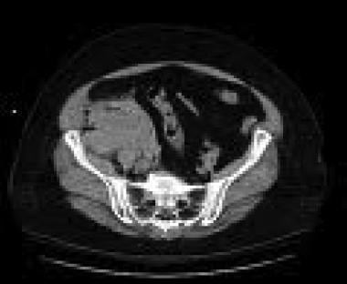
X-ray computed tomography (CT or CAT scan) is a medical imaging method that uses computer-processed combinations of many X-ray images taken from different angles to produce cross-sectional (tomographic) images (virtual "slices") of the body. These cross-sectional images can then be used to display detailed internal views of organs, bones, and soft tissues in the body.
The term "computed tomography" is used instead of "CT scan" or "CAT scan" because the machines take a series of X-ray measurements from different angles around the body and then use a computer to process these data to create detailed images of internal structures within the body.
CT scanning is a noninvasive, painless medical test that helps physicians diagnose and treat medical conditions. CT imaging provides detailed information about many types of tissue including lung, bone, soft tissue and blood vessels. CT examinations can be performed on every part of the body for a variety of reasons including diagnosis, surgical planning, and monitoring of therapeutic responses.
In computed tomography (CT), an X-ray source and detector rotate around the patient, measuring the X-ray attenuation at many different angles. A computer uses this data to construct a cross-sectional image by the process of reconstruction. This technique is called "tomography". The term "computed" refers to the use of a computer to reconstruct the images.
CT has become an important tool in medical imaging and diagnosis, allowing radiologists and other physicians to view detailed internal images of the body. It can help identify many different medical conditions including cancer, heart disease, lung nodules, liver tumors, and internal injuries from trauma. CT is also commonly used for guiding biopsies and other minimally invasive procedures.
In summary, X-ray computed tomography (CT or CAT scan) is a medical imaging technique that uses computer-processed combinations of many X-ray images taken from different angles to produce cross-sectional images of the body. It provides detailed internal views of organs, bones, and soft tissues in the body, allowing physicians to diagnose and treat medical conditions.
Three-dimensional (3D) imaging in medicine refers to the use of technologies and techniques that generate a 3D representation of internal body structures, organs, or tissues. This is achieved by acquiring and processing data from various imaging modalities such as X-ray computed tomography (CT), magnetic resonance imaging (MRI), ultrasound, or confocal microscopy. The resulting 3D images offer a more detailed visualization of the anatomy and pathology compared to traditional 2D imaging techniques, allowing for improved diagnostic accuracy, surgical planning, and minimally invasive interventions.
In 3D imaging, specialized software is used to reconstruct the acquired data into a volumetric model, which can be manipulated and viewed from different angles and perspectives. This enables healthcare professionals to better understand complex anatomical relationships, detect abnormalities, assess disease progression, and monitor treatment response. Common applications of 3D imaging include neuroimaging, orthopedic surgery planning, cancer staging, dental and maxillofacial reconstruction, and interventional radiology procedures.
Reproducibility of results in a medical context refers to the ability to obtain consistent and comparable findings when a particular experiment or study is repeated, either by the same researcher or by different researchers, following the same experimental protocol. It is an essential principle in scientific research that helps to ensure the validity and reliability of research findings.
In medical research, reproducibility of results is crucial for establishing the effectiveness and safety of new treatments, interventions, or diagnostic tools. It involves conducting well-designed studies with adequate sample sizes, appropriate statistical analyses, and transparent reporting of methods and findings to allow other researchers to replicate the study and confirm or refute the results.
The lack of reproducibility in medical research has become a significant concern in recent years, as several high-profile studies have failed to produce consistent findings when replicated by other researchers. This has led to increased scrutiny of research practices and a call for greater transparency, rigor, and standardization in the conduct and reporting of medical research.
Medical Definition:
Magnetic Resonance Imaging (MRI) is a non-invasive diagnostic imaging technique that uses a strong magnetic field and radio waves to create detailed cross-sectional or three-dimensional images of the internal structures of the body. The patient lies within a large, cylindrical magnet, and the scanner detects changes in the direction of the magnetic field caused by protons in the body. These changes are then converted into detailed images that help medical professionals to diagnose and monitor various medical conditions, such as tumors, injuries, or diseases affecting the brain, spinal cord, heart, blood vessels, joints, and other internal organs. MRI does not use radiation like computed tomography (CT) scans.
'Homing behavior' is not a term typically used in medical definitions. However, it is commonly used to describe an animal's innate ability to return to its home territory or nest after traveling large distances. This behavior has been observed in various species including birds, insects, and mammals. It is not a medical condition or disease.
Anatomic models are three-dimensional representations of body structures used for educational, training, or demonstration purposes. They can be made from various materials such as plastic, wax, or rubber and may depict the entire body or specific regions, organs, or systems. These models can be used to provide a visual aid for understanding anatomy, physiology, and pathology, and can be particularly useful in situations where actual human specimens are not available or practical to use. They may also be used for surgical planning and rehearsal, as well as in medical research and product development.
Cephalometry is a medical term that refers to the measurement and analysis of the skull, particularly the head face relations. It is commonly used in orthodontics and maxillofacial surgery to assess and plan treatment for abnormalities related to the teeth, jaws, and facial structures. The process typically involves taking X-ray images called cephalograms, which provide a lateral view of the head, and then using various landmarks and reference lines to make measurements and evaluate skeletal and dental relationships. This information can help clinicians diagnose problems, plan treatment, and assess treatment outcomes.
The "chin" is the lower, prominent part of the front portion of the jaw in humans and other animals. In medical terms, it is often referred to as the mentum or the symphysis of the mandible. The chin helps in protecting the soft tissues of the mouth and throat during activities such as eating, speaking, and swallowing. It also plays a role in shaping the overall appearance of the face. Anatomically, the chin is formed by the fusion of the two halves of the mandible (lower jawbone) at the symphysis menti.
Mesopic vision is a term used to describe the intermediate level of vision that occurs in conditions of decreased illumination, specifically between 0.02 and 3 candelas per square meter (cd/m²). This range falls between photopic vision, which is vision in bright light (>3 cd/m²), and scotopic vision, which is vision in very low light (
An encyclopedia is a comprehensive reference work containing articles on various topics, usually arranged in alphabetical order. In the context of medicine, a medical encyclopedia is a collection of articles that provide information about a wide range of medical topics, including diseases and conditions, treatments, tests, procedures, and anatomy and physiology. Medical encyclopedias may be published in print or electronic formats and are often used as a starting point for researching medical topics. They can provide reliable and accurate information on medical subjects, making them useful resources for healthcare professionals, students, and patients alike. Some well-known examples of medical encyclopedias include the Merck Manual and the Stedman's Medical Dictionary.
The retina is the innermost, light-sensitive layer of tissue in the eye of many vertebrates and some cephalopods. It receives light that has been focused by the cornea and lens, converts it into neural signals, and sends these to the brain via the optic nerve. The retina contains several types of photoreceptor cells including rods (which handle vision in low light) and cones (which are active in bright light and are capable of color vision).
In medical terms, any pathological changes or diseases affecting the retinal structure and function can lead to visual impairment or blindness. Examples include age-related macular degeneration, diabetic retinopathy, retinal detachment, and retinitis pigmentosa among others.
Photoreceptor cells in vertebrates are specialized types of neurons located in the retina of the eye that are responsible for converting light stimuli into electrical signals. These cells are primarily responsible for the initial process of vision and have two main types: rods and cones.
Rods are more numerous and are responsible for low-light vision or scotopic vision, enabling us to see in dimly lit conditions. They do not contribute to color vision but provide information about the shape and movement of objects.
Cones, on the other hand, are less numerous and are responsible for color vision and high-acuity vision or photopic vision. There are three types of cones, each sensitive to different wavelengths of light: short (S), medium (M), and long (L) wavelengths, which correspond to blue, green, and red, respectively. The combination of signals from these three types of cones allows us to perceive a wide range of colors.
Both rods and cones contain photopigments that consist of a protein called opsin and a light-sensitive chromophore called retinal. When light hits the photopigment, it triggers a series of chemical reactions that ultimately lead to the generation of an electrical signal that is transmitted to the brain via the optic nerve. This process enables us to see and perceive our visual world.
Ocular vision refers to the ability to process and interpret visual information that is received by the eyes. This includes the ability to see clearly and make sense of the shapes, colors, and movements of objects in the environment. The ocular system, which includes the eye and related structures such as the optic nerve and visual cortex of the brain, works together to enable vision.
There are several components of ocular vision, including:
* Visual acuity: the clarity or sharpness of vision
* Field of vision: the extent of the visual world that is visible at any given moment
* Color vision: the ability to distinguish different colors
* Depth perception: the ability to judge the distance of objects in three-dimensional space
* Contrast sensitivity: the ability to distinguish an object from its background based on differences in contrast
Disorders of ocular vision can include refractive errors such as nearsightedness or farsightedness, as well as more serious conditions such as cataracts, glaucoma, and macular degeneration. These conditions can affect one or more aspects of ocular vision and may require medical treatment to prevent further vision loss.
In the context of medical terminology, "light" doesn't have a specific or standardized definition on its own. However, it can be used in various medical terms and phrases. For example, it could refer to:
1. Visible light: The range of electromagnetic radiation that can be detected by the human eye, typically between wavelengths of 400-700 nanometers. This is relevant in fields such as ophthalmology and optometry.
2. Therapeutic use of light: In some therapies, light is used to treat certain conditions. An example is phototherapy, which uses various wavelengths of ultraviolet (UV) or visible light for conditions like newborn jaundice, skin disorders, or seasonal affective disorder.
3. Light anesthesia: A state of reduced consciousness in which the patient remains responsive to verbal commands and physical stimulation. This is different from general anesthesia where the patient is completely unconscious.
4. Pain relief using light: Certain devices like transcutaneous electrical nerve stimulation (TENS) units have a 'light' setting, indicating lower intensity or frequency of electrical impulses used for pain management.
Without more context, it's hard to provide a precise medical definition of 'light'.
Retinal cone photoreceptor cells are specialized neurons located in the retina of the eye, responsible for visual phototransduction and color vision. They are one of the two types of photoreceptors, with the other being rods, which are more sensitive to low light levels. Cones are primarily responsible for high-acuity, color vision during daylight or bright-light conditions.
There are three types of cone cells, each containing different photopigments that absorb light at distinct wavelengths: short (S), medium (M), and long (L) wavelengths, which correspond to blue, green, and red light, respectively. The combination of signals from these three types of cones allows the human visual system to perceive a wide range of colors and discriminate between them. Cones are densely packed in the central region of the retina, known as the fovea, which provides the highest visual acuity.
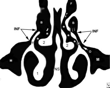
 CT Scan of the Paranasal Sinuses: History, Basic Concepts, Anatomy
 Anatomic space - Wikipedia
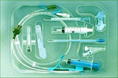
Femoral Central Venous Access Periprocedural Care: Equipment, Patient Preparation
 Anatomic landmarks and availability of bone for placement of orthodontic mini-implants for normal and short maxillary body...
 Foregut Content Review - SAGES
 Criteria for radiologic diagnosis of hypochondroplasia in neonates
 Meghan N Imrie | Stanford Medicine
 Davies Review Questions For Liver - ProProfs Quiz
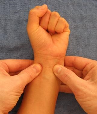
Trigger Finger Clinical Presentation: History, Physical Examination
Retina - Wikipedia
 General Recommendations on Immunization
ACIP Vaccine Administration Guidelines for Immunization | CDC
 Thieme E-Journals - Journal of Reconstructive Microsurgery / Abstract
CT Radiation Dose for Computer-Assisted Endoscopic Sinus Surgery: Dose Survey and Determination of Dose-Reduction Limits |...
Plus it
 Adult Thoracentesis Simulator
 Primary Endoscopic Carpal Tunnel Release - Approaches - Orthobullets
 Frontal language areas do not emerge in the absence of temporal language areas: A case study of an individual born without a...
 Jagdish MENON | Professor | MS(Ortho) MRCS(Edin) DNB MNAMS FACS PGDHQM | Jawaharlal Institute of Postgraduate Medical Education...
 Incident and Recurrent Cases of Central Serous Chorioretinopathy, Active Component, U.S. Armed Forces, 2001-2018 | Health.mil
Inferior Alveolar Nerve Block: Overview, Indications, Contraindications
dental
 Diffusion Tensor MR Imaging of the Brain and White Matter Tractography | AJR
 Single-Tray Impression Technique for Implant-Supported Overdentures | Journal of Oral Implantology
 Frontiers | Brain Shift in Neuronavigation of Brain Tumors: An Updated Review of Intra-Operative Ultrasound Applications
Left ventricular diastolic function assessed using Doppler tissue imaging in patients with hypertrophic cardiomyopathy:...
 Piriformis : Wheeless' Textbook of Orthopaedics
NIOSHTIC-2 Search Results - Basic View
 MRI Practice Potsdam SmartSpeed | FieldStrength | Philips
Anatomical landmarks2
- Aim: To evaluate the position, presence, appearance and extent of various anatomical landmarks in the mandibular interforaminal region of Brazilian patients using cone-beam computed tomography (CBCT). (bvsalud.org)
- In recent years, several studies have analyzed the characteristics of anatomical landmarks in the mandibular anterior region in various populations around the world 2,8-10 . (bvsalud.org)
Structures4
- If sufficiently thin axial sections (1-2 mm) are available, sagittal reconstructions can also be helpful for teaching purposes and further delineating anatomic structures. (medscape.com)
- Anatomic spaces are often landmarks to find other important structures. (wikipedia.org)
- 6 In FESS, only a limited view of the anatomy is warranted, and the use of these systems aims at avoiding injury to important anatomic structures around the orbit and the anterior skull base, especially the internal carotid artery, the optic nerve, and the floor of the anterior skull base. (ajnr.org)
- Last, prominence of anatomic structures needed for intraoperative orientation by ENT surgeons could suffer. (ajnr.org)
Medial2
- What anatomical landmark can you use to identify the left medial segment separate from the right anterior segment of the liver? (proprofs.com)
- The middle hepatic vein can be used as an anatomical landmark to identify the left medial segment separate from the right anterior segment of the liver. (proprofs.com)
Orientation2
- 5 Although these systems improve orientation in narrow anatomic compartments, they lead to an increased intervention time 3 and the need for preoperative imaging. (ajnr.org)
- Results indicated both curvilinear-array (microconvex) and phased-array transducers can be used by experienced sonographers to obtain diagnostic ultrasonographic images of the lungs in dogs with acute or resolving left-sided congestive heart failure and suggested the former transducer may be preferred, particularly to aid identification of anatomic landmarks for orientation. (avma.org)
Anatomy2
- Familiarization with the radiologic landmarks and cross-sectional anatomy on patient CT scans, along with clinical correlation, can further enhance the reader's ability to understand sinus CT findings. (medscape.com)
- In anatomy, a spatium or anatomic space is a space (cavity or gap). (wikipedia.org)
Identify2
- Identify anatomic landmarks. (medscape.com)
- ENT surgeons were able to identify anatomic landmarks on scans with a dose as low as 3.1 mGy. (ajnr.org)
Considerations2
- This activity provides knowledge related to indications, anatomic landmarks, probe considerations and techniques, and pathologic findings associated with scanning the gallbladder. (acponline.org)
- the focus of this article is on anatomic considerations relevant to the clinical diagnosis and care of patients, with particular emphasis on procedural and urgent medicine. (medscape.com)
Clinical1
- With experience, CT findings can be accurately correlated with the anatomic and clinical realities of the particular patient. (medscape.com)
Bone1
- Anatomic landmarks and availability of bone for placement of orthodontic mini-implants for normal and short maxillary body lengths. (bvsalud.org)
Scans1
- SmartExam automatically detects anatomic landmarks in a survey scan of the patient, then plans the diagnostic scans related to the patient's actual position in the magnet. (indianweb2.com)
Lateral1
- Lateral radiographic view of a dog with a VAP after surgery was completed to implant the VAP in a jugular vein by use of recommended anatomic landmarks. (avma.org)
Relation2
- 45°) in relation to its normal anatomic position. (msdmanuals.com)
- The position of the adrenal glands in relation to regional vasculature is very consistent, making vascular landmarks quite helpful for identification of adrenal glands. (vin.com)
Site1
- Students use anatomic landmarks on the manikin to locate the site where a specific sound should be heard. (gaumard.com)
Structure2
- Which anatomic structure is a useful landmark in location of this structure? (proprofs.com)
- Any structure, anatomic or architectural, requires a foundation or scaffold. (medscape.com)
Important1
- [ 5 ] When femoral central vascular access is desired, the inguinal ligament may serve as an important landmark in adequately perfused nonobese patients. (medscape.com)
Location1
- Superficial anatomic landmarks can be used to triangulate the location of canine peripheral lymphocentrums: superficial cervical, axillary, and superficial inguinal. (ufl.edu)
Students1
- This e-module is designed to introduce dental and dental hygiene students to anatomic landmarks on a panoramic radiograph. (unmc.edu)
Identification1
- Due to their small size, a good anatomic understanding of where the adrenal glands are located is crucial for adrenal gland identification with ultrasound. (vin.com)
Visual3
- Fluid accumulation causes anatomic and functional changes affecting visual function. (health.mil)
- Seven anatomic landmarks were marked permanently on the palatal surface of the duplicate model only, which acted as visual cues for landmark localisation. (annals.edu.sg)
- Anatomic landmarks with the clearest visual cue were the least variable after ten rounds of scanning. (annals.edu.sg)
Potential1
- Many anatomic spaces are potential spaces, which means that they are potential rather than realized (with their realization being dynamic according to physiologic or pathophysiologic events). (wikipedia.org)
Technique2
- Geometric, landmark-guided technique reduces tissue trauma, surgery time, and subjective difficulty for canine peripheral lymphadenectomies: an educational crossover study. (ufl.edu)
- This study determined the reliability of the surface laser scanning technique and assessed the reliability of interactive three dimensional landmark localisation. (annals.edu.sg)
Allows the practitioner1
- The portable ultrasound allows the practitioner to identify these anatomic variations prior to unintentionally sticking the carotid artery. (apsf.org)
Surgical2
- 9. The sternomastoid branch of the occipital artery: a surgical landmark for the spinal accessory nerve in selective neck dissections. (nih.gov)
- Prevention of groin pain may be the most effective solution to this management problem and necessitates careful anatomic dissection and precise knowledge of surgical anatomy of the groin as well as potential pitfalls of surgical intervention. (mssm.edu)
Neck2
- 2. Anatomic relationship between the spinal accessory nerve and internal jugular vein in the upper neck. (nih.gov)
- disfiguring and could lead to permanent scarring, such as superficial strawberry-appearing lesions that can cause skin changes, and other lesions in visible locations on the head and neck that can distort anatomic landmarks, such as cartilage of the nose or ear or shape of the lip. (medscape.com)
Knee2
- This study examines stress transmitted to anatomic landmarks of the knee (patella, combined patella tendon and tibial tubercle) while in static kneeling postures without kneepads and while wearing two kneepads commonly worn in the mining industry. (cdc.gov)
- For each posture, peak and mean pressure on the anatomic landmarks of the knee were obtained. (cdc.gov)
Functional1
- Fluid accumulation causes anatomic and functional changes affecting visual function. (health.mil)
Variations1
- On the other hand, anatomic standardization analyzes subject groups, for which individual variations are treated statistically. (snmjournals.org)
Surfaces1
- We describe the use of variational implicit surfaces (level sets of an embedded generating function modeled using radial basis interpolants) in anatomic modeling. (nih.gov)
Study1
- 6. Anatomic relationship between the spinal accessory nerve and the jugular vein: a cadaveric study. (nih.gov)
Techniques1
- Both landmark and fluoroscopic guided techniques are utilized for the blockade of the intercostal nerve. (asra.com)
Studies1
- The specific goals of the MAGUD Projects will be: 1) to develop a low resolution gene expression atlas of all genes expressed within the developing murine GU tract, 2) to perform high resolution anatomic gene expression studies using available or newly generated molecular tools, and 3) to produce an integrated, continuously updated database that will provide the entire research community with access to the data as it is generated. (nih.gov)
Patients2
- Many investigators refuse to apply anatomic standardization to patients because every patient is unique, even if he or she has the same disease as another patient. (snmjournals.org)
- This article aims to provide a thorough review of pertinent anatomic landmarks for the proper identification of the nerves that, if injured, result in chronic groin pain and to provide a treatment algorithm for patients suffering with this morbidity. (mssm.edu)
Identify1
- The ability to glean demographic information from chest radiographs may aid forensic investigations, as well as help identify novel anatomic landmarks for gender and age," which may be a "useful tool in 'forensic' radiology," the researchers concluded in their abstract. (auntminnie.com)
Figure1
- Figure 1 outlines the landmarks for the seven sections in the rat. (nih.gov)
Region1
- The loss of bone tissue varies according to the anatomic region [ 3 ], and the most frequently fractured sites are the distal segment of the femur and the proximal segment of the tibia, where the bone tissue is predominantly trabecular [ 2 ]. (hindawi.com)
Method1
- Therefore, no universal criteria can determine that a method of anatomic standardization is superior to another. (snmjournals.org)
Solution1
- However, no single true solution exists for anatomic standardization. (snmjournals.org)
Sites1
- Trephine sites should be selected carefully, using appropriate anatomic landmarks. (msdvetmanual.com)
Provide1
- For traumatologists, intensivists, vascular surgeons, interventional radiologists, and cardiologists, the AIS and the pubic tubercle provide a relatively constant set of landmarks by which to gauge the course of the femoral artery or vein when central vascular access is required. (medscape.com)