Agrin
Receptors, Cholinergic
Receptor Aggregation
Dystroglycans
Synaptic Membranes
Muscle Fibers, Skeletal
Globular domains of agrin are functional units that collaborate to induce acetylcholine receptor clustering. (1/304)
Agrin, an extracellular matrix protein involved in neuromuscular junction formation, directs clustering of postsynaptic molecules, including acetylcholine receptors (AChRs). This activity resides entirely in the C-terminal portion of the protein, which consists of three laminin-like globular domains (G-domains: G1, G2 and G3) and four EGF-like repeats. Additionally, alternate mRNA splicing yields G-domain variants G2(0,4) with 0- or 4-amino-acid inserts, and G3(0, 8,11,19) with 0-, 8-, 11- or 19-amino-acid inserts. In order to better understand the contributions of individual domains and alternate splicing to agrin activity, single G-domains and covalently linked pairs of G-domains were expressed as soluble proteins and their AChR clustering activity measured on cultured C2 myotubes. These analyses reveal the following: (1) While only G3(8) exhibits detectable activity by itself, all G-domains studied (G1, G2(0), G2(4), G3(0) and G3(8)) enhance G3(8) activity when physically linked to G3(8). This effect is most pronounced when G2(4) is linked to G3(8) and is independent of the order of the G-domains. (2) The deletion of EGF-like repeats enhances activity. (3) Increasing the physical separation between linked G1 and G3(8) domains produces a significant increase in activity; similar alterations to linked G2 and G3(8) domains are without effect. (4) Clusters induced by two concatenated G3(8) domains are significantly smaller than all other agrin forms studied. These data suggest that agrin G-domains are the functional units which interact independently of their specific organization to yield AChR clustering. G-domain synergism resulting in biological output could be due to physical interactions between G-domains or, alternatively, independent interactions of G-domains with cell surface receptors which require spatially localized coactivation for optimal signal transduction. (+info)Constitutively active MuSK is clustered in the absence of agrin and induces ectopic postsynaptic-like membranes in skeletal muscle fibers. (2/304)
In skeletal muscle fibers, neural agrin can direct the accumulation of acetylcholine receptors (AChR) and transcription of AChR subunit genes from the subsynaptic nuclei. Although the receptor tyrosine kinase MuSK is required for AChR clustering, it is less clear whether MuSK regulates gene transcription. To elucidate the role of MuSK in these processes, we constructed a constitutively active MuSK receptor, MuSKneuTMuSK, taking advantage of the spontaneous homodimerization of the transmembrane domain of neuT, an oncogenic variant of the neu/erbB2 receptor. In the extrasynaptic region of innervated muscle fibers, MuSKneuTMuSK formed highly concentrated aggregates that colocalized with AChR clusters. Associated with MuSK-induced AChR clusters was a normal complement of synaptic proteins. Moreover, transcription of the AChR-epsilon subunit gene was increased, albeit via an indirect mechanism by MuSK-induced aggregation of erbB receptors and neuregulin. Although neural agrin was not required, the activity of MuSKneuTMuSK was nevertheless potentiated by ectopic expression of a muscle agrin isoform inactive in AChR clustering. To define the role of the kinase domain in the formation of a postsynaptic-like membrane, a second fusion receptor, neuneuTMuSK, which included the MuSK kinase but not the MuSK extracellular domain, was expressed. Significantly, neuneuTMuSK induced AChR clusters that colocalized with aggregates of endogenous MuSK. Taken together, it was concluded that the MuSK kinase domain is sufficient to initiate the recruitment of additional MuSK receptors, which then develop into highly concentrated aggregates by means of a positive feedback loop to induce a postsynaptic membrane in the absence of neural agrin. (+info)Agrin in Alzheimer's disease: altered solubility and abnormal distribution within microvasculature and brain parenchyma. (3/304)
Agrin is a heparan sulfate proteoglycan that is widely expressed in neurons and microvascular basal lamina in the rodent and avian central nervous system. Agrin induces the differentiation of nerve-muscle synapses, but its function in either normal or diseased brains is not known. Alzheimer's disease (AD) is characterized by loss of synapses, changes in microvascular architecture, and formation of neurofibrillary tangles and senile plaques. Here we have asked whether AD causes changes in the distribution and biochemical properties of agrin. Immunostaining of normal, aged human central nervous system revealed that agrin is expressed in neurons in multiple brain areas. Robust agrin immunoreactivity was observed uniformly in the microvascular basal lamina. In AD brains, agrin is highly concentrated in both diffuse and neuritic plaques as well as neurofibrillary tangles; neuronal expression of agrin also was observed. Furthermore, patients with AD had microvascular alterations characterized by thinning and fragmentation of the basal lamina. Detergent extraction and Western blotting showed that virtually all the agrin in normal brain is soluble in 1% SDS. In contrast, a large fraction of the agrin in AD brains is insoluble under these conditions, suggesting that it is tightly associated with beta-amyloid. Together, these data indicate that the agrin abnormalities observed in AD are closely linked to beta-amyloid deposition. These observations suggest that altered agrin expression in the microvasculature and the brain parenchyma contribute to the pathogenesis of AD. (+info)Alternatively spliced isoforms of nerve- and muscle-derived agrin: their roles at the neuromuscular junction. (4/304)
Agrin induces synaptic differentiation at the skeletal neuromuscular junction (NMJ); both pre- and postsynaptic differentiation are drastically impaired in its absence. Multiple alternatively spliced forms of agrin that differ in binding characteristics and bioactivity are synthesized by nerve and muscle cells. We used surgical chimeras, isoform-specific mutant mice, and nerve-muscle cocultures to determine the origins and nature of the agrin required for synaptogenesis. We show that agrin containing Z exons (Z+) is a critical nerve-derived inducer of postsynaptic differentiation, whereas neural isoforms containing a heparin binding site (Y+) and all muscle-derived isoforms are dispensable for major steps in synaptogenesis. Our results also suggest that the requirement of agrin for presynaptic differentiation is mediated indirectly by its ability to promote postsynaptic production or localization of appropriate retrograde signals. (+info)Roles of rapsyn and agrin in interaction of postsynaptic proteins with acetylcholine receptors. (5/304)
At the neuromuscular junction, aggregates of acetylcholine receptors (AChRs) are anchored in the muscle membrane by association with rapsyn and other postsynaptic proteins. We have investigated the interactions between the AChR and these proteins in cultured C2 myotubes before and after treatment with agrin, a nerve-derived protein that induces AChRs to cluster. When AChRs were isolated from detergent extracts of untreated C2 myotubes, they were associated with rapsyn and, to a lesser degree, with utrophin, beta-dystroglycan, MuSK, and src-related kinases, but not with syntrophin. Treatment with agrin increased the association of AChRs with MuSK, a receptor tyrosine kinase that forms part of the agrin receptor complex, without affecting other interactions. Analysis of rapsyn-deficient myotubes, which do not form protein clusters in response to agrin, revealed that rapsyn is required for association of the AChR with utrophin and beta-dystroglycan, and for the agrin-induced increase in association with MuSK, but not for constitutive interactions with MuSK and src-related kinases. In rapsyn -/- myotubes, agrin caused normal tyrosine phosphorylation of AChR-associated and total MuSK, whereas phosphorylation of the AChR beta subunit, both constitutive and agrin-induced, was strongly reduced. These results show first that aneural myotubes contain preassembled AChR protein complexes that may function in the assembly of the postsynaptic apparatus, and second that rapsyn, in addition to its role in AChR phosphorylation, mediates selected protein interactions with the AChR and serves as a link between the AChR and the dystrophin/utrophin glycoprotein complex. (+info)Evidence of an agrin receptor in cortical neurons. (6/304)
Agrin plays a key role in directing the differentiation of the vertebrate neuromuscular junction. Understanding agrin function at the neuromuscular junction has come via molecular genetic analyses of agrin as well as identification of its receptor and associated signal transduction pathways. Agrin is also expressed by many populations of neurons in brain, but its role remains unknown. Here we show, in cultured cortical neurons, that agrin induces expression of the immediate early gene c-fos in a concentration-dependent and saturable manner, as expected for a signal transduction pathway activated by a cell surface receptor. Agrin is active in cortical neurons at picomolar concentrations, is Ca(2+) dependent, and is inhibited by heparin and staurosporine. Despite marked differences in acetylcholine receptor (AChR)-clustering activity, all alternatively spliced forms of agrin are equally potent inducers of c-fos in cortical neurons. A similar, isoform-independent response to agrin was also observed in cultures prepared from the hippocampus and cerebellum. Only agrin with high AChR-clustering activity was effective in cultured muscle, whereas non-neuronal cells were agrin insensitive. Although consistent with a receptor tyrosine kinase model similar to the muscle-specific kinase-myotube-associated specificity component complex in muscle, our data suggest that CNS neurons express a unique agrin receptor. Evidence that neuronal signal transduction is mediated via an increase in intracellular Ca(2+) means that agrin is well situated to influence important Ca(2+)-dependent functions in brain, including neuronal growth, differentiation, and adaptive changes in gene expression associated with synaptic remodeling. (+info)Distinct domains of MuSK mediate its abilities to induce and to associate with postsynaptic specializations. (7/304)
Agrin released from motor nerve terminals activates a muscle-specific receptor tyrosine kinase (MuSK) in muscle cells to trigger formation of the skeletal neuromuscular junction. A key step in synaptogenesis is the aggregation of acetylcholine receptors (AChRs) in the postsynaptic membrane, a process that requires the AChR-associated protein, rapsyn. Here, we mapped domains on MuSK necessary for its interactions with agrin and rapsyn. Myotubes from MuSK(-/)- mutant mice form no AChR clusters in response to agrin, but agrin-responsiveness is restored by the introduction of rat MuSK or a Torpedo orthologue. Thus, MuSK(-/)- myotubes provide an assay system for the structure-function analysis of MuSK. Using this system, we found that sequences in or near the first of four extracellular immunoglobulin-like domains in MuSK are required for agrin responsiveness, whereas sequences in or near the fourth immunoglobulin-like domain are required for interaction with rapsyn. Analysis of the cytoplasmic domain revealed that a recognition site for the phosphotyrosine binding domain-containing proteins is essential for MuSK activity, whereas consensus binding sites for the PSD-95/Dlg/ZO-1-like domain-containing proteins and phosphatidylinositol-3-kinase are dispensable. Together, our results indicate that the ectodomain of MuSK mediates both agrin- dependent activation of a complex signal transduction pathway and agrin-independent association of the kinase with other postsynaptic components. These interactions allow MuSK not only to induce a multimolecular AChR-containing complex, but also to localize that complex to a primary scaffold in the postsynaptic membrane. (+info)Formation of the neuromuscular junction. Agrin and its unusual receptors. (8/304)
Synapses are essential relay stations for the transmission of information between neurones and other cells. An ordered and tightly regulated formation of these structures is crucial for the functioning of the nervous system. The induction of the intensively studied synapse between nerve and muscle is initiated by the binding of neurone-specific isoforms of the basal membrane protein agrin to receptors on the surface of myotubes. Agrin activates a receptor complex that includes the muscle-specific kinase and most likely additional, yet to be identified, components. Receptor activation leads to the aggregation of acetylcholine receptors (AChR) and other proteins of the postsynaptic apparatus. This activation process has unique features which distinguish it from other receptor tyrosine kinases. In particular, the autophosphorylation of the kinase domain, which usually induces the recruitment of adaptor and signalling molecules, is not sufficient for AChR aggregation. Apparently, interactions of the extracellular domain with unknown components are also required for this process. Agrin binds to a second protein complex on the muscle surface known as the dystrophin-associated glycoprotein complex. This binding forms one end of a molecular link between the extracellular matrix and the cytoskeleton. While many components of the machinery triggering postsynaptic differentiation have now been identified, our picture of the molecular pathway causing the redistribution of synaptic proteins is still incomplete. (+info)Agrin is a protein that plays a crucial role in the formation and maintenance of the neuromuscular junction, which is the specialized synapse between motor neurons and muscle fibers. It is produced by the motor neuron and released into the synaptic cleft, where it helps to cluster acetylcholine receptors on the muscle fiber membrane. This clustering of receptors is essential for efficient neuromuscular transmission and normal muscle function.
Agrin is a large heparan sulfate proteoglycan that contains a number of functional domains, including a unique alternatively spliced region that determines its activity in acetylcholine receptor clustering. Mutations in the gene encoding agrin have been associated with certain forms of congenital myasthenic syndrome, a group of inherited disorders characterized by muscle weakness and fatigability.
Cholinergic receptors are a type of receptor in the body that are activated by the neurotransmitter acetylcholine. Acetylcholine is a chemical that nerve cells use to communicate with each other and with muscles. There are two main types of cholinergic receptors: muscarinic and nicotinic.
Muscarinic receptors are found in the heart, smooth muscle, glands, and the central nervous system. They are activated by muscarine, a type of alkaloid found in certain mushrooms. When muscarinic receptors are activated, they can cause changes in heart rate, blood pressure, and other bodily functions.
Nicotinic receptors are found in the nervous system and at the junction between nerves and muscles (the neuromuscular junction). They are activated by nicotine, a type of alkaloid found in tobacco plants. When nicotinic receptors are activated, they can cause the release of neurotransmitters and the contraction of muscles.
Cholinergic receptors play an important role in many physiological processes, including learning, memory, and movement. They are also targets for drugs used to treat a variety of medical conditions, such as Alzheimer's disease, Parkinson's disease, and myasthenia gravis (a disorder that causes muscle weakness).
Receptor aggregation, also known as receptor clustering or patching, is a process that occurs when multiple receptor proteins, which are typically found dispersed on the cell membrane, come together and form a cluster or aggregate in response to a stimulus. This can occur through various mechanisms such as ligand-induced dimerization, conformational changes, or interactions with intracellular signaling molecules.
Receptor aggregation can lead to changes in receptor function, including increased sensitivity, altered signaling properties, and internalization of the receptors. This process plays an important role in various physiological processes such as cell signaling, immune response, and neuronal communication. However, abnormal receptor aggregation has also been implicated in several diseases, including cancer and neurological disorders.
Dystroglycans are a type of protein that play a crucial role in the structure and function of the muscle membrane (sarcolemma). They are an essential component of the dystrophin-glycoprotein complex, which helps maintain the stability and integrity of the sarcolemma during muscle contraction and relaxation.
Dystroglycans consist of two subunits: alpha-dystroglycan and beta-dystroglycan. Alpha-dystroglycan is a large, heavily glycosylated protein that extends from the intracellular space to the extracellular matrix, where it interacts with various extracellular matrix proteins such as laminin and agrin. Beta-dystroglycan, on the other hand, spans the muscle membrane and binds to dystrophin, a cytoskeletal protein that helps maintain the structural integrity of the sarcolemma.
Mutations in genes encoding for proteins involved in the glycosylation of alpha-dystroglycan can lead to a group of genetic disorders known as congenital muscular dystrophies, which are characterized by muscle weakness, hypotonia, and developmental delays. These disorders include Walker-Warburg syndrome, Fukuyama congenital muscular dystrophy, and Muscle-Eye-Brain disease, among others.
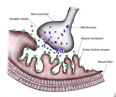
The neuromuscular junction (NMJ) is the specialized synapse or chemical communication point, where the motor neuron's nerve terminal (presynaptic element) meets the muscle fiber's motor end plate (postsynaptic element). This junction plays a crucial role in controlling muscle contraction and relaxation.
At the NMJ, the neurotransmitter acetylcholine is released from the presynaptic nerve terminal into the synaptic cleft, following an action potential. Acetylcholine then binds to nicotinic acetylcholine receptors on the postsynaptic membrane of the muscle fiber, leading to the generation of an end-plate potential. If sufficient end-plate potentials are generated and summate, they will trigger an action potential in the muscle fiber, ultimately causing muscle contraction.
Dysfunction at the neuromuscular junction can result in various neuromuscular disorders, such as myasthenia gravis, where autoantibodies attack acetylcholine receptors, leading to muscle weakness and fatigue.
Synaptic membranes, also known as presynaptic and postsynaptic membranes, are specialized structures in neurons where synaptic transmission occurs. The presynaptic membrane is the portion of the neuron's membrane where neurotransmitters are released into the synaptic cleft, a small gap between two neurons. The postsynaptic membrane, on the other hand, is the portion of the neighboring neuron's membrane that contains receptors for the neurotransmitters released by the presynaptic neuron. Together, these structures facilitate the transmission of electrical signals from one neuron to another through the release and binding of chemical messengers.
Skeletal muscle fibers, also known as striated muscle fibers, are the type of muscle cells that make up skeletal muscles, which are responsible for voluntary movements of the body. These muscle fibers are long, cylindrical, and multinucleated, meaning they contain multiple nuclei. They are surrounded by a connective tissue layer called the endomysium, and many fibers are bundled together into fascicles, which are then surrounded by another layer of connective tissue called the perimysium.
Skeletal muscle fibers are composed of myofibrils, which are long, thread-like structures that run the length of the fiber. Myofibrils contain repeating units called sarcomeres, which are responsible for the striated appearance of skeletal muscle fibers. Sarcomeres are composed of thick and thin filaments, which slide past each other during muscle contraction to shorten the sarcomere and generate force.
Skeletal muscle fibers can be further classified into two main types based on their contractile properties: slow-twitch (type I) and fast-twitch (type II). Slow-twitch fibers have a high endurance capacity and are used for sustained, low-intensity activities such as maintaining posture. Fast-twitch fibers, on the other hand, have a higher contractile speed and force generation capacity but fatigue more quickly and are used for powerful, explosive movements.
 Agrin
Agrin
EGF-like domain
Olfactory memory
Barney Josephson
Markus Rüegg
Dok-7
Neuromuscular junction
George Yancopoulos
MuSK protein
Torpedo (genus)
Behavior mutation
Development of the nervous system
Dystroglycan
Integrin alpha 7
Heparan sulfate
Myasthenia gravis
Turtles Can Fly
RAPSN
Omigapil
INaturalist
Pikachurin
Joshua Sanes
Synaptogenesis
Glomerular basement membrane
Ocellated electric ray
Veronica Maggio
Congenital myasthenic syndrome
Neurexin
Patch clamp
Mucin-16
Agrin - Wikipedia
 Agrin and LRP4 antibodies as new biomarkers of myasthenia gravis - PubMed
 Agrin in the Muscularis Mucosa Serves as a Biomarker Distinguishing Hyperplastic Polyps from Sessile Serrated Lesions |...
 JCI Insight -
Congenital myasthenic syndrome-associated agrin variants affect clustering of acetylcholine receptors in a domain...
JCI Insight -
Congenital myasthenic syndrome-associated agrin variants affect clustering of acetylcholine receptors in a domain...
Novel SEA and LG2 Agrin mutations causing congenital Myasthenic syndrome - Nuffield Department of Clinical Neurosciences
 Agrin
Agrin
 Merchant - Agrin
 Products | Opex Agrin
 Sugar sorghum - Agrin Maroc
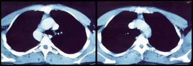
 Myasthenia Gravis Workup: Laboratory Tests, Radiography, CT, and MRI, Electrodiagnostic Studies
 Rotary oven (AGRIN R) - سحر
 SMP N 1 Kaliwungu: Agrin Setyo Palupi
Agrin Free All to Divx MPEG FLV MOV WMV Activation Key
 Dr. Richard Agrin, MD - New brunswick, NJ - Reviews and Ratings - DiabetesIQ.com
 looking for a map | HIVE
 Agrin and CD34 immunohistochemistry for the discrimination of benign versus malignant hepatocellular lesions. | Read by QxMD
 RCSB PDB - 1PXU: Crystal structure of chicken NtA from a eukaryotic source at 2.2A resolution
 Neuroscience Elisa | Gentaur Elisa Kits | US - UK & Europe Supply
The extracellular matrix protein agrin is essential for epicardial epithelial-to-mesenchymal transition during heart...
The nature and biology of basement membranes
LG2 agrin mutation causing severe congenital myasthenic syndrome mimics functional characteristics of non-neural (z−) agrin -...
 Autoantibodies to cortactin and agrin in sera of patients with myasthenia gravis. Doppler K, Hemprich A, Haarmann A, Brecht I,...
 Schwann cell-specific PTEN and EGFR dysfunctions affect neuromuscular junction development by impairing Agrin signalingand...
 Avaz Latif movie reviews & film summaries | Roger Ebert
 Frontiers | Extracellular Control of Radial Glia Proliferation and Scaffolding During Cortical Development and Pathology
 No healing in a vacuum, study finds | ScienceDaily
Sv2a MGI Mouse Gene Detail - MGI:1927139 - synaptic vesicle glycoprotein 2a
 Turtles Can Fly (2004)
Neuromuscular junction8
- Agrin is a large proteoglycan whose best-characterised role is in the development of the neuromuscular junction during embryogenesis. (wikipedia.org)
- Agrin functions by activating the MuSK protein (for Muscle-Specific Kinase), which is a receptor tyrosine kinase required for the formation and maintenance of the neuromuscular junction. (wikipedia.org)
- Agrin is also required for neuromuscular junction formation. (wikipedia.org)
- Most notably, Agrin is responsible for the clustering of acetylcholine receptors (AChRs) on the cell surface and their localization to the neuromuscular junction. (thermofisher.com)
- Agrin, a protein extracted from the electric organ of Torpedo californica, induces the formation of specializations on cultured chick myotubes that resemble the postsynaptic apparatus at the neuromuscular junction. (rupress.org)
- Agrin is a key organizer of acetylcholine receptor (AChR) clustering at the neuromuscular junction. (unibas.ch)
- Here we show that agrin, a proteoglycan involved in the neuromuscular junction, is a critical niche-derived signal that controls survival and proliferation of HSCs. (ox.ac.uk)
- MuSK mediates the agrin-induced clustering of AChRs during synapse formation, and is also expressed at the mature neuromuscular junction. (nih.gov)
Receptor13
- The nerve secretes agrin, resulting in phosphorylation of the MuSK receptor. (wikipedia.org)
- Since agrin fragments induce acetylcholine receptor aggregation as well as phosphorylation of the MuSK receptor, researchers spliced them and found that the variant did not trigger phosphorylation. (wikipedia.org)
- Agrin acts via a MuSK receptor complex. (nature.com)
- Bowe, M. A., Deyst, K. A., Leszyk, J. D. & Fallon, J. R. Identification and purification of an agrin receptor from Torpedo postsynaptic membranes: a heteromeric complex related to the dystroglycans. (nature.com)
- Interestingly, a unique receptor tyrosine kinase, designated MuSK, has been discovered that interacts with Agrin and is specifically localized to developing muscle. (thermofisher.com)
- Regulation of agrin-induced acetylcholine receptor aggregation by Ca++ and phorbol ester. (rupress.org)
- Agrin-induced receptor aggregation also was inhibited by phorbol 12-myristate 13-acetate, an activator of protein kinase C, and by inhibitors of energy metabolism. (rupress.org)
- Agrin is expressed by multipotent nonhematopoietic mesenchymal stem cells (MSCs) and by differentiated osteoblasts lining the endosteal bone surface, whereas Lin−Sca1+c-Kit+ (LSK) cells express the α-dystroglycan receptor for agrin. (ox.ac.uk)
- Lrp4 is a receptor for Agrin and forms a complex with MuSK. (ox.ac.uk)
- MuSK, a receptor tyrosine kinase that is expressed in skeletal muscle, and Agrin, a motor neuron-derived ligand that stimulates MuSK phosphorylation, play critical roles in synaptic differentiation, as synapses do not form in their absence, and mutations in MuSK or downstream effectors are a major cause of a group of neuromuscular disorders, termed congenital myasthenic syndromes (CMS). (ox.ac.uk)
- Here, we report that Lrp4, a member of the LDLR family, is a receptor for Agrin, forms a complex with MuSK, and mediates MuSK activation by Agrin. (ox.ac.uk)
- Here, we define a phosphorylation-dependent binding site on the receptor that mediates agrin-induced clustering. (jneurosci.org)
- Together, these findings suggest that agrin-induced phosphorylation of the β subunit motif increases the stoichiometry of rapsyn binding to the AChR, thereby helping to stably cluster the receptor and anchor it at high density in the postsynaptic membrane. (jneurosci.org)
MuSK7
- Agrin is required to activate MuSK. (wikipedia.org)
- In addition to MuSK, agrin binds several other proteins on the surface of muscle, including dystroglycan and laminin. (wikipedia.org)
- The requirement for Agrin and MuSK in the formation of the NMJ was demonstrated primarily by knockout mouse studies. (wikipedia.org)
- Furthermore, Agrin signalling through Lrp4-muscle-specific tyrosine kinase (MuSK) forms a critical oncogenic axis. (a-star.edu.sg)
- Although the agrin/MuSK signaling pathway remains largely unknown, changes in intracellular calcium levels are required for agrin-induced AChR aggregation (Megeath and Fallon [1998]: J Neurosci 18: 672-678). (nih.gov)
- Blockade of L-CaChs in muscle cultures inhibited agrin-induced AChR aggregation but not tyrosine phosphorylation of MuSK or AChR beta subunits. (nih.gov)
- How Agrin activates MuSK and stimulates synaptic differentiation is not known and remains a fundamental gap in our understanding of signaling at neuromuscular synapses. (ox.ac.uk)
Inhibited agrin-induced AChR aggregation1
- The formation and maintenance of agrin-induced AChR aggregates required Ca++, Co++ and Mn++ inhibited agrin-induced AChR aggregation and increased the rate of aggregate dispersal. (rupress.org)
Differentiation2
- Agrin is a molecule that resides in the basal lamina of muscle cells and directs key events in post synaptic differentiation. (thermofisher.com)
- The similarities between agrin's effects on cultured myotubes and events that occur during formation of neuromuscular junctions support the hypothesis that axon terminals release molecules similar to agrin that induce the differentiation of the postsynaptic apparatus. (rupress.org)
Phosphorylation3
- The sequence contains a conserved tyrosine (Y390) whose phosphorylation is induced by agrin and whose mutation abolished clustering of β loop chimeras and their ability to inhibit agrin-induced clustering of the endogenous AChR. (jneurosci.org)
- Indeed, we found that rapsyn associated with CD4-β loop chimeras in a phosphorylation-dependent manner, and that agrin increased the ratio of rapsyn binding to wild type AChR but not to AChR-β 3F/3F , which lacks β loop tyrosine phosphorylation sites. (jneurosci.org)
- Indeed, we have shown previously that mutation of a tyrosine phosphorylation site in the long cytoplasmic loop of the β subunit impairs agrin-induced cytoskeletal anchoring and aggregation of mutant AChR in muscle cells ( Borges and Ferns, 2001 ). (jneurosci.org)
AChR3
- Heparan sulfate glycosaminoglycans covalently linked to the agrin protein have been shown to play a role in the clustering of AChR. (wikipedia.org)
- Here, we show that L-type calcium channels (L-CaChs) are required for full agrin-induced aggregation of AChRs and sufficient to induce agrin-independent AChR aggregation. (nih.gov)
- Using chimeric proteins in which CD4 is fused to the large intracellular loop of each of the AChR subunits we found that agrin induced clustering of only chimeras containing the β subunit loop. (jneurosci.org)
Laminin2
Proteoglycan3
- In addition, by its ability to activate the Hippo signaling pathway, agrin is emerging as a key proteoglycan in the tumor microenvironment. (wikipedia.org)
- His longstanding research interest has been the role of the extracellular matrix in nervous system development, especially the function of the heparan sulfate proteoglycan agrin. (nccu.edu)
- Retina development in zebrafish requires the heparan sulfate proteoglycan agrin. (nccu.edu)
Mice3
- In vivo , a single administration of agrin promotes cardiac regeneration in adult mice after myocardial infarction, although the degree of cardiomyocyte proliferation observed in this model suggests that there are additional therapeutic mechanisms. (nature.com)
- Figure 3: Agrin induces cardiac regeneration in adult mice. (nature.com)
- Agrin-deficient mice displayed in vivo apoptosis of CD34+CD135− LSK cells and impaired hematopoiesis, both of which were reverted by an agrin-sufficient stroma. (ox.ac.uk)
Synaptic1
- Agrin may play an important role in the basement membrane of the microvasculature as well as in synaptic plasticity. (wikipedia.org)
Proliferation5
- Figure 1: Identification of agrin in a screen for mouse cardiac ECM-mediated cardiomyocyte proliferation. (nature.com)
- Figure 4: Agrin promotes cardiomyocyte proliferation through Dag1, ERK and Yap signalling. (nature.com)
- Figure 5: Agrin promotes proliferation and attenuates maturation of human iPSC-CMs. (nature.com)
- In vitro, agrin-deficient MSCs were less efficient in supporting proliferation of mouse Lin−c-Kit+ cells, suggesting that agrin plays a role in the hematopoietic cell development. (ox.ac.uk)
- Agrin enhances cellular proliferation, migration and oncogenic signalling. (a-star.edu.sg)
Rahmani2
- The 22 year old's ingénue looks come with the requisite street swagger to pull off seductively shaded songs that capture night life ambience with a noir-ish flair that she and her producer/collaborator, Agrin Rahmani, have dubbed 'future retro. (deezer.com)
- LÉON herself, along with Agrin Rahmani. (scandipop.co.uk)
Recombinant2
- In vitro , recombinant agrin promotes the division of cardiomyocytes that are derived from mouse and human induced pluripotent stem cells through a mechanism that involves the disassembly of the dystrophin-glycoprotein complex, and Yap- and ERK-mediated signalling. (nature.com)
- Recombinant rat agrin, C-terminal construct. (thermofisher.com)
Skeletal muscle1
- When secreted, agrin binds to several receptors on the surface of skeletal muscle. (wikipedia.org)
Extracellular matrix1
- We identify agrin, a component of neonatal extracellular matrix, as required for the full regenerative capacity of neonatal mouse hearts. (nature.com)
Acetylcholine receptors2
- Agrin is named based on its involvement in the aggregation of acetylcholine receptors during synaptogenesis. (wikipedia.org)
- The aim of the studies reported here was to characterize the effects of agrin on the distribution of acetylcholine receptors (AChRs) and cholinesterase as a step toward determining agrin's mechanism of action. (rupress.org)
Vivo1
- Importantly, antibodies targeting Agrin reduced oncogenic signalling and tumour growth in vivo. (a-star.edu.sg)
Heparan2
- There are three potential heparan sulfate (HS) attachment sites within the primary structure of agrin, but it is thought that only two of these actually carry HS chains when the protein is expressed. (wikipedia.org)
- It may be that rather than solely binding directly to the agrin protein core a number of components of the secondary scaffold may also interact with its heparan sulfate side-chains. (wikipedia.org)
Aggregates3
- When agrin was added to the medium bathing chick myotubes small (less than 4 micron 2) aggregates of AChRs began to appear within 2 h and increased rapidly in number until 4 h. (rupress.org)
- The accumulation of AChRs into agrin-induced aggregates occurred primarily by lateral migration of AChRs already in the myotube plasma membrane at the time agrin was added to the cultures. (rupress.org)
- if agrin was removed the number of aggregates declined slowly. (rupress.org)
Congenital1
- Agrin mutations lead to a congenital myasthenic syndrome with distal muscle weakness and atrophy. (genomeweb.com)
Myotubes1
- and at agrin-induced clusters in cultured myotubes ( Wallace, 1989 ). (jneurosci.org)
Regeneration1
- Figure 2: Agrin delays neonatal cardiomyocyte maturation and is required for P1 cardiac regeneration following surgical resection. (nature.com)
Producer1
- Agrin is the largest producer and processor of diverse varieties of herbs, spices and seeds in Morocco. (agrinmaroc.ma)
Role5
- A role in the retention of anionic macromolecules within the vasculature has also been suggested for agrin-linked HS at the glomerular or alveolar basement membrane. (wikipedia.org)
- These data unveil a crucial role of agrin in the hematopoietic niches and in the cross-talk between stromal and hematopoietic stem cells. (ox.ac.uk)
- An oncogenic role of Agrin in. (a-star.edu.sg)
- Here, combining SILAC quantitative proteomics and biochemical approaches, we uncover a critical oncogenic role of Agrin, which is overexpressed and secreted in HCC. (a-star.edu.sg)
- The major emphasis of the laboratory is the investigation of the role of agrin in fetal alcohol spectrum disorders (FASD) and how agrin function via Fgfs and Shh is a target of embryonal ethanol and cannabinoid exposure in zebrafish. (nccu.edu)
Cultures1
- Agrin responsiveness was significantly reduced in primary muscle cultures from the muscular dysgenesis mouse, a natural mutant, which does not express the L-CaCh. (nih.gov)
Laboratory1
- Agrin was first identified by the U.J. McMahan laboratory, Stanford University. (wikipedia.org)
Domain1
- It has also been shown that the G3 domain of agrin is very plastic, meaning it can discriminate between binding partners for a better fit. (wikipedia.org)
Function4
- Also, agrin may be involved in blood-brain barrier (BBB) formation and/or function and it influences Aβ homeostasis. (wikipedia.org)
- These studies are currently focusing on elucidating the mechanisms by which agrin modulates the function of heparin-binding growth factors and morphogens, such as fibroblast growth factors (Fgfs) and sonic hedgehog (Shh), during zebrafish nervous system development. (nccu.edu)
- Forebrain and hindbrain development in zebrafish is sensitive to ethanol exposure involving agrin, Fgf and Sonic hedgehog function. (nccu.edu)
- Agrin function associated with ocular development is a target of ethanol exposure in embryonic zebrafish. (nccu.edu)
Development3
- During development in humans, the growing end of motor neuron axons secrete a protein called agrin. (wikipedia.org)
- Agrin signaling related to NMJ development, was downregulated in TA muscle. (edu.hk)
- These studies are employing genetic and molecular approaches to examine ocular, forebrain, and hindbrain development in response to ethanol exposure, and the subsequent changes in agrin, Fgf or Shh signaling. (nccu.edu)
Forms1
- Agrin splice forms having inserts at two sites in the carboxy terminus designated "y" and "z" display a high affinity for AChRs, while splice forms lacking these inserts associate with AChRs weakly. (thermofisher.com)
Website1
- Step one for the Nashville-based startup is getting an initial cohort of consumers to create their personalized Health eBiography® via the Agrin website . (venturenashville.com)
Heart1
- When Satellite first sees Agrin, it is as though Cupid has shot an arrow directly into the young man's heart. (clevescene.com)
Products1
- From specification to treatment and blending, Agrin offers to its customers the opportunity to develop a fully customized ingredients and products that suits their business requirements. (agrinmaroc.ma)
Point2
- At no point will Agrin Health "diagnose or prescribe" treatments, said Thomas, though the company is certain to have some partnerships with other healthcare or healthtech organizations that provide medical or nursing care, telemedicine or other goods and services. (venturenashville.com)
- Reaching the point at which Agrin begins providing actionable intelligence for consumers will be a major inflection point for the company, she added. (venturenashville.com)
Notice1
- Too lost in her own trauma to notice, Agrin gazes back with grave, unsmiling eyes. (clevescene.com)