Absorptiometry, Photon
Photons
Bone Density
Body Composition
Osteoporosis
Radius
Bone Diseases, Metabolic
Adipose Tissue
Lumbar Vertebrae
Adiposity
Bone and Bones
Tomography, Emission-Computed, Single-Photon
Osteoporosis, Postmenopausal
Anthropometry
Body Mass Index
Minerals
Body Fat Distribution
Hip
Skinfold Thickness
Body Weight
Obesity
Neutron Activation Analysis
Bone Remodeling
Calcaneus
Abdominal Fat
Puberty
Intra-Abdominal Fat
Sarcopenia
Calcium, Dietary
Bone Development
Densitometry
Cross-Sectional Studies
Activation Analysis
Body Height
Body Fluid Compartments
Postmenopause
Tomography, X-Ray Computed
Reproducibility of Results
Regression Analysis
Osteocalcin
Elementary Particles
HIV-Associated Lipodystrophy Syndrome
Bone Density Conservation Agents
Premenopause
Tibia
Whole Body Imaging
Aging
Scattering, Radiation
Reference Values
Linear Models
Radiometry
Exercise
Phantoms, Imaging
Sex Characteristics
Muscle, Skeletal
European Continental Ancestry Group
Monte Carlo Method
Scintillation Counting
Pelvic Bones
Basal Metabolism
Anorexia Nervosa
Longitudinal Studies
Vitamin D
Sex Factors
Radionuclide Imaging
Thinness
Sensitivity and Specificity
Hip Joint
Bone Demineralization, Pathologic
Prospective Studies
Muscle Strength
Osteoporotic Fractures
Microscopy, Fluorescence, Multiphoton
Energy Metabolism
Organotechnetium Compounds
Age Factors
Calorimetry, Indirect
Alendronate
Potassium Radioisotopes
Risk Factors
Insulin Resistance
Particle Accelerators
Radiation Dosage
Parathyroid Hormone
Overweight
Hand Bones
Waist Circumference
Biological Markers
Diphosphonates
Leptin
Bone Lengthening
Radiopharmaceuticals
Physical Fitness
Technetium Tc 99m Exametazime
Cohort Studies
Thallium Radioisotopes
Predictive Value of Tests
Body Constitution
Technetium
Analysis of Variance
Magnetic Resonance Imaging
Follow-Up Studies
Alkaline Phosphatase
Case-Control Studies
African Continental Ancestry Group
Tomography, Emission-Computed
Oximes
Obesity, Abdominal
Menopause
Equipment Failure Analysis
Radiotherapy Planning, Computer-Assisted
Human Growth Hormone
Relative Biological Effectiveness
Resistance Training
Models, Biological
Image Processing, Computer-Assisted
Glucose Tolerance Test
Body Weights and Measures
Tellurium
Collagen Type I
Hormone Replacement Therapy
Radiotherapy, High-Energy
Adiponectin
Fractures, Spontaneous
Lipodystrophy
Models, Structural
Waist-Hip Ratio
African Americans
Double-Blind Method
Iodine Radioisotopes
Calcium
Viscera
Insulin
Tomography, Optical
Osteomalacia
Female Athlete Triad Syndrome
Gymnastics
Semiconductors
Teriparatide
Calibration
Optics and Photonics
Athletes
Gamma Cameras
Hip Fractures
Algorithms
Fluoride Poisoning
Weight-Bearing
Lasers
Insulin-Like Growth Factor I
Lipids
Radioisotopes
Calcification, Physiologic
Testosterone
Renal Osteodystrophy
Iofetamine
Radiation
Dose-Response Relationship, Radiation
Glucocorticoids
Electrons
Spectrometry, Gamma
Statistics, Nonparametric
Luminescent Measurements
Lutetium
Association of polymorphism at the type I collagen (COL1A1) locus with reduced bone mineral density, increased fracture risk, and increased collagen turnover. (1/2834)
OBJECTIVE: To examine the relationship between a common polymorphism within intron 1 of the COL1A1 gene and osteoporosis in a nested case-control study. METHODS: We studied 185 healthy women (mean +/- SD age 54.3+/-4.6 years). Bone mineral density (BMD) was measured using dual x-ray absorptiometry, and fractures were determined radiographically. The COL1A1 genotype was assessed using the polymerase chain reaction and Bal I endonuclease digestion. RESULTS: Genotype frequencies were similar to those previously observed and in Hardy-Weinberg equilibrium: SS 61.1%, Ss 36.2%, and ss 2.7%. Carriage of at least one copy of the "s" allele was associated with a significant reduction in lumbar spine BMD (P = 0.02) and an increased risk of total fracture (P = 0.04). Urinary pyridinoline levels were significantly elevated in those with the risk allele (P < 0.05). CONCLUSION: These data support the findings that the COL1A1 gene polymorphism is associated with low BMD and fracture risk, and suggest a possible physiologic effect on total body turnover of type I collagen. (+info)Transplantation of osteoblast-like cells to the distracted callus in rabbits. (2/2834)
We carried out limb lengthening in rabbits and then transplanted osteoblast-like cells derived from the tibial periosteum to the centres of distracted callus immediately after distraction had been terminated. Two weeks later the transaxial area ratio at the centre of the distracted callus and the bone mineral density (BMD) were significantly higher in the transplanted group, by 21% and 42%, respectively, than in the non-injected group or the group injected with physiological saline (p < 0.05). Callus BMD as a percentage of density in uninvolved bone was also significantly higher in the transplanted group (p < 0.05) than in the other two groups, by 27% and 20% in the second and fourth weeks, respectively (p < 0.05). Mechanically, the callus in the transplanted group tended to be stronger as shown by the three-point bending test although the difference in fracture strength was not statistically significant. Our results show that transplantation of osteoblast-like cells promotes maturity of the distracted callus as observed at the second and fourth weeks after lengthening. The method appears promising as a means of shortening the consolidation period of callus distraction and decreasing complications during limb lengthening with an external fixator. (+info)Subsidence of a non-polished stem in revisions of the hip using impaction allograft. Evaluation with radiostereometry and dual-energy X-ray absorptiometry. (3/2834)
We revised 24 consecutive hips with loosening of the femoral stem using impaction allograft and a cemented stem with an unpolished proximal surface. Repeated radiostereometric examinations for up to two years showed a slow rate of subsidence with a mean of 0.32 mm (-2.0 to +0.31). Fifteen cases followed for a further year showed the same mean subsidence after three years, indicating stabilisation. A tendency to retroversion of the stems was noted between the operation and the last follow-up. Retroversion was also recorded when displacement of the stem was studied in ten of the patients after two years. Repeated determination of bone mineral density showed an initial loss after six months, followed by recovery to the postoperative level at two years. Defects in the cement mantle and malalignment of the stem were often noted on postoperative radiographs, but did not correlate with the degrees of migration or displacement. After one year, increasing frequency of trabecular remodelling or resorption of the graft was observed in the greater trochanter and distal to the tip of the stem. Cortical repair was noted distally and medially (Gruen regions 3, 5 and 6). Migration of the stems was the lowest reported to date, which we attribute to the improved grafting technique and to the hardness of the graft. (+info)Plasma leptin concentrations in obese children: changes during 4-mo periods with and without physical training. (4/2834)
BACKGROUND: Little is known about the effects of physical training on plasma leptin concentrations in children. OBJECTIVE: We sought to determine the effects of 4-mo periods with and without physical training on leptin in obese children and to explore the determinants of leptin at baseline and in response to physical training. DESIGN: Participants were 34 obese 7-11-y-old children randomly assigned to engage in physical training during either the first or second 4 mo of the 8-mo study. RESULTS: Total body composition, visceral adiposity, and insulin were all positively correlated with leptin at baseline (P < or = 0.05); however, only fat mass was retained in the final stepwise regression (P = 0.0001, R2 = 0.57). Leptin decreased during the 4-mo periods of physical training and increased in the 4 mo after cessation of physical training (P < 0.001 for the time by group interaction). Decreases in leptin were greatest in children with higher pretraining leptin concentrations, those whose total mass increased least, and those whose insulin concentrations decreased most (P < or = 0.05); only pretraining leptin concentration (P = 0.009) and change in total mass (P = 0.0002) were retained in the final regression (R2 = 0.53). CONCLUSIONS: In obese children, leptin concentration decreased during 4 mo of physical training and increased during a subsequent 4-mo period without physical training, fat mass was highly correlated with baseline leptin, and greater reductions in leptin during 4 mo of physical training were seen in children with higher pretraining leptin and in those whose total mass increased least. (+info)Use of ultrasonography in the diagnosis of osteomalacia: preliminary results on experimental osteomalacia in the rat. (5/2834)
This study was performed to investigate the ability of ultrasonographic technique to distinguish osteomalacia from normal bone with the same mineral content. Ten rats with experimentally induced osteomalacia (group A) and 12 control rats having similar body size and weight (group B) were studied. Histomorphometric analysis confirmed the presence of osteomalacia in two rats from group A and showed normally mineralized bone in two rats from group B. Whole body bone mineral density, measured by dual-energy x-ray absorptiometry, was similar in the two groups (86 +/- 6 mg/cm2 in group A and 89 +/- 4 mg/cm2 in group B). The velocity of the ultrasound beam in bone was measured by densitometer at the first caudal vertebra of each rat. The velocity was measured when the first peak of the waveform reached a predetermined minimum amplitude value (amplitude-dependent speed of sound) as well as at the lowest point of this curve before it reaches the predetermined minimum amplitude (first minimum speed of sound). Although the amplitude-dependent speed of sound was similar in the two groups (1381.9 +/- 11.8 m/s in group A and 1390.9 +/- 17.8 m/s in group B), the first minimum speed of sound was clearly different (1446.1 +/- 8.9 m/s in group A and 1503.3 +/- 10.9 m/s in group B; P < 0.001). This study shows that ultrasonography could be used to identify alterations in bone quality, such as osteomalacia, but further studies need to be carried out before this method can be introduced into clinical practice. (+info)Bone densitometry at a district general hospital: evaluation of service by doctors and patients. (6/2834)
OBJECTIVE: To assess doctors' and patients' views about a district general hospital bone densitometry service and to examine existing practice to influence future provision. DESIGN: Three postal surveys: (a) of doctors potentially using the service, (b) of patients undergoing a bone densitometry test during a six month period, and (c) of the referring doctors of the patients undergoing the test. SETTING: Bone densitometry service at South Cleveland Hospital, Middlesbrough and two district health authorities: South Tees and Northallerton. SUBJECTS: All general practitioners (n=201) and hospital consultants in general medicine, rheumatology, obstetrics and gynaecology, orthopaedics, radio therapy and oncology, haematology, and radiology (n=61); all patients undergoing an initial bone densitometry test (n=309) during a six month period; and their referring doctors. MAIN MEASURES: Service awareness and use, knowledge of clinical indications, test results, influence of test results on patient management, satisfaction with the service and its future provision. RESULTS: The overall response rates for the three surveys were 87%, 70%, and 61%. There was a high awareness of the service among doctors and patients; 219(84%) doctors were aware and 155 of them (71%) had used it, and patients often (40%) suggested the test to their doctor. The test was used for a range of reasons including screening although the general use was consistent with current guidelines. Two hundred (65%) bone densitometry measurements were normal, 71(23%) were low normal, and 38(12%) were low. Although doctors reported that management of patients had been influenced by the test results, the algorithm for decision making was unclear. Patients and doctors were satisfied with the service and most (n=146, 68%) doctors wanted referral guidelines for the service. CONCLUSIONS: There was a high awareness of, use of, and satisfaction with the service. Patients were being referred for a range of reasons and a few of these could not be justified, many tests were normal, and clinical decision making was not always influenced by the test result. It is concluded that bone densitometry services should be provided but only for patients whose management will be influenced by test results and subject to guidelines to ensure appropriate use of the technology. (+info)Cardiovascular disease risk factors in 2 distinct ethnic groups: Indian and Pakistani compared with American premenopausal women. (7/2834)
BACKGROUND: Although people from the Indian subcontinent have high rates of cardiovascular disease (CVD), studies of such in Indian and Pakistani women living in the United States are lacking. OBJECTIVE: This study accounted for variability in serum lipid (total cholesterol and triacylglycerol) and lipoprotein [LDL cholesterol, lipoprotein(a), and HDL cholesterol] concentrations in Indian and Pakistani compared with American premenopausal women in the United States. Body composition, regional fat distribution, dietary intake, and energy expenditure were compared between groups. DESIGN: The 2 groups were 47 Indian and Pakistani and 47 American women. Health was assessed via medical history, physical activity, body composition (via anthropometry and dual-energy X-ray absorptiometry), dietary intake (via 7-d food records), and serum lipids. RESULTS: Serum total cholesterol, triacylglycerol, LDL cholesterol, lipoprotein(a), the ratio of total to HDL cholesterol, and the ratio of LDL to HDL cholesterol were greater (P <0.03), whereas HDL-cholesterol values were lower (P = 0.011) in Indians and Pakistanis than in Americans. Multiple regression analysis indicated that approximately 18% of the variance in total cholesterol (P = 0.0010) and LDL cholesterol (P = 0.0009) was accounted for by ethnicity, energy expenditure, and the ratio of the sum of central to the sum of peripheral skinfold thicknesses. Ethnicity, sum of central skinfold thicknesses, ratio of polyunsaturated to saturated fat, and monounsaturated fat intake accounted for approximately 43% of the variance in triacylglycerol concentration (P < 0.0001). Monounsaturated fat, percentage body fat, and alcohol intake accounted for approximately 26% of variance in HDL cholesterol. Ethnicity contributed approximately 22% of the 25% overall variance in lipoprotein(a). CONCLUSIONS: Results suggest that these Indian and Pakistani women are at higher CVD risk than their American counterparts, but that increasing their physical activity is likely to decrease overall and regional adiposity, thereby improving their serum lipid profiles. (+info)Riboflavin and riboflavin-derived cofactors in adolescent girls with anorexia nervosa. (8/2834)
BACKGROUND: Thyroid hormones, riboflavin, riboflavin cofactors, and organic acids were assessed in girls with anorexia nervosa. OBJECTIVE: The objective was to examine the effect of malnutrition and low thyroid hormone concentrations on erythrocyte and plasma riboflavin metabolism and their relation with urinary organic acid excretion. DESIGN: Seventeen adolescent girls with anorexia nervosa [body mass index (BMI; in kg/m2): 14.8 +/- 2.2] and 17 age-matched, healthy girls (control subjects; BMI: 20.5 +/- 2.2) took part in the feeding study. Erythrocyte and plasma riboflavin as well as riboflavin cofactors (flavin mononucleotide and flavin adenine dinucleotide) were assessed by HPLC, whereas urinary organic acids were assessed by gas chromatography-mass spectrometry. RESULTS: Anorectic patients who began a feeding program had higher erythrocyte riboflavin (3.5 +/- 2.2 compared with <0.1 nmol/mol hemoglobin; P < 0.001), lower plasma flavin adenine dinucleotide (57.8 +/- 18.5 compared with 78.5 +/- 54.3 nmol/L; P < 0.05), and higher urinary ethylmalonic acid (7.12 +/- 4.39 compared with 1.3 +/- 2.8 micromol/mmol creatinine; P < 0.001) and isovalerylglycine (7.65 +/- 4.78 compared with 3.8 +/- 0.9 micromol/mmol creatinine; P < 0.05) concentrations than did control subjects. Triiodothyronine concentrations were low and negatively correlated with plasma riboflavin concentrations (r = -0.69, P < 0.01). Not all patients showed improvements in these biochemical indexes after 30 d of refeeding. CONCLUSIONS: The low triiodothyronine concentrations observed in anorexia nervosa could alter the extent of riboflavin conversion into cofactors, thus leading to high erythrocyte riboflavin concentrations, low plasma flavin adenine dinucleotide concentrations, and high rates of ethylmalonic acid and isovalerylglycine excretion. (+info)Photon Absorptiometry is a medical technique used to measure the absorption of photons (light particles) by tissues or materials. In clinical practice, it is often used as a non-invasive method for measuring bone mineral density (BMD). This technique uses a low-energy X-ray beam or gamma ray to penetrate the tissue and then measures the amount of radiation absorbed by the bone. The amount of absorption is related to the density and thickness of the bone, allowing for an assessment of BMD. It can be used to diagnose osteoporosis and monitor treatment response in patients with bone diseases. There are two types of photon absorptiometry: single-photon absorptiometry (SPA) and dual-photon absorptiometry (DPA). SPA uses one energy level, while DPA uses two different energy levels to measure BMD, providing more precise measurements.
A photon is not a term that has a specific medical definition, as it is a fundamental concept in physics. Photons are elementary particles that carry electromagnetic energy, such as light. They have no mass or electric charge and exhibit both particle-like and wave-like properties. In the context of medicine, photons are often discussed in relation to various medical imaging techniques (e.g., X-ray imaging, CT scans, and PET scans) and therapeutic interventions like laser therapy and radiation therapy, where photons are used to diagnose or treat medical conditions.
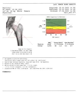
Bone density refers to the amount of bone mineral content (usually measured in grams) in a given volume of bone (usually measured in cubic centimeters). It is often used as an indicator of bone strength and fracture risk. Bone density is typically measured using dual-energy X-ray absorptiometry (DXA) scans, which provide a T-score that compares the patient's bone density to that of a young adult reference population. A T-score of -1 or above is considered normal, while a T-score between -1 and -2.5 indicates osteopenia (low bone mass), and a T-score below -2.5 indicates osteoporosis (porous bones). Regular exercise, adequate calcium and vitamin D intake, and medication (if necessary) can help maintain or improve bone density and prevent fractures.
Body composition refers to the relative proportions of different components that make up a person's body, including fat mass, lean muscle mass, bone mass, and total body water. It is an important measure of health and fitness, as changes in body composition can indicate shifts in overall health status. For example, an increase in fat mass and decrease in lean muscle mass can be indicative of poor nutrition, sedentary behavior, or certain medical conditions.
There are several methods for measuring body composition, including:
1. Bioelectrical impedance analysis (BIA): This method uses low-level electrical currents to estimate body fat percentage based on the conductivity of different tissues.
2. Dual-energy X-ray absorptiometry (DXA): This method uses low-dose X-rays to measure bone density and body composition, including lean muscle mass and fat distribution.
3. Hydrostatic weighing: This method involves submerging a person in water and measuring their weight underwater to estimate body density and fat mass.
4. Air displacement plethysmography (ADP): This method uses air displacement to measure body volume and density, which can be used to estimate body composition.
Understanding body composition can help individuals make informed decisions about their health and fitness goals, as well as provide valuable information for healthcare providers in the management of chronic diseases such as obesity, diabetes, and heart disease.
Osteoporosis is a systemic skeletal disease characterized by low bone mass, deterioration of bone tissue, and disruption of bone architecture, leading to increased risk of fractures, particularly in the spine, wrist, and hip. It mainly affects older people, especially postmenopausal women, due to hormonal changes that reduce bone density. Osteoporosis can also be caused by certain medications, medical conditions, or lifestyle factors such as smoking, alcohol abuse, and a lack of calcium and vitamin D in the diet. The diagnosis is often made using bone mineral density testing, and treatment may include medication to slow bone loss, promote bone formation, and prevent fractures.
The radius is one of the two bones in the forearm in humans and other vertebrates. In humans, it runs from the lateral side of the elbow to the thumb side of the wrist. It is responsible for rotation of the forearm and articulates with the humerus at the elbow and the carpals at the wrist. Any medical condition or injury that affects the radius can impact the movement and function of the forearm and hand.
The "femur neck" is the narrow, upper part of the femur (thigh bone) where it connects to the pelvis. It is the region through which the femoral head articulates with the acetabulum to form the hip joint. The femur neck is a common site for fractures, especially in older adults with osteoporosis.
Metabolic bone diseases are a group of conditions that affect the bones and are caused by disorders in the body's metabolism. These disorders can result in changes to the bone structure, density, and strength, leading to an increased risk of fractures and other complications. Some common examples of metabolic bone diseases include:
1. Osteoporosis: a condition characterized by weak and brittle bones that are more likely to break, often as a result of age-related bone loss or hormonal changes.
2. Paget's disease of bone: a chronic disorder that causes abnormal bone growth and deformities, leading to fragile and enlarged bones.
3. Osteomalacia: a condition caused by a lack of vitamin D or problems with the body's ability to absorb it, resulting in weak and soft bones.
4. Hyperparathyroidism: a hormonal disorder that causes too much parathyroid hormone to be produced, leading to bone loss and other complications.
5. Hypoparathyroidism: a hormonal disorder that results in low levels of parathyroid hormone, causing weak and brittle bones.
6. Renal osteodystrophy: a group of bone disorders that occur as a result of chronic kidney disease, including osteomalacia, osteoporosis, and high turnover bone disease.
Treatment for metabolic bone diseases may include medications to improve bone density and strength, dietary changes, exercise, and lifestyle modifications. In some cases, surgery may be necessary to correct bone deformities or fractures.
Adipose tissue, also known as fatty tissue, is a type of connective tissue that is composed mainly of adipocytes (fat cells). It is found throughout the body, but is particularly abundant in the abdominal cavity, beneath the skin, and around organs such as the heart and kidneys.
Adipose tissue serves several important functions in the body. One of its primary roles is to store energy in the form of fat, which can be mobilized and used as an energy source during periods of fasting or exercise. Adipose tissue also provides insulation and cushioning for the body, and produces hormones that help regulate metabolism, appetite, and reproductive function.
There are two main types of adipose tissue: white adipose tissue (WAT) and brown adipose tissue (BAT). WAT is the more common form and is responsible for storing energy as fat. BAT, on the other hand, contains a higher number of mitochondria and is involved in heat production and energy expenditure.
Excessive accumulation of adipose tissue can lead to obesity, which is associated with an increased risk of various health problems such as diabetes, heart disease, and certain types of cancer.
The lumbar vertebrae are the five largest and strongest vertebrae in the human spine, located in the lower back region. They are responsible for bearing most of the body's weight and providing stability during movement. The lumbar vertebrae have a characteristic shape, with a large body in the front, which serves as the main weight-bearing structure, and a bony ring in the back, formed by the pedicles, laminae, and processes. This ring encloses and protects the spinal cord and nerves. The lumbar vertebrae are numbered L1 to L5, starting from the uppermost one. They allow for flexion, extension, lateral bending, and rotation movements of the trunk.
"Adiposity" is a medical term that refers to the condition of having an excessive amount of fat in the body. It is often used to describe obesity or being significantly overweight. Adipose tissue, which is the technical name for body fat, is important for many bodily functions, such as storing energy and insulating the body. However, an excess of adipose tissue can lead to a range of health problems, including heart disease, diabetes, and certain types of cancer.
There are different ways to measure adiposity, including body mass index (BMI), waist circumference, and skinfold thickness. BMI is the most commonly used method and is calculated by dividing a person's weight in kilograms by their height in meters squared. A BMI of 30 or higher is considered obese, while a BMI between 25 and 29.9 is considered overweight. However, it's important to note that BMI may not accurately reflect adiposity in some individuals, such as those with a lot of muscle mass.
In summary, adiposity refers to the condition of having too much body fat, which can increase the risk of various health problems.
"Bone" is the hard, dense connective tissue that makes up the skeleton of vertebrate animals. It provides support and protection for the body's internal organs, and serves as a attachment site for muscles, tendons, and ligaments. Bone is composed of cells called osteoblasts and osteoclasts, which are responsible for bone formation and resorption, respectively, and an extracellular matrix made up of collagen fibers and mineral crystals.
Bones can be classified into two main types: compact bone and spongy bone. Compact bone is dense and hard, and makes up the outer layer of all bones and the shafts of long bones. Spongy bone is less dense and contains large spaces, and makes up the ends of long bones and the interior of flat and irregular bones.
The human body has 206 bones in total. They can be further classified into five categories based on their shape: long bones, short bones, flat bones, irregular bones, and sesamoid bones.
Emission-Computed Tomography, Single-Photon (SPECT) is a type of nuclear medicine imaging procedure that generates detailed, three-dimensional images of the distribution of radioactive pharmaceuticals within the body. It uses gamma rays emitted by a radiopharmaceutical that is introduced into the patient's body, and a specialized gamma camera to detect these gamma rays and create tomographic images. The data obtained from the SPECT imaging can be used to diagnose various medical conditions, evaluate organ function, and guide treatment decisions. It is commonly used to image the heart, brain, and bones, among other organs and systems.
The femur is the medical term for the thigh bone, which is the longest and strongest bone in the human body. It connects the hip bone to the knee joint and plays a crucial role in supporting the weight of the body and allowing movement during activities such as walking, running, and jumping. The femur is composed of a rounded head, a long shaft, and two condyles at the lower end that articulate with the tibia and patella to form the knee joint.
Postmenopausal osteoporosis is a specific type of osteoporosis that occurs in women after they have gone through menopause. It is defined as a skeletal disorder characterized by compromised bone strength, leading to an increased risk of fractures. In this condition, the decline in estrogen levels that occurs during menopause accelerates bone loss, resulting in a decrease in bone density and quality, which can lead to fragility fractures, particularly in the hips, wrists, and spine.
It's important to note that while postmenopausal osteoporosis is more common in women, men can also develop osteoporosis due to other factors such as aging, lifestyle choices, and medical conditions.
Anthropometry is the scientific study of measurements and proportions of the human body. It involves the systematic measurement and analysis of various physical characteristics, such as height, weight, blood pressure, waist circumference, and other body measurements. These measurements are used in a variety of fields, including medicine, ergonomics, forensics, and fashion design, to assess health status, fitness level, or to design products and environments that fit the human body. In a medical context, anthropometry is often used to assess growth and development, health status, and disease risk factors in individuals and populations.
Body Mass Index (BMI) is a measure used to assess whether a person has a healthy weight for their height. It's calculated by dividing a person's weight in kilograms by the square of their height in meters. Here is the medical definition:
Body Mass Index (BMI) = weight(kg) / [height(m)]^2
According to the World Health Organization, BMI categories are defined as follows:
* Less than 18.5: Underweight
* 18.5-24.9: Normal or healthy weight
* 25.0-29.9: Overweight
* 30.0 and above: Obese
It is important to note that while BMI can be a useful tool for identifying weight issues in populations, it does have limitations when applied to individuals. For example, it may not accurately reflect body fat distribution or muscle mass, which can affect health risks associated with excess weight. Therefore, BMI should be used as one of several factors when evaluating an individual's health status and risk for chronic diseases.
In the context of nutrition and health, minerals are inorganic elements that are essential for various bodily functions, such as nerve impulse transmission, muscle contraction, maintaining fluid and electrolyte balance, and bone structure. They are required in small amounts compared to macronutrients (carbohydrates, proteins, and fats) and are obtained from food and water.
Some of the major minerals include calcium, phosphorus, magnesium, sodium, potassium, and chloride, while trace minerals or microminerals are required in even smaller amounts and include iron, zinc, copper, manganese, iodine, selenium, and fluoride.
It's worth noting that the term "minerals" can also refer to geological substances found in the earth, but in medical terminology, it specifically refers to the essential inorganic elements required for human health.
Body fat distribution refers to the way in which adipose tissue (fat) is distributed throughout the body. There are two main types of body fat distribution: android or central/abdominal distribution and gynoid or peripheral distribution.
Android or central/abdominal distribution is characterized by a higher proportion of fat deposited in the abdominal area, surrounding internal organs (visceral fat) and between muscle fibers (intramuscular fat). This pattern is more common in men and is associated with an increased risk of metabolic diseases such as type 2 diabetes, hypertension, dyslipidemia, and cardiovascular disease.
Gynoid or peripheral distribution is characterized by a higher proportion of fat deposited in the hips, thighs, and buttocks. This pattern is more common in women and is generally considered less harmful to health than android distribution. However, excessive accumulation of body fat, regardless of its distribution, can lead to obesity-related health problems.
It's important to note that body fat distribution can be influenced by various factors, including genetics, hormones, lifestyle, and environmental factors. Assessing body fat distribution is an essential aspect of evaluating overall health and disease risk.
In medical terms, the hip is a ball-and-socket joint where the rounded head of the femur (thigh bone) fits into the cup-shaped socket, also known as the acetabulum, of the pelvis. This joint allows for a wide range of movement in the lower extremities and supports the weight of the upper body during activities such as walking, running, and jumping. The hip joint is surrounded by strong ligaments, muscles, and tendons that provide stability and enable proper functioning.
Electric impedance is a measure of opposition to the flow of alternating current (AC) in an electrical circuit or component, caused by both resistance (ohmic) and reactance (capacitive and inductive). It is expressed as a complex number, with the real part representing resistance and the imaginary part representing reactance. The unit of electric impedance is the ohm (Ω).
In the context of medical devices, electric impedance may be used to measure various physiological parameters, such as tissue conductivity or fluid composition. For example, bioelectrical impedance analysis (BIA) uses electrical impedance to estimate body composition, including fat mass and lean muscle mass. Similarly, electrical impedance tomography (EIT) is a medical imaging technique that uses electric impedance to create images of internal organs and tissues.
Skinfold thickness is a measurement of the thickness of the subcutaneous fat layer (the layer of fat directly beneath the skin) at specific sites on the body. It's typically measured using calipers and is expressed in millimeters (mm). This measurement is often used in health assessments to estimate body fat percentage and overall health status. The most commonly used sites for skinfold thickness measurements are the triceps, biceps, suprailiac (just above the iliac crest), subscapular (underneath the shoulder blade), and abdominal areas.
A bone fracture is a medical condition in which there is a partial or complete break in the continuity of a bone due to external or internal forces. Fractures can occur in any bone in the body and can vary in severity from a small crack to a shattered bone. The symptoms of a bone fracture typically include pain, swelling, bruising, deformity, and difficulty moving the affected limb. Treatment for a bone fracture may involve immobilization with a cast or splint, surgery to realign and stabilize the bone, or medication to manage pain and prevent infection. The specific treatment approach will depend on the location, type, and severity of the fracture.
The spine, also known as the vertebral column, is a complex structure in the human body that is part of the axial skeleton. It is composed of 33 individual vertebrae (except in some people where there are fewer due to fusion of certain vertebrae), intervertebral discs, facet joints, ligaments, muscles, and nerves.
The spine has several important functions:
1. Protection: The spine protects the spinal cord, which is a major component of the nervous system, by enclosing it within a bony canal.
2. Support: The spine supports the head and upper body, allowing us to maintain an upright posture and facilitating movement of the trunk and head.
3. Movement: The spine enables various movements such as flexion (bending forward), extension (bending backward), lateral flexion (bending sideways), and rotation (twisting).
4. Weight-bearing: The spine helps distribute weight and pressure evenly across the body, reducing stress on individual vertebrae and other structures.
5. Blood vessel and nerve protection: The spine protects vital blood vessels and nerves that pass through it, including the aorta, vena cava, and spinal nerves.
The spine is divided into five regions: cervical (7 vertebrae), thoracic (12 vertebrae), lumbar (5 vertebrae), sacrum (5 fused vertebrae), and coccyx (4 fused vertebrae, also known as the tailbone). Each region has unique characteristics that allow for specific functions and adaptations to the body's needs.
Body weight is the measure of the force exerted on a scale or balance by an object's mass, most commonly expressed in units such as pounds (lb) or kilograms (kg). In the context of medical definitions, body weight typically refers to an individual's total weight, which includes their skeletal muscle, fat, organs, and bodily fluids.
Healthcare professionals often use body weight as a basic indicator of overall health status, as it can provide insights into various aspects of a person's health, such as nutritional status, metabolic function, and risk factors for certain diseases. For example, being significantly underweight or overweight can increase the risk of developing conditions like malnutrition, diabetes, heart disease, and certain types of cancer.
It is important to note that body weight alone may not provide a complete picture of an individual's health, as it does not account for factors such as muscle mass, bone density, or body composition. Therefore, healthcare professionals often use additional measures, such as body mass index (BMI), waist circumference, and blood tests, to assess overall health status more comprehensively.
Obesity is a complex disease characterized by an excess accumulation of body fat to the extent that it negatively impacts health. It's typically defined using Body Mass Index (BMI), a measure calculated from a person's weight and height. A BMI of 30 or higher is indicative of obesity. However, it's important to note that while BMI can be a useful tool for identifying obesity in populations, it does not directly measure body fat and may not accurately reflect health status in individuals. Other factors such as waist circumference, blood pressure, cholesterol levels, and blood sugar levels should also be considered when assessing health risks associated with weight.
Neutron Activation Analysis (NAA) is not strictly a medical definition, but it's a technique used in the field of nuclear medicine and forensic medicine for material analysis and identification. Here's a general definition:
Neutron Activation Analysis is a non-destructive analytical method that uses nuclear reactions to identify and determine the concentration of elements within a sample. The sample is irradiated with neutrons, which induce nuclear reactions that produce radioactive isotopes of the elements present in the sample. The gamma radiation emitted by these radioisotopes is then measured and analyzed to quantify the elemental composition of the sample. This technique is particularly useful for detecting and measuring trace elements and isotopes, making it valuable in various fields such as archaeology, geology, nuclear medicine, and forensic science.
Body water refers to the total amount of water present in the human body. It is an essential component of life and makes up about 60-70% of an adult's body weight. Body water is distributed throughout various fluid compartments within the body, including intracellular fluid (water inside cells), extracellular fluid (water outside cells), and transcellular fluid (water found in specific bodily spaces such as the digestive tract, eyes, and joints). Maintaining proper hydration and balance of body water is crucial for various physiological processes, including temperature regulation, nutrient transportation, waste elimination, and overall health.
Bone remodeling is the normal and continuous process by which bone tissue is removed from the skeleton (a process called resorption) and new bone tissue is formed (a process called formation). This ongoing cycle allows bones to repair microdamage, adjust their size and shape in response to mechanical stress, and maintain mineral homeostasis. The cells responsible for bone resorption are osteoclasts, while the cells responsible for bone formation are osteoblasts. These two cell types work together to maintain the structural integrity and health of bones throughout an individual's life.
During bone remodeling, the process can be divided into several stages:
1. Activation: The initiation of bone remodeling is triggered by various factors such as microdamage, hormonal changes, or mechanical stress. This leads to the recruitment and activation of osteoclast precursor cells.
2. Resorption: Osteoclasts attach to the bone surface and create a sealed compartment called a resorption lacuna. They then secrete acid and enzymes that dissolve and digest the mineralized matrix, creating pits or cavities on the bone surface. This process helps remove old or damaged bone tissue and releases calcium and phosphate ions into the bloodstream.
3. Reversal: After resorption is complete, the osteoclasts undergo apoptosis (programmed cell death), and mononuclear cells called reversal cells appear on the resorbed surface. These cells prepare the bone surface for the next stage by cleaning up debris and releasing signals that attract osteoblast precursors.
4. Formation: Osteoblasts, derived from mesenchymal stem cells, migrate to the resorbed surface and begin producing a new organic matrix called osteoid. As the osteoid mineralizes, it forms a hard, calcified structure that gradually replaces the resorbed bone tissue. The osteoblasts may become embedded within this newly formed bone as they differentiate into osteocytes, which are mature bone cells responsible for maintaining bone homeostasis and responding to mechanical stress.
5. Mineralization: Over time, the newly formed bone continues to mineralize, becoming stronger and more dense. This process helps maintain the structural integrity of the skeleton and ensures adequate calcium storage.
Throughout this continuous cycle of bone remodeling, hormones, growth factors, and mechanical stress play crucial roles in regulating the balance between resorption and formation. Disruptions to this delicate equilibrium can lead to various bone diseases, such as osteoporosis, where excessive resorption results in weakened bones and increased fracture risk.
The calcaneus is the largest tarsal bone in the human foot, and it is commonly known as the heel bone. It articulates with the cuboid bone anteriorly, the talus bone superiorly, and several tendons and ligaments that help to form the posterior portion of the foot's skeletal structure. The calcaneus plays a crucial role in weight-bearing and movement, as it forms the lower part of the leg's ankle joint and helps to absorb shock during walking or running.
Abdominal fat, also known as visceral fat, is the fat that is stored in the abdominal cavity and surrounds the internal organs such as the liver, pancreas, and intestines. It is different from subcutaneous fat, which is the fat located just under the skin, and is often measured using techniques such as CT scans or MRI to assess health risks. Excess abdominal fat has been linked to an increased risk of various health conditions, including type 2 diabetes, heart disease, and stroke.
Puberty is the period of sexual maturation, generally occurring between the ages of 10 and 16 in females and between 12 and 18 in males. It is characterized by a series of events including rapid growth, development of secondary sexual characteristics, and the acquisition of reproductive capabilities. Puberty is initiated by the activation of the hypothalamic-pituitary-gonadal axis, leading to the secretion of hormones such as estrogen and testosterone that drive the physical changes associated with this stage of development.
In females, puberty typically begins with the onset of breast development (thelarche) and the appearance of pubic hair (pubarche), followed by the start of menstruation (menarche). In males, puberty usually starts with an increase in testicular size and the growth of pubic hair, followed by the deepening of the voice, growth of facial hair, and the development of muscle mass.
It's important to note that the onset and progression of puberty can vary widely among individuals, and may be influenced by genetic, environmental, and lifestyle factors.
Intra-abdominal fat, also known as visceral fat, is the fat that is stored within the abdominal cavity and surrounds the internal organs such as the liver, pancreas, and intestines. It's different from subcutaneous fat, which is the fat found just under the skin. Intra-abdominal fat is metabolically active and has been linked to an increased risk of various health conditions, including type 2 diabetes, heart disease, high blood pressure, and stroke. The accumulation of intra-abdominal fat can be influenced by factors such as diet, physical activity, genetics, and age. Waist circumference and imaging tests, such as CT scans and MRIs, are commonly used to measure intra-abdominal fat.
Sarcopenia is a medical term that refers to the age-related decline in muscle mass, strength, and function. It's a natural process that occurs as we get older, but it can also be accelerated by various factors such as sedentary lifestyle, poor nutrition, and chronic diseases. Sarcopenia can lead to decreased mobility, weakness, frailty, and increased risk of falls and fractures in older adults. It's important to note that sarcopenia is different from cachexia, which is a muscle wasting condition associated with chronic illnesses such as cancer, HIV/AIDS, and heart failure.
Dietary calcium is a type of calcium that is obtained through food sources. Calcium is an essential mineral that is necessary for many bodily functions, including bone formation and maintenance, muscle contraction, nerve impulse transmission, and blood clotting.
The recommended daily intake of dietary calcium varies depending on age, sex, and other factors. For example, the recommended daily intake for adults aged 19-50 is 1000 mg, while women over 50 and men over 70 require 1200 mg per day.
Good dietary sources of calcium include dairy products such as milk, cheese, and yogurt; leafy green vegetables like broccoli and kale; fortified cereals and juices; and certain types of fish, such as salmon and sardines. It is important to note that some foods can inhibit the absorption of calcium, including oxalates found in spinach and rhubarb, and phytates found in whole grains and legumes.
If a person is unable to get enough calcium through their diet, they may need to take calcium supplements. However, it is important to talk to a healthcare provider before starting any new supplement regimen, as excessive intake of calcium can lead to negative health effects.
Bone development, also known as ossification, is the process by which bone tissue is formed and grows. This complex process involves several different types of cells, including osteoblasts, which produce new bone matrix, and osteoclasts, which break down and resorb existing bone tissue.
There are two main types of bone development: intramembranous and endochondral ossification. Intramembranous ossification occurs when bone tissue forms directly from connective tissue, while endochondral ossification involves the formation of a cartilage model that is later replaced by bone.
During fetal development, most bones develop through endochondral ossification, starting as a cartilage template that is gradually replaced by bone tissue. However, some bones, such as those in the skull and clavicles, develop through intramembranous ossification.
Bone development continues after birth, with new bone tissue being laid down and existing tissue being remodeled throughout life. This ongoing process helps to maintain the strength and integrity of the skeleton, allowing it to adapt to changing mechanical forces and repair any damage that may occur.
Densitometry is a medical technique used to measure the density or degree of opacity of various structures, particularly bones and tissues. It is often used in the diagnosis and monitoring of osteoporosis, a condition characterized by weak and brittle bones. Bone densitometry measures the amount of calcium and other minerals in a segment of bone to determine its strength and density. This information can help doctors assess a patient's risk of fractures and make treatment recommendations. Densitometry is also used in other medical fields, such as mammography, where it is used to measure the density of breast tissue to detect abnormalities and potential signs of cancer.
A cross-sectional study is a type of observational research design that examines the relationship between variables at one point in time. It provides a snapshot or a "cross-section" of the population at a particular moment, allowing researchers to estimate the prevalence of a disease or condition and identify potential risk factors or associations.
In a cross-sectional study, data is collected from a sample of participants at a single time point, and the variables of interest are measured simultaneously. This design can be used to investigate the association between exposure and outcome, but it cannot establish causality because it does not follow changes over time.
Cross-sectional studies can be conducted using various data collection methods, such as surveys, interviews, or medical examinations. They are often used in epidemiology to estimate the prevalence of a disease or condition in a population and to identify potential risk factors that may contribute to its development. However, because cross-sectional studies only provide a snapshot of the population at one point in time, they cannot account for changes over time or determine whether exposure preceded the outcome.
Therefore, while cross-sectional studies can be useful for generating hypotheses and identifying potential associations between variables, further research using other study designs, such as cohort or case-control studies, is necessary to establish causality and confirm any findings.
Activation analysis is a technique used in medical and scientific research to analyze the composition of materials by measuring the radiation emitted from a sample that has been exposed to an intense source of radiation, such as a nuclear reactor or a particle accelerator. This process causes some of the atoms in the sample to become "activated," meaning they transform into unstable isotopes that emit gamma rays or other subatomic particles as they decay back to a stable state.
By measuring the energy and intensity of these emissions, researchers can identify the elements present in the sample and determine their relative abundances. Activation analysis can be used to analyze a wide range of materials, including biological tissues, environmental samples, and archaeological artifacts, and it has applications in fields such as forensics, geology, and nuclear medicine.
It is important to note that activation analysis involves handling radioactive materials and requires specialized training and equipment to ensure safety and accuracy.
"Body height" is a measure of the vertical length of a person's body from the top of their head to the bottom of their feet. It is typically measured in units such as centimeters (cm) or inches (in). In medical settings, body height is often used as a basic anthropometric measurement to assess overall health status, growth and development, nutritional status, and aging-related changes.
There are different methods for measuring body height, but the most common one involves having the person stand upright against a vertical surface (such as a wall or a stadiometer) with their heels, buttocks, shoulders, and head touching the surface. The measurement is taken at the point where the top of the person's head meets the surface.
Body height can be influenced by various factors, including genetics, nutrition, health status, and environmental conditions. Changes in body height over time can provide important insights into a person's health trajectory and potential health risks. For example, a significant decrease in body height may indicate bone loss or spinal compression, while a rapid increase in height during childhood or adolescence may suggest optimal growth and development.
Body fluid compartments refer to the distribution of body fluids in the human body, which are divided into two main compartments: the intracellular fluid compartment and the extracellular fluid compartment. The intracellular fluid compartment contains fluid that is inside the cells, while the extracellular fluid compartment contains fluid that is outside the cells.
The extracellular fluid compartment is further divided into two sub-compartments: the interstitial fluid compartment and the intravascular fluid compartment. The interstitial fluid compartment is the space between the cells, while the intravascular fluid compartment is the fluid inside the blood vessels.
These body fluid compartments are essential for maintaining homeostasis in the human body, as they help to regulate the balance of water and electrolytes, transport nutrients and waste products, and provide a medium for immune cells to travel through the body. Abnormalities in the distribution of body fluids can lead to various medical conditions, such as edema, dehydration, and heart failure.
Postmenopause is a stage in a woman's life that follows 12 months after her last menstrual period (menopause) has occurred. During this stage, the ovaries no longer release eggs and produce lower levels of estrogen and progesterone hormones. The reduced levels of these hormones can lead to various physical changes and symptoms, such as hot flashes, vaginal dryness, and mood changes. Postmenopause is also associated with an increased risk of certain health conditions, including osteoporosis and heart disease. It's important for women in postmenopause to maintain a healthy lifestyle, including regular exercise, a balanced diet, and routine medical check-ups to monitor their overall health and manage any potential risks.
X-ray computed tomography (CT or CAT scan) is a medical imaging method that uses computer-processed combinations of many X-ray images taken from different angles to produce cross-sectional (tomographic) images (virtual "slices") of the body. These cross-sectional images can then be used to display detailed internal views of organs, bones, and soft tissues in the body.
The term "computed tomography" is used instead of "CT scan" or "CAT scan" because the machines take a series of X-ray measurements from different angles around the body and then use a computer to process these data to create detailed images of internal structures within the body.
CT scanning is a noninvasive, painless medical test that helps physicians diagnose and treat medical conditions. CT imaging provides detailed information about many types of tissue including lung, bone, soft tissue and blood vessels. CT examinations can be performed on every part of the body for a variety of reasons including diagnosis, surgical planning, and monitoring of therapeutic responses.
In computed tomography (CT), an X-ray source and detector rotate around the patient, measuring the X-ray attenuation at many different angles. A computer uses this data to construct a cross-sectional image by the process of reconstruction. This technique is called "tomography". The term "computed" refers to the use of a computer to reconstruct the images.
CT has become an important tool in medical imaging and diagnosis, allowing radiologists and other physicians to view detailed internal images of the body. It can help identify many different medical conditions including cancer, heart disease, lung nodules, liver tumors, and internal injuries from trauma. CT is also commonly used for guiding biopsies and other minimally invasive procedures.
In summary, X-ray computed tomography (CT or CAT scan) is a medical imaging technique that uses computer-processed combinations of many X-ray images taken from different angles to produce cross-sectional images of the body. It provides detailed internal views of organs, bones, and soft tissues in the body, allowing physicians to diagnose and treat medical conditions.
Reproducibility of results in a medical context refers to the ability to obtain consistent and comparable findings when a particular experiment or study is repeated, either by the same researcher or by different researchers, following the same experimental protocol. It is an essential principle in scientific research that helps to ensure the validity and reliability of research findings.
In medical research, reproducibility of results is crucial for establishing the effectiveness and safety of new treatments, interventions, or diagnostic tools. It involves conducting well-designed studies with adequate sample sizes, appropriate statistical analyses, and transparent reporting of methods and findings to allow other researchers to replicate the study and confirm or refute the results.
The lack of reproducibility in medical research has become a significant concern in recent years, as several high-profile studies have failed to produce consistent findings when replicated by other researchers. This has led to increased scrutiny of research practices and a call for greater transparency, rigor, and standardization in the conduct and reporting of medical research.
Regression analysis is a statistical technique used in medicine, as well as in other fields, to examine the relationship between one or more independent variables (predictors) and a dependent variable (outcome). It allows for the estimation of the average change in the outcome variable associated with a one-unit change in an independent variable, while controlling for the effects of other independent variables. This technique is often used to identify risk factors for diseases or to evaluate the effectiveness of medical interventions. In medical research, regression analysis can be used to adjust for potential confounding variables and to quantify the relationship between exposures and health outcomes. It can also be used in predictive modeling to estimate the probability of a particular outcome based on multiple predictors.
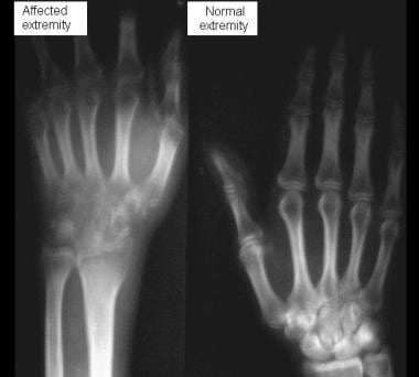
The metacarpus is the medical term for the part of the hand located between the carpus (wrist) and the digits (fingers). It consists of five bones, known as the metacarpal bones, which are numbered 1 to 5 from the thumb side to the little finger side. Each metacarpal bone has a base, a shaft, and a head. The bases of the metacarpal bones articulate with the carpal bones to form the wrist joint, while the heads of the metacarpal bones form the knuckles at the back of the hand.
The metacarpus plays an essential role in hand function as it provides stability and support for the movement of the fingers and thumb. Injuries or conditions affecting the metacarpus can significantly impact hand function, causing pain, stiffness, weakness, or deformity.
Osteocalcin is a protein that is produced by osteoblasts, which are the cells responsible for bone formation. It is one of the most abundant non-collagenous proteins found in bones and plays a crucial role in the regulation of bone metabolism. Osteocalcin contains a high affinity for calcium ions, making it essential for the mineralization of the bone matrix.
Once synthesized, osteocalcin is secreted into the extracellular matrix, where it binds to hydroxyapatite crystals, helping to regulate their growth and contributing to the overall strength and integrity of the bones. Osteocalcin also has been found to play a role in other physiological processes outside of bone metabolism, such as modulating insulin sensitivity, energy metabolism, and male fertility.
In summary, osteocalcin is a protein produced by osteoblasts that plays a critical role in bone formation, mineralization, and turnover, and has been implicated in various other physiological processes.
Elementary particles are the fundamental building blocks that make up all matter and energy in the universe. They are called "elementary" because they cannot be broken down into smaller, simpler components. According to our current understanding of particle physics, there are two main types of elementary particles: fermions and bosons.
Fermions include quarks and leptons, which make up matter. There are six types of each, known as flavors: up and down quarks, charm and strange quarks, top and bottom quarks, and electron, muon, and tau leptons (also called "electron-type," "muon-type," and "tau-type" leptons). Each fermion also has an associated antiparticle.
Bosons are the force carriers that mediate the fundamental forces of nature: gravity, electromagnetism, the strong nuclear force, and the weak nuclear force. These include the photon (which carries the electromagnetic force), the gluon (which carries the strong nuclear force), and the W and Z bosons (which carry the weak nuclear force). The Higgs boson is also a type of boson, associated with the Higgs field that gives other particles their mass.
It's important to note that our understanding of elementary particles and their properties is still evolving, as new experiments and theories continue to shape our knowledge of the universe's smallest constituents.
HIV-Associated Lipodystrophy Syndrome is a term used to describe a range of body shape changes and metabolic abnormalities that can occur in some individuals receiving long-term combination antiretroviral therapy (cART) for HIV infection. The syndrome is characterized by the abnormal distribution of fat, including:
1. Lipoatrophy: Loss of subcutaneous fat from the face, limbs, and buttocks, leading to a gaunt appearance.
2. Lipohypertrophy: Accumulation of fat in the abdomen, breasts, and dorsocervical region (buffalo hump), resulting in an altered body shape.
3. Metabolic abnormalities: Insulin resistance, hyperlipidemia, and lactic acidosis, which can increase the risk of developing cardiovascular disease and diabetes mellitus.
The exact pathogenesis of HIV-Associated Lipodystrophy Syndrome is not fully understood, but it is believed to be related to a combination of factors, including the direct effects of HIV infection on adipose tissue, mitochondrial toxicity caused by certain antiretroviral medications, and chronic inflammation. The syndrome can have significant psychological and social consequences for affected individuals, and management typically involves a multidisciplinary approach that includes switching to alternative antiretroviral regimens, addressing metabolic abnormalities, and providing cosmetic interventions as needed.
Bone density conservation agents, also known as anti-resorptive agents or bone-sparing drugs, are a class of medications that help to prevent the loss of bone mass and reduce the risk of fractures. They work by inhibiting the activity of osteoclasts, the cells responsible for breaking down and reabsorbing bone tissue during the natural remodeling process.
Examples of bone density conservation agents include:
1. Bisphosphonates (e.g., alendronate, risedronate, ibandronate, zoledronic acid) - These are the most commonly prescribed class of bone density conservation agents. They bind to hydroxyapatite crystals in bone tissue and inhibit osteoclast activity, thereby reducing bone resorption.
2. Denosumab (Prolia) - This is a monoclonal antibody that targets RANKL (Receptor Activator of Nuclear Factor-κB Ligand), a key signaling molecule involved in osteoclast differentiation and activation. By inhibiting RANKL, denosumab reduces osteoclast activity and bone resorption.
3. Selective estrogen receptor modulators (SERMs) (e.g., raloxifene) - These medications act as estrogen agonists or antagonists in different tissues. In bone tissue, SERMs mimic the bone-preserving effects of estrogen by inhibiting osteoclast activity and reducing bone resorption.
4. Hormone replacement therapy (HRT) - Estrogen hormone replacement therapy has been shown to preserve bone density in postmenopausal women; however, its use is limited due to increased risks of breast cancer, cardiovascular disease, and thromboembolic events.
5. Calcitonin - This hormone, secreted by the thyroid gland, inhibits osteoclast activity and reduces bone resorption. However, it has largely been replaced by other more effective bone density conservation agents.
These medications are often prescribed for individuals at high risk of fractures due to conditions such as osteoporosis or metabolic disorders that affect bone health. It is essential to follow the recommended dosage and administration guidelines to maximize their benefits while minimizing potential side effects. Regular monitoring of bone density, blood calcium levels, and other relevant parameters is also necessary during treatment with these medications.
Bone resorption is the process by which bone tissue is broken down and absorbed into the body. It is a normal part of bone remodeling, in which old or damaged bone tissue is removed and new tissue is formed. However, excessive bone resorption can lead to conditions such as osteoporosis, in which bones become weak and fragile due to a loss of density. This process is carried out by cells called osteoclasts, which break down the bone tissue and release minerals such as calcium into the bloodstream.
A spinal fracture, also known as a vertebral compression fracture, is a break in one or more bones (vertebrae) of the spine. This type of fracture often occurs due to weakened bones caused by osteoporosis, but it can also result from trauma such as a car accident or a fall.
In a spinal fracture, the front part of the vertebra collapses, causing the height of the vertebra to decrease, while the back part of the vertebra remains intact. This results in a wedge-shaped deformity of the vertebra. Multiple fractures can lead to a hunched forward posture known as kyphosis or dowager's hump.
Spinal fractures can cause pain, numbness, tingling, or weakness in the back, legs, or arms, depending on the location and severity of the fracture. In some cases, spinal cord compression may occur, leading to more severe symptoms such as paralysis or loss of bladder and bowel control.
Premenopause is not a formal medical term, but it's often informally used to refer to the time period in a woman's life leading up to menopause. During this stage, which can last for several years, hormonal changes begin to occur in preparation for menopause. The ovaries start to produce less estrogen and progesterone, which can lead to various symptoms such as irregular periods, hot flashes, mood swings, and sleep disturbances. However, it's important to note that not all women will experience these symptoms.
The official medical term for the stage when a woman's period becomes irregular and less frequent, but hasn't stopped completely, is perimenopause. This stage typically lasts from two to eight years and ends with menopause, which is defined as the point when a woman has not had a period for 12 consecutive months. After menopause, women enter postmenopause.
The tibia, also known as the shin bone, is the larger of the two bones in the lower leg and part of the knee joint. It supports most of the body's weight and is a major insertion point for muscles that flex the foot and bend the leg. The tibia articulates with the femur at the knee joint and with the fibula and talus bone at the ankle joint. Injuries to the tibia, such as fractures, are common in sports and other activities that put stress on the lower leg.
Whole Body Imaging (WBI) is a diagnostic technique that involves obtaining images of the entire body or significant portions of it, typically for the purpose of detecting abnormalities such as tumors, fractures, infections, or other diseases. This can be achieved through various imaging modalities including:
1. Whole Body Computed Tomography (WBCT): This is a series of CT scans taken from head to toe to create detailed cross-sectional images of the body. It's often used in trauma situations to identify internal injuries.
2. Whole Body Magnetic Resonance Imaging (WBMRI): This uses magnetic fields and radio waves to produce detailed images of the body's internal structures. It's particularly useful for detecting soft tissue abnormalities.
3. Positron Emission Tomography - Computed Tomography (PET-CT): This combines PET and CT scans to create detailed, 3D images of the body's functional processes, such as metabolism or blood flow. It's often used in cancer diagnosis and staging.
4. Whole Body Bone Scan: This uses a small amount of radioactive material to highlight areas of increased bone turnover, which can indicate conditions like fractures, tumors, or infections.
5. Whole Body PET: Similar to WBMRI, this uses positron emission tomography to create detailed images of the body's metabolic processes, but it doesn't provide the same level of anatomical detail as PET-CT.
It's important to note that while WBI can be a powerful diagnostic tool, it also involves higher doses of radiation (in the case of WBCT and Whole Body Bone Scan) and greater costs compared to single or limited area imaging studies. Therefore, its use is typically reserved for specific clinical scenarios where the benefits outweigh the risks and costs.
Aging is a complex, progressive and inevitable process of bodily changes over time, characterized by the accumulation of cellular damage and degenerative changes that eventually lead to increased vulnerability to disease and death. It involves various biological, genetic, environmental, and lifestyle factors that contribute to the decline in physical and mental functions. The medical field studies aging through the discipline of gerontology, which aims to understand the underlying mechanisms of aging and develop interventions to promote healthy aging and extend the human healthspan.
Radiation scattering is a physical process in which radiation particles or waves deviate from their original direction due to interaction with matter. This phenomenon can occur through various mechanisms such as:
1. Elastic Scattering: Also known as Thomson scattering or Rayleigh scattering, it occurs when the energy of the scattered particle or wave remains unchanged after the collision. In the case of electromagnetic radiation (e.g., light), this results in a change of direction without any loss of energy.
2. Inelastic Scattering: This type of scattering involves an exchange of energy between the scattered particle and the target medium, leading to a change in both direction and energy of the scattered particle or wave. An example is Compton scattering, where high-energy photons (e.g., X-rays or gamma rays) interact with charged particles (usually electrons), resulting in a decrease in photon energy and an increase in electron kinetic energy.
3. Coherent Scattering: In this process, the scattered radiation maintains its phase relationship with the incident radiation, leading to constructive and destructive interference patterns. An example is Bragg scattering, which occurs when X-rays interact with a crystal lattice, resulting in diffraction patterns that reveal information about the crystal structure.
In medical contexts, radiation scattering can have both beneficial and harmful effects. For instance, in diagnostic imaging techniques like computed tomography (CT) scans, radiation scattering contributes to image noise and reduces contrast resolution. However, in radiation therapy for cancer treatment, controlled scattering of therapeutic radiation beams can help ensure that the tumor receives a uniform dose while minimizing exposure to healthy tissues.
Reference values, also known as reference ranges or reference intervals, are the set of values that are considered normal or typical for a particular population or group of people. These values are often used in laboratory tests to help interpret test results and determine whether a patient's value falls within the expected range.
The process of establishing reference values typically involves measuring a particular biomarker or parameter in a large, healthy population and then calculating the mean and standard deviation of the measurements. Based on these statistics, a range is established that includes a certain percentage of the population (often 95%) and excludes extreme outliers.
It's important to note that reference values can vary depending on factors such as age, sex, race, and other demographic characteristics. Therefore, it's essential to use reference values that are specific to the relevant population when interpreting laboratory test results. Additionally, reference values may change over time due to advances in measurement technology or changes in the population being studied.
I'm sorry for any confusion, but "Linear Models" is actually a term from the field of statistics and machine learning, rather than medicine. A linear model is a type of statistical model that is used to analyze the relationship between two or more variables. In a linear model, the relationship between the dependent variable (the outcome or result) and the independent variable(s) (the factors being studied) is assumed to be linear, meaning that it can be described by a straight line on a graph.
The equation for a simple linear model with one independent variable (x) and one dependent variable (y) looks like this:
y = β0 + β1*x + ε
In this equation, β0 is the y-intercept or the value of y when x equals zero, β1 is the slope or the change in y for each unit increase in x, and ε is the error term or the difference between the actual values of y and the predicted values of y based on the linear model.
Linear models are widely used in medical research to study the relationship between various factors (such as exposure to a risk factor or treatment) and health outcomes (such as disease incidence or mortality). They can also be used to adjust for confounding variables, which are factors that may influence both the independent variable and the dependent variable, and thus affect the observed relationship between them.
Radiometry is the measurement of electromagnetic radiation, including visible light. It quantifies the amount and characteristics of radiant energy in terms of power or intensity, wavelength, direction, and polarization. In medical physics, radiometry is often used to measure therapeutic and diagnostic radiation beams used in various imaging techniques and cancer treatments such as X-rays, gamma rays, and ultraviolet or infrared light. Radiometric measurements are essential for ensuring the safe and effective use of these medical technologies.
Exercise is defined in the medical context as a physical activity that is planned, structured, and repetitive, with the primary aim of improving or maintaining one or more components of physical fitness. Components of physical fitness include cardiorespiratory endurance, muscular strength, muscular endurance, flexibility, and body composition. Exercise can be classified based on its intensity (light, moderate, or vigorous), duration (length of time), and frequency (number of times per week). Common types of exercise include aerobic exercises, such as walking, jogging, cycling, and swimming; resistance exercises, such as weightlifting; flexibility exercises, such as stretching; and balance exercises. Exercise has numerous health benefits, including reducing the risk of chronic diseases, improving mental health, and enhancing overall quality of life.
The ulna is one of the two long bones in the forearm, the other being the radius. It runs from the elbow to the wrist and is located on the medial side of the forearm, next to the bone called the humerus in the upper arm. The ulna plays a crucial role in the movement of the forearm and also serves as an attachment site for various muscles.
In the field of medical imaging, "phantoms" refer to physical objects that are specially designed and used for calibration, quality control, and evaluation of imaging systems. These phantoms contain materials with known properties, such as attenuation coefficients or spatial resolution, which allow for standardized measurement and comparison of imaging parameters across different machines and settings.
Imaging phantoms can take various forms depending on the modality of imaging. For example, in computed tomography (CT), a common type of phantom is the "water-equivalent phantom," which contains materials with similar X-ray attenuation properties as water. This allows for consistent measurement of CT dose and image quality. In magnetic resonance imaging (MRI), phantoms may contain materials with specific relaxation times or magnetic susceptibilities, enabling assessment of signal-to-noise ratio, spatial resolution, and other imaging parameters.
By using these standardized objects, healthcare professionals can ensure the accuracy, consistency, and reliability of medical images, ultimately contributing to improved patient care and safety.
In the context of medical terminology, "light" doesn't have a specific or standardized definition on its own. However, it can be used in various medical terms and phrases. For example, it could refer to:
1. Visible light: The range of electromagnetic radiation that can be detected by the human eye, typically between wavelengths of 400-700 nanometers. This is relevant in fields such as ophthalmology and optometry.
2. Therapeutic use of light: In some therapies, light is used to treat certain conditions. An example is phototherapy, which uses various wavelengths of ultraviolet (UV) or visible light for conditions like newborn jaundice, skin disorders, or seasonal affective disorder.
3. Light anesthesia: A state of reduced consciousness in which the patient remains responsive to verbal commands and physical stimulation. This is different from general anesthesia where the patient is completely unconscious.
4. Pain relief using light: Certain devices like transcutaneous electrical nerve stimulation (TENS) units have a 'light' setting, indicating lower intensity or frequency of electrical impulses used for pain management.
Without more context, it's hard to provide a precise medical definition of 'light'.
"Sex characteristics" refer to the anatomical, chromosomal, and genetic features that define males and females. These include both primary sex characteristics (such as reproductive organs like ovaries or testes) and secondary sex characteristics (such as breasts or facial hair) that typically develop during puberty. Sex characteristics are primarily determined by the presence of either X or Y chromosomes, with XX individuals usually developing as females and XY individuals usually developing as males, although variations and exceptions to this rule do occur.
Skeletal muscle, also known as striated or voluntary muscle, is a type of muscle that is attached to bones by tendons or aponeuroses and functions to produce movements and support the posture of the body. It is composed of long, multinucleated fibers that are arranged in parallel bundles and are characterized by alternating light and dark bands, giving them a striped appearance under a microscope. Skeletal muscle is under voluntary control, meaning that it is consciously activated through signals from the nervous system. It is responsible for activities such as walking, running, jumping, and lifting objects.
The term "European Continental Ancestry Group" is a medical/ethnic classification that refers to individuals who trace their genetic ancestry to the continent of Europe. This group includes people from various ethnic backgrounds and nationalities, such as Northern, Southern, Eastern, and Western European descent. It is often used in research and medical settings for population studies or to identify genetic patterns and predispositions to certain diseases that may be more common in specific ancestral groups. However, it's important to note that this classification can oversimplify the complex genetic diversity within and between populations, and should be used with caution.
I'm sorry for any confusion, but the "Monte Carlo method" is actually a term from the field of mathematics and computer science, not medicine. It refers to a statistical technique that allows for the modeling of complex systems by running multiple simulations with random inputs. This method is widely used in various fields such as physics, engineering, and finance, but it is not a medical concept or term.
Scintillation counting is a method used in medical physics and nuclear medicine to detect and quantify radioactivity. It relies on the principle that certain materials, known as scintillators, emit light flashes (scintillations) when they absorb ionizing radiation. This light can then be detected and measured to determine the amount of radiation present.
In a scintillation counting system, the sample containing radioisotopes is placed in close proximity to the scintillator. When radiation is emitted from the sample, it interacts with the scintillator material, causing it to emit light. This light is then detected by a photomultiplier tube (PMT), which converts the light into an electrical signal that can be processed and counted by electronic circuits.
The number of counts recorded over a specific period of time is proportional to the amount of radiation emitted by the sample, allowing for the quantification of radioactivity. Scintillation counting is widely used in various applications such as measuring radioactive decay rates, monitoring environmental radiation levels, and analyzing radioisotopes in biological samples.
The pelvic bones, also known as the hip bones, are a set of three irregularly shaped bones that connect to form the pelvic girdle in the lower part of the human body. They play a crucial role in supporting the spine and protecting the abdominal and pelvic organs.
The pelvic bones consist of three bones:
1. The ilium: This is the largest and uppermost bone, forming the majority of the hip bone and the broad, flaring part of the pelvis known as the wing of the ilium or the iliac crest, which can be felt on the side of the body.
2. The ischium: This is the lower and back portion of the pelvic bone that forms part of the sitting surface or the "sit bones."
3. The pubis: This is the front part of the pelvic bone, which connects to the other side at the pubic symphysis in the midline of the body.
The pelvic bones are joined together at the acetabulum, a cup-shaped socket that forms the hip joint and articulates with the head of the femur (thigh bone). The pelvic bones also have several openings for the passage of blood vessels, nerves, and reproductive and excretory organs.
The shape and size of the pelvic bones differ between males and females due to their different roles in childbirth and locomotion. Females typically have a wider and shallower pelvis than males to accommodate childbirth, while males usually have a narrower and deeper pelvis that is better suited for weight-bearing and movement.
Basal metabolism, also known as basal metabolic rate (BMR) or resting metabolic rate (RMR), is the amount of energy expended by an organism at rest, in a neutrally temperate environment, while in the post-absorptive state. It is the minimum amount of energy required to maintain basic bodily functions such as breathing, heartbeat, and maintenance of body temperature.
The BMR is typically measured in units of energy per unit time, such as kilocalories per day (kcal/day) or watts (W). In humans, the BMR is usually around 10-15% of a person's total daily energy expenditure. It can vary depending on factors such as age, sex, body size and composition, and genetics.
The BMR can be measured in a variety of ways, including direct calorimetry, indirect calorimetry, or by using predictive equations based on factors such as age, weight, and height. It is an important concept in the study of energy balance, nutrition, and metabolism.
Anorexia nervosa is a psychological eating disorder characterized by an intense fear of gaining weight, a distorted body image, and extremely restrictive eating behaviors leading to significantly low body weight. It primarily affects adolescent girls and young women but can also occur in boys and men. The diagnostic criteria for anorexia nervosa, as per the Diagnostic and Statistical Manual of Mental Disorders (DSM-5), include:
1. Restriction of energy intake relative to requirements leading to a significantly low body weight in the context of age, sex, developmental trajectory, and physical health. Significantly low weight is defined as a weight that is less than minimally normal or, for children and adolescents, less than that expected.
2. Intense fear of gaining weight or becoming fat, even though underweight.
3. Disturbance in the way in which one's body weight or shape is experienced, undue influence of body weight or shape on self-evaluation, or denial of the seriousness of the current low body weight.
4. In postmenarcheal females, amenorrhea (the absence of at least three consecutive menstrual cycles). A woman is considered to have amenorrhea if her periods occur only following hormone replacement therapy.
Anorexia nervosa can manifest in two subtypes: the restricting type and the binge-eating/purging type. The restricting type involves limiting food intake without engaging in binge eating or purging behaviors, while the binge-eating/purging type includes recurrent episodes of binge eating or purging through self-induced vomiting or misuse of laxatives, diuretics, or enemas.
Anorexia nervosa can lead to severe medical complications, including but not limited to malnutrition, electrolyte imbalances, heart problems, bone density loss, and hormonal disturbances. Early identification, intervention, and comprehensive treatment, which often involve a combination of psychotherapy, nutrition counseling, and medication management, are crucial for improving outcomes and reducing the risk of long-term health consequences.
Longitudinal studies are a type of research design where data is collected from the same subjects repeatedly over a period of time, often years or even decades. These studies are used to establish patterns of changes and events over time, and can help researchers identify causal relationships between variables. They are particularly useful in fields such as epidemiology, psychology, and sociology, where the focus is on understanding developmental trends and the long-term effects of various factors on health and behavior.
In medical research, longitudinal studies can be used to track the progression of diseases over time, identify risk factors for certain conditions, and evaluate the effectiveness of treatments or interventions. For example, a longitudinal study might follow a group of individuals over several decades to assess their exposure to certain environmental factors and their subsequent development of chronic diseases such as cancer or heart disease. By comparing data collected at multiple time points, researchers can identify trends and correlations that may not be apparent in shorter-term studies.
Longitudinal studies have several advantages over other research designs, including their ability to establish temporal relationships between variables, track changes over time, and reduce the impact of confounding factors. However, they also have some limitations, such as the potential for attrition (loss of participants over time), which can introduce bias and affect the validity of the results. Additionally, longitudinal studies can be expensive and time-consuming to conduct, requiring significant resources and a long-term commitment from both researchers and study participants.
Vitamin D is a fat-soluble secosteroid that is crucial for the regulation of calcium and phosphate levels in the body, which are essential for maintaining healthy bones and teeth. It can be synthesized by the human body when skin is exposed to ultraviolet-B (UVB) rays from sunlight, or it can be obtained through dietary sources such as fatty fish, fortified dairy products, and supplements. There are two major forms of vitamin D: vitamin D2 (ergocalciferol), which is found in some plants and fungi, and vitamin D3 (cholecalciferol), which is produced in the skin or obtained from animal-derived foods. Both forms need to undergo two hydroxylations in the body to become biologically active as calcitriol (1,25-dihydroxyvitamin D3), the hormonally active form of vitamin D. This activated form exerts its effects by binding to the vitamin D receptor (VDR) found in various tissues, including the small intestine, bone, kidney, and immune cells, thereby influencing numerous physiological processes such as calcium homeostasis, bone metabolism, cell growth, and immune function.
"Sex factors" is a term used in medicine and epidemiology to refer to the differences in disease incidence, prevalence, or response to treatment that are observed between males and females. These differences can be attributed to biological differences such as genetics, hormones, and anatomy, as well as social and cultural factors related to gender.
For example, some conditions such as autoimmune diseases, depression, and osteoporosis are more common in women, while others such as cardiovascular disease and certain types of cancer are more prevalent in men. Additionally, sex differences have been observed in the effectiveness and side effects of various medications and treatments.
It is important to consider sex factors in medical research and clinical practice to ensure that patients receive appropriate and effective care.
Radionuclide imaging, also known as nuclear medicine, is a medical imaging technique that uses small amounts of radioactive material, called radionuclides or radiopharmaceuticals, to diagnose and treat various diseases and conditions. The radionuclides are introduced into the body through injection, inhalation, or ingestion and accumulate in specific organs or tissues. A special camera then detects the gamma rays emitted by these radionuclides and converts them into images that provide information about the structure and function of the organ or tissue being studied.
Radionuclide imaging can be used to evaluate a wide range of medical conditions, including heart disease, cancer, neurological disorders, gastrointestinal disorders, and bone diseases. The technique is non-invasive and generally safe, with minimal exposure to radiation. However, it should only be performed by qualified healthcare professionals in accordance with established guidelines and regulations.
"Thinness" is not a term that is typically used in medical definitions. However, it generally refers to having a lower than average body weight or low body mass index (BMI) for a person's height. In medical terms, being significantly underweight might be defined as having a BMI of less than 18.5. It's important to note that while low body weight can be a sign of health issues like malnutrition or eating disorders, being thin does not necessarily equate to being healthy. A person's overall health is determined by a variety of factors, including diet, exercise, genetics, and the presence or absence of chronic diseases.
Sensitivity and specificity are statistical measures used to describe the performance of a diagnostic test or screening tool in identifying true positive and true negative results.
* Sensitivity refers to the proportion of people who have a particular condition (true positives) who are correctly identified by the test. It is also known as the "true positive rate" or "recall." A highly sensitive test will identify most or all of the people with the condition, but may also produce more false positives.
* Specificity refers to the proportion of people who do not have a particular condition (true negatives) who are correctly identified by the test. It is also known as the "true negative rate." A highly specific test will identify most or all of the people without the condition, but may also produce more false negatives.
In medical testing, both sensitivity and specificity are important considerations when evaluating a diagnostic test. High sensitivity is desirable for screening tests that aim to identify as many cases of a condition as possible, while high specificity is desirable for confirmatory tests that aim to rule out the condition in people who do not have it.
It's worth noting that sensitivity and specificity are often influenced by factors such as the prevalence of the condition in the population being tested, the threshold used to define a positive result, and the reliability and validity of the test itself. Therefore, it's important to consider these factors when interpreting the results of a diagnostic test.
The hip joint, also known as the coxal joint, is a ball-and-socket type synovial joint that connects the femur (thigh bone) to the pelvis. The "ball" is the head of the femur, while the "socket" is the acetabulum, a concave surface on the pelvic bone.
The hip joint is surrounded by a strong fibrous capsule and is reinforced by several ligaments, including the iliofemoral, ischiofemoral, and pubofemoral ligaments. The joint allows for flexion, extension, abduction, adduction, medial and lateral rotation, and circumduction movements, making it one of the most mobile joints in the body.
The hip joint is also supported by various muscles, including the gluteus maximus, gluteus medius, gluteus minimus, iliopsoas, and other hip flexors and extensors. These muscles provide stability and strength to the joint, allowing for weight-bearing activities such as walking, running, and jumping.
In the context of human anatomy, the thigh is the part of the lower limb that extends from the hip to the knee. It is the upper and largest portion of the leg and is primarily composed of the femur bone, which is the longest and strongest bone in the human body, as well as several muscles including the quadriceps femoris (front thigh), hamstrings (back thigh), and adductors (inner thigh). The major blood vessels and nerves that supply the lower limb also pass through the thigh.
Pathologic bone demineralization is a condition characterized by the loss of minerals, such as calcium and phosphate, from the bones. This process makes the bones more porous, weaker, and more susceptible to fractures. It can occur due to various medical conditions, including osteoporosis, hyperparathyroidism, Paget's disease of bone, and cancer that has spread to the bones (metastatic cancer).
In a healthy individual, the body constantly remodels the bones by removing old bone tissue (resorption) and replacing it with new tissue. This process is regulated by two types of cells: osteoclasts, which are responsible for bone resorption, and osteoblasts, which produce new bone tissue. In pathologic bone demineralization, there is an imbalance between the activity of these two cell types, with excessive resorption and inadequate formation of new bone tissue.
Pathologic bone demineralization can lead to a range of symptoms, including bone pain, fractures, loss of height, and a decreased ability to perform daily activities. Treatment for this condition depends on the underlying cause but may include medications that slow down bone resorption or promote bone formation, as well as lifestyle changes such as exercise and dietary modifications.
Prospective studies, also known as longitudinal studies, are a type of cohort study in which data is collected forward in time, following a group of individuals who share a common characteristic or exposure over a period of time. The researchers clearly define the study population and exposure of interest at the beginning of the study and follow up with the participants to determine the outcomes that develop over time. This type of study design allows for the investigation of causal relationships between exposures and outcomes, as well as the identification of risk factors and the estimation of disease incidence rates. Prospective studies are particularly useful in epidemiology and medical research when studying diseases with long latency periods or rare outcomes.
Muscle strength, in a medical context, refers to the amount of force a muscle or group of muscles can produce during contraction. It is the maximum amount of force that a muscle can generate through its full range of motion and is often measured in units of force such as pounds or newtons. Muscle strength is an important component of physical function and mobility, and it can be assessed through various tests, including manual muscle testing, dynamometry, and isokinetic testing. Factors that can affect muscle strength include age, sex, body composition, injury, disease, and physical activity level.
Osteoporotic fractures are breaks or cracks in bones that occur as a result of osteoporosis, a condition characterized by weak and brittle bones. Osteoporosis causes bones to lose density and strength, making them more susceptible to fractures, even from minor injuries or falls.
The most common types of osteoporotic fractures are:
1. Hip fractures: These occur when the upper part of the thigh bone (femur) breaks, often due to a fall. Hip fractures can be serious and may require surgery and hospitalization.
2. Vertebral compression fractures: These occur when the bones in the spine (vertebrae) collapse, causing height loss, back pain, and deformity. They are often caused by everyday activities, such as bending or lifting.
3. Wrist fractures: These occur when the bones in the wrist break, often due to a fall. Wrist fractures are common in older adults with osteoporosis.
4. Other fractures: Osteoporotic fractures can also occur in other bones, such as the pelvis, ribs, and humerus (upper arm bone).
Prevention is key in managing osteoporosis and reducing the risk of osteoporotic fractures. This includes getting enough calcium and vitamin D, engaging in regular weight-bearing exercise, avoiding smoking and excessive alcohol consumption, and taking medications as prescribed by a healthcare provider.
Fluorescence microscopy is a type of optical microscopy that uses fluorescent probes to highlight and visualize specific components or structures within a sample. When these probes are excited by light of a specific wavelength, they emit light at longer wavelengths, creating a bright contrast against the dark background. This allows for high-resolution imaging of cells, tissues, and subcellular structures.
Multiphoton microscopy is a type of fluorescence microscopy that uses multiple photons of lower energy to excite the fluorophores, rather than a single high-energy photon. This technique offers several advantages over traditional fluorescence microscopy, including reduced photodamage and improved depth penetration in thick samples. Additionally, multiphoton microscopy can be used for techniques such as second harmonic generation (SHG) and third harmonic generation (THG), which provide additional contrast mechanisms for imaging.
In summary, fluorescence multiphoton microscopy is a powerful tool for high-resolution imaging of biological samples, offering improved depth penetration, reduced photodamage, and additional contrast mechanisms compared to traditional fluorescence microscopy.
In the field of medicine, "time factors" refer to the duration of symptoms or time elapsed since the onset of a medical condition, which can have significant implications for diagnosis and treatment. Understanding time factors is crucial in determining the progression of a disease, evaluating the effectiveness of treatments, and making critical decisions regarding patient care.
For example, in stroke management, "time is brain," meaning that rapid intervention within a specific time frame (usually within 4.5 hours) is essential to administering tissue plasminogen activator (tPA), a clot-busting drug that can minimize brain damage and improve patient outcomes. Similarly, in trauma care, the "golden hour" concept emphasizes the importance of providing definitive care within the first 60 minutes after injury to increase survival rates and reduce morbidity.
Time factors also play a role in monitoring the progression of chronic conditions like diabetes or heart disease, where regular follow-ups and assessments help determine appropriate treatment adjustments and prevent complications. In infectious diseases, time factors are crucial for initiating antibiotic therapy and identifying potential outbreaks to control their spread.
Overall, "time factors" encompass the significance of recognizing and acting promptly in various medical scenarios to optimize patient outcomes and provide effective care.
Subcutaneous fat, also known as hypodermic fat, is the layer of fat found beneath the skin and above the muscle fascia, which is the fibrous connective tissue covering the muscles. It serves as an energy reserve, insulation to maintain body temperature, and a cushion to protect underlying structures. Subcutaneous fat is distinct from visceral fat, which is found surrounding internal organs in the abdominal cavity.
Energy metabolism is the process by which living organisms produce and consume energy to maintain life. It involves a series of chemical reactions that convert nutrients from food, such as carbohydrates, fats, and proteins, into energy in the form of adenosine triphosphate (ATP).
The process of energy metabolism can be divided into two main categories: catabolism and anabolism. Catabolism is the breakdown of nutrients to release energy, while anabolism is the synthesis of complex molecules from simpler ones using energy.
There are three main stages of energy metabolism: glycolysis, the citric acid cycle (also known as the Krebs cycle), and oxidative phosphorylation. Glycolysis occurs in the cytoplasm of the cell and involves the breakdown of glucose into pyruvate, producing a small amount of ATP and nicotinamide adenine dinucleotide (NADH). The citric acid cycle takes place in the mitochondria and involves the further breakdown of pyruvate to produce more ATP, NADH, and carbon dioxide. Oxidative phosphorylation is the final stage of energy metabolism and occurs in the inner mitochondrial membrane. It involves the transfer of electrons from NADH and other electron carriers to oxygen, which generates a proton gradient across the membrane. This gradient drives the synthesis of ATP, producing the majority of the cell's energy.
Overall, energy metabolism is a complex and essential process that allows organisms to grow, reproduce, and maintain their bodily functions. Disruptions in energy metabolism can lead to various diseases, including diabetes, obesity, and neurodegenerative disorders.
Organotechnetium compounds are chemical substances that contain carbon-technetium bonds, where technetium is an element with the symbol Tc and atomic number 43. These types of compounds are primarily used in medical imaging as radioactive tracers due to the ability of technetium-99m to emit gamma rays. The organotechnetium compounds help in localizing specific organs, tissues, or functions within the body, making them useful for diagnostic purposes in nuclear medicine.
It is important to note that most organotechnetium compounds are synthesized from technetium-99m, which is generated from the decay of molybdenum-99. The use of these compounds requires proper handling and administration by trained medical professionals due to their radioactive nature.
Weight loss is a reduction in body weight attributed to loss of fluid, fat, muscle, or bone mass. It can be intentional through dieting and exercise or unintentional due to illness or disease. Unintentional weight loss is often a cause for concern and should be evaluated by a healthcare professional to determine the underlying cause and develop an appropriate treatment plan. Rapid or significant weight loss can also have serious health consequences, so it's important to approach any weight loss plan in a healthy and sustainable way.
"Age factors" refer to the effects, changes, or differences that age can have on various aspects of health, disease, and medical care. These factors can encompass a wide range of issues, including:
1. Physiological changes: As people age, their bodies undergo numerous physical changes that can affect how they respond to medications, illnesses, and medical procedures. For example, older adults may be more sensitive to certain drugs or have weaker immune systems, making them more susceptible to infections.
2. Chronic conditions: Age is a significant risk factor for many chronic diseases, such as heart disease, diabetes, cancer, and arthritis. As a result, age-related medical issues are common and can impact treatment decisions and outcomes.
3. Cognitive decline: Aging can also lead to cognitive changes, including memory loss and decreased decision-making abilities. These changes can affect a person's ability to understand and comply with medical instructions, leading to potential complications in their care.
4. Functional limitations: Older adults may experience physical limitations that impact their mobility, strength, and balance, increasing the risk of falls and other injuries. These limitations can also make it more challenging for them to perform daily activities, such as bathing, dressing, or cooking.
5. Social determinants: Age-related factors, such as social isolation, poverty, and lack of access to transportation, can impact a person's ability to obtain necessary medical care and affect their overall health outcomes.
Understanding age factors is critical for healthcare providers to deliver high-quality, patient-centered care that addresses the unique needs and challenges of older adults. By taking these factors into account, healthcare providers can develop personalized treatment plans that consider a person's age, physical condition, cognitive abilities, and social circumstances.
Indirect calorimetry is a method used to estimate an individual's energy expenditure or metabolic rate. It does not directly measure the heat produced by the body, but instead calculates it based on the amount of oxygen consumed and carbon dioxide produced during respiration. The principle behind indirect calorimetry is that the body's energy production is closely related to its consumption of oxygen and production of carbon dioxide during cellular metabolism.
The most common application of indirect calorimetry is in measuring an individual's resting metabolic rate (RMR), which is the amount of energy required to maintain basic bodily functions while at rest. This measurement can be used to determine an individual's daily caloric needs and help guide weight loss or gain strategies, as well as assess nutritional status and health.
Indirect calorimetry can also be used in clinical settings to monitor the energy expenditure of critically ill patients, who may have altered metabolic rates due to illness or injury. This information can help healthcare providers optimize nutrition support and monitor recovery.
Overall, indirect calorimetry is a valuable tool for assessing an individual's energy needs and metabolic status in both healthy and clinical populations.
Alendronate is a medication that falls under the class of bisphosphonates. It is commonly used in the treatment and prevention of osteoporosis in postmenopausal women and men, as well as in the management of glucocorticoid-induced osteoporosis and Paget's disease of bone.
Alendronate works by inhibiting the activity of osteoclasts, which are cells responsible for breaking down and reabsorbing bone tissue. By reducing the activity of osteoclasts, alendronate helps to slow down bone loss and increase bone density, thereby reducing the risk of fractures.
The medication is available in several forms, including tablets and oral solutions, and is typically taken once a week for osteoporosis prevention and treatment. It is important to follow the dosing instructions carefully, as improper administration can reduce the drug's effectiveness or increase the risk of side effects. Common side effects of alendronate include gastrointestinal symptoms such as heartburn, stomach pain, and nausea.
Potassium radioisotopes refer to unstable isotopes or variants of the element potassium that emit radiation as they decay towards a stable form. A common example is Potassium-40 (40K), which occurs naturally in small amounts in potassium-containing substances. It decays through beta decay and positron emission, as well as electron capture, with a half-life of approximately 1.25 billion years.
Radioisotopes like 40K have medical applications such as in dating archaeological artifacts or studying certain biological processes. However, exposure to high levels of radiation from potassium radioisotopes can be harmful and potentially lead to health issues like radiation sickness or cancer.
Medical Definition:
"Risk factors" are any attribute, characteristic or exposure of an individual that increases the likelihood of developing a disease or injury. They can be divided into modifiable and non-modifiable risk factors. Modifiable risk factors are those that can be changed through lifestyle choices or medical treatment, while non-modifiable risk factors are inherent traits such as age, gender, or genetic predisposition. Examples of modifiable risk factors include smoking, alcohol consumption, physical inactivity, and unhealthy diet, while non-modifiable risk factors include age, sex, and family history. It is important to note that having a risk factor does not guarantee that a person will develop the disease, but rather indicates an increased susceptibility.
Insulin resistance is a condition in which the body's cells become less responsive to insulin, a hormone produced by the pancreas that regulates blood sugar levels. In response to this decreased sensitivity, the pancreas produces more insulin to help glucose enter the cells. However, over time, the pancreas may not be able to keep up with the increased demand for insulin, leading to high levels of glucose in the blood and potentially resulting in type 2 diabetes, prediabetes, or other health issues such as metabolic syndrome, cardiovascular disease, and non-alcoholic fatty liver disease. Insulin resistance is often associated with obesity, physical inactivity, and genetic factors.
I'm sorry for any confusion, but "Particle Accelerators" are not a medical term. Particle accelerators are large pieces of scientific equipment that use electromagnetic fields to propel charged particles to high speeds and contain them in well-defined beams. They are used for a variety of purposes, including research in particle physics, nuclear physics, and synchrotron light sources in a wide range of disciplines, such as condensed matter physics, chemistry, biology, and materials science. If you have any questions about medical terminology or concepts, I would be happy to help with those!
Radiation dosage, in the context of medical physics, refers to the amount of radiation energy that is absorbed by a material or tissue, usually measured in units of Gray (Gy), where 1 Gy equals an absorption of 1 Joule of radiation energy per kilogram of matter. In the clinical setting, radiation dosage is used to plan and assess the amount of radiation delivered to a patient during treatments such as radiotherapy. It's important to note that the biological impact of radiation also depends on other factors, including the type and energy level of the radiation, as well as the sensitivity of the irradiated tissues or organs.
Parathyroid hormone (PTH) is a polypeptide hormone that plays a crucial role in the regulation of calcium and phosphate levels in the body. It is produced and secreted by the parathyroid glands, which are four small endocrine glands located on the back surface of the thyroid gland.
The primary function of PTH is to maintain normal calcium levels in the blood by increasing calcium absorption from the gut, mobilizing calcium from bones, and decreasing calcium excretion by the kidneys. PTH also increases phosphate excretion by the kidneys, which helps to lower serum phosphate levels.
In addition to its role in calcium and phosphate homeostasis, PTH has been shown to have anabolic effects on bone tissue, stimulating bone formation and preventing bone loss. However, chronic elevations in PTH levels can lead to excessive bone resorption and osteoporosis.
Overall, Parathyroid Hormone is a critical hormone that helps maintain mineral homeostasis and supports healthy bone metabolism.
Medically, 'overweight' is a term used to describe a person whose body weight is greater than what is considered healthy for their height. This excess weight often comes from fat, muscle, bone, or water accumulation. The most commonly used measure to define overweight is the Body Mass Index (BMI), which is calculated by dividing a person's weight in kilograms by the square of their height in meters. A BMI of 25.0 to 29.9 is considered overweight, while a BMI of 30.0 or higher is considered obese. However, it's important to note that BMI doesn't directly measure body fat and may not accurately reflect health status for all individuals, such as athletes with high muscle mass.
The term "hand bones" refers to the skeletal components that make up the human hand. These bones are divided into three categories: carpals, metacarpals, and phalanges.
1. Carpals: There are eight carpal bones arranged in two rows in the wrist region. The proximal row consists of the scaphoid, lunate, triquetral, and pisiform bones, while the distal row includes the trapezium, trapezoid, capitate, and hamate bones.
2. Metacarpals: There are five metacarpal bones, one for each finger, located in the middle part of the hand between the carpals and phalanges. They are numbered 1 to 5 from the thumb side to the little finger side.
3. Phalanges: These are the bones found in the fingers and thumb. Each finger has three phalanges (proximal, middle, and distal), while the thumb only has two (proximal and distal). In total, there are 14 phalangeal bones in the hand.
Together, these hand bones provide structure, support, and mobility to the hand, enabling various complex movements essential for daily activities.
Waist circumference is a measurement of the distance around a person's waist. It is typically taken at the narrowest point between the bottom of the ribcage and the top of the hips, also known as the natural waist. This measurement is used as an indicator of abdominal obesity and health status. A high waist circumference (generally 35 inches or more for women and 40 inches or more for men) is associated with an increased risk of conditions such as type 2 diabetes, heart disease, and stroke. It is often used in conjunction with other measures like blood pressure, body mass index (BMI), and cholesterol levels to assess overall health.
A biological marker, often referred to as a biomarker, is a measurable indicator that reflects the presence or severity of a disease state, or a response to a therapeutic intervention. Biomarkers can be found in various materials such as blood, tissues, or bodily fluids, and they can take many forms, including molecular, histologic, radiographic, or physiological measurements.
In the context of medical research and clinical practice, biomarkers are used for a variety of purposes, such as:
1. Diagnosis: Biomarkers can help diagnose a disease by indicating the presence or absence of a particular condition. For example, prostate-specific antigen (PSA) is a biomarker used to detect prostate cancer.
2. Monitoring: Biomarkers can be used to monitor the progression or regression of a disease over time. For instance, hemoglobin A1c (HbA1c) levels are monitored in diabetes patients to assess long-term blood glucose control.
3. Predicting: Biomarkers can help predict the likelihood of developing a particular disease or the risk of a negative outcome. For example, the presence of certain genetic mutations can indicate an increased risk for breast cancer.
4. Response to treatment: Biomarkers can be used to evaluate the effectiveness of a specific treatment by measuring changes in the biomarker levels before and after the intervention. This is particularly useful in personalized medicine, where treatments are tailored to individual patients based on their unique biomarker profiles.
It's important to note that for a biomarker to be considered clinically valid and useful, it must undergo rigorous validation through well-designed studies, including demonstrating sensitivity, specificity, reproducibility, and clinical relevance.
The abdomen refers to the portion of the body that lies between the thorax (chest) and the pelvis. It is a musculo-fascial cavity containing the digestive, urinary, and reproductive organs. The abdominal cavity is divided into several regions and quadrants for medical description and examination purposes. These include the upper and lower abdomen, as well as nine quadrants formed by the intersection of the midline and a horizontal line drawn at the level of the umbilicus (navel).
The major organs located within the abdominal cavity include:
1. Stomach - muscular organ responsible for initial digestion of food
2. Small intestine - long, coiled tube where most nutrient absorption occurs
3. Large intestine - consists of the colon and rectum; absorbs water and stores waste products
4. Liver - largest internal organ, involved in protein synthesis, detoxification, and metabolism
5. Pancreas - secretes digestive enzymes and hormones such as insulin
6. Spleen - filters blood and removes old red blood cells
7. Kidneys - pair of organs responsible for filtering waste products from the blood and producing urine
8. Adrenal glands - sit atop each kidney, produce hormones that regulate metabolism, immune response, and stress response
The abdomen is an essential part of the human body, playing a crucial role in digestion, absorption, and elimination of food and waste materials, as well as various metabolic processes.
Diphosphonates are a class of medications that are used to treat bone diseases, such as osteoporosis and Paget's disease. They work by binding to the surface of bones and inhibiting the activity of bone-resorbing cells called osteoclasts. This helps to slow down the breakdown and loss of bone tissue, which can help to reduce the risk of fractures.
Diphosphonates are typically taken orally in the form of tablets, but some forms may be given by injection. Commonly prescribed diphosphonates include alendronate (Fosamax), risedronate (Actonel), and ibandronate (Boniva). Side effects of diphosphonates can include gastrointestinal symptoms such as nausea, heartburn, and abdominal pain. In rare cases, they may also cause esophageal ulcers or osteonecrosis of the jaw.
It is important to follow the instructions for taking diphosphonates carefully, as they must be taken on an empty stomach with a full glass of water and the patient must remain upright for at least 30 minutes after taking the medication to reduce the risk of esophageal irritation. Regular monitoring of bone density and kidney function is also recommended while taking these medications.
Leptin is a hormone primarily produced and released by adipocytes, which are the fat cells in our body. It plays a crucial role in regulating energy balance and appetite by sending signals to the brain when the body has had enough food. This helps control body weight by suppressing hunger and increasing energy expenditure. Leptin also influences various metabolic processes, including glucose homeostasis, neuroendocrine function, and immune response. Defects in leptin signaling can lead to obesity and other metabolic disorders.
Bone lengthening is a surgical procedure that involves cutting and then gradually stretching the bone apart, allowing new bone to grow in its place. This process is also known as distraction osteogenesis. The goal of bone lengthening is to increase the length of a bone, either to improve function or to correct a deformity.
The procedure typically involves making an incision in the skin over the bone and using specialized tools to cut through the bone. Once the bone is cut, a device called an external fixator is attached to the bone on either side of the cut. The external fixator is then gradually adjusted over time to slowly stretch the bone apart, creating a gap between the two ends of the bone. As the bone is stretched, new bone tissue begins to grow in the space between the two ends, eventually filling in the gap and lengthening the bone.
Bone lengthening can be used to treat a variety of conditions, including limb length discrepancies, congenital deformities, and injuries that result in bone loss. It is typically performed by an orthopedic surgeon and may require several months of follow-up care to ensure proper healing and growth of the new bone tissue.
Radiopharmaceuticals are defined as pharmaceutical preparations that contain radioactive isotopes and are used for diagnosis or therapy in nuclear medicine. These compounds are designed to interact specifically with certain biological targets, such as cells, tissues, or organs, and emit radiation that can be detected and measured to provide diagnostic information or used to destroy abnormal cells or tissue in therapeutic applications.
The radioactive isotopes used in radiopharmaceuticals have carefully controlled half-lives, which determine how long they remain radioactive and how long the pharmaceutical preparation remains effective. The choice of radioisotope depends on the intended use of the radiopharmaceutical, as well as factors such as its energy, range of emission, and chemical properties.
Radiopharmaceuticals are used in a wide range of medical applications, including imaging, cancer therapy, and treatment of other diseases and conditions. Examples of radiopharmaceuticals include technetium-99m for imaging the heart, lungs, and bones; iodine-131 for treating thyroid cancer; and samarium-153 for palliative treatment of bone metastases.
The use of radiopharmaceuticals requires specialized training and expertise in nuclear medicine, as well as strict adherence to safety protocols to minimize radiation exposure to patients and healthcare workers.
Physical fitness is a state of being able to perform various physical activities that require endurance, strength, flexibility, balance, and coordination. According to the American Heart Association (AHA), physical fitness is defined as "a set of attributes that people have or achieve that relates to the ability to perform physical activity."
The AHA identifies five components of physical fitness:
1. Cardiorespiratory endurance: The ability of the heart, lungs, and blood vessels to supply oxygen to muscles during sustained physical activity.
2. Muscular strength: The amount of force a muscle can exert in a single effort.
3. Muscular endurance: The ability of a muscle or group of muscles to sustain repeated contractions or to continue to apply force against an external resistance over time.
4. Flexibility: The range of motion possible at a joint.
5. Body composition: The proportion of fat-free mass (muscle, bone, and organs) to fat mass in the body.
Being physically fit can help reduce the risk of chronic diseases such as heart disease, diabetes, and some types of cancer. It can also improve mental health, increase energy levels, and enhance overall quality of life.
Technetium Tc 99m Exametazime is a radiopharmaceutical agent used in nuclear medicine imaging procedures. The compound consists of the radioisotope Technetium-99m (^99m^Tc) bonded to Exametazime, also known as HMPAO (hexamethylpropyleneamine oxime).
Once injected into the patient's bloodstream, Technetium Tc 99m Exametazime distributes evenly throughout the brain, crossing the blood-brain barrier and entering cells. The radioactive decay of Technetium-99m emits gamma rays that can be detected by a gamma camera, creating images of the brain's blood flow and distribution of the tracer.
This imaging technique is often used in cerebral perfusion studies to assess conditions such as stroke, epilepsy, or dementia, providing valuable information about regional cerebral blood flow and potential areas of injury or abnormality.
"Energy intake" is a medical term that refers to the amount of energy or calories consumed through food and drink. It is an important concept in the study of nutrition, metabolism, and energy balance, and is often used in research and clinical settings to assess an individual's dietary habits and health status.
Energy intake is typically measured in kilocalories (kcal) or joules (J), with one kcal equivalent to approximately 4.184 J. The recommended daily energy intake varies depending on factors such as age, sex, weight, height, physical activity level, and overall health status.
It's important to note that excessive energy intake, particularly when combined with a sedentary lifestyle, can lead to weight gain and an increased risk of chronic diseases such as obesity, type 2 diabetes, and cardiovascular disease. On the other hand, inadequate energy intake can lead to malnutrition, decreased immune function, and other health problems. Therefore, it's essential to maintain a balanced energy intake that meets individual nutritional needs while promoting overall health and well-being.
A cohort study is a type of observational study in which a group of individuals who share a common characteristic or exposure are followed up over time to determine the incidence of a specific outcome or outcomes. The cohort, or group, is defined based on the exposure status (e.g., exposed vs. unexposed) and then monitored prospectively to assess for the development of new health events or conditions.
Cohort studies can be either prospective or retrospective in design. In a prospective cohort study, participants are enrolled and followed forward in time from the beginning of the study. In contrast, in a retrospective cohort study, researchers identify a cohort that has already been assembled through medical records, insurance claims, or other sources and then look back in time to assess exposure status and health outcomes.
Cohort studies are useful for establishing causality between an exposure and an outcome because they allow researchers to observe the temporal relationship between the two. They can also provide information on the incidence of a disease or condition in different populations, which can be used to inform public health policy and interventions. However, cohort studies can be expensive and time-consuming to conduct, and they may be subject to bias if participants are not representative of the population or if there is loss to follow-up.
Thallium radioisotopes are radioactive isotopes or variants of the element thallium (Tl), which decays and emits radiation. Thallium has several radioisotopes, with the most commonly used being thallium-201 (^201Tl). This radioisotope is used in medical imaging, specifically in myocardial perfusion scintigraphy, to evaluate blood flow to the heart muscle. It decays by electron capture and emits gamma radiation with a half-life of 73 hours, making it suitable for diagnostic procedures.
It's important to note that handling and using radioisotopes require proper training and safety measures due to their ionizing radiation properties.
The Predictive Value of Tests, specifically the Positive Predictive Value (PPV) and Negative Predictive Value (NPV), are measures used in diagnostic tests to determine the probability that a positive or negative test result is correct.
Positive Predictive Value (PPV) is the proportion of patients with a positive test result who actually have the disease. It is calculated as the number of true positives divided by the total number of positive results (true positives + false positives). A higher PPV indicates that a positive test result is more likely to be a true positive, and therefore the disease is more likely to be present.
Negative Predictive Value (NPV) is the proportion of patients with a negative test result who do not have the disease. It is calculated as the number of true negatives divided by the total number of negative results (true negatives + false negatives). A higher NPV indicates that a negative test result is more likely to be a true negative, and therefore the disease is less likely to be present.
The predictive value of tests depends on the prevalence of the disease in the population being tested, as well as the sensitivity and specificity of the test. A test with high sensitivity and specificity will generally have higher predictive values than a test with low sensitivity and specificity. However, even a highly sensitive and specific test can have low predictive values if the prevalence of the disease is low in the population being tested.
The term "body constitution" is often used in traditional systems of medicine, such as Traditional Chinese Medicine (TCM) and Ayurveda. It refers to the unique combination of physical and psychological characteristics that make up an individual's inherent nature and predisposition to certain health conditions. In TCM, for example, a person's body constitution may be classified as being predominantly hot, cold, damp, or dry, which can influence their susceptibility to certain diseases and their response to treatment. Similarly, in Ayurveda, an individual's constitution is determined by the balance of three fundamental energies or doshas: Vata, Pitta, and Kapha. Understanding a person's body constitution is thought to be essential for developing a personalized approach to healthcare that addresses their unique needs and tendencies. However, it should be noted that this concept is not widely recognized in modern Western medicine.
Technetium is not a medical term itself, but it is a chemical element with the symbol Tc and atomic number 43. However, in the field of nuclear medicine, which is a branch of medicine that uses small amounts of radioactive material to diagnose or treat diseases, Technetium-99m (a radioisotope of technetium) is commonly used for various diagnostic procedures.
Technetium-99m is a metastable nuclear isomer of technetium-99, and it emits gamma rays that can be detected outside the body to create images of internal organs or tissues. It has a short half-life of about 6 hours, which makes it ideal for diagnostic imaging since it decays quickly and reduces the patient's exposure to radiation.
Technetium-99m is used in a variety of medical procedures, such as bone scans, lung scans, heart scans, liver-spleen scans, brain scans, and kidney scans, among others. It can be attached to different pharmaceuticals or molecules that target specific organs or tissues, allowing healthcare professionals to assess their function or identify any abnormalities.
Analysis of Variance (ANOVA) is a statistical technique used to compare the means of two or more groups and determine whether there are any significant differences between them. It is a way to analyze the variance in a dataset to determine whether the variability between groups is greater than the variability within groups, which can indicate that the groups are significantly different from one another.
ANOVA is based on the concept of partitioning the total variance in a dataset into two components: variance due to differences between group means (also known as "between-group variance") and variance due to differences within each group (also known as "within-group variance"). By comparing these two sources of variance, ANOVA can help researchers determine whether any observed differences between groups are statistically significant, or whether they could have occurred by chance.
ANOVA is a widely used technique in many areas of research, including biology, psychology, engineering, and business. It is often used to compare the means of two or more experimental groups, such as a treatment group and a control group, to determine whether the treatment had a significant effect. ANOVA can also be used to compare the means of different populations or subgroups within a population, to identify any differences that may exist between them.
Nutritional status is a concept that refers to the condition of an individual in relation to their nutrient intake, absorption, metabolism, and excretion. It encompasses various aspects such as body weight, muscle mass, fat distribution, presence of any deficiencies or excesses of specific nutrients, and overall health status.
A comprehensive assessment of nutritional status typically includes a review of dietary intake, anthropometric measurements (such as height, weight, waist circumference, blood pressure), laboratory tests (such as serum albumin, total protein, cholesterol levels, vitamin and mineral levels), and clinical evaluation for signs of malnutrition or overnutrition.
Malnutrition can result from inadequate intake or absorption of nutrients, increased nutrient requirements due to illness or injury, or excessive loss of nutrients due to medical conditions. On the other hand, overnutrition can lead to obesity and related health problems such as diabetes, cardiovascular disease, and certain types of cancer.
Therefore, maintaining a good nutritional status is essential for overall health and well-being, and it is an important consideration in the prevention, diagnosis, and treatment of various medical conditions.
Bone diseases is a broad term that refers to various medical conditions that affect the bones. These conditions can be categorized into several groups, including:
1. Developmental and congenital bone diseases: These are conditions that affect bone growth and development before or at birth. Examples include osteogenesis imperfecta (brittle bone disease), achondroplasia (dwarfism), and cleidocranial dysostosis.
2. Metabolic bone diseases: These are conditions that affect the body's ability to maintain healthy bones. They are often caused by hormonal imbalances, vitamin deficiencies, or problems with mineral metabolism. Examples include osteoporosis, osteomalacia, and Paget's disease of bone.
3. Inflammatory bone diseases: These are conditions that cause inflammation in the bones. They can be caused by infections, autoimmune disorders, or other medical conditions. Examples include osteomyelitis, rheumatoid arthritis, and ankylosing spondylitis.
4. Degenerative bone diseases: These are conditions that cause the bones to break down over time. They can be caused by aging, injury, or disease. Examples include osteoarthritis, avascular necrosis, and diffuse idiopathic skeletal hyperostosis (DISH).
5. Tumors and cancers of the bone: These are conditions that involve abnormal growths in the bones. They can be benign or malignant. Examples include osteosarcoma, chondrosarcoma, and Ewing sarcoma.
6. Fractures and injuries: While not strictly a "disease," fractures and injuries are common conditions that affect the bones. They can result from trauma, overuse, or weakened bones. Examples include stress fractures, compound fractures, and dislocations.
Overall, bone diseases can cause a wide range of symptoms, including pain, stiffness, deformity, and decreased mobility. Treatment for these conditions varies depending on the specific diagnosis but may include medication, surgery, physical therapy, or lifestyle changes.
Medical Definition:
Magnetic Resonance Imaging (MRI) is a non-invasive diagnostic imaging technique that uses a strong magnetic field and radio waves to create detailed cross-sectional or three-dimensional images of the internal structures of the body. The patient lies within a large, cylindrical magnet, and the scanner detects changes in the direction of the magnetic field caused by protons in the body. These changes are then converted into detailed images that help medical professionals to diagnose and monitor various medical conditions, such as tumors, injuries, or diseases affecting the brain, spinal cord, heart, blood vessels, joints, and other internal organs. MRI does not use radiation like computed tomography (CT) scans.
Follow-up studies are a type of longitudinal research that involve repeated observations or measurements of the same variables over a period of time, in order to understand their long-term effects or outcomes. In medical context, follow-up studies are often used to evaluate the safety and efficacy of medical treatments, interventions, or procedures.
In a typical follow-up study, a group of individuals (called a cohort) who have received a particular treatment or intervention are identified and then followed over time through periodic assessments or data collection. The data collected may include information on clinical outcomes, adverse events, changes in symptoms or functional status, and other relevant measures.
The results of follow-up studies can provide important insights into the long-term benefits and risks of medical interventions, as well as help to identify factors that may influence treatment effectiveness or patient outcomes. However, it is important to note that follow-up studies can be subject to various biases and limitations, such as loss to follow-up, recall bias, and changes in clinical practice over time, which must be carefully considered when interpreting the results.
Alkaline phosphatase (ALP) is an enzyme found in various body tissues, including the liver, bile ducts, digestive system, bones, and kidneys. It plays a role in breaking down proteins and minerals, such as phosphate, in the body.
The medical definition of alkaline phosphatase refers to its function as a hydrolase enzyme that removes phosphate groups from molecules at an alkaline pH level. In clinical settings, ALP is often measured through blood tests as a biomarker for various health conditions.
Elevated levels of ALP in the blood may indicate liver or bone diseases, such as hepatitis, cirrhosis, bone fractures, or cancer. Therefore, physicians may order an alkaline phosphatase test to help diagnose and monitor these conditions. However, it is essential to interpret ALP results in conjunction with other diagnostic tests and clinical findings for accurate diagnosis and treatment.
A case-control study is an observational research design used to identify risk factors or causes of a disease or health outcome. In this type of study, individuals with the disease or condition (cases) are compared with similar individuals who do not have the disease or condition (controls). The exposure history or other characteristics of interest are then compared between the two groups to determine if there is an association between the exposure and the disease.
Case-control studies are often used when it is not feasible or ethical to conduct a randomized controlled trial, as they can provide valuable insights into potential causes of diseases or health outcomes in a relatively short period of time and at a lower cost than other study designs. However, because case-control studies rely on retrospective data collection, they are subject to biases such as recall bias and selection bias, which can affect the validity of the results. Therefore, it is important to carefully design and conduct case-control studies to minimize these potential sources of bias.
The term "African Continental Ancestry Group" is a racial category used in the field of genetics and population health to describe individuals who have ancestral origins in the African continent. This group includes people from diverse ethnic backgrounds, cultures, and languages across the African continent. It's important to note that this term is used for genetic and epidemiological research purposes and should not be used to make assumptions about an individual's personal identity, culture, or experiences.
It's also worth noting that there is significant genetic diversity within Africa, and using a single category to describe all individuals with African ancestry can oversimplify this diversity. Therefore, it's more accurate and informative to specify the particular population or region of African ancestry when discussing genetic research or health outcomes.
Emission computed tomography (ECT) is a type of tomographic imaging technique in which an emission signal from within the body is detected to create cross-sectional images of that signal's distribution. In Emission-Computed Tomography (ECT), a radionuclide is introduced into the body, usually through injection, inhalation or ingestion. The radionuclide emits gamma rays that are then detected by external gamma cameras.
The data collected from these cameras is then used to create cross-sectional images of the distribution of the radiopharmaceutical within the body. This allows for the identification and quantification of functional information about specific organs or systems within the body, such as blood flow, metabolic activity, or receptor density.
One common type of Emission-Computed Tomography is Single Photon Emission Computed Tomography (SPECT), which uses a single gamma camera that rotates around the patient to collect data from multiple angles. Another type is Positron Emission Tomography (PET), which uses positron-emitting radionuclides and detects the coincident gamma rays emitted by the annihilation of positrons and electrons.
Overall, ECT is a valuable tool in medical imaging for diagnosing and monitoring various diseases, including cancer, heart disease, and neurological disorders.
Oximes are a class of chemical compounds that contain the functional group =N-O-, where two organic groups are attached to the nitrogen atom. In a clinical context, oximes are used as antidotes for nerve agent and pesticide poisoning. The most commonly used oxime in medicine is pralidoxime (2-PAM), which is used to reactivate acetylcholinesterase that has been inhibited by organophosphorus compounds, such as nerve agents and certain pesticides. These compounds work by forming a bond with the phosphoryl group of the inhibited enzyme, allowing for its reactivation and restoration of normal neuromuscular function.
Abdominal obesity is a type of obesity that is defined by an excessive accumulation of fat in the abdominal region. It is often assessed through the measurement of waist circumference or the waist-to-hip ratio. Abdominal obesity has been linked to an increased risk of various health conditions, including type 2 diabetes, cardiovascular disease, and certain types of cancer.
In medical terms, abdominal obesity is also known as central obesity or visceral obesity. It is characterized by the accumulation of fat around internal organs in the abdomen, such as the liver and pancreas, rather than just beneath the skin (subcutaneous fat). This type of fat distribution is thought to be more harmful to health than the accumulation of fat in other areas of the body.
Abdominal obesity can be caused by a variety of factors, including genetics, lifestyle choices, and certain medical conditions. Treatment typically involves making lifestyle changes, such as eating a healthy diet and getting regular exercise, as well as addressing any underlying medical conditions that may be contributing to the problem. In some cases, medication or surgery may also be recommended.
Menopause is a natural biological process that typically occurs in women in their mid-40s to mid-50s. It marks the end of menstrual cycles and fertility, defined as the absence of menstruation for 12 consecutive months. This transition period can last several years and is often accompanied by various physical and emotional symptoms such as hot flashes, night sweats, mood changes, sleep disturbances, and vaginal dryness. The hormonal fluctuations during this time, particularly the decrease in estrogen levels, contribute to these symptoms. It's essential to monitor and manage these symptoms to maintain overall health and well-being during this phase of life.
A diet, in medical terms, refers to the planned and regular consumption of food and drinks. It is a balanced selection of nutrient-rich foods that an individual eats on a daily or periodic basis to meet their energy needs and maintain good health. A well-balanced diet typically includes a variety of fruits, vegetables, whole grains, lean proteins, and low-fat dairy products.
A diet may also be prescribed for therapeutic purposes, such as in the management of certain medical conditions like diabetes, hypertension, or obesity. In these cases, a healthcare professional may recommend specific restrictions or modifications to an individual's regular diet to help manage their condition and improve their overall health.
It is important to note that a healthy and balanced diet should be tailored to an individual's age, gender, body size, activity level, and any underlying medical conditions. Consulting with a healthcare professional, such as a registered dietitian or nutritionist, can help ensure that an individual's dietary needs are being met in a safe and effective way.
Equipment Failure Analysis is a process of identifying the cause of failure in medical equipment or devices. This involves a systematic examination and evaluation of the equipment, its components, and operational history to determine why it failed. The analysis may include physical inspection, chemical testing, and review of maintenance records, as well as assessment of design, manufacturing, and usage factors that may have contributed to the failure.
The goal of Equipment Failure Analysis is to identify the root cause of the failure, so that corrective actions can be taken to prevent similar failures in the future. This is important in medical settings to ensure patient safety and maintain the reliability and effectiveness of medical equipment.
Computer-assisted radiotherapy planning (CARP) is the use of computer systems and software to assist in the process of creating a treatment plan for radiotherapy. The goal of radiotherapy is to deliver a precise and effective dose of radiation to a tumor while minimizing exposure to healthy tissue. CARP involves using imaging data, such as CT or MRI scans, to create a 3D model of the patient's anatomy. This model is then used to simulate the delivery of radiation from different angles and determine the optimal treatment plan. The use of computers in this process allows for more accurate and efficient planning, as well as the ability to easily adjust the plan as needed.
Human Growth Hormone (HGH), also known as somatotropin, is a peptide hormone produced in the pituitary gland. It plays a crucial role in human development and growth by stimulating the production of another hormone called insulin-like growth factor 1 (IGF-1). IGF-1 promotes the growth and reproduction of cells throughout the body, particularly in bones and other tissues. HGH also helps regulate body composition, body fluids, muscle and bone growth, sugar and fat metabolism, and possibly heart function. It is essential for human development and continues to have important effects throughout life. The secretion of HGH decreases with age, which is thought to contribute to the aging process.
Relative Biological Effectiveness (RBE) is a term used in radiation biology and medicine to describe the relative effectiveness of different types or energies of ionizing radiation in causing biological damage, compared to a reference radiation such as high-energy photons (X-rays or gamma rays). RBE takes into account the differences in biological impact between various types of radiation, which can be due to differences in linear energy transfer (LET), quality factor, and other factors. It is used to estimate the biological effects of mixed radiation fields, such as those encountered in radiotherapy treatments that combine different types or energies of radiation. The RBE value for a specific type of radiation is determined through experimental studies that compare its biological impact to that of the reference radiation.
Resistance training is a form of exercise that involves working your muscles against some form of external resistance, such as free weights, resistance bands, or your own body weight. The goal of resistance training is to increase muscle strength, power, endurance, and size. It can also help improve overall physical function, bone density, and metabolic health.
In a medical context, resistance training may be recommended as part of a treatment plan for various conditions, such as chronic pain, arthritis, or mobility limitations. When performed regularly and with proper form, resistance training can help reduce symptoms, improve functional ability, and enhance quality of life for individuals with these conditions.
It is important to note that resistance training should be tailored to the individual's fitness level, goals, and any medical considerations. It is always recommended to consult with a healthcare provider or a qualified fitness professional before starting a new exercise program.
The forearm is the region of the upper limb between the elbow and the wrist. It consists of two bones, the radius and ulna, which are located side by side and run parallel to each other. The forearm is responsible for movements such as flexion, extension, supination, and pronation of the hand and wrist.
Equipment design, in the medical context, refers to the process of creating and developing medical equipment and devices, such as surgical instruments, diagnostic machines, or assistive technologies. This process involves several stages, including:
1. Identifying user needs and requirements
2. Concept development and brainstorming
3. Prototyping and testing
4. Design for manufacturing and assembly
5. Safety and regulatory compliance
6. Verification and validation
7. Training and support
The goal of equipment design is to create safe, effective, and efficient medical devices that meet the needs of healthcare providers and patients while complying with relevant regulations and standards. The design process typically involves a multidisciplinary team of engineers, clinicians, designers, and researchers who work together to develop innovative solutions that improve patient care and outcomes.
Biological models, also known as physiological models or organismal models, are simplified representations of biological systems, processes, or mechanisms that are used to understand and explain the underlying principles and relationships. These models can be theoretical (conceptual or mathematical) or physical (such as anatomical models, cell cultures, or animal models). They are widely used in biomedical research to study various phenomena, including disease pathophysiology, drug action, and therapeutic interventions.
Examples of biological models include:
1. Mathematical models: These use mathematical equations and formulas to describe complex biological systems or processes, such as population dynamics, metabolic pathways, or gene regulation networks. They can help predict the behavior of these systems under different conditions and test hypotheses about their underlying mechanisms.
2. Cell cultures: These are collections of cells grown in a controlled environment, typically in a laboratory dish or flask. They can be used to study cellular processes, such as signal transduction, gene expression, or metabolism, and to test the effects of drugs or other treatments on these processes.
3. Animal models: These are living organisms, usually vertebrates like mice, rats, or non-human primates, that are used to study various aspects of human biology and disease. They can provide valuable insights into the pathophysiology of diseases, the mechanisms of drug action, and the safety and efficacy of new therapies.
4. Anatomical models: These are physical representations of biological structures or systems, such as plastic models of organs or tissues, that can be used for educational purposes or to plan surgical procedures. They can also serve as a basis for developing more sophisticated models, such as computer simulations or 3D-printed replicas.
Overall, biological models play a crucial role in advancing our understanding of biology and medicine, helping to identify new targets for therapeutic intervention, develop novel drugs and treatments, and improve human health.
Computer-assisted image processing is a medical term that refers to the use of computer systems and specialized software to improve, analyze, and interpret medical images obtained through various imaging techniques such as X-ray, CT (computed tomography), MRI (magnetic resonance imaging), ultrasound, and others.
The process typically involves several steps, including image acquisition, enhancement, segmentation, restoration, and analysis. Image processing algorithms can be used to enhance the quality of medical images by adjusting contrast, brightness, and sharpness, as well as removing noise and artifacts that may interfere with accurate diagnosis. Segmentation techniques can be used to isolate specific regions or structures of interest within an image, allowing for more detailed analysis.
Computer-assisted image processing has numerous applications in medical imaging, including detection and characterization of lesions, tumors, and other abnormalities; assessment of organ function and morphology; and guidance of interventional procedures such as biopsies and surgeries. By automating and standardizing image analysis tasks, computer-assisted image processing can help to improve diagnostic accuracy, efficiency, and consistency, while reducing the potential for human error.
A Glucose Tolerance Test (GTT) is a medical test used to diagnose prediabetes, type 2 diabetes, and gestational diabetes. It measures how well your body is able to process glucose, which is a type of sugar.
During the test, you will be asked to fast (not eat or drink anything except water) for at least eight hours before the test. Then, a healthcare professional will take a blood sample to measure your fasting blood sugar level. After that, you will be given a sugary drink containing a specific amount of glucose. Your blood sugar levels will be measured again after two hours and sometimes also after one hour.
The results of the test will indicate how well your body is able to process the glucose and whether you have normal, impaired, or diabetic glucose tolerance. If your blood sugar levels are higher than normal but not high enough to be diagnosed with diabetes, you may have prediabetes, which means that you are at increased risk of developing type 2 diabetes in the future.
It is important to note that a Glucose Tolerance Test should be performed under the supervision of a healthcare professional, as high blood sugar levels can be dangerous if not properly managed.
"Body weights and measures" is a general term that refers to the various methods used to quantify an individual's physical characteristics, particularly those related to health and fitness. This can include:
1. Body weight: The total amount of weight that a person's body possesses, typically measured in pounds or kilograms.
2. Height: The vertical distance from the bottom of the feet to the top of the head, usually measured in inches or centimeters.
3. Blood pressure: The force exerted by the blood on the walls of the arteries as it circulates through the body, typically measured in millimeters of mercury (mmHg).
4. Body mass index (BMI): A measure of body fat based on an individual's weight and height, calculated by dividing a person's weight in kilograms by their height in meters squared.
5. Waist circumference: The distance around the narrowest part of the waist, typically measured at the level of the belly button.
6. Hip circumference: The distance around the widest part of the hips, usually measured at the level of the greatest protrusion of the buttocks.
7. Blood glucose levels: The concentration of glucose in the blood, typically measured in milligrams per deciliter (mg/dL) or millimoles per liter (mmol/L).
8. Cholesterol levels: The amount of cholesterol present in the blood, usually measured in milligrams per deciliter (mg/dL) or millimoles per liter (mmol/L).
These and other body weights and measures are commonly used by healthcare professionals to assess an individual's health status, identify potential health risks, and guide treatment decisions.
Subcutaneous fat in the abdominal area refers to the adipose tissue located beneath the skin and above the abdominal muscles in the stomach region. It is the layer of fat that you can pinch between your fingers. While some level of subcutaneous fat is normal and healthy, excessive amounts can increase the risk of various health conditions such as obesity, diabetes, cardiovascular disease, and metabolic disorders.
It's worth noting that there is another type of fat called visceral fat, which is found deeper within the abdominal cavity, surrounding the internal organs. Visceral fat is often referred to as "active" fat because it releases hormones and inflammatory substances that can have a negative impact on health, even if overall body weight is normal. High levels of visceral fat are associated with an increased risk of developing conditions such as metabolic syndrome, heart disease, and certain types of cancer.
While subcutaneous fat is less metabolically active than visceral fat, excessive amounts can still contribute to health problems. Therefore, it's important to maintain a healthy body weight through regular exercise and a balanced diet.
I'm sorry for any confusion, but "Tellurium" is not a medical term. Tellurium is a chemical element with the symbol "Te" and atomic number 52. It is a rare, silver-white metalloid that is chemically related to selenium and sulfur. It is not used in medicine or healthcare. If you have any medical concerns or questions, I'd be happy to try to help answer those for you!
Collagen Type I is the most abundant form of collagen in the human body, found in various connective tissues such as tendons, ligaments, skin, and bones. It is a structural protein that provides strength and integrity to these tissues. Collagen Type I is composed of three alpha chains, two alpha-1(I) chains, and one alpha-2(I) chain, arranged in a triple helix structure. This type of collagen is often used in medical research and clinical applications, such as tissue engineering and regenerative medicine, due to its excellent mechanical properties and biocompatibility.
Hormone Replacement Therapy (HRT) is a medical treatment that involves the use of hormones to replace or supplement those that the body is no longer producing or no longer producing in sufficient quantities. It is most commonly used to help manage symptoms associated with menopause and conditions related to hormonal imbalances.
In women, HRT typically involves the use of estrogen and/or progesterone to alleviate hot flashes, night sweats, vaginal dryness, and mood changes that can occur during menopause. In some cases, testosterone may also be prescribed to help improve energy levels, sex drive, and overall sense of well-being.
In men, HRT is often used to treat low testosterone levels (hypogonadism) and related symptoms such as fatigue, decreased muscle mass, and reduced sex drive.
It's important to note that while HRT can be effective in managing certain symptoms, it also carries potential risks, including an increased risk of blood clots, stroke, breast cancer (in women), and cardiovascular disease. Therefore, the decision to undergo HRT should be made carefully and discussed thoroughly with a healthcare provider.
High-energy radiotherapy, also known as external beam radiation therapy (EBRT), is a type of cancer treatment that uses high-energy radiation beams to destroy cancer cells and shrink tumors. The radiation beams are produced by a machine called a linear accelerator (LINAC) and are directed at the tumor site from outside the body. High-energy radiotherapy can be used to treat many different types of cancer, either alone or in combination with other treatments such as surgery or chemotherapy.
The high-energy radiation beams used in this type of radiotherapy are able to penetrate deep into the body and target large areas, making it an effective treatment for cancers that have spread or are too large to be removed surgically. The dose and duration of treatment will depend on the type and stage of cancer being treated, as well as the patient's overall health.
High-energy radiotherapy works by damaging the DNA of cancer cells, which prevents them from dividing and growing. This ultimately leads to the death of the cancer cells. While radiation therapy can also damage normal cells, they are generally better able to repair themselves compared to cancer cells. Therefore, the goal of high-energy radiotherapy is to deliver a high enough dose to destroy the cancer cells while minimizing harm to surrounding healthy tissue.
It's important to note that high-energy radiotherapy requires careful planning and delivery to ensure that the radiation beams are focused on the tumor site and avoid healthy tissues as much as possible. This is typically done using imaging techniques such as CT, MRI, or PET scans to create a treatment plan that maps out the exact location and shape of the tumor. The patient will then undergo a series of treatments, usually scheduled daily over several weeks.
Adiponectin is a hormone that is produced and secreted by adipose tissue, which is another name for body fat. This hormone plays an important role in regulating metabolism and energy homeostasis. It helps to regulate glucose levels, break down fatty acids, and has anti-inflammatory effects.
Adiponectin is unique because it is exclusively produced by adipose tissue, and its levels are inversely related to body fat mass. This means that lean individuals tend to have higher levels of adiponectin than obese individuals. Low levels of adiponectin have been associated with an increased risk of developing various metabolic disorders, such as insulin resistance, type 2 diabetes, and cardiovascular disease.
Overall, adiponectin is an important hormone that plays a crucial role in maintaining metabolic health, and its levels may serve as a useful biomarker for assessing metabolic risk.
Spontaneous fractures are bone breaks that occur without any identifiable trauma or injury. They are typically caused by underlying medical conditions that weaken the bones, making them more susceptible to breaking under normal stress or weight. The most common cause of spontaneous fractures is osteoporosis, a condition characterized by weak and brittle bones. Other potential causes include various bone diseases, certain cancers, long-term use of corticosteroids, and genetic disorders affecting bone strength.
It's important to note that while the term "spontaneous" implies that the fracture occurred without any apparent cause, it is usually the result of an underlying medical condition. Therefore, if you experience a spontaneous fracture, seeking medical attention is crucial to diagnose and manage the underlying cause to prevent future fractures and related complications.
Lipodystrophy is a medical condition characterized by abnormal distribution or absence of fat (adipose tissue) in the body. It can lead to metabolic complications such as insulin resistance, diabetes mellitus, high levels of fats in the blood (dyslipidemia), and liver disease. There are different types of lipodystrophy, including congenital generalized lipodystrophy, acquired generalized lipodystrophy, and partial lipodystrophy, which can affect different parts of the body and have varying symptoms and causes.
Structural models in medicine and biology are theoretical or physical representations used to explain the arrangement, organization, and relationship of various components or parts of a living organism or its systems. These models can be conceptual, graphical, mathematical, or computational and are used to understand complex biological structures and processes, such as molecular interactions, cell signaling pathways, organ system functions, and whole-body physiology. Structural models help researchers and healthcare professionals form hypotheses, design experiments, interpret data, and develop interventions for various medical conditions and diseases.
Waist-hip ratio (WHR) is a measurement of the proportion of fat distribution around the waist and hips. It's calculated by dividing the circumference of the waist by the circumference of the hips. A higher waist-hip ratio indicates an increased risk for obesity-related health conditions, such as cardiovascular disease and type 2 diabetes. Generally, a healthy WHR is considered to be less than 0.9 for men and less than 0.8 for women.
African Americans are defined as individuals who have ancestry from any of the black racial groups of Africa. This term is often used to describe people living in the United States who have total or partial descent from enslaved African peoples. The term does not refer to a single ethnicity but is a broad term that includes various ethnic groups with diverse cultures, languages, and traditions. It's important to note that some individuals may prefer to identify as Black or of African descent rather than African American, depending on their personal identity and background.
Radiotherapy dosage refers to the total amount of radiation energy that is absorbed by tissues or organs, typically measured in units of Gray (Gy), during a course of radiotherapy treatment. It is the product of the dose rate (the amount of radiation delivered per unit time) and the duration of treatment. The prescribed dosage for cancer treatments can range from a few Gray to more than 70 Gy, depending on the type and location of the tumor, the patient's overall health, and other factors. The goal of radiotherapy is to deliver a sufficient dosage to destroy the cancer cells while minimizing damage to surrounding healthy tissues.
The double-blind method is a study design commonly used in research, including clinical trials, to minimize bias and ensure the objectivity of results. In this approach, both the participants and the researchers are unaware of which group the participants are assigned to, whether it be the experimental group or the control group. This means that neither the participants nor the researchers know who is receiving a particular treatment or placebo, thus reducing the potential for bias in the evaluation of outcomes. The assignment of participants to groups is typically done by a third party not involved in the study, and the codes are only revealed after all data have been collected and analyzed.
Iodine radioisotopes are radioactive isotopes of the element iodine, which decays and emits radiation in the form of gamma rays. Some commonly used iodine radioisotopes include I-123, I-125, I-131. These radioisotopes have various medical applications such as in diagnostic imaging, therapy for thyroid disorders, and cancer treatment.
For example, I-131 is commonly used to treat hyperthyroidism and differentiated thyroid cancer due to its ability to destroy thyroid tissue. On the other hand, I-123 is often used in nuclear medicine scans of the thyroid gland because it emits gamma rays that can be detected by a gamma camera, allowing for detailed images of the gland's structure and function.
It is important to note that handling and administering radioisotopes require specialized training and safety precautions due to their radiation-emitting properties.
A diet that is reduced in calories or portion sizes, often specifically designed to help a person achieve weight loss. A reducing diet typically aims to create a caloric deficit, where the body takes in fewer calories than it uses, leading to a reduction in body fat stores and overall body weight. These diets may also focus on limiting certain types of foods, such as those high in sugar or unhealthy fats, while encouraging increased consumption of fruits, vegetables, lean proteins, and whole grains. It is important to consult with a healthcare professional before starting any reducing diet to ensure it is safe, appropriate, and nutritionally balanced for the individual's needs.
Calcium is an essential mineral that is vital for various physiological processes in the human body. The medical definition of calcium is as follows:
Calcium (Ca2+) is a crucial cation and the most abundant mineral in the human body, with approximately 99% of it found in bones and teeth. It plays a vital role in maintaining structural integrity, nerve impulse transmission, muscle contraction, hormonal secretion, blood coagulation, and enzyme activation.
Calcium homeostasis is tightly regulated through the interplay of several hormones, including parathyroid hormone (PTH), calcitonin, and vitamin D. Dietary calcium intake, absorption, and excretion are also critical factors in maintaining optimal calcium levels in the body.
Hypocalcemia refers to low serum calcium levels, while hypercalcemia indicates high serum calcium levels. Both conditions can have detrimental effects on various organ systems and require medical intervention to correct.
Blood glucose, also known as blood sugar, is the concentration of glucose in the blood. Glucose is a simple sugar that serves as the main source of energy for the body's cells. It is carried to each cell through the bloodstream and is absorbed into the cells with the help of insulin, a hormone produced by the pancreas.
The normal range for blood glucose levels in humans is typically between 70 and 130 milligrams per deciliter (mg/dL) when fasting, and less than 180 mg/dL after meals. Levels that are consistently higher than this may indicate diabetes or other metabolic disorders.
Blood glucose levels can be measured through a variety of methods, including fingerstick blood tests, continuous glucose monitoring systems, and laboratory tests. Regular monitoring of blood glucose levels is important for people with diabetes to help manage their condition and prevent complications.
Viscera is a medical term that refers to the internal organs of the body, specifically those contained within the chest and abdominal cavities. These include the heart, lungs, liver, pancreas, spleen, kidneys, and intestines. In some contexts, it may also refer to the reproductive organs. The term viscera is often used in anatomical or surgical descriptions, and is derived from the Latin word "viscus," meaning "an internal organ."
Insulin is a hormone produced by the beta cells of the pancreatic islets, primarily in response to elevated levels of glucose in the circulating blood. It plays a crucial role in regulating blood glucose levels and facilitating the uptake and utilization of glucose by peripheral tissues, such as muscle and adipose tissue, for energy production and storage. Insulin also inhibits glucose production in the liver and promotes the storage of excess glucose as glycogen or triglycerides.
Deficiency in insulin secretion or action leads to impaired glucose regulation and can result in conditions such as diabetes mellitus, characterized by chronic hyperglycemia and associated complications. Exogenous insulin is used as a replacement therapy in individuals with diabetes to help manage their blood glucose levels and prevent long-term complications.
Optical Tomography (OT) is a non-invasive imaging technique that uses light to visualize and measure the optical properties of tissue, such as absorption and scattering coefficients. This modality can be used to produce cross-sectional or three-dimensional images of internal structures, providing functional information about tissue physiology. It has applications in various fields including biomedical research, dermatology, and oncology for the detection and monitoring of diseases. There are different types of optical tomography, such as diffuse optical tomography (DOT) and near-infrared spectroscopy (NIRS), which differ in their light sources, detection schemes, and data analysis methods.
Organ size refers to the volume or physical measurement of an organ in the body of an individual. It can be described in terms of length, width, and height or by using specialized techniques such as imaging studies (like CT scans or MRIs) to determine the volume. The size of an organ can vary depending on factors such as age, sex, body size, and overall health status. Changes in organ size may indicate various medical conditions, including growths, inflammation, or atrophy.
Osteomalacia is a medical condition characterized by the softening of bones due to defective bone mineralization, resulting from inadequate vitamin D, phosphate, or calcium. It mainly affects adults and is different from rickets, which occurs in children. The primary symptom is bone pain, but muscle weakness can also occur. Prolonged osteomalacia may lead to skeletal deformities and an increased risk of fractures. Treatment typically involves supplementation with vitamin D, calcium, and sometimes phosphate.
Proton therapy, also known as proton beam therapy, is a type of radiation therapy used in the treatment of various types of cancer. It uses a focused beam of high-energy protons instead of X-rays (photons) to deliver radiation directly to the tumor site, minimizing exposure to healthy tissues surrounding the tumor.
The main advantage of proton therapy is its ability to precisely target the tumor while sparing nearby organs and critical structures, potentially reducing side effects and complications associated with conventional radiation therapy. Proton therapy is particularly beneficial for treating tumors located close to sensitive tissues, such as those found in the brain, base of the skull, spine, eye, or prostate gland.
During proton therapy, a cyclotron or synchrotron accelerates protons to nearly the speed of light, creating a high-energy proton beam. The proton beam is then carefully aimed and directed at the tumor using advanced imaging techniques, such as computed tomography (CT), magnetic resonance imaging (MRI), or positron emission tomography (PET) scans.
The depth of penetration and energy deposition of protons within tissue are controlled by adjusting the beam's intensity and energy. This allows for a highly conformal dose distribution, where most of the radiation is deposited directly within the tumor while minimizing exposure to healthy tissues beyond it. The Bragg peak, a characteristic feature of proton therapy, describes this distinct energy deposition pattern, where the majority of the radiation energy is released at a specific depth, just prior to stopping inside the tumor.
Proton therapy has been shown to be effective in treating various types of cancer, including brain tumors, head and neck cancers, base-of-skull tumors, spinal cord tumors, prostate cancer, lung cancer, liver cancer, and pediatric cancers. While it offers several advantages over conventional radiation therapy, proton therapy is generally more expensive and less widely available. However, its unique properties make it an increasingly popular treatment option for patients with specific types of cancer who may benefit from reduced side effects and improved quality of life during and after treatment.
The Female Athlete Triad Syndrome is a medical condition that affects physically active females, particularly athletes. It is characterized by the interrelatedness of three components: energy availability, menstrual function, and bone mineral density.
1. Energy availability refers to the amount of energy remaining for bodily functions, growth, and repair after exercise and dietary energy intake have been considered. Inadequate energy availability can lead to a range of issues, including menstrual dysfunction and decreased bone mineral density.
2. Menstrual function is often disrupted in this syndrome, with athletes experiencing amenorrhea (the absence of menstruation) or oligomenorrhea (infrequent or light menstruation). This disruption can be caused by low energy availability and is associated with a range of health issues, including decreased bone mineral density.
3. Bone mineral density refers to the amount of mineral content in bones. In the Female Athlete Triad Syndrome, low energy availability and menstrual dysfunction can lead to decreased bone mineral density, which increases the risk of stress fractures and other injuries.
The Female Athlete Triad Syndrome is a serious medical condition that requires prompt diagnosis and treatment. Athletes who are experiencing any of the symptoms associated with this syndrome should seek medical attention from a healthcare provider who is knowledgeable about the condition. Treatment typically involves addressing the underlying causes of the syndrome, such as improving energy availability and restoring menstrual function, as well as addressing any related health issues, such as stress fractures or nutritional deficiencies.
I'm sorry for any confusion, but "Gymnastics" is not a medical term. It is an athletic discipline consisting of physical exercises requiring balance, strength, flexibility, agility, coordination, and endurance. If you have any questions related to medical terminology or health concerns, I would be happy to try to help answer those for you!
I'm sorry for any confusion, but "semiconductors" is a term from the field of materials science and electronics rather than medicine. Semiconductors are materials, often silicon-based, that have properties between conductors and insulators. They are used in various electronic devices due to their unique property of controlling the flow of electrical current. If you have any medical questions, I'd be happy to help with those instead!
Teriparatide is a synthetic form of parathyroid hormone (PTH), which is a natural hormone produced by the parathyroid glands in the body. The medication contains the active fragment of PTH, known as 1-34 PTH, and it is used in medical treatment to stimulate new bone formation and increase bone density.
Teriparatide is primarily prescribed for the management of osteoporosis in postmenopausal women and men with a high risk of fractures who have not responded well to other osteoporosis therapies, such as bisphosphonates. It is administered via subcutaneous injection, typically once daily.
By increasing bone formation and reducing bone resorption, teriparatide helps improve bone strength and structure, ultimately decreasing the risk of fractures in treated individuals. The medication's effects on bone metabolism can lead to improvements in bone mineral density (BMD) and microarchitecture, making it an essential tool for managing severe osteoporosis and reducing fracture risk.
In medical terms, "heel" generally refers to the posterior and largest part of the foot, specifically the calcaneus bone. The heel is the first part of the foot to make contact with the ground during walking or running, and it plays a crucial role in supporting the body's weight and absorbing shock during movement.
The term "heel" can also be used to describe a structure or device that is attached to the back of a shoe or boot to provide additional height, support, or protection to the wearer's heel. These types of heels are often worn for fashion purposes or to compensate for differences in leg length.
"Optical processes" is not a specific medical term, but rather a general term that refers to various phenomena and techniques involving the use of light in physics and engineering, which can have applications in medicine. Here are some examples of optical processes that may be relevant to medical fields:
1. Optical imaging: This refers to the use of light to create images of structures within the body. Examples include endoscopy, microscopy, and ophthalmoscopy.
2. Optical spectroscopy: This involves analyzing the interaction between light and matter to identify the chemical composition or physical properties of a sample. It can be used in medical diagnostics to detect abnormalities in tissues or fluids.
3. Laser therapy: Lasers are highly concentrated beams of light that can be used for a variety of medical applications, including surgery, pain relief, and skin treatments.
4. Optogenetics: This is a technique that involves using light to control the activity of specific cells in living organisms. It has potential applications in neuroscience and other fields of medicine.
5. Photodynamic therapy: This is a treatment that uses light to activate a photosensitizing agent, which then produces a chemical reaction that can destroy abnormal cells or tissues.
Overall, optical processes are an important part of many medical technologies and techniques, enabling doctors and researchers to diagnose and treat diseases with greater precision and effectiveness.
Weight gain is defined as an increase in body weight over time, which can be attributed to various factors such as an increase in muscle mass, fat mass, or total body water. It is typically measured in terms of pounds or kilograms and can be intentional or unintentional. Unintentional weight gain may be a cause for concern if it's significant or accompanied by other symptoms, as it could indicate an underlying medical condition such as hypothyroidism, diabetes, or heart disease.
It is important to note that while body mass index (BMI) can be used as a general guideline for weight status, it does not differentiate between muscle mass and fat mass. Therefore, an increase in muscle mass through activities like strength training could result in a higher BMI, but this may not necessarily be indicative of increased health risks associated with excess body fat.
"Body size" is a general term that refers to the overall physical dimensions and proportions of an individual's body. It can encompass various measurements, including height, weight, waist circumference, hip circumference, blood pressure, and other anthropometric measures.
In medical and public health contexts, body size is often used to assess health status, risk factors for chronic diseases, and overall well-being. For example, a high body mass index (BMI) may indicate excess body fat and increase the risk of conditions such as diabetes, hypertension, and cardiovascular disease. Similarly, a large waist circumference or high blood pressure may also be indicators of increased health risks.
It's important to note that body size is just one aspect of health and should not be used as the sole indicator of an individual's overall well-being. A holistic approach to health that considers multiple factors, including diet, physical activity, mental health, and social determinants of health, is essential for promoting optimal health outcomes.
In the context of medicine and medical devices, calibration refers to the process of checking, adjusting, or confirming the accuracy of a measurement instrument or system. This is typically done by comparing the measurements taken by the device being calibrated to those taken by a reference standard of known accuracy. The goal of calibration is to ensure that the medical device is providing accurate and reliable measurements, which is critical for making proper diagnoses and delivering effective treatment. Regular calibration is an important part of quality assurance and helps to maintain the overall performance and safety of medical devices.
I'm sorry for any confusion, but "Optics and Photonics" is a broad field that encompasses various scientific and engineering disciplines. It doesn't have a specific medical definition, as it's not a term typically used in a medical context.
However, I can provide a general definition:
Optics is the study of light and its interactions with matter. This includes how light is produced, controlled, transmitted, and detected. It involves phenomena such as reflection, refraction, diffraction, and interference.
Photonics, on the other hand, is a branch of optics that deals with the generation, detection, and manipulation of individual photons, the basic units of light. Photonics is often applied to technologies such as lasers, fiber optics, and optical communications.
In a medical context, these fields might be used in various diagnostic and therapeutic applications, such as endoscopes, ophthalmic devices, laser surgery, and imaging technologies like MRI and CT scans. But the terms "Optics" and "Photonics" themselves are not medical conditions or treatments.
An "athlete" is defined in the medical field as an individual who actively participates in sports, physical training, or other forms of exercise that require a significant amount of physical exertion and stamina. Athletes are often divided into different categories based on the specific type of sport or activity they engage in, such as:
1. Professional athletes: These are individuals who compete in organized sports at the highest level and earn a living from their athletic pursuits. Examples include professional football players, basketball players, golfers, tennis players, and soccer players.
2. Collegiate athletes: These are students who participate in intercollegiate sports at the university or college level. They may receive scholarships or other forms of financial aid to support their athletic and academic pursuits.
3. Amateur athletes: These are individuals who engage in sports or physical activity for recreation, fitness, or personal enjoyment rather than as a profession. Examples include weekend warriors, joggers, swimmers, and hikers.
4. Elite athletes: These are individuals who have achieved a high level of skill and performance in their chosen sport or activity. They may compete at the national or international level and represent their country in competitions.
5. Para-athletes: These are athletes with disabilities who compete in sports specifically adapted for their abilities. Examples include wheelchair basketball, blind soccer, and deaf swimming.
Regardless of the category, athletes are prone to various medical conditions related to their physical exertion, including musculoskeletal injuries, cardiovascular issues, respiratory problems, and nutritional deficiencies. Therefore, it is essential for athletes to receive regular medical check-ups, maintain a healthy lifestyle, and follow proper training and nutrition guidelines to prevent injuries and optimize their performance.
"Motor activity" is a general term used in the field of medicine and neuroscience to refer to any kind of physical movement or action that is generated by the body's motor system. The motor system includes the brain, spinal cord, nerves, and muscles that work together to produce movements such as walking, talking, reaching for an object, or even subtle actions like moving your eyes.
Motor activity can be voluntary, meaning it is initiated intentionally by the individual, or involuntary, meaning it is triggered automatically by the nervous system without conscious control. Examples of voluntary motor activity include deliberately lifting your arm or kicking a ball, while examples of involuntary motor activity include heartbeat, digestion, and reflex actions like jerking your hand away from a hot stove.
Abnormalities in motor activity can be a sign of neurological or muscular disorders, such as Parkinson's disease, cerebral palsy, or multiple sclerosis. Assessment of motor activity is often used in the diagnosis and treatment of these conditions.
In medical terms, the leg refers to the lower portion of the human body that extends from the knee down to the foot. It includes the thigh (femur), lower leg (tibia and fibula), foot, and ankle. The leg is primarily responsible for supporting the body's weight and enabling movements such as standing, walking, running, and jumping.
The leg contains several important structures, including bones, muscles, tendons, ligaments, blood vessels, nerves, and joints. These structures work together to provide stability, support, and mobility to the lower extremity. Common medical conditions that can affect the leg include fractures, sprains, strains, infections, peripheral artery disease, and neurological disorders.
A gamma camera, also known as a scintillation camera, is a device used in nuclear medicine to image gamma-emitting radionuclides in the body. It detects gamma radiation emitted by radioisotopes that have been introduced into the body, usually through injection or ingestion. The camera consists of a large flat crystal (often sodium iodide) that scintillates when struck by gamma rays, producing light flashes that are detected by an array of photomultiplier tubes.
The resulting signals are then processed by a computer to generate images that reflect the distribution and concentration of the radionuclide in the body. Gamma cameras are used in a variety of medical imaging procedures, including bone scans, lung scans, heart scans (such as myocardial perfusion imaging), and brain scans. They can help diagnose conditions such as cancer, heart disease, and neurological disorders.
A hip fracture is a medical condition referring to a break in the upper part of the femur (thigh) bone, which forms the hip joint. The majority of hip fractures occur due to falls or direct trauma to the area. They are more common in older adults, particularly those with osteoporosis, a condition that weakens bones and makes them more prone to breaking. Hip fractures can significantly impact mobility and quality of life, often requiring surgical intervention and rehabilitation.
An algorithm is not a medical term, but rather a concept from computer science and mathematics. In the context of medicine, algorithms are often used to describe step-by-step procedures for diagnosing or managing medical conditions. These procedures typically involve a series of rules or decision points that help healthcare professionals make informed decisions about patient care.
For example, an algorithm for diagnosing a particular type of heart disease might involve taking a patient's medical history, performing a physical exam, ordering certain diagnostic tests, and interpreting the results in a specific way. By following this algorithm, healthcare professionals can ensure that they are using a consistent and evidence-based approach to making a diagnosis.
Algorithms can also be used to guide treatment decisions. For instance, an algorithm for managing diabetes might involve setting target blood sugar levels, recommending certain medications or lifestyle changes based on the patient's individual needs, and monitoring the patient's response to treatment over time.
Overall, algorithms are valuable tools in medicine because they help standardize clinical decision-making and ensure that patients receive high-quality care based on the latest scientific evidence.
Fluoride poisoning, also known as fluoride toxicity, is a condition that occurs when someone ingests too much fluoride. This can lead to a variety of symptoms, including nausea, vomiting, diarrhea, abdominal pain, excessive saliva, and weakness. In severe cases, it can cause more serious problems, such as seizures, coma, or even death.
Fluoride is a naturally occurring mineral that is often added to drinking water and toothpaste in order to help prevent tooth decay. However, consuming too much fluoride can be harmful. The amount of fluoride that is considered safe for human consumption depends on a number of factors, including age, weight, and overall health.
Fluoride poisoning is usually caused by accidental ingestion of large amounts of fluoride-containing products, such as toothpaste or mouthwash. It can also occur if someone drinks water that has been contaminated with high levels of fluoride. In some cases, fluoride poisoning may be the result of industrial accidents or intentional poisoning.
If you suspect that you or someone else has ingested too much fluoride, it is important to seek medical attention immediately. Treatment for fluoride poisoning typically involves supportive care, such as administering fluids to help flush the fluoride out of the body. In severe cases, more invasive treatments may be necessary.
"Weight-bearing" is a term used in the medical field to describe the ability of a body part or limb to support the weight or pressure exerted upon it, typically while standing, walking, or performing other physical activities. In a clinical setting, healthcare professionals often use the term "weight-bearing exercise" to refer to physical activities that involve supporting one's own body weight, such as walking, jogging, or climbing stairs. These exercises can help improve bone density, muscle strength, and overall physical function, particularly in individuals with conditions affecting the bones, joints, or muscles.
In addition, "weight-bearing" is also used to describe the positioning of a body part during medical imaging studies, such as X-rays or MRIs. For example, a weight-bearing X-ray of the foot or ankle involves taking an image while the patient stands on the affected limb, allowing healthcare providers to assess any alignment or stability issues that may not be apparent in a non-weight-bearing position.
Menarche is the first occurrence of menstruation in a female adolescent, indicating the onset of reproductive capability. It usually happens between the ages of 10 and 16, with an average age of around 12-13 years old, but it can vary widely from one individual to another due to various factors such as genetics, nutrition, and overall health.
Achieving menarche is a significant milestone in a girl's life, signaling the transition from childhood to adolescence. It is also an essential indicator of sexual maturation, often used in conjunction with other physical changes to assess pubertal development. However, it does not necessarily mean that a girl is psychologically or emotionally prepared for menstruation and sexual activity; therefore, appropriate education and support are crucial during this period.
A laser is not a medical term per se, but a physical concept that has important applications in medicine. The term "LASER" stands for "Light Amplification by Stimulated Emission of Radiation." It refers to a device that produces and amplifies light with specific characteristics, such as monochromaticity (single wavelength), coherence (all waves moving in the same direction), and high intensity.
In medicine, lasers are used for various therapeutic and diagnostic purposes, including surgery, dermatology, ophthalmology, and dentistry. They can be used to cut, coagulate, or vaporize tissues with great precision, minimizing damage to surrounding structures. Additionally, lasers can be used to detect and measure physiological parameters, such as blood flow and oxygen saturation.
It's important to note that while lasers are powerful tools in medicine, they must be used by trained professionals to ensure safe and effective treatment.
Insulin-like growth factor I (IGF-I) is a hormone that plays a crucial role in growth and development. It is a small protein with structural and functional similarity to insulin, hence the name "insulin-like." IGF-I is primarily produced in the liver under the regulation of growth hormone (GH).
IGF-I binds to its specific receptor, the IGF-1 receptor, which is widely expressed throughout the body. This binding activates a signaling cascade that promotes cell proliferation, differentiation, and survival. In addition, IGF-I has anabolic effects on various tissues, including muscle, bone, and cartilage, contributing to their growth and maintenance.
IGF-I is essential for normal growth during childhood and adolescence, and it continues to play a role in maintaining tissue homeostasis throughout adulthood. Abnormal levels of IGF-I have been associated with various medical conditions, such as growth disorders, diabetes, and certain types of cancer.
Photometry is the measurement and study of light, specifically its brightness or luminous intensity. In a medical context, photometry is often used in ophthalmology to describe diagnostic tests that measure the amount and type of light that is perceived by the eye. This can help doctors diagnose and monitor various eye conditions and diseases, such as cataracts, glaucoma, and retinal disorders. Photometry may also be used in other medical fields, such as dermatology, to evaluate the effects of different types of light on skin conditions.
Lipids are a broad group of organic compounds that are insoluble in water but soluble in nonpolar organic solvents. They include fats, waxes, sterols, fat-soluble vitamins (such as vitamins A, D, E, and K), monoglycerides, diglycerides, triglycerides, and phospholipids. Lipids serve many important functions in the body, including energy storage, acting as structural components of cell membranes, and serving as signaling molecules. High levels of certain lipids, particularly cholesterol and triglycerides, in the blood are associated with an increased risk of cardiovascular disease.
Amenorrhea is a medical condition characterized by the absence or cessation of menstrual periods in women of reproductive age. It can be categorized as primary amenorrhea, when a woman who has not yet had her first period at the expected age (usually around 16 years old), or secondary amenorrhea, when a woman who has previously had regular periods stops getting them for six months or more.
There are various causes of amenorrhea, including hormonal imbalances, pregnancy, breastfeeding, menopause, extreme weight loss or gain, eating disorders, intense exercise, stress, chronic illness, tumors, and certain medications or medical treatments. In some cases, amenorrhea may indicate an underlying medical condition that requires further evaluation and treatment.
Amenorrhea can have significant impacts on a woman's health and quality of life, including infertility, bone loss, and emotional distress. Therefore, it is essential to consult with a healthcare provider if you experience amenorrhea or missed periods to determine the underlying cause and develop an appropriate treatment plan.
Radioisotopes, also known as radioactive isotopes or radionuclides, are variants of chemical elements that have unstable nuclei and emit radiation in the form of alpha particles, beta particles, gamma rays, or conversion electrons. These isotopes are formed when an element's nucleus undergoes natural or artificial radioactive decay.
Radioisotopes can be produced through various processes, including nuclear fission, nuclear fusion, and particle bombardment in a cyclotron or other types of particle accelerators. They have a wide range of applications in medicine, industry, agriculture, research, and energy production. In the medical field, radioisotopes are used for diagnostic imaging, radiation therapy, and in the labeling of molecules for research purposes.
It is important to note that handling and using radioisotopes requires proper training, safety measures, and regulatory compliance due to their ionizing radiation properties, which can pose potential health risks if not handled correctly.
Physiologic calcification is the normal deposit of calcium salts in body tissues and organs. It is a natural process that occurs as part of the growth and development of the human body, as well as during the repair and remodeling of tissues.
Calcium is an essential mineral that plays a critical role in many bodily functions, including bone formation, muscle contraction, nerve impulse transmission, and blood clotting. In order to maintain proper levels of calcium in the body, excess calcium that is not needed for these functions may be deposited in various tissues as a normal part of the aging process.
Physiologic calcification typically occurs in areas such as the walls of blood vessels, the lungs, and the heart valves. While these calcifications are generally harmless, they can sometimes lead to complications, particularly if they occur in large amounts or in sensitive areas. For example, calcification of the coronary arteries can increase the risk of heart disease, while calcification of the lung tissue can cause respiratory symptoms.
It is important to note that pathologic calcification, on the other hand, refers to the abnormal deposit of calcium salts in tissues and organs, which can be caused by various medical conditions such as chronic kidney disease, hyperparathyroidism, and certain infections. Pathologic calcification is not a normal process and can lead to serious health complications if left untreated.
Testosterone is a steroid hormone that belongs to androsten class of hormones. It is primarily secreted by the Leydig cells in the testes of males and, to a lesser extent, by the ovaries and adrenal glands in females. Testosterone is the main male sex hormone and anabolic steroid. It plays a key role in the development of masculine characteristics, such as body hair and muscle mass, and contributes to bone density, fat distribution, red cell production, and sex drive. In females, testosterone contributes to sexual desire and bone health. Testosterone is synthesized from cholesterol and its production is regulated by luteinizing hormone (LH) and follicle-stimulating hormone (FSH).
Renal osteodystrophy is a bone disease that occurs in individuals with chronic kidney disease (CKD). It is characterized by abnormalities in the bones' structure and mineral composition due to disturbances in the metabolism of calcium, phosphorus, and vitamin D. These metabolic disturbances result from the kidneys' decreased ability to maintain balance in the levels of these minerals and hormones.
Renal osteodystrophy can manifest as several bone disorders, including:
1. Osteitis fibrosa cystica: Increased bone turnover due to excessive parathyroid hormone (PTH) production, leading to high levels of alkaline phosphatase and increased resorption of bones.
2. Adynamic bone disease: Decreased bone turnover due to reduced PTH levels, resulting in low bone formation rates and increased fracture risk.
3. Mixed uremic osteodystrophy: A combination of high and low bone turnover, with varying degrees of mineralization defects.
4. Osteomalacia: Defective mineralization of bones due to vitamin D deficiency or resistance, leading to soft and weak bones.
Symptoms of renal osteodystrophy may include bone pain, muscle weakness, fractures, deformities, and growth retardation in children. Diagnosis typically involves laboratory tests, imaging studies, and sometimes bone biopsies. Treatment focuses on correcting the metabolic imbalances through dietary modifications, medications (such as phosphate binders, vitamin D analogs, and calcimimetics), and addressing any secondary hyperparathyroidism if present.
Iofetamine is a radiopharmaceutical agent used in myocardial perfusion imaging, a type of nuclear stress test. It is a derivative of the amphetamine family and functions as a vasoconstrictor when administered. Iofetamine is labeled with technetium-99m (^99mTc) before use, which allows for the detection and imaging of the heart's blood flow and function during rest and stress conditions. This information helps physicians diagnose and assess coronary artery disease and evaluate the effectiveness of treatments.
The medical definition of Iofetamine is:
A radiopharmaceutical agent, (^99mTc)Tc-sestamibi or (^99mTc)Tc-MIBI, used in myocardial perfusion imaging for the assessment of coronary artery disease. Iofetamine is a lipophilic cation that accumulates in myocardial cells in proportion to regional blood flow. The technetium-99m label enables gamma camera detection and imaging, providing information about the heart's blood flow and function during rest and stress conditions.
Medical Definition:
Radiation is the emission of energy as electromagnetic waves or as moving subatomic particles, especially high-energy particles that cause ionization, which can occur naturally (e.g., sunlight) or be produced artificially (e.g., x-rays, radioisotopes). In medicine, radiation is used diagnostically and therapeutically in various forms, such as X-rays, gamma rays, and radiopharmaceuticals, to diagnose and treat diseases like cancer. However, excessive exposure to radiation can pose health risks, including radiation sickness and increased risk of cancer.
Medical Definition of Rest:
1. A state of motionless, inactivity, or repose of the body.
2. A period during which such a state is experienced, usually as a result of sleep or relaxation.
3. The cessation of mental or physical activity; a pause or interval of rest is a period of time in which one does not engage in work or exertion.
4. In medical contexts, rest may also refer to the treatment or management strategy that involves limiting physical activity or exertion in order to allow an injury or illness to heal, reduce pain or prevent further harm. This can include bed rest, where a person is advised to stay in bed for a certain period of time.
5. In physiology, rest refers to the state of the body when it is not engaged in physical activity and the muscles are at their resting length and tension. During rest, the body's systems have an opportunity to recover from the demands placed on them during activity, allowing for optimal functioning and overall health.
A dose-response relationship in radiation refers to the correlation between the amount of radiation exposure (dose) and the biological response or adverse health effects observed in exposed individuals. As the level of radiation dose increases, the severity and frequency of the adverse health effects also tend to increase. This relationship is crucial in understanding the risks associated with various levels of radiation exposure and helps inform radiation protection standards and guidelines.
The effects of ionizing radiation can be categorized into two types: deterministic and stochastic. Deterministic effects have a threshold dose below which no effect is observed, and above this threshold, the severity of the effect increases with higher doses. Examples include radiation-induced cataracts or radiation dermatitis. Stochastic effects, on the other hand, do not have a clear threshold and are based on probability; as the dose increases, so does the likelihood of the adverse health effect occurring, such as an increased risk of cancer.
Understanding the dose-response relationship in radiation exposure is essential for setting limits on occupational and public exposure to ionizing radiation, optimizing radiation protection practices, and developing effective medical countermeasures in case of radiation emergencies.
Glucocorticoids are a class of steroid hormones that are naturally produced in the adrenal gland, or can be synthetically manufactured. They play an essential role in the metabolism of carbohydrates, proteins, and fats, and have significant anti-inflammatory effects. Glucocorticoids suppress immune responses and inflammation by inhibiting the release of inflammatory mediators from various cells, such as mast cells, eosinophils, and lymphocytes. They are frequently used in medical treatment for a wide range of conditions, including allergies, asthma, rheumatoid arthritis, dermatological disorders, and certain cancers. Prolonged use or high doses of glucocorticoids can lead to several side effects, such as weight gain, mood changes, osteoporosis, and increased susceptibility to infections.
An electron is a subatomic particle, symbol e-, with a negative electric charge. Electrons are fundamental components of atoms and are responsible for the chemical bonding between atoms to form molecules. They are located in an atom's electron cloud, which is the outermost region of an atom and contains negatively charged electrons that surround the positively charged nucleus.
Electrons have a mass that is much smaller than that of protons or neutrons, making them virtually weightless on the atomic scale. They are also known to exhibit both particle-like and wave-like properties, which is a fundamental concept in quantum mechanics. Electrons play a crucial role in various physical phenomena, such as electricity, magnetism, and chemical reactions.
Gamma spectrometry is a type of spectrometry used to identify and measure the energy and intensity of gamma rays emitted by radioactive materials. It utilizes a device called a gamma spectrometer, which typically consists of a scintillation detector or semiconductor detector, coupled with electronic circuitry that records and analyzes the energy of each detected gamma ray.
Gamma rays are a form of ionizing radiation, characterized by their high energy and short wavelength. When they interact with matter, such as the detector in a gamma spectrometer, they can cause the ejection of electrons from atoms or molecules, leading to the creation of charged particles that can be detected and measured.
In gamma spectrometry, the energy of each detected gamma ray is used to identify the radioactive isotope that emitted it, based on the characteristic energy levels associated with different isotopes. The intensity of the gamma rays can also be measured, providing information about the quantity or activity of the radioactive material present.
Gamma spectrometry has a wide range of applications in fields such as nuclear medicine, radiation protection, environmental monitoring, and nuclear non-proliferation.
Nonparametric statistics is a branch of statistics that does not rely on assumptions about the distribution of variables in the population from which the sample is drawn. In contrast to parametric methods, nonparametric techniques make fewer assumptions about the data and are therefore more flexible in their application. Nonparametric tests are often used when the data do not meet the assumptions required for parametric tests, such as normality or equal variances.
Nonparametric statistical methods include tests such as the Wilcoxon rank-sum test (also known as the Mann-Whitney U test) for comparing two independent groups, the Wilcoxon signed-rank test for comparing two related groups, and the Kruskal-Wallis test for comparing more than two independent groups. These tests use the ranks of the data rather than the actual values to make comparisons, which allows them to be used with ordinal or continuous data that do not meet the assumptions of parametric tests.
Overall, nonparametric statistics provide a useful set of tools for analyzing data in situations where the assumptions of parametric methods are not met, and can help researchers draw valid conclusions from their data even when the data are not normally distributed or have other characteristics that violate the assumptions of parametric tests.
Luminescent measurements refer to the quantitative assessment of the emission of light from a substance that has been excited, typically through some form of energy input such as electrical energy or radiation. In the context of medical diagnostics and research, luminescent measurements can be used in various applications, including bioluminescence imaging, which is used to study biological processes at the cellular and molecular level.
Bioluminescence occurs when a chemical reaction produces light within a living organism, often through the action of enzymes such as luciferase. By introducing a luciferase gene into cells or organisms, researchers can use bioluminescent measurements to track cellular processes and monitor gene expression in real time.
Luminescent measurements may also be used in medical research to study the properties of materials used in medical devices, such as LEDs or optical fibers, or to develop new diagnostic tools based on light-emitting nanoparticles or other luminescent materials.
In summary, luminescent measurements are a valuable tool in medical research and diagnostics, providing a non-invasive way to study biological processes and develop new technologies for disease detection and treatment.
Lutetium is a chemical element with the symbol Lu and atomic number 71. It is a rare earth metal that belongs to the lanthanide series. In its pure form, lutetium is a silvery-white metal that is solid at room temperature.
Medically, lutetium is used in the form of radioactive isotopes for diagnostic and therapeutic purposes. For example, lutetium-177 (^177Lu) is a radiopharmaceutical agent that can be used to treat certain types of cancer, such as neuroendocrine tumors. The radioactivity of ^177Lu can be harnessed to destroy cancer cells while minimizing damage to healthy tissue.
It's important to note that the use of lutetium in medical treatments should only be performed under the supervision of trained medical professionals, and with appropriate safety measures in place to protect patients and healthcare workers from radiation exposure.
The Musculoskeletal System is a complex system composed of the bones, joints, muscles, tendons, ligaments, and associated tissues that work together to provide form, support, stability, and movement to the body. It serves various functions including:
1. Protection: The musculoskeletal system protects vital organs by encasing them in bones, such as the ribcage protecting the lungs and heart, and the skull protecting the brain.
2. Support and Movement: Muscles and bones work together to enable movement and maintain posture. Muscles contract to pull on bones, causing joint motion and producing movements like walking, running, or jumping.
3. Storage: Bones act as a reservoir for essential minerals like calcium and phosphorus, which can be released into the bloodstream when needed.
4. Hematopoiesis: Within the bone marrow, hematopoietic cells produce blood cells, including red blood cells, white blood cells, and platelets.
5. Endocrine Function: Bone tissue is also an endocrine organ, producing hormones like osteocalcin and FGF23 that regulate various physiological processes, such as energy metabolism and mineral homeostasis.
Dysfunctions or injuries in the musculoskeletal system can result in conditions like arthritis, fractures, muscle strains, tendonitis, and other painful or debilitating ailments that impact an individual's quality of life and mobility.
I believe you are asking for a medical explanation or definition of "candy," which is not a term typically used in the medical field. However, I can provide some context regarding how it may be discussed in a medical setting.
Candy, also known as sweets or confectionery, generally refers to sugary snacks and treats. In a medical context, excessive consumption of candy can contribute to dental caries (cavities), obesity, type 2 diabetes, and other health issues related to poor nutrition and high sugar intake.
Healthcare professionals may advise patients, particularly children, to limit their candy consumption due to these potential health risks. Additionally, candies that contain ingredients like nuts or allergens can pose a risk for individuals with specific food allergies.
Hispanic Americans, also known as Latino Americans, are individuals in the United States who are of Spanish-speaking origin or whose ancestors came from Spain, Mexico, Cuba, the Caribbean, Central and South America. This group includes various cultures, races, and nationalities. It is important to note that "Hispanic" refers to a cultural and linguistic affiliation rather than a racial category. Therefore, Hispanic Americans can be of any race, including White, Black, Asian, Native American, or mixed races.
Cadmium compounds refer to combinations of the chemical element cadmium (Cd) with one or more other elements. Cadmium is a naturally occurring heavy metal that is commonly found in zinc ores and is often produced as a byproduct of mining and smelting operations for other metals.
Cadmium compounds can take many forms, including cadmium chloride (CdCl2), cadmium sulfate (CdSO4), cadmium oxide (CdO), and cadmium carbonate (CdCO3). These compounds are often used in a variety of industrial applications, such as electroplating, pigments, and batteries.
Exposure to cadmium compounds can be harmful to human health, as they can accumulate in the body over time and cause damage to the kidneys, liver, bones, and respiratory system. Long-term exposure to cadmium has been linked to an increased risk of cancer, particularly lung cancer. As a result, occupational exposure to cadmium compounds is regulated by various governmental agencies, and efforts are underway to reduce the use of cadmium in consumer products.
A hip prosthesis, also known as a total hip replacement, is a surgical implant designed to replace the damaged or diseased components of the human hip joint. The procedure involves replacing the femoral head (the ball at the top of the thigh bone) and the acetabulum (the socket in the pelvis) with artificial parts, typically made from materials such as metal, ceramic, or plastic.
The goal of a hip prosthesis is to relieve pain, improve joint mobility, and restore function, allowing patients to return to their normal activities and enjoy an improved quality of life. The procedure is most commonly performed in individuals with advanced osteoarthritis, rheumatoid arthritis, or other degenerative conditions that have caused significant damage to the hip joint.
There are several different types of hip prostheses available, each with its own unique design and set of benefits and risks. The choice of prosthesis will depend on a variety of factors, including the patient's age, activity level, overall health, and specific medical needs. In general, however, all hip prostheses are designed to provide a durable, long-lasting solution for patients suffering from debilitating joint pain and stiffness.
A synchrotron is not a medical term, but rather a type of particle accelerator used in physics and related fields. Therefore, it doesn't have a specific medical definition. However, synchrotrons do have important applications in medicine, particularly in the field of medical imaging and radiation therapy.
In brief, a synchrotron is a large circular accelerator that uses magnetic fields to bend and focus a beam of charged particles (such as electrons) into a narrow, intense beam. The particles are then accelerated to very high speeds using electric fields. As the particles pass through special devices called insertion devices, they emit light in the form of X-rays or other forms of electromagnetic radiation. These X-rays can be used for a variety of scientific and medical applications, including:
1. Medical imaging: Synchrotron X-rays can produce high-resolution images of the body's internal structures, such as bones, tissues, and organs. This is particularly useful in the study of complex anatomical structures or diseases that affect them.
2. Radiation therapy: Synchrotron radiation can be used to deliver highly targeted doses of radiation to cancer cells while minimizing damage to surrounding healthy tissue. This technique, known as synchrotron-based radiotherapy, is still in the experimental stage but shows promise for improving the effectiveness and safety of radiation therapy.
3. Biomedical research: Synchrotron X-rays can be used to study the structure and function of biological molecules, such as proteins and DNA, at a molecular level. This information can help researchers better understand the mechanisms of diseases and develop new drugs and therapies.
In summary, while synchrotrons are not medical terms themselves, they have important applications in medicine, particularly in medical imaging, radiation therapy, and biomedical research.
Diagnostic imaging is a medical specialty that uses various technologies to produce visual representations of the internal structures and functioning of the body. These images are used to diagnose injury, disease, or other abnormalities and to monitor the effectiveness of treatment. Common modalities of diagnostic imaging include:
1. Radiography (X-ray): Uses ionizing radiation to produce detailed images of bones, teeth, and some organs.
2. Computed Tomography (CT) Scan: Combines X-ray technology with computer processing to create cross-sectional images of the body.
3. Magnetic Resonance Imaging (MRI): Uses a strong magnetic field and radio waves to generate detailed images of soft tissues, organs, and bones.
4. Ultrasound: Employs high-frequency sound waves to produce real-time images of internal structures, often used for obstetrics and gynecology.
5. Nuclear Medicine: Involves the administration of radioactive tracers to assess organ function or detect abnormalities within the body.
6. Positron Emission Tomography (PET) Scan: Uses a small amount of radioactive material to produce detailed images of metabolic activity in the body, often used for cancer detection and monitoring treatment response.
7. Fluoroscopy: Utilizes continuous X-ray imaging to observe moving structures or processes within the body, such as swallowing studies or angiography.
Diagnostic imaging plays a crucial role in modern medicine, allowing healthcare providers to make informed decisions about patient care and treatment plans.
Retinal rod photoreceptor cells are specialized neurons in the retina of the eye that are primarily responsible for vision in low light conditions. They contain a light-sensitive pigment called rhodopsin, which undergoes a chemical change when struck by a single photon of light. This triggers a cascade of biochemical reactions that ultimately leads to the generation of electrical signals, which are then transmitted to the brain via the optic nerve.
Rod cells do not provide color vision or fine detail, but they allow us to detect motion and see in dim light. They are more sensitive to light than cone cells, which are responsible for color vision and detailed sight in bright light conditions. Rod cells are concentrated at the outer edges of the retina, forming a crescent-shaped region called the peripheral retina, with fewer rod cells located in the central region of the retina known as the fovea.
Heavy Ion Radiotherapy is a type of external beam radiation therapy used in the treatment of cancer. It uses beams of heavy, charged particles such as carbon or lead ions to deliver high doses of radiation directly to tumor cells while minimizing exposure and damage to surrounding healthy tissues. This is achieved by taking advantage of the unique physical properties of these particles, which can deposit their energy more densely in tissue and stop closer to the tumor site compared to conventional photon or electron beams.
The process involves accelerating the heavy ions to near-light speeds using a particle accelerator, then directing them at the tumor with precision. Upon interaction with the tumor cells, these high-energy particles cause ionization and DNA damage, leading to cell death and shrinkage or eradication of the tumor. Heavy Ion Radiotherapy has been shown to be effective in treating certain types of cancer, including some radioresistant tumors, due to its increased biological effectiveness compared to conventional radiotherapy techniques.
Treatment outcome is a term used to describe the result or effect of medical treatment on a patient's health status. It can be measured in various ways, such as through symptoms improvement, disease remission, reduced disability, improved quality of life, or survival rates. The treatment outcome helps healthcare providers evaluate the effectiveness of a particular treatment plan and make informed decisions about future care. It is also used in clinical research to compare the efficacy of different treatments and improve patient care.
I'm sorry for any confusion, but "Quantum Theory" is not a term that is typically used in medical definitions. Quantum theory, also known as quantum mechanics, is a branch of physics that deals with phenomena on a very small scale, such as molecules, atoms, and subatomic particles like electrons and photons (which are particles of light).
Quantum theory introduces the concept of wave-particle duality, where particles can exhibit both wave-like and particle-like properties. It also includes principles like superposition, which suggests that a physical system—such as an electron in an atom—can exist in multiple states or places at the same time until it is measured.
While quantum mechanics has had profound implications for our understanding of the physical world, its concepts are not directly applicable to medical definitions or human health. If you have any questions related to medicine or health, I'd be happy to help with those instead!
Overnutrition is a state that occurs when an individual consumes food and drinks in quantities that exceed their energy needs, leading to an excessive accumulation of nutrients, particularly macronutrients (carbohydrates, fats, and proteins) and energy. This condition can result in an imbalance between nutrient intake and energy expenditure, which can contribute to the development of various health issues such as obesity, type 2 diabetes, cardiovascular diseases, non-alcoholic fatty liver disease, and certain types of cancer. It is important to note that overnutrition does not only refer to excessive calorie intake but also encompasses the consumption of nutrients in disproportionate amounts, such as an excessively high intake of saturated fats or sugars, which can have detrimental effects on health.
Positron-Emission Tomography (PET) is a type of nuclear medicine imaging that uses small amounts of radioactive material, called a radiotracer, to produce detailed, three-dimensional images. This technique measures metabolic activity within the body, such as sugar metabolism, to help distinguish between healthy and diseased tissue, identify cancerous cells, or examine the function of organs.
During a PET scan, the patient is injected with a radiotracer, typically a sugar-based compound labeled with a positron-emitting radioisotope, such as fluorine-18 (^18^F). The radiotracer accumulates in cells that are metabolically active, like cancer cells. As the radiotracer decays, it emits positrons, which then collide with electrons in nearby tissue, producing gamma rays. A special camera, called a PET scanner, detects these gamma rays and uses this information to create detailed images of the body's internal structures and processes.
PET is often used in conjunction with computed tomography (CT) or magnetic resonance imaging (MRI) to provide both functional and anatomical information, allowing for more accurate diagnosis and treatment planning. Common applications include detecting cancer recurrence, staging and monitoring cancer, evaluating heart function, and assessing brain function in conditions like dementia and epilepsy.
Physical education and training (PE/PT) is not a term typically used in medical terminology, but it generally refers to the process of teaching and learning physical skills, knowledge, and behaviors that contribute to an individual's overall health and well-being. According to the World Health Organization (WHO), physical education can be defined as:
"Education through physical activity that is planned, structured, and purposeful. It aims to develop and maintain physical competence, improve health and fitness, enhance personal and social skills, and promote enjoyment of physical activity."
Physical training, on the other hand, typically refers to a more focused and structured approach to improving physical fitness through exercise and other activities. Physical trainers or coaches may work with individuals or groups to develop specific training programs that target areas such as strength, flexibility, endurance, balance, and agility.
In medical contexts, PE/PT may be used to describe interventions aimed at improving physical function, reducing disability, or promoting overall health in patients with various medical conditions. For example, a physical therapy program might be prescribed for someone recovering from an injury or surgery, while a regular exercise routine might be recommended as part of a treatment plan for managing chronic diseases such as diabetes or heart disease.
Rhodopsin, also known as visual purple, is a light-sensitive pigment found in the rods of the vertebrate retina. It is a complex protein molecule made up of two major components: an opsin protein and retinal, a form of vitamin A. When light hits the retinal in rhodopsin, it changes shape, which initiates a series of chemical reactions leading to the activation of the visual pathway and ultimately results in vision. This process is known as phototransduction. Rhodopsin plays a crucial role in low-light vision or scotopic vision.
Thallium is a chemical element with the symbol Tl and atomic number 81. It is a soft, malleable, silver-like metal that is highly toxic. In the context of medicine, thallium may be used as a component in medical imaging tests, such as thallium stress tests, which are used to evaluate blood flow to the heart and detect coronary artery disease. Thallium-201 is a radioactive isotope of thallium that is used as a radiopharmaceutical in these tests. When administered to a patient, it is taken up by heart muscle tissue in proportion to its blood flow, allowing doctors to identify areas of the heart that may not be receiving enough oxygen-rich blood. However, due to concerns about its potential toxicity and the availability of safer alternatives, thallium stress tests are less commonly used today than they were in the past.
Weight lifting, also known as resistance training, is a form of exercise that involves working against an external force, such as gravity or elastic bands, to build strength, power, and endurance. In a medical context, weight lifting can be used as a therapeutic intervention to improve physical function, mobility, and overall health.
Weight lifting typically involves the use of free weights, weight machines, or resistance bands to target specific muscle groups in the body. The exercises may include movements such as bicep curls, bench presses, squats, lunges, and deadlifts, among others. These exercises can be performed at varying intensities, repetitions, and sets to achieve different fitness goals, such as increasing muscle mass, improving muscular endurance, or enhancing athletic performance.
It is important to note that weight lifting should be performed with proper form and technique to avoid injury. It is recommended to seek the guidance of a certified personal trainer or physical therapist to ensure safe and effective exercise practices.
Luminescence is not a term that has a specific medical definition. However, in general terms, luminescence refers to the emission of light by a substance that has absorbed energy. This phenomenon can occur in some medical contexts, such as in medical imaging techniques like bioluminescence imaging (BLI) and chemiluminescence immunoassays (CLIA).
In BLI, genetically modified organisms or cells are used to produce light at specific wavelengths that can be detected and measured. This technique is often used in preclinical research to study biological processes such as gene expression, cell proliferation, and metastasis.
In CLIA, an enzymatic reaction produces light that is used to detect and quantify the presence of a specific analyte or target molecule. This technique is commonly used in clinical laboratories for the detection of various biomarkers, such as hormones, drugs, and infectious agents.
Therefore, while luminescence is not a medical term per se, it has important applications in medical research and diagnostics.
A computer simulation is a process that involves creating a model of a real-world system or phenomenon on a computer and then using that model to run experiments and make predictions about how the system will behave under different conditions. In the medical field, computer simulations are used for a variety of purposes, including:
1. Training and education: Computer simulations can be used to create realistic virtual environments where medical students and professionals can practice their skills and learn new procedures without risk to actual patients. For example, surgeons may use simulation software to practice complex surgical techniques before performing them on real patients.
2. Research and development: Computer simulations can help medical researchers study the behavior of biological systems at a level of detail that would be difficult or impossible to achieve through experimental methods alone. By creating detailed models of cells, tissues, organs, or even entire organisms, researchers can use simulation software to explore how these systems function and how they respond to different stimuli.
3. Drug discovery and development: Computer simulations are an essential tool in modern drug discovery and development. By modeling the behavior of drugs at a molecular level, researchers can predict how they will interact with their targets in the body and identify potential side effects or toxicities. This information can help guide the design of new drugs and reduce the need for expensive and time-consuming clinical trials.
4. Personalized medicine: Computer simulations can be used to create personalized models of individual patients based on their unique genetic, physiological, and environmental characteristics. These models can then be used to predict how a patient will respond to different treatments and identify the most effective therapy for their specific condition.
Overall, computer simulations are a powerful tool in modern medicine, enabling researchers and clinicians to study complex systems and make predictions about how they will behave under a wide range of conditions. By providing insights into the behavior of biological systems at a level of detail that would be difficult or impossible to achieve through experimental methods alone, computer simulations are helping to advance our understanding of human health and disease.
Xenon radioisotopes are unstable isotopes of the element xenon that emit radiation as they decay into more stable forms. These isotopes can be produced through various nuclear reactions and have a wide range of applications, including medical imaging and cancer treatment. Examples of commonly used xenon radioisotopes include xenon-127, xenon-131m, xenon-133, and xenon-135.
It's important to note that the use of radioisotopes in medical settings must be carefully regulated and monitored to ensure safety and minimize potential risks to patients and healthcare workers.
Delayed puberty is a condition where the typical physical changes of puberty, such as the development of secondary sexual characteristics, growth spurt, and fertility, do not begin to occur during the expected age range. In medical terms, delayed puberty is defined as the absence of signs of puberty by age 13 in girls (such as breast development or menstruation) and by age 14 in boys (such as testicular enlargement or growth of facial hair).
There are various factors that can contribute to delayed puberty, including genetic conditions, chronic illnesses, hormonal imbalances, eating disorders, and excessive exercise. In some cases, the cause may be unknown. Delayed puberty can have significant emotional and social consequences for affected individuals, so it is important to seek medical evaluation and treatment if there are concerns about delayed puberty. Treatment options may include hormone replacement therapy or other interventions to support normal pubertal development.
Biomechanics is the application of mechanical laws to living structures and systems, particularly in the field of medicine and healthcare. A biomechanical phenomenon refers to a observable event or occurrence that involves the interaction of biological tissues or systems with mechanical forces. These phenomena can be studied at various levels, from the molecular and cellular level to the tissue, organ, and whole-body level.
Examples of biomechanical phenomena include:
1. The way that bones and muscles work together to produce movement (known as joint kinematics).
2. The mechanical behavior of biological tissues such as bone, cartilage, tendons, and ligaments under various loads and stresses.
3. The response of cells and tissues to mechanical stimuli, such as the way that bone tissue adapts to changes in loading conditions (known as Wolff's law).
4. The biomechanics of injury and disease processes, such as the mechanisms of joint injury or the development of osteoarthritis.
5. The use of mechanical devices and interventions to treat medical conditions, such as orthopedic implants or assistive devices for mobility impairments.
Understanding biomechanical phenomena is essential for developing effective treatments and prevention strategies for a wide range of medical conditions, from musculoskeletal injuries to neurological disorders.
Linear Energy Transfer (LET) is a concept in radiation physics that describes the amount of energy that is transferred from an ionizing particle to a medium per unit length along its path. It is usually expressed in units of keV/μm (kiloelectron volts per micrometer). High-LET radiations, such as alpha particles and heavy ions, transfer more energy to the medium per unit length than low-LET radiations, such as X-rays and gamma rays. This results in a higher probability of producing dense ionizations and causing biological damage along the particle's path. Therefore, LET is an important factor in determining the relative biological effectiveness (RBE) of different types of radiation.
A Severity of Illness Index is a measurement tool used in healthcare to assess the severity of a patient's condition and the risk of mortality or other adverse outcomes. These indices typically take into account various physiological and clinical variables, such as vital signs, laboratory values, and co-morbidities, to generate a score that reflects the patient's overall illness severity.
Examples of Severity of Illness Indices include the Acute Physiology and Chronic Health Evaluation (APACHE) system, the Simplified Acute Physiology Score (SAPS), and the Mortality Probability Model (MPM). These indices are often used in critical care settings to guide clinical decision-making, inform prognosis, and compare outcomes across different patient populations.
It is important to note that while these indices can provide valuable information about a patient's condition, they should not be used as the sole basis for clinical decision-making. Rather, they should be considered in conjunction with other factors, such as the patient's overall clinical presentation, treatment preferences, and goals of care.
I'm sorry for any confusion, but "Trinidad and Tobago" is not a medical term or concept. It is the name of a country located in the southern Caribbean Sea, off the northeastern coast of Venezuela. The country is an archipelago, consisting of two main islands, Trinidad and Tobago, and numerous smaller islands.
If you have any questions related to medical terminology or health-related topics, I'd be happy to help!
X-rays, also known as radiographs, are a type of electromagnetic radiation with higher energy and shorter wavelength than visible light. In medical imaging, X-rays are used to produce images of the body's internal structures, such as bones and organs, by passing the X-rays through the body and capturing the resulting shadows or patterns on a specialized film or digital detector.
The amount of X-ray radiation used is carefully controlled to minimize exposure and ensure patient safety. Different parts of the body absorb X-rays at different rates, allowing for contrast between soft tissues and denser structures like bone. This property makes X-rays an essential tool in diagnosing and monitoring a wide range of medical conditions, including fractures, tumors, infections, and foreign objects within the body.
Diet records are documents used to track and record an individual's food and beverage intake over a specific period. These records may include details such as the type and quantity of food consumed, time of consumption, and any related observations or notes. Diet records can be used for various purposes, including assessing dietary habits and patterns, identifying potential nutritional deficiencies or excesses, and developing personalized nutrition plans. They are often used in research, clinical settings, and weight management programs.
Multivariate analysis is a statistical method used to examine the relationship between multiple independent variables and a dependent variable. It allows for the simultaneous examination of the effects of two or more independent variables on an outcome, while controlling for the effects of other variables in the model. This technique can be used to identify patterns, associations, and interactions among multiple variables, and is commonly used in medical research to understand complex health outcomes and disease processes. Examples of multivariate analysis methods include multiple regression, factor analysis, cluster analysis, and discriminant analysis.
An exercise test, also known as a stress test or an exercise stress test, is a medical procedure used to evaluate the heart's function and response to physical exertion. It typically involves walking on a treadmill or pedaling a stationary bike while being monitored for changes in heart rate, blood pressure, electrocardiogram (ECG), and sometimes other variables such as oxygen consumption or gas exchange.
During the test, the patient's symptoms, such as chest pain or shortness of breath, are also closely monitored. The exercise test can help diagnose coronary artery disease, assess the severity of heart-related symptoms, and evaluate the effectiveness of treatments for heart conditions. It may also be used to determine a person's safe level of physical activity and fitness.
There are different types of exercise tests, including treadmill stress testing, stationary bike stress testing, nuclear stress testing, and stress echocardiography. The specific type of test used depends on the patient's medical history, symptoms, and overall health status.
Vitamin D deficiency is a condition characterized by insufficient levels of vitamin D in the body, typically defined as a serum 25-hydroxyvitamin D level below 20 nanograms per milliliter (ng/mL) or 50 nanomoles per liter (nmol/L). Vitamin D is an essential fat-soluble vitamin that plays a crucial role in maintaining healthy bones and teeth by regulating the absorption of calcium and phosphorus. It also has various other functions in the body, including modulation of cell growth, immune function, and neuromuscular activity.
Vitamin D can be obtained through dietary sources such as fatty fish, fortified dairy products, and supplements, but the majority of vitamin D is produced in the skin upon exposure to sunlight. Deficiency can occur due to inadequate dietary intake, insufficient sun exposure, or impaired absorption or metabolism of vitamin D.
Risk factors for vitamin D deficiency include older age, darker skin tone, obesity, malabsorption syndromes, liver or kidney disease, and certain medications. Symptoms of vitamin D deficiency can be subtle and nonspecific, such as fatigue, bone pain, muscle weakness, and mood changes. However, prolonged deficiency can lead to more severe health consequences, including osteoporosis, osteomalacia, and increased risk of fractures.
I'm sorry for any confusion, but "spectrum analysis" is not a commonly used medical term. Spectrum analysis is a term that is more frequently used in the fields of physics, mathematics, and engineering to describe the process of breaking down a signal or a wave into its different frequencies and amplitudes, creating a visual representation called a spectrum.
If you have any concerns about a medical issue, I would recommend consulting with a healthcare professional for accurate information and guidance.
I'm sorry for any confusion, but "pilot projects" is not a medical term per se. Instead, it is a general term used in various fields, including healthcare and medicine, to describe a small-scale initiative that is implemented on a temporary basis to evaluate its feasibility, effectiveness, or impact before deciding whether to expand or continue it.
In the context of healthcare, pilot projects might involve testing new treatment protocols, implementing innovative care models, or introducing technology solutions in a limited setting to assess their potential benefits and drawbacks. The results of these projects can help inform decisions about broader implementation and provide valuable insights for improving the quality and efficiency of healthcare services.
A dietary supplement is a product that contains nutrients, such as vitamins, minerals, amino acids, herbs or other botanicals, and is intended to be taken by mouth, to supplement the diet. Dietary supplements can include a wide range of products, such as vitamin and mineral supplements, herbal supplements, and sports nutrition products.
Dietary supplements are not intended to treat, diagnose, cure, or alleviate the effects of diseases. They are intended to be used as a way to add extra nutrients to the diet or to support specific health functions. It is important to note that dietary supplements are not subject to the same rigorous testing and regulations as drugs, so it is important to choose products carefully and consult with a healthcare provider if you have any questions or concerns about using them.
Technetium Tc 99m Sestamibi is a radiopharmaceutical compound used in medical imaging, specifically in myocardial perfusion scintigraphy. It is a technetium-labeled isonitrile chelate that is taken up by mitochondria in cells with high metabolic activity, such as cardiomyocytes (heart muscle cells).
Once injected into the patient's body, Technetium Tc 99m Sestamibi emits gamma rays, which can be detected by a gamma camera. This allows for the creation of images that reflect the distribution and function of the radiopharmaceutical within the heart muscle. The images can help identify areas of reduced blood flow or ischemia, which may indicate coronary artery disease.
The uptake of Technetium Tc 99m Sestamibi in other organs, such as the breast and thyroid, can also be used for imaging purposes, although its primary use remains in cardiac imaging.
X-ray microtomography, often referred to as micro-CT, is a non-destructive imaging technique used to visualize and analyze the internal structure of objects with high spatial resolution. It is based on the principles of computed tomography (CT), where multiple X-ray images are acquired at different angles and then reconstructed into cross-sectional slices using specialized software. These slices can be further processed to create 3D visualizations, allowing researchers and clinicians to examine the internal structure and composition of samples in great detail. Micro-CT is widely used in materials science, biology, medicine, and engineering for various applications such as material characterization, bone analysis, and defect inspection.
A muscle strength dynamometer is a medical device used to measure the force or strength of a muscle or group of muscles. It typically consists of a handheld handle connected to a spring scale or digital force gauge, which measures the amount of force applied by the individual being tested. The person being tested pushes or pulls against the handle with as much force as possible, and the dynamometer provides an objective measurement of their muscle strength in units such as pounds or kilograms.
Muscle strength dynamometers are commonly used in clinical settings to assess muscle weakness or dysfunction, monitor changes in muscle strength over time, and evaluate the effectiveness of rehabilitation interventions. They can be used to test various muscle groups, including the handgrip, quadriceps, hamstrings, biceps, triceps, and shoulder muscles.
When using a muscle strength dynamometer, it is important to follow standardized testing protocols to ensure accurate and reliable measurements. This may include positioning the individual in a specific way, providing standardized instructions, and averaging multiple trials to obtain an accurate measure of their muscle strength.
Muscular atrophy is a condition characterized by a decrease in the size and mass of muscles due to lack of use, disease, or injury. This occurs when there is a disruption in the balance between muscle protein synthesis and degradation, leading to a net loss of muscle proteins. There are two main types of muscular atrophy:
1. Disuse atrophy: This type of atrophy occurs when muscles are not used or are immobilized for an extended period, such as after an injury, surgery, or prolonged bed rest. In this case, the nerves that control the muscles may still be functioning properly, but the muscles themselves waste away due to lack of use.
2. Neurogenic atrophy: This type of atrophy is caused by damage to the nerves that supply the muscles, leading to muscle weakness and wasting. Conditions such as amyotrophic lateral sclerosis (ALS), spinal cord injuries, and peripheral neuropathies can cause neurogenic atrophy.
In both cases, the affected muscles may become weak, shrink in size, and lose their tone and mass. Treatment for muscular atrophy depends on the underlying cause and may include physical therapy, exercise, and medication to manage symptoms and improve muscle strength and function.
Intensity-modulated radiotherapy (IMRT) is a type of external beam radiation therapy that uses advanced technology to precisely target tumors while minimizing exposure to healthy tissues. In IMRT, the intensity of the radiation beam is modulated or varied during treatment, allowing for more conformal dose distributions and better sparing of normal structures. This is achieved through the use of computer-controlled linear accelerators that shape the radiation beam to match the three-dimensional shape of the tumor. The result is improved treatment accuracy, reduced side effects, and potentially higher cure rates compared to conventional radiotherapy techniques.
Ocular vision refers to the ability to process and interpret visual information that is received by the eyes. This includes the ability to see clearly and make sense of the shapes, colors, and movements of objects in the environment. The ocular system, which includes the eye and related structures such as the optic nerve and visual cortex of the brain, works together to enable vision.
There are several components of ocular vision, including:
* Visual acuity: the clarity or sharpness of vision
* Field of vision: the extent of the visual world that is visible at any given moment
* Color vision: the ability to distinguish different colors
* Depth perception: the ability to judge the distance of objects in three-dimensional space
* Contrast sensitivity: the ability to distinguish an object from its background based on differences in contrast
Disorders of ocular vision can include refractive errors such as nearsightedness or farsightedness, as well as more serious conditions such as cataracts, glaucoma, and macular degeneration. These conditions can affect one or more aspects of ocular vision and may require medical treatment to prevent further vision loss.
Technetium Tc 99m Pyrophosphate (Tc-99m PYP) is a radiopharmaceutical agent used in nuclear medicine imaging, specifically myocardial perfusion imaging. It is a complex of technetium-99m, a metastable isotope of technetium, with pyrophosphate, a molecule that accumulates in damaged heart muscle tissue.
When injected into the patient's bloodstream, Tc-99m PYP is taken up by the heart muscle in proportion to its blood flow and the degree of damage or scarring (fibrosis). This allows for the detection and evaluation of conditions such as myocardial infarction (heart attack), cardiomyopathy, and heart transplant rejection.
The imaging procedure involves the injection of Tc-99m PYP, followed by the acquisition of images using a gamma camera, which detects the gamma rays emitted by the technetium-99m isotope. The resulting images provide information about the distribution and extent of heart muscle damage, helping physicians to make informed decisions regarding diagnosis and treatment planning.
Photoreceptor cells are specialized neurons in the retina of the eye that convert light into electrical signals. These cells consist of two types: rods and cones. Rods are responsible for vision at low light levels and provide black-and-white, peripheral, and motion sensitivity. Cones are active at higher light levels and are capable of color discrimination and fine detail vision. Both types of photoreceptor cells contain light-sensitive pigments that undergo chemical changes when exposed to light, triggering a series of electrical signals that ultimately reach the brain and contribute to visual perception.
Sex Hormone-Binding Globulin (SHBG) is a protein produced mainly in the liver that plays a crucial role in regulating the active forms of the sex hormones, testosterone and estradiol, in the body. SHBG binds to these hormones in the bloodstream, creating a reservoir of bound hormones. Only the unbound (or "free") fraction of testosterone and estradiol is considered biologically active and can easily enter cells to exert its effects.
By binding to sex hormones, SHBG helps control their availability and transport in the body. Factors such as age, sex, infection with certain viruses (like hepatitis or HIV), liver disease, obesity, and various medications can influence SHBG levels and, consequently, impact the amount of free testosterone and estradiol in circulation.
SHBG is an essential factor in maintaining hormonal balance and has implications for several physiological processes, including sexual development, reproduction, bone health, muscle mass, and overall well-being. Abnormal SHBG levels can contribute to various medical conditions, such as hypogonadism (low testosterone levels), polycystic ovary syndrome (PCOS), and certain types of cancer.
Osteoprotegerin (OPG) is a soluble decoy receptor for the receptor activator of nuclear factor kappa-B ligand (RANKL). It is a member of the tumor necrosis factor (TNF) receptor superfamily and plays a crucial role in regulating bone metabolism. By binding to RANKL, OPG prevents it from interacting with its signaling receptor RANK on the surface of osteoclast precursor cells, thereby inhibiting osteoclast differentiation, activation, and survival. This results in reduced bone resorption and increased bone mass.
In addition to its role in bone homeostasis, OPG has also been implicated in various physiological and pathological processes, including immune regulation, cancer progression, and cardiovascular disease.
Ovariectomy is a surgical procedure in which one or both ovaries are removed. It is also known as "ovary removal" or "oophorectomy." This procedure is often performed as a treatment for various medical conditions, including ovarian cancer, endometriosis, uterine fibroids, and pelvic pain. Ovariectomy can also be part of a larger surgical procedure called an hysterectomy, in which the uterus is also removed.
In some cases, an ovariectomy may be performed as a preventative measure for individuals at high risk of developing ovarian cancer. This is known as a prophylactic ovariectomy. After an ovariectomy, a person will no longer have menstrual periods and will be unable to become pregnant naturally. Hormone replacement therapy may be recommended in some cases to help manage symptoms associated with the loss of hormones produced by the ovaries.
Hand strength refers to the measure of force or power that an individual can generate using the muscles of the hand and forearm. It is often assessed through various tests, such as grip strength dynamometry, which measures the maximum force exerted by the hand when squeezing a device called a handgrip dynanometer. Hand strength is important for performing daily activities, maintaining independence, and can be indicative of overall health and well-being. Reduced hand strength may be associated with conditions such as neuromuscular disorders, arthritis, or injuries.
A Nutrition Assessment is a systematic and comprehensive evaluation of an individual's nutritional status, which is carried out by healthcare professionals such as registered dietitians or nutritionists. The assessment typically involves collecting and analyzing data related to various factors that influence nutritional health, including:
1. Anthropometric measurements: These include height, weight, waist circumference, blood pressure, and other physical measures that can provide insights into an individual's overall health status and risk of chronic diseases.
2. Dietary intake assessment: This involves evaluating an individual's dietary patterns, food preferences, and eating habits to determine whether they are meeting their nutritional needs through their diet.
3. Biochemical assessments: These include blood tests and other laboratory measures that can provide information about an individual's nutrient status, such as serum levels of vitamins, minerals, and other nutrients.
4. Clinical assessment: This involves reviewing an individual's medical history, current medications, and any symptoms or health conditions that may be impacting their nutritional health.
5. Social and economic assessment: This includes evaluating an individual's access to food, income, education level, and other social determinants of health that can affect their ability to obtain and consume a healthy diet.
The goal of a Nutrition Assessment is to identify any nutritional risks or deficiencies and develop a personalized nutrition plan to address them. This may involve making dietary recommendations, providing education and counseling, or referring the individual to other healthcare professionals for further evaluation and treatment.
Age determination by skeleton, also known as skeletal aging or skeletal maturation, is the process of estimating a person's age based on the analysis of their skeletal remains. This technique is commonly used in forensic anthropology to help identify unknown individuals or determine the time since death.
The method involves examining various features of the skeleton, such as the degree of fusion of epiphyseal growth plates, the shape and size of certain bones, and the presence or absence of degenerative changes. These features change in a predictable way as a person grows and develops, allowing for an estimation of their age at death.
It is important to note that while skeletal aging can provide useful information, it is not always possible to determine an exact age. Instead, forensic anthropologists typically provide a range of ages that the individual may have fallen into based on the skeletal evidence. Additionally, factors such as genetics, nutrition, and health can affect the rate at which skeletal features develop, making it difficult to provide a precise estimate in some cases.
Photosynthesis is not strictly a medical term, but it is a fundamental biological process with significant implications for medicine, particularly in understanding energy production in cells and the role of oxygen in sustaining life. Here's a general biological definition:
Photosynthesis is a process by which plants, algae, and some bacteria convert light energy, usually from the sun, into chemical energy in the form of organic compounds, such as glucose (or sugar), using water and carbon dioxide. This process primarily takes place in the chloroplasts of plant cells, specifically in structures called thylakoids. The overall reaction can be summarized as:
6 CO2 + 6 H2O + light energy → C6H12O6 + 6 O2
In this equation, carbon dioxide (CO2) and water (H2O) are the reactants, while glucose (C6H12O6) and oxygen (O2) are the products. Photosynthesis has two main stages: the light-dependent reactions and the light-independent reactions (Calvin cycle). The light-dependent reactions occur in the thylakoid membrane and involve the conversion of light energy into ATP and NADPH, which are used to power the Calvin cycle. The Calvin cycle takes place in the stroma of chloroplasts and involves the synthesis of glucose from CO2 and water using the ATP and NADPH generated during the light-dependent reactions.
Understanding photosynthesis is crucial for understanding various biological processes, including cellular respiration, plant metabolism, and the global carbon cycle. Additionally, research into artificial photosynthesis has potential applications in renewable energy production and environmental remediation.
Iodobenzenes are organic compounds that contain a iodine atom (I) attached to a benzene ring. The general formula for iodobenzenes is C6H5I. They can be considered as aryl halides and can undergo various chemical reactions such as nucleophilic substitution, electrophilic aromatic substitution, and reduction. Iodobenzenes are less reactive than other aryl halides due to the larger size and lower electronegativity of iodine compared to other halogens. They are used in organic synthesis as building blocks or reagents for various chemical transformations.
I'm sorry for any confusion, but neutrons are not a medical term. They are a fundamental particle in physics and chemistry. Neutrons are neutral particles found in the nucleus of an atom, alongside protons. Together, they make up the atomic nucleus, while electrons orbit around it.
Neutrons are not typically discussed in the context of medical definitions unless it's in relation to nuclear medicine, radiation therapy or other specialized medical fields that involve ionizing radiation and nuclear reactions. If you have any questions related to medical terminology, I would be happy to help!
The Radioisotope Dilution Technique is a method used in nuclear medicine to measure the volume and flow rate of a particular fluid in the body. It involves introducing a known amount of a radioactive isotope, or radioisotope, into the fluid, such as blood. The isotope mixes with the fluid, and samples are then taken from the fluid at various time points.
By measuring the concentration of the radioisotope in each sample, it is possible to calculate the total volume of the fluid based on the amount of the isotope introduced and the dilution factor. The flow rate can also be calculated by measuring the concentration of the isotope over time and using the formula:
Flow rate = Volume/Time
This technique is commonly used in medical research and clinical settings to measure cardiac output, cerebral blood flow, and renal function, among other applications. It is a safe and reliable method that has been widely used for many years. However, it does require the use of radioactive materials and specialized equipment, so it should only be performed by trained medical professionals in appropriate facilities.
Osseointegration is a direct structural and functional connection between living bone and the surface of an implant. It's a process where the bone grows in and around the implant, which is typically made of titanium or another biocompatible material. This process provides a solid foundation for dental prosthetics, such as crowns, bridges, or dentures, or for orthopedic devices like artificial limbs. The success of osseointegration depends on various factors, including the patient's overall health, the quality and quantity of available bone, and the surgical technique used for implant placement.
The term "Asian Continental Ancestry Group" is a medical/ethnic classification used to describe a person's genetic background and ancestry. According to this categorization, individuals with origins in the Asian continent are grouped together. This includes populations from regions such as East Asia (e.g., China, Japan, Korea), South Asia (e.g., India, Pakistan, Bangladesh), Southeast Asia (e.g., Philippines, Indonesia, Thailand), and Central Asia (e.g., Kazakhstan, Uzbekistan, Tajikistan). It is important to note that this broad categorization may not fully capture the genetic diversity within these regions or accurately reflect an individual's specific ancestral origins.
Prevalence, in medical terms, refers to the total number of people in a given population who have a particular disease or condition at a specific point in time, or over a specified period. It is typically expressed as a percentage or a ratio of the number of cases to the size of the population. Prevalence differs from incidence, which measures the number of new cases that develop during a certain period.
Fluorescence is not a medical term per se, but it is widely used in the medical field, particularly in diagnostic tests, medical devices, and research. Fluorescence is a physical phenomenon where a substance absorbs light at a specific wavelength and then emits light at a longer wavelength. This process, often referred to as fluorescing, results in the emission of visible light that can be detected and measured.
In medical terms, fluorescence is used in various applications such as:
1. In-vivo imaging: Fluorescent dyes or probes are introduced into the body to highlight specific structures, cells, or molecules during imaging procedures. This technique can help doctors detect and diagnose diseases such as cancer, inflammation, or infection.
2. Microscopy: Fluorescence microscopy is a powerful tool for visualizing biological samples at the cellular and molecular level. By labeling specific proteins, nucleic acids, or other molecules with fluorescent dyes, researchers can observe their distribution, interactions, and dynamics within cells and tissues.
3. Surgical guidance: Fluorescence-guided surgery is a technique where surgeons use fluorescent markers to identify critical structures such as blood vessels, nerves, or tumors during surgical procedures. This helps ensure precise and safe surgical interventions.
4. Diagnostic tests: Fluorescence-based assays are used in various diagnostic tests to detect and quantify specific biomarkers or analytes. These assays can be performed using techniques such as enzyme-linked immunosorbent assay (ELISA), polymerase chain reaction (PCR), or flow cytometry.
In summary, fluorescence is a physical process where a substance absorbs and emits light at different wavelengths. In the medical field, this phenomenon is harnessed for various applications such as in-vivo imaging, microscopy, surgical guidance, and diagnostic tests.
Etidronic acid is a type of medication known as a bisphosphonate. It is used to treat conditions such as Paget's disease, osteoporosis, and certain types of cancer that have spread to the bones.
Etidronic acid works by inhibiting the activity of cells called osteoclasts, which are responsible for breaking down bone tissue. This helps to slow down the process of bone loss and can increase bone density, making bones stronger and less likely to break.
The medication is available in the form of a solution that is given intravenously (through a vein) in a hospital or clinic setting. It may be given as a single dose or as multiple doses over a period of time, depending on the condition being treated and the individual patient's needs.
As with any medication, etidronic acid can have side effects, including nausea, vomiting, diarrhea, and bone pain. It is important for patients to discuss the potential risks and benefits of this medication with their healthcare provider before starting treatment.
Adolescent development is a phase of growth and development that occurs after childhood and before adulthood, typically between the ages of 10-24 years old. This stage is characterized by significant physical, cognitive, emotional, and social changes as an individual transitions from dependence to independence.
Physical development during adolescence includes significant growth spurts, hormonal changes, and sexual maturation, leading to puberty. Cognitive development involves the acquisition of abstract thinking, problem-solving, and decision-making skills. Emotional development is marked by increased self-awareness, self-esteem, and the ability to regulate emotions. Social development includes the formation of peer relationships, romantic relationships, and the development of a sense of identity and independence from family.
It's important to note that adolescent development can vary widely among individuals, and cultural, social, and environmental factors can significantly influence the course and outcome of this stage.
Rheumatoid arthritis (RA) is a systemic autoimmune disease that primarily affects the joints. It is characterized by persistent inflammation, synovial hyperplasia, and subsequent damage to the articular cartilage and bone. The immune system mistakenly attacks the body's own tissues, specifically targeting the synovial membrane lining the joint capsule. This results in swelling, pain, warmth, and stiffness in affected joints, often most severely in the hands and feet.
RA can also have extra-articular manifestations, affecting other organs such as the lungs, heart, skin, eyes, and blood vessels. The exact cause of RA remains unknown, but it is believed to involve a complex interplay between genetic susceptibility and environmental triggers. Early diagnosis and treatment are crucial in managing rheumatoid arthritis to prevent joint damage, disability, and systemic complications.
Plethysmography is a non-invasive medical technique used to measure changes in volume or blood flow within an organ or body part, typically in the lungs or extremities. There are several types of plethysmography, including:
1. **Whole Body Plethysmography (WBP):** This type of plethysmography is used to assess lung function and volumes by measuring changes in pressure within a sealed chamber that contains the patient's entire body except for their head. The patient breathes normally while wearing a nose clip, allowing technicians to analyze respiratory patterns, airflow, and lung volume changes.
2. **Segmental or Local Plethysmography:** This technique measures volume or blood flow changes in specific body parts, such as the limbs or digits. It can help diagnose and monitor conditions affecting peripheral circulation, like deep vein thrombosis, arterial occlusive disease, or Raynaud's phenomenon.
3. **Impedance Plethysmography (IPG):** This non-invasive method uses electrical impedance to estimate changes in blood volume within an organ or body part. By applying a small electrical current and measuring the opposition to flow (impedance), technicians can determine variations in blood volume, which can help diagnose conditions like deep vein thrombosis or heart failure.
4. **Optical Plethysmography:** This technique uses light to measure changes in blood volume, typically in the skin or mucous membranes. By shining a light on the area and analyzing the reflected or transmitted light, technicians can detect variations in blood volume related to cardiac output, respiration, or other physiological factors.
Overall, plethysmography is an essential tool for diagnosing and monitoring various medical conditions affecting circulation, respiratory function, and organ volumes.
X-ray therapy, also known as radiation therapy, is a medical treatment that uses high-energy radiation to destroy cancer cells and shrink or control the growth of tumors. The radiation used in x-ray therapy can come from a machine outside the body (external beam radiation) or from radioactive material placed in or near the tumor (internal radiation or brachytherapy).
The goal of x-ray therapy is to kill cancer cells while minimizing harm to normal cells. The treatment is carefully planned and tailored to the size, shape, and location of the tumor, as well as the patient's overall health. X-ray therapy can be used alone or in combination with other cancer treatments, such as surgery and chemotherapy.
It is important to note that x-ray therapy itself does not cause cancer, but it can increase the risk of developing secondary cancers in the future. This risk is generally low and will be weighed against the potential benefits of treatment. Patients should discuss any concerns about this risk with their healthcare provider.
Abdominal radiography, also known as a KUB (kidneys, ureters, bladder) X-ray, is a medical imaging technique used to examine the abdominal cavity. It involves using ionizing radiation to produce images of the internal structures of the abdomen, including the bones, organs, and soft tissues.
The procedure typically involves the patient lying down on a table while a specialized X-ray machine captures images of the abdomen from different angles. The images produced can help doctors diagnose and monitor a variety of conditions, such as kidney stones, intestinal obstructions, and abnormalities in the spine or other bones.
Abdominal radiography is a quick, painless, and non-invasive procedure that requires little preparation on the part of the patient. However, it does involve exposure to radiation, so it is typically only used when necessary and when other imaging techniques are not appropriate.
Computer-Aided Design (CAD) is the use of computer systems to aid in the creation, modification, analysis, or optimization of a design. CAD software is used to create and manage designs in a variety of fields, such as architecture, engineering, and manufacturing. It allows designers to visualize their ideas in 2D or 3D, simulate how the design will function, and make changes quickly and easily. This can help to improve the efficiency and accuracy of the design process, and can also facilitate collaboration and communication among team members.
Cerebrovascular circulation refers to the network of blood vessels that supply oxygenated blood and nutrients to the brain tissue, and remove waste products. It includes the internal carotid arteries, vertebral arteries, circle of Willis, and the intracranial arteries that branch off from them.
The internal carotid arteries and vertebral arteries merge to form the circle of Willis, a polygonal network of vessels located at the base of the brain. The anterior cerebral artery, middle cerebral artery, posterior cerebral artery, and communicating arteries are the major vessels that branch off from the circle of Willis and supply blood to different regions of the brain.
Interruptions or abnormalities in the cerebrovascular circulation can lead to various neurological conditions such as stroke, transient ischemic attack (TIA), and vascular dementia.
Contraceptive agents, female, are medications or devices specifically designed to prevent pregnancy in women. They work by interfering with the normal process of ovulation, fertilization, or implantation of a fertilized egg in the uterus. Some common examples of female contraceptive agents include:
1. Hormonal methods: These include combined oral contraceptives (COCs), progestin-only pills, patches, vaginal rings, and hormonal implants. They contain synthetic forms of the female hormones estrogen and/or progesterone, which work by preventing ovulation, thickening cervical mucus to make it harder for sperm to reach the egg, or thinning the lining of the uterus to prevent implantation of a fertilized egg.
2. Intrauterine devices (IUDs): These are small, T-shaped devices made of plastic or copper that are inserted into the uterus by a healthcare provider. They release hormones or copper ions that interfere with sperm movement and prevent fertilization or implantation.
3. Barrier methods: These include condoms, diaphragms, cervical caps, and sponges. They work by physically preventing sperm from reaching the egg.
4. Emergency contraception: This includes medications such as Plan B or Ella, which can be taken up to 5 days after unprotected sex to prevent pregnancy. They work by delaying ovulation or preventing fertilization of the egg.
5. Fertility awareness-based methods (FABMs): These involve tracking a woman's menstrual cycle and avoiding sexual intercourse during her fertile window. Some FABMs also involve using barrier methods during this time.
It is important to note that different contraceptive agents have varying levels of effectiveness, side effects, and risks. Women should consult with their healthcare provider to determine the best method for their individual needs and circumstances.
Osteogenesis is the process of bone formation or development. It involves the differentiation and maturation of osteoblasts, which are bone-forming cells that synthesize and deposit the organic matrix of bone tissue, composed mainly of type I collagen. This organic matrix later mineralizes to form the inorganic crystalline component of bone, primarily hydroxyapatite.
There are two primary types of osteogenesis: intramembranous and endochondral. Intramembranous osteogenesis occurs directly within connective tissue, where mesenchymal stem cells differentiate into osteoblasts and form bone tissue without an intervening cartilage template. This process is responsible for the formation of flat bones like the skull and clavicles.
Endochondral osteogenesis, on the other hand, involves the initial development of a cartilaginous model or template, which is later replaced by bone tissue. This process forms long bones, such as those in the limbs, and occurs through several stages involving chondrocyte proliferation, hypertrophy, and calcification, followed by invasion of blood vessels and osteoblasts to replace the cartilage with bone tissue.
Abnormalities in osteogenesis can lead to various skeletal disorders and diseases, such as osteogenesis imperfecta (brittle bone disease), achondroplasia (a form of dwarfism), and cleidocranial dysplasia (a disorder affecting skull and collarbone development).
In the context of medicine, growth generally refers to the increase in size or mass of an organism or a specific part of the body over time. This can be quantified through various methods such as measuring height, weight, or the dimensions of particular organs or tissues. In children, normal growth is typically assessed using growth charts that plot measurements like height and weight against age to determine whether a child's growth is following a typical pattern.
Growth can be influenced by a variety of factors, including genetics, nutrition, hormonal regulation, and overall health status. Abnormalities in growth patterns may indicate underlying medical conditions or developmental disorders that require further evaluation and treatment.
Conformal radiotherapy is a type of external beam radiation therapy that uses advanced technology to conform the radiation beam to the shape of the tumor, allowing for more precise and targeted treatment while minimizing exposure to healthy surrounding tissue. This can help reduce the risk of side effects and improve the therapeutic ratio. Conformal radiotherapy techniques include 3D conformal radiation therapy (3D-CRT), intensity-modulated radiation therapy (IMRT), and volumetric modulated arc therapy (VMAT). These techniques use sophisticated imaging and treatment planning systems to create a personalized treatment plan for each patient, based on the size, shape, and location of their tumor.
The ilium is the largest and broadest of the three parts that make up the hip bone or coxal bone. It is the uppermost portion of the pelvis and forms the side of the waist. The ilium has a curved, fan-like shape and articulates with the sacrum at the back to form the sacroiliac joint. The large, concave surface on the top of the ilium is called the iliac crest, which can be felt as a prominent ridge extending from the front of the hip to the lower back. This region is significant in orthopedics and physical examinations for its use in assessing various medical conditions and performing certain maneuvers during the physical examination.
The lumbosacral region is the lower part of the back where the lumbar spine (five vertebrae in the lower back) connects with the sacrum (a triangular bone at the base of the spine). This region is subject to various conditions such as sprains, strains, herniated discs, and degenerative disorders that can cause pain and discomfort. It's also a common site for surgical intervention when non-surgical treatments fail to provide relief.
A femoral neck fracture is a type of hip fracture that occurs in the narrow, vertical section of bone just below the ball of the femur (thigh bone) that connects to the hip socket. This area is called the femoral neck. Femoral neck fractures can be categorized into different types based on their location and the direction of the fractured bone.
These fractures are typically caused by high-energy trauma, such as car accidents or falls from significant heights, in younger individuals. However, in older adults, particularly those with osteoporosis, femoral neck fractures can also result from low-energy trauma, like a simple fall from standing height.
Femoral neck fractures are often serious and require prompt medical attention. Treatment usually involves surgery to realign and stabilize the broken bone fragments, followed by rehabilitation to help regain mobility and strength. Potential complications of femoral neck fractures include avascular necrosis (loss of blood flow to the femoral head), nonunion or malunion (improper healing), and osteoarthritis in the hip joint.
A Receiver Operating Characteristic (ROC) curve is a graphical representation used in medical decision-making and statistical analysis to illustrate the performance of a binary classifier system, such as a diagnostic test or a machine learning algorithm. It's a plot that shows the tradeoff between the true positive rate (sensitivity) and the false positive rate (1 - specificity) for different threshold settings.
The x-axis of an ROC curve represents the false positive rate (the proportion of negative cases incorrectly classified as positive), while the y-axis represents the true positive rate (the proportion of positive cases correctly classified as positive). Each point on the curve corresponds to a specific decision threshold, with higher points indicating better performance.
The area under the ROC curve (AUC) is a commonly used summary measure that reflects the overall performance of the classifier. An AUC value of 1 indicates perfect discrimination between positive and negative cases, while an AUC value of 0.5 suggests that the classifier performs no better than chance.
ROC curves are widely used in healthcare to evaluate diagnostic tests, predictive models, and screening tools for various medical conditions, helping clinicians make informed decisions about patient care based on the balance between sensitivity and specificity.
Spinal cord injuries (SCI) refer to damage to the spinal cord that results in a loss of function, such as mobility or feeling. This injury can be caused by direct trauma to the spine or by indirect damage resulting from disease or degeneration of surrounding bones, tissues, or blood vessels. The location and severity of the injury on the spinal cord will determine which parts of the body are affected and to what extent.
The effects of SCI can range from mild sensory changes to severe paralysis, including loss of motor function, autonomic dysfunction, and possible changes in sensation, strength, and reflexes below the level of injury. These injuries are typically classified as complete or incomplete, depending on whether there is any remaining function below the level of injury.
Immediate medical attention is crucial for spinal cord injuries to prevent further damage and improve the chances of recovery. Treatment usually involves immobilization of the spine, medications to reduce swelling and pressure, surgery to stabilize the spine, and rehabilitation to help regain lost function. Despite advances in treatment, SCI can have a significant impact on a person's quality of life and ability to perform daily activities.
Oxygen consumption, also known as oxygen uptake, is the amount of oxygen that is consumed or utilized by the body during a specific period of time, usually measured in liters per minute (L/min). It is a common measurement used in exercise physiology and critical care medicine to assess an individual's aerobic metabolism and overall health status.
In clinical settings, oxygen consumption is often measured during cardiopulmonary exercise testing (CPET) to evaluate cardiovascular function, pulmonary function, and exercise capacity in patients with various medical conditions such as heart failure, chronic obstructive pulmonary disease (COPD), and other respiratory or cardiac disorders.
During exercise, oxygen is consumed by the muscles to generate energy through a process called oxidative phosphorylation. The amount of oxygen consumed during exercise can provide important information about an individual's fitness level, exercise capacity, and overall health status. Additionally, measuring oxygen consumption can help healthcare providers assess the effectiveness of treatments and rehabilitation programs in patients with various medical conditions.
 Single photon absorptiometry
Danielle Laraque-Arena
Dual-energy X-ray absorptiometry
Pencil-beam scanning
Ignac Fogelman
Menatetrenone
List of MeSH codes (E05)
List of MeSH codes (E01)
Bone density
Isotopes of gadolinium
Spectral imaging (radiography)
John Shepherd (physicist)
Medical physics
Single photon absorptiometry - Wikipedia
 IMSEAR Repository :: Browsing Theses and Dissertations by Subject "Absorptiometry, Photon"
 Comparison of a low carbohydrate-low fiber diet and a moderate carbohydrate-high fiber diet in the management of feline...
The relationship between mineral status of the organism and the number of teeth present and periodontal condition in...
 Medical Abbreviations - R - GlobalRPH
 Advanced Search Results - Public Health Image Library(PHIL)
 Osteoporosis Imaging: Practice Essentials, Radiography, Computed Tomography
 Orthopaedics (Lund)<...
 Nutrition Flashcards
 DEXA, Dual-Energy X-Ray Absorptiometry
 WTS database | WHO FCTC
Diagnostic criteria for sarcopenia and physical performance<...
 Role of osteodensitometry in osteoporosis and osteopenia in psoriatic arthritis
 Bone mineral density in patients with Crohn's disease during long-term treatment with azathioprine | Lund University...
 Bone mass and breast cancer risk in older women: differences by stage
 Baby’s Pregnancy Calendar
Gallagher, John C. | Creighton University
 What happens to body composition during dieting? - Moose and Doc
 Wm. LeRoy Heinrichs' Profile | Stanford Profiles
Wm. LeRoy Heinrichs' Profile | Stanford Profiles
 Afhandlinger - DOS
Mycelex-g 100 mg - Safe Mycelex-g online OTC
 Bone mass measurement, fracture risk, and screening for osteoporosis.
 Biblio | College of Health | Oregon State University
Katsuhiko Suzuki - Research output - Waseda University
Bone Mineral Density and Associated Genetic Variants in High-level Caucasian Marathon Runners</em>...
Association between regional body fat and cardiovascular disease risk among postmenopausal women with normal body mass index<...
Can we improve the prediction of hip fracture by assessing bone structure using shape and appearance modelling?<...
Single-photon13
- Single photon absorptiometry is a measuring method for bone density invented by John R. Cameron and James A. Sorenson in 1963. (wikipedia.org)
- Single photon absorptiometry (SPA) was developed in 1963 by Steichen et al. (wikipedia.org)
- The single photon absorption method operates when a certain amount of gamma rays emitted by isotopes, pass through human tissues. (wikipedia.org)
- In 1963, the single photon absorption Assay (SPA) invented by Cameron and Sorenson was the first quantitative analysis method applied to the diagnosis of osteoporosis. (wikipedia.org)
- The basic principle of single-photon bone mineral density measuring instrument is to calculate the attenuation degree of single-energy gamma photon beam through bone tissue. (wikipedia.org)
- This method is called gamma-ray absorption method, which is also called single-photon absorption method. (wikipedia.org)
- Single photon absorption is the earliest method to measure bone mineral density accurately. (wikipedia.org)
- The thickness of soft tissue measured by single photon absorption method is the same. (wikipedia.org)
- The next improvement in the field of bone mineral density is the single-photon absorption (SPA) method invented by Cameron and Sorenson in 1963. (wikipedia.org)
- At study entry, we used single-photon absorptiometry to measure each woman's BMD at three skeletal sites: the wrist, forearm, and heel. (greenmedinfo.com)
- SINGLE-PHOTON EMISSION-COMPUTED TOMOGRAPHY is closely related to positron emission tomography , but uses isotopes with longer half-lives and resolution is lower. (lookformedical.com)
- A method of computed tomography that uses radionuclides which emit a single photon of a given energy. (lookformedical.com)
- Common methods include conventional radiography, quantitative CT (QCT), single-photon absorptiometry (SPA), dual-photon absorptiometry (DPA), quantitative ultrasonography (QUS), and dual-energy x-ray absorptiometry (DEXA). (medscape.com)
DEXA6
- Dual-energy x-ray absorptiometry (DEXA) detects small changes in bone mass by comparing the patient's bone density to that of healthy adults (T score) and to age-matched adults (Z score). (medscape.com)
- [ 12 ] and dual-energy x-ray absorptiometry (DEXA). (medscape.com)
- Body composition measurement can be assessed using a DEXA machine, which stands for Dual-Energy X-Ray Absorptiometry. (topendsports.com)
- Dual‑energy X‑ray Absorptiometry (DEXA) determines the mineral contents for the spine, the proximal femur and the entire body. (spandidos-publications.com)
- dual-energy X-ray absorptiometry (DEXA) and quantitative computed tomography (QCT). (spandidos-publications.com)
- The most common and accurate way uses a procedure called dual-energy x-ray absorptiometry, also known as a DEXA scan. (medlineplus.gov)
Osteoporosis1
- A total of 117 female patients were divided according to lumbar spine BMD measurements via dual-energy x-ray absorptiometry in three groups: 47 with osteopenia, 21 with osteoporosis and 49 with normal BMD. (kbco.hr)
Spine1
- METHODS: BMD (g/cm(2)) was measured by dual-energy X-ray absorptiometry (DXA) of the lumbar spine (L1-L4), femoral neck and total hip in 25 adults (12 male) who fulfilled the 1996 MFS diagnostic criteria. (ox.ac.uk)
Metabolic1
- Data included the LEAF-Q, SCOFF Questionnaire for disordered eating, dual-energy X-ray absorptiometry-derived body composition and bone mineral density, Mini International Neuropsychiatric Interview, blood pressure, and blood metabolic and reproductive hormones. (edu.au)
Methods1
- METHODS: Bone mass was assessed by dual photon X-ray absorptiometry at the level of L2-L4. (lu.se)
Dual5
- The expensive and potentially dangerous radioactive sources used in SPA and DPA have been replaced by single X-ray absorptiometry (SXA) and dual-energy X-ray absorptiometry (DXA) since the late 1980s. (wikipedia.org)
- Body composition was determined by dual energy X-ray absorptiometry. (elsevierpure.com)
- For this reason, chronic HCV-infected participants (n = 69) were recruited into a prospective cohort study and underwent dual-energy X-ray absorptiometry for determination of bone mineral density (BMD). (tmu.edu.tw)
- Measurements: Appendicular lean mass (aLM) determined by dual-energy X-ray absorptiometry and normalised to height squared (aLM/Ht2), weight (aLM/Wt) or body mass index (aLM/BMI). (korea.ac.kr)
- Dual-energy X-ray absorptiometry (DXA) can accurately measure body composition with high-precision, low X-ray exposure, and short-scanning time. (elsevierpure.com)
Measure1
- The study was initiated to examine the feasibility of using this nuclear related method based on X-Ray absorptiometry to measure Pb levels in aerosols and to compare the validity of this method with EDXRF, AAS and dithizone extraction. (who.int)
Energy3
- BMC and BMD are obtained by calculating the density of photon energy emitted and emitted. (wikipedia.org)
- BMC (g/cm) can be obtained by synthesizing the measured bone gamma photon absorption energy. (wikipedia.org)
- The person is scanned with photons that are generated by two low-dose x-rays at different energy levels. (topendsports.com)
Results1
- The disadvantage is that, unlike positron-emission tomography where the positron-electron annihilation results in the emission of 2 photons at 180 degrees from each other, SPECT requires physical collimation to line up the photons, which results in the loss of many available photons and hence degrades the image. (lookformedical.com)
Levels1
- The body's absorption of the photons at the two levels is measured. (topendsports.com)
Machine1
- At the same time, another scanning machine called a photon generator will pass beneath you. (medlineplus.gov)
Measurements4
- Usefulness of bone mass measurements by photon absorptiometry. (cdc.gov)
- Bone mineral density measurements were made by dual-energy x-ray absorptiometry at the proximal femur, lumbar spine (L2-4) and contralateral distal radius, and the T-scores were generated for comparison. (nih.gov)
- The technique relies on transmission measurements made at two photon energies to allow calcium, and thereby bone mineral, be assessed. (iaea.org)
- dual-energy x-ray absorptiometry measurements of aBMD at the lumbar spine and total hip were also measured. (ox.ac.uk)
Measurement1
- The disease is common and readily diagnosed by the measurement of bone mineral with single- or dual-energy absorptiometry. (springer.com)
Density3
- BMC and BMD are obtained by calculating the density of photon energy emitted and emitted. (wikipedia.org)
- Dual energy x-ray absorptiometry (DXA) was evaluated for its ability to measure changes in bone mineral density in isolated rat tibiae. (elsevierpure.com)
- Bone density was assessed using dual-energy X-ray absorptiometry. (uni-luebeck.de)
Scans1
- No tissue bank performs dual energy X-ray absorptiometry scans of potential bone donors. (elsevierpure.com)
Total body1
- A variety of methods are now available for assessing body composition in CF, including bioelectric impedance analysis, dual photon and dual x-ray absorptiometry, and total body electrical conductivity. (nih.gov)
Source1
- Iodine or Americium gamma photons are used as a light source to penetrate the forearm. (wikipedia.org)